Connective Tissue - Seattle Central...
26
Connective Tissue Functions Classification Components
Transcript of Connective Tissue - Seattle Central...
Connective Tissue
Functions Classification Components
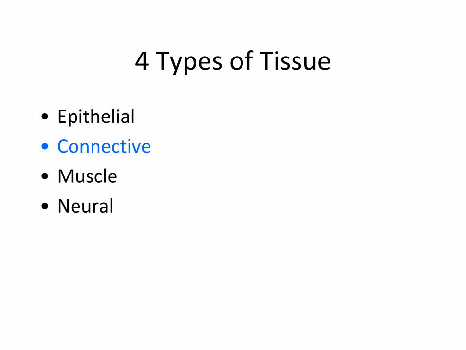
4 Types of Tissue
• Epithelial • Connective • Muscle • Neural
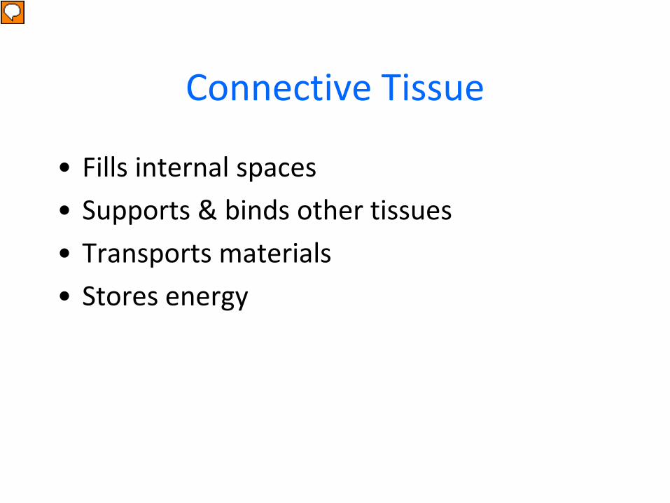
Connective Tissue
• Fills internal spaces • Supports & binds other tissues • Transports materials • Stores energy
Presenter
Presentation Notes
Internal spaces =
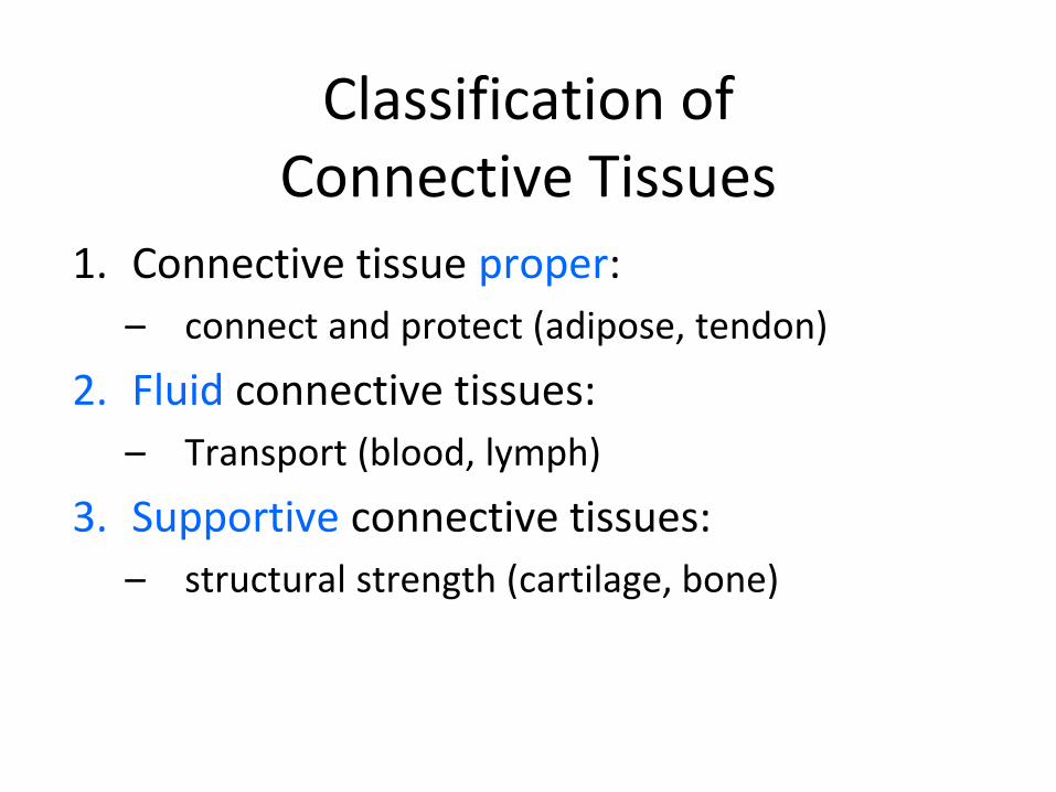
Classification of Connective Tissues
1. Connective tissue proper: – connect and protect (adipose, tendon)
2. Fluid connective tissues: – Transport (blood, lymph)
3. Supportive connective tissues: – structural strength (cartilage, bone)
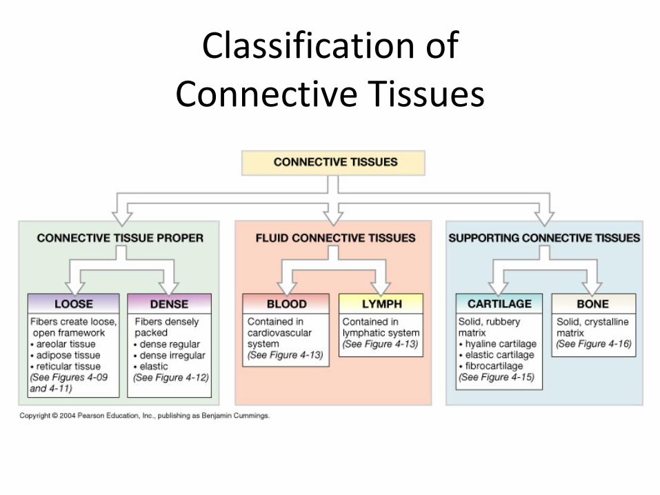
Classification of Connective Tissues
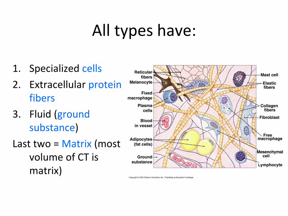
All types have:
1. Specialized cells 2. Extracellular protein
fibers 3. Fluid (ground
substance) Last two = Matrix (most
volume of CT is matrix)
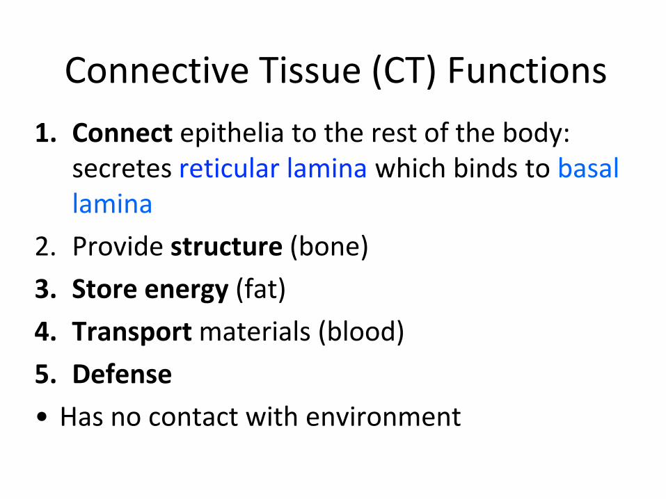
Connective Tissue (CT) Functions 1. Connect epithelia to the rest of the body:
secretes reticular lamina which binds to basal lamina
2. Provide structure (bone) 3. Store energy (fat) 4. Transport materials (blood) 5. Defense • Has no contact with environment
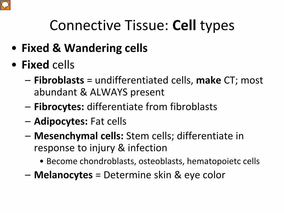
Connective Tissue: Cell types • Fixed & Wandering cells • Fixed cells
– Fibroblasts = undifferentiated cells, make CT; most abundant & ALWAYS present
– Fibrocytes: differentiate from fibroblasts – Adipocytes: Fat cells – Mesenchymal cells: Stem cells; differentiate in
response to injury & infection • Become chondroblasts, osteoblasts, hematopoietc cells
– Melanocytes = Determine skin & eye color
Presenter
Presentation Notes
What do macrophages do?Stringent hall monitors What are adipocytes? What are melanocytes?
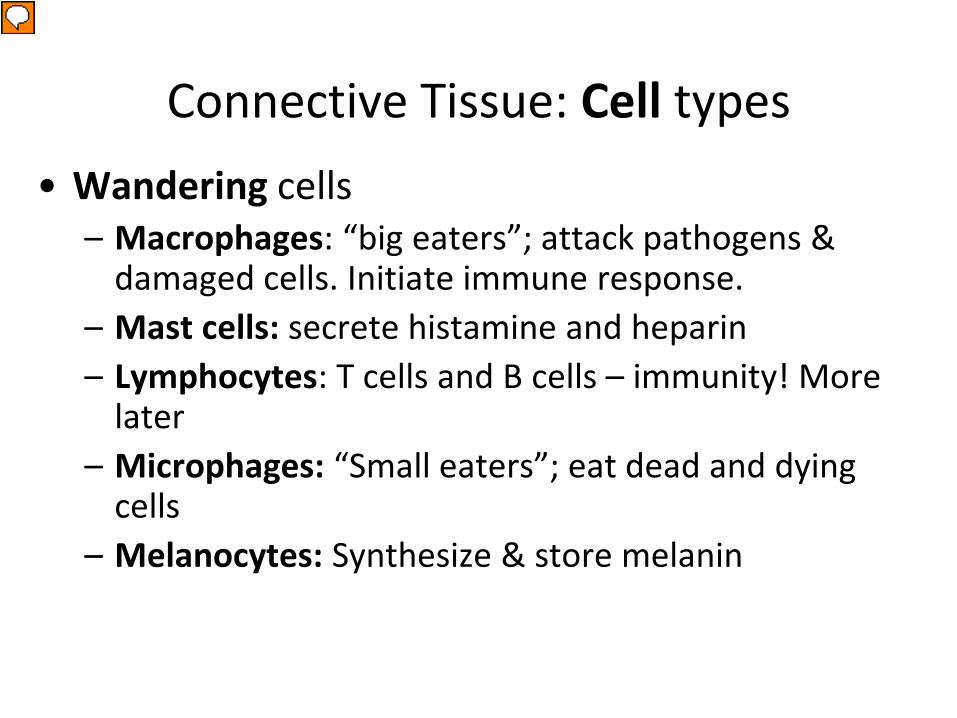
Connective Tissue: Cell types • Wandering cells
– Macrophages: “big eaters”; attack pathogens & damaged cells. Initiate immune response.
– Mast cells: secrete histamine and heparin – Lymphocytes: T cells and B cells – immunity! More
later – Microphages: “Small eaters”; eat dead and dying
cells – Melanocytes: Synthesize & store melanin
Presenter
Presentation Notes
What do macrophages do?Stringent hall monitors What are adipocytes? What are melanocytes?
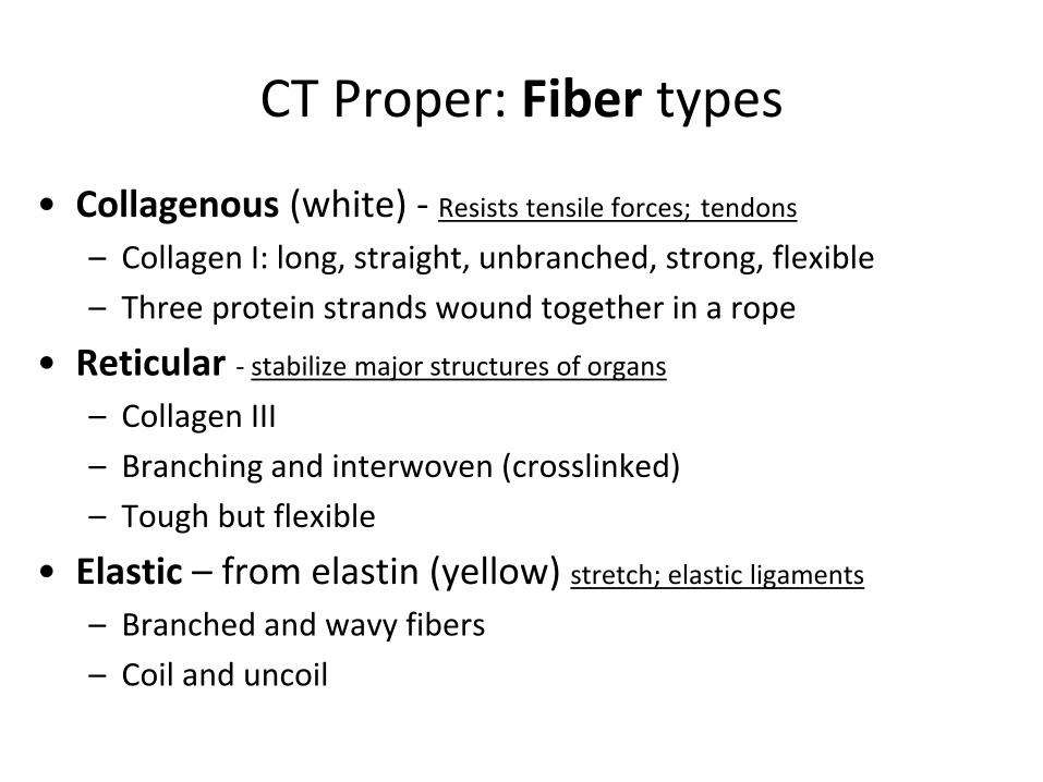
CT Proper: Fiber types
• Collagenous (white) - Resists tensile forces; tendons
– Collagen I: long, straight, unbranched, strong, flexible – Three protein strands wound together in a rope
• Reticular - stabilize major structures of organs – Collagen III – Branching and interwoven (crosslinked) – Tough but flexible
• Elastic – from elastin (yellow) stretch; elastic ligaments
– Branched and wavy fibers – Coil and uncoil
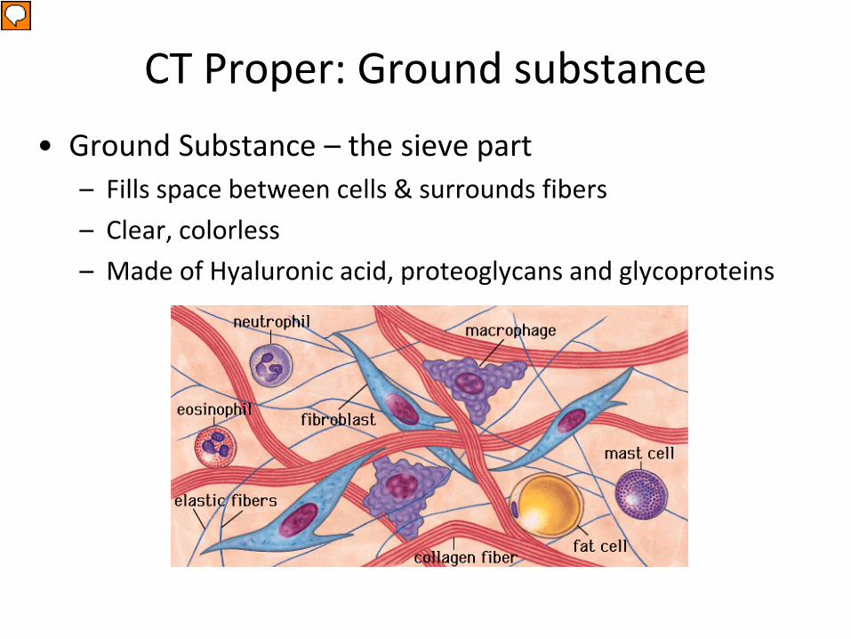
CT Proper: Ground substance • Ground Substance – the sieve part
– Fills space between cells & surrounds fibers – Clear, colorless – Made of Hyaluronic acid, proteoglycans and glycoproteins
Presenter
Presentation Notes
Which cells secrete hyaluronic acid, proteoglycans and glycoproteins??
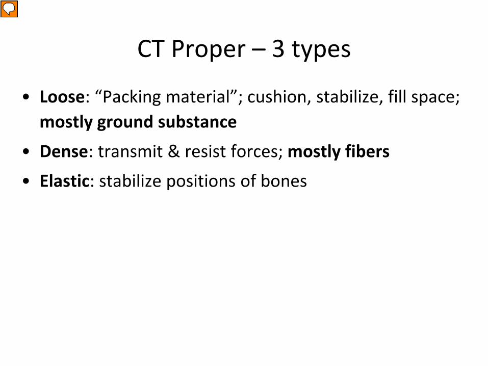
CT Proper – 3 types
• Loose: “Packing material”; cushion, stabilize, fill space; mostly ground substance
• Dense: transmit & resist forces; mostly fibers • Elastic: stabilize positions of bones
Presenter
Presentation Notes
where is areolar commonly found?
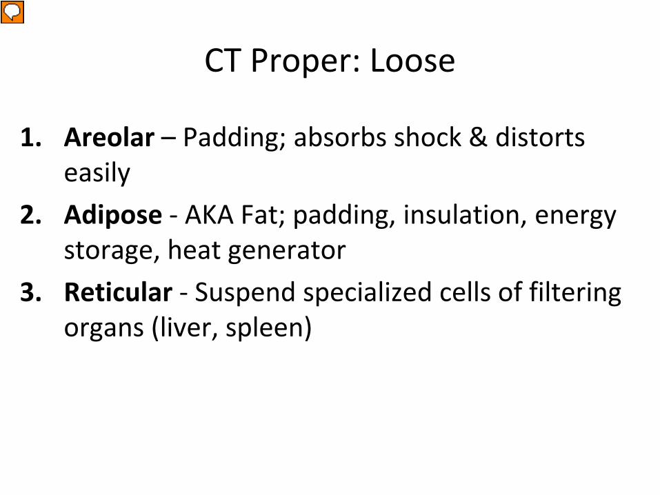
CT Proper: Loose
1. Areolar – Padding; absorbs shock & distorts easily
2. Adipose - AKA Fat; padding, insulation, energy storage, heat generator
3. Reticular - Suspend specialized cells of filtering organs (liver, spleen)
Presenter
Presentation Notes
where is areolar commonly found?
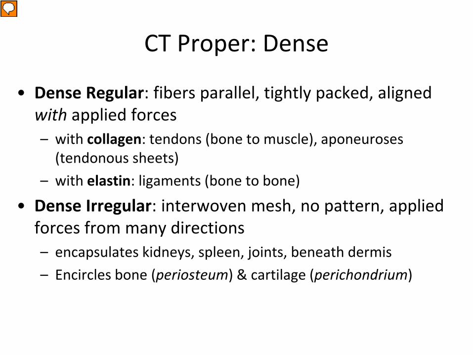
CT Proper: Dense
• Dense Regular: fibers parallel, tightly packed, aligned with applied forces – with collagen: tendons (bone to muscle), aponeuroses
(tendonous sheets) – with elastin: ligaments (bone to bone)
• Dense Irregular: interwoven mesh, no pattern, applied forces from many directions – encapsulates kidneys, spleen, joints, beneath dermis – Encircles bone (periosteum) & cartilage (perichondrium)
Presenter
Presentation Notes
where is areolar commonly found?
CT Proper: Elastic
• Elastic – Dense Regular – Vocal cords & between vertebral bodies
Presenter
Presentation Notes
where is areolar commonly found?
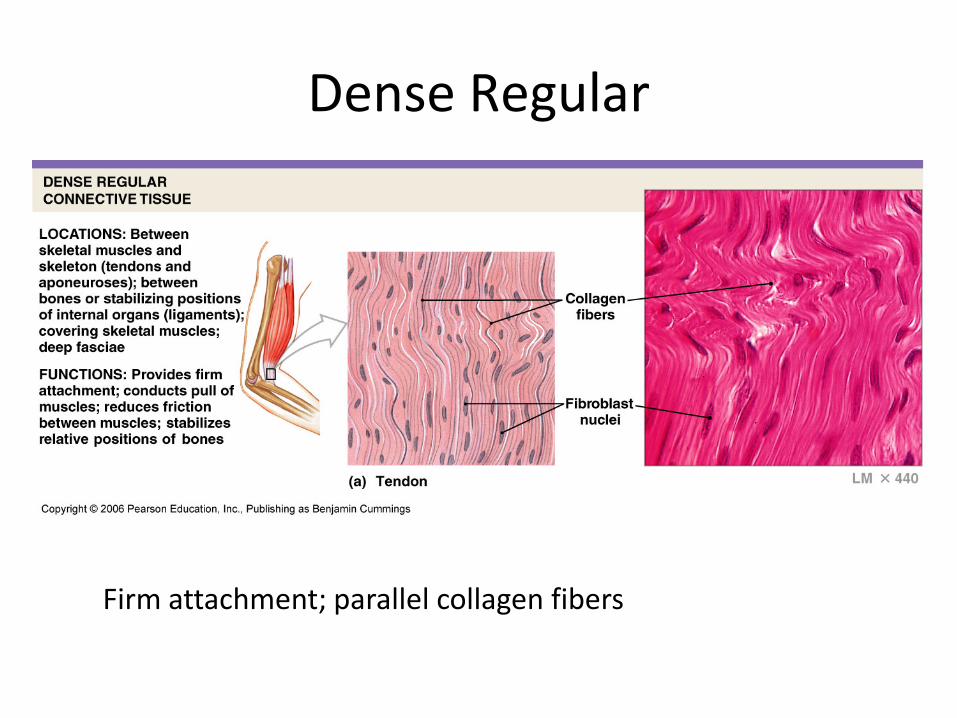
Dense Regular
Firm attachment; parallel collagen fibers
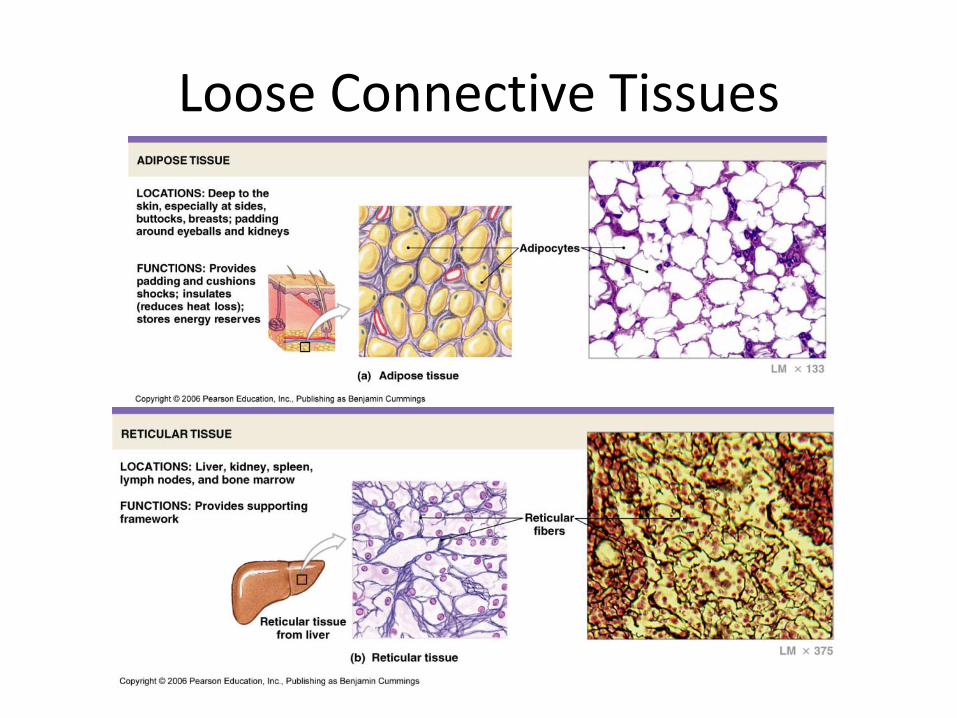
Loose Connective Tissues
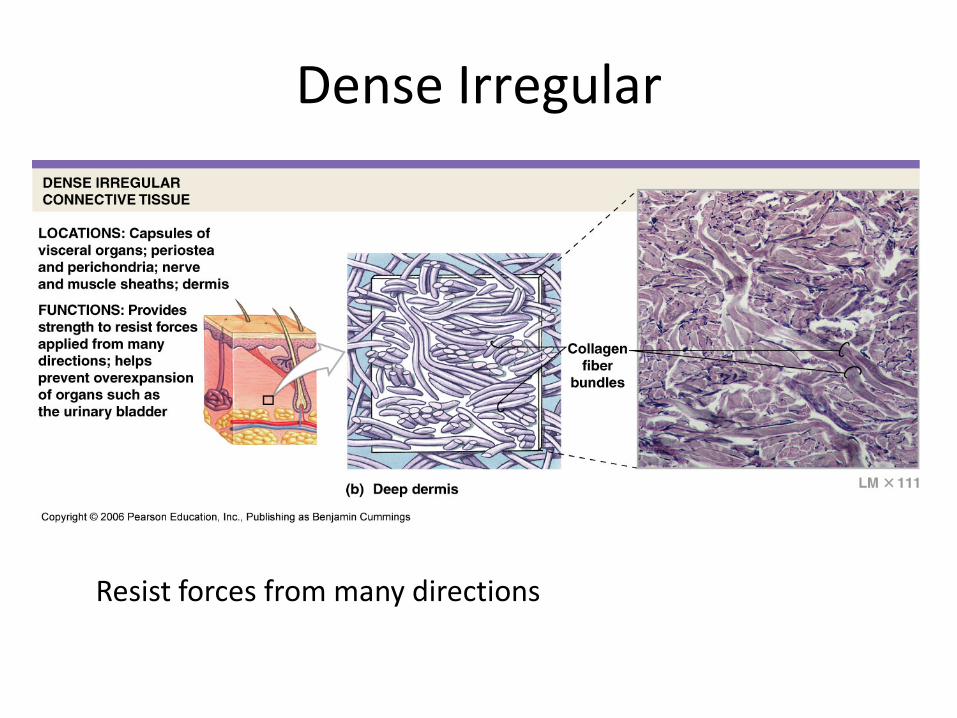
Dense Irregular
Resist forces from many directions
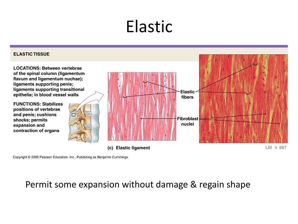
Elastic
Permit some expansion without damage & regain shape
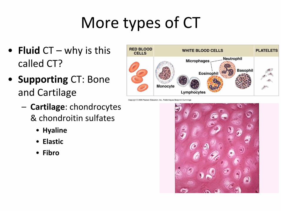
More types of CT • Fluid CT – why is this
called CT? • Supporting CT: Bone
and Cartilage – Cartilage: chondrocytes
& chondroitin sulfates • Hyaline • Elastic • Fibro
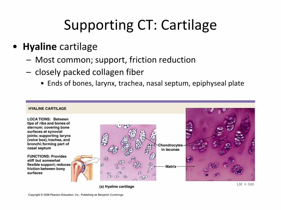
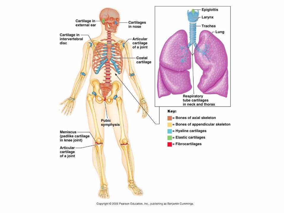
Supporting CT: Cartilage • Hyaline cartilage
– Most common; support, friction reduction – closely packed collagen fiber
• Ends of bones, larynx, trachea, nasal septum, epiphyseal plate
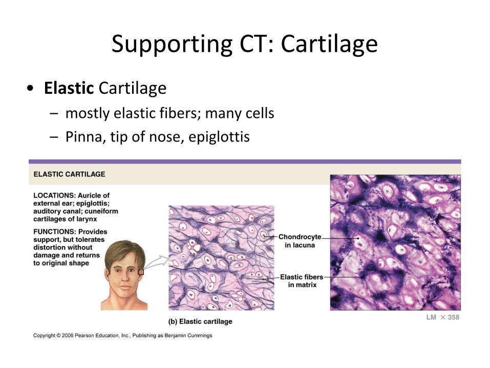
Supporting CT: Cartilage • Elastic Cartilage
– mostly elastic fibers; many cells – Pinna, tip of nose, epiglottis
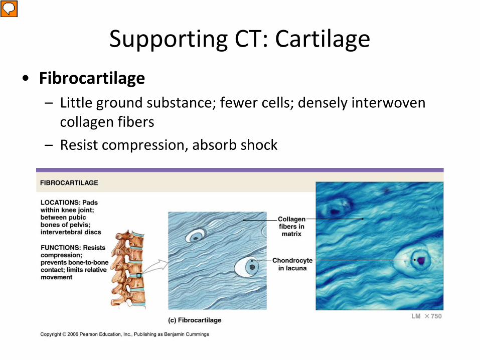
Supporting CT: Cartilage • Fibrocartilage
– Little ground substance; fewer cells; densely interwoven collagen fibers
– Resist compression, absorb shock
Presenter
Presentation Notes
What’s the ground substance?
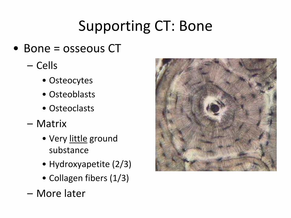
Supporting CT: Bone • Bone = osseous CT
– Cells • Osteocytes • Osteoblasts • Osteoclasts
– Matrix • Very little ground
substance • Hydroxyapetite (2/3) • Collagen fibers (1/3)
– More later
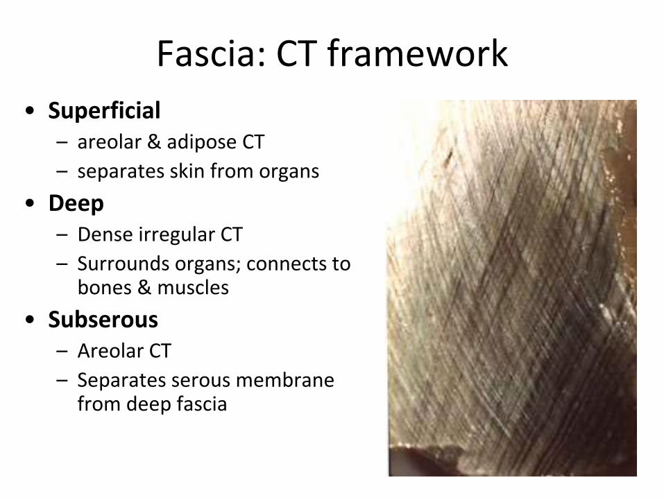
Fascia: CT framework • Superficial
– areolar & adipose CT – separates skin from organs
• Deep – Dense irregular CT – Surrounds organs; connects to
bones & muscles • Subserous
– Areolar CT – Separates serous membrane
from deep fascia