
Congenital lobar emphysema concurrent with pneumothorax and pneumomediastinum … · 2017-08-30 ·...
4
NOTE Surgery Congenital lobar emphysema concurrent with pneumothorax and pneumomediastinum in a dog Sungho YUN 1) , Hohyun LEE 1) , Jaehyun LIM 2) , Kija LEE 3) , Kwangho JANG 1) , Nozomi SHIWA 4) , Hassadin BOONSRIROJ 4) , Kazunori KIMITSUKI 4) , Chunho PARK 4) and Youngsam KWON 1) * 1) Department of Veterinary Surgery, College of Veterinary Medicine, Kyungpook National University, Daegu 702–701, Republic of Korea 2) Daegu Animal Medical Center, Daegu 706–842, Republic of Korea 3) Department of Veterinary Medical Imaging, College of Veterinary Medicine, Kyungpook National University, Daegu 702–701, Republic of Korea 4) Department of Veterinary Pathology, School of Veterinary Medicine, Kitasato University, 23–35–1 Higashi, Towada, Aomori 034–8628, Japan (Received 22 July 2015/Accepted 27 January 2016/Published online in J-STAGE 9 February 2016) ABSTRACT. A two-year-old castrated male Pomeranian dog was referred with the chief complaints of coughing and subcutaneous emphy- sema. On physical examination, the crepitant areas were palpable. When auscultated, the right chest was absent of respiratory sound, while the sound of the opposite side was enhanced. Radiographs presented pneumothorax and pneumomediastinum. On computed tomography, hypoattenuated bulla-like lesion at right middle lung lobe and trapped air in mediastinum were shown. After patient stabilization, surgery for excision of affected lobe was performed. During follow-up period, there were no recurrence and complication on radiographic examination. Based on clinical and pathological findings, the dog was diagnosed as congenital lobar emphysema. KEY WORDS: canine, congenital lobar emphysema, pneumomediastinum, pneumothorax doi: 10.1292/jvms.15-0362; J. Vet. Med. Sci. 78(5): 909–912, 2016 Congenital lobar emphysema (CLE) is rare pulmonary disease in dogs, and the majority of affected dogs were between 6 weeks to 6 months of age [1, 2, 10]. The most common clinical sign is progressive dyspnea with coughing and exercise intolerance [2, 5]. It predominantly occurs in unilateral cranial lobe or right middle lobe, and has distinct histopathological characteristics [1, 2, 10]. We present a case of CLE with concurrent pneumothorax, pneumomediasti- num and subcutaneous emphysema, that was successfully corrected with surgical treatment and definitively diagnosed through histopathological staining, in an adult dog. A two-year-old castrated male Pomeranian dog, weighing 2.7 kg, was referred to the Kyungpook National Univer- sity Veterinary Medical Teaching Hospital with the chief complaints of subcutaneous emphysema and acute onset of coughing. No previous clinical sign related with respiratory tract had been noted. On physical examination, the crepitant areas were palpable on head, neck and lumbar regions. When auscultated, the right chest was absent of respiratory sound, while the sound of the opposite side was enhanced. Despite such changes, only mild tachypnea with coughing was ob- served. Visible mucous membrane color was normal. The dog was normothermic and had regular pulse. There were no history of trauma and noticeable superficial soft tissue injury. Complete blood count and blood chemistry values were normal. Lateral radiographs presented the radiolucent space between heart and sternum. The cranial mediastinum had heterogeneous opacity, and major vessels of mediastinum were visualized, which can be the evidence of pneumo- mediastinum. In ventrodorsal radiographs, radiolucent bulla-like lesion with left shift of heart and mediastinum was visualized in the right thorax. Caudal displacement of right hemidiaphragm and hyperlucency of right thorax, implying pneumothorax, were seen. Subcutaneous emphysema was extended from head to the lumbar region (Fig. 1). On computed tomography (CT), pneumothorax was obviously observed in right thorax. The hypoattenuated bullae-like lesion on the right middle lung lobe was shown. Pneumomediastinum was seen as trapped air around trachea, esophagus and major vessels (Fig. 2). After patient stabilization and sufficient oxygenation, lo- bectomy was performed with right intercostal thoracotomy. The incision was made between 5th and 6th ribs. Lack of negative pressure within thoracic cavity was identified dur- ing thoracotomy. Grossly, the middle lung lobe had extreme- ly scanty parenchyma (Fig. 3). After the ligation of blood vessels and bronchus, affected lung lobe was excised. When positive pressure was given with ventilation machine, col- lapsed cranial, caudal and accessory lobes were recovered to normal shape. Prior to closing the thorax, warm sterile saline was filled within the thoracic space to confirm the absence of air leakage, and Jackson-Pratt drain tube was placed. Tissue samples were fixed in 10% buffered formalin, em- bedded in paraffin wax and sectioned. Sections were stained with hematoxylin and eosin (HE), and serial sections were *CORRESPONDENCE TO: KWON, Y., Department of Veterinary Surgery, College of Veterinary Medicine, Kyungpook National University, Daegu 702–701, Republic of Korea. e-mail: [email protected] ©2016 The Japanese Society of Veterinary Science This is an open-access article distributed under the terms of the Creative Commons Attribution Non-Commercial No Derivatives (by-nc-nd) License <http://creativecommons.org/licenses/by-nc-nd/4.0/>.
Transcript of Congenital lobar emphysema concurrent with pneumothorax and pneumomediastinum … · 2017-08-30 ·...

NOTE Surgery
Congenital lobar emphysema concurrent with pneumothorax and pneumomediastinum in a dog
Sungho YUN1), Hohyun LEE1), Jaehyun LIM2), Kija LEE3), Kwangho JANG1), Nozomi SHIWA4), Hassadin BOONSRIROJ4), Kazunori KIMITSUKI4), Chunho PARK4) and Youngsam KWON1)*
1)Department of Veterinary Surgery, College of Veterinary Medicine, Kyungpook National University, Daegu 702–701, Republic of Korea2)Daegu Animal Medical Center, Daegu 706–842, Republic of Korea3)Department of Veterinary Medical Imaging, College of Veterinary Medicine, Kyungpook National University, Daegu 702–701, Republic
of Korea4)Department of Veterinary Pathology, School of Veterinary Medicine, Kitasato University, 23–35–1 Higashi, Towada, Aomori 034–8628,
Japan
(Received 22 July 2015/Accepted 27 January 2016/Published online in J-STAGE 9 February 2016)
ABSTRACT. A two-year-old castrated male Pomeranian dog was referred with the chief complaints of coughing and subcutaneous emphy-sema. On physical examination, the crepitant areas were palpable. When auscultated, the right chest was absent of respiratory sound, while the sound of the opposite side was enhanced. Radiographs presented pneumothorax and pneumomediastinum. On computed tomography, hypoattenuated bulla-like lesion at right middle lung lobe and trapped air in mediastinum were shown. After patient stabilization, surgery for excision of affected lobe was performed. During follow-up period, there were no recurrence and complication on radiographic examination. Based on clinical and pathological findings, the dog was diagnosed as congenital lobar emphysema.KEY WORDS: canine, congenital lobar emphysema, pneumomediastinum, pneumothorax
doi: 10.1292/jvms.15-0362; J. Vet. Med. Sci. 78(5): 909–912, 2016
Congenital lobar emphysema (CLE) is rare pulmonary disease in dogs, and the majority of affected dogs were between 6 weeks to 6 months of age [1, 2, 10]. The most common clinical sign is progressive dyspnea with coughing and exercise intolerance [2, 5]. It predominantly occurs in unilateral cranial lobe or right middle lobe, and has distinct histopathological characteristics [1, 2, 10]. We present a case of CLE with concurrent pneumothorax, pneumomediasti-num and subcutaneous emphysema, that was successfully corrected with surgical treatment and definitively diagnosed through histopathological staining, in an adult dog.
A two-year-old castrated male Pomeranian dog, weighing 2.7 kg, was referred to the Kyungpook National Univer-sity Veterinary Medical Teaching Hospital with the chief complaints of subcutaneous emphysema and acute onset of coughing. No previous clinical sign related with respiratory tract had been noted. On physical examination, the crepitant areas were palpable on head, neck and lumbar regions. When auscultated, the right chest was absent of respiratory sound, while the sound of the opposite side was enhanced. Despite such changes, only mild tachypnea with coughing was ob-served. Visible mucous membrane color was normal. The dog was normothermic and had regular pulse. There were no history of trauma and noticeable superficial soft tissue
injury. Complete blood count and blood chemistry values were normal.
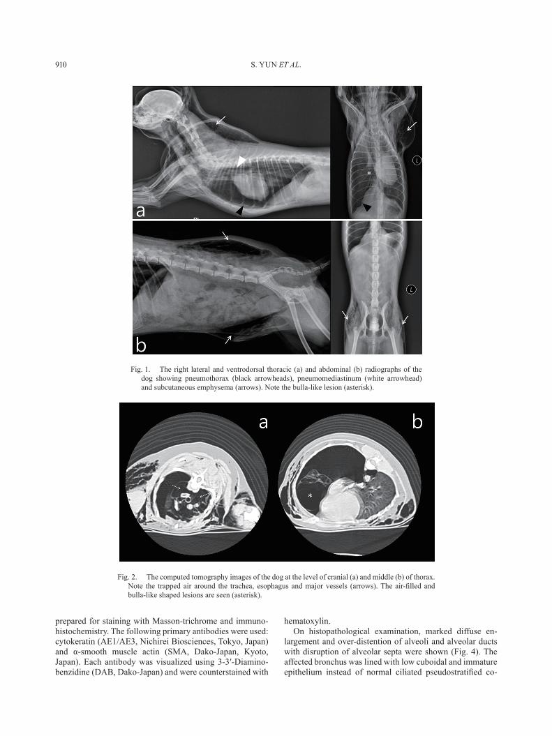
Lateral radiographs presented the radiolucent space between heart and sternum. The cranial mediastinum had heterogeneous opacity, and major vessels of mediastinum were visualized, which can be the evidence of pneumo-mediastinum. In ventrodorsal radiographs, radiolucent bulla-like lesion with left shift of heart and mediastinum was visualized in the right thorax. Caudal displacement of right hemidiaphragm and hyperlucency of right thorax, implying pneumothorax, were seen. Subcutaneous emphysema was extended from head to the lumbar region (Fig. 1).
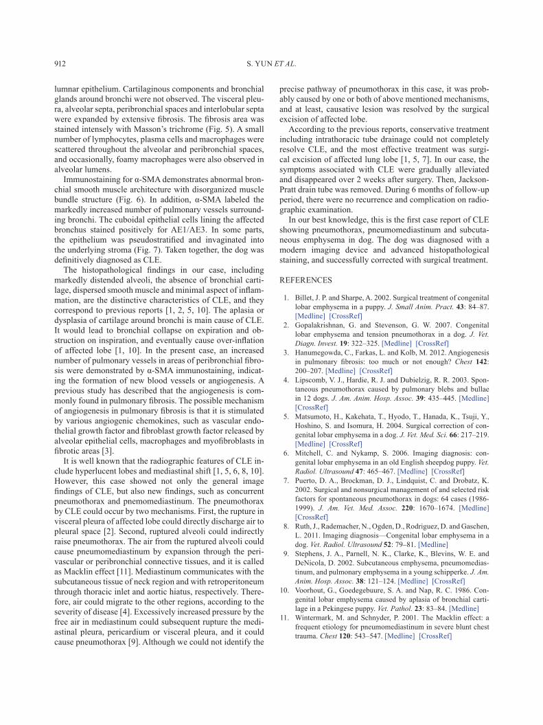
On computed tomography (CT), pneumothorax was obviously observed in right thorax. The hypoattenuated bullae-like lesion on the right middle lung lobe was shown. Pneumomediastinum was seen as trapped air around trachea, esophagus and major vessels (Fig. 2).
After patient stabilization and sufficient oxygenation, lo-bectomy was performed with right intercostal thoracotomy. The incision was made between 5th and 6th ribs. Lack of negative pressure within thoracic cavity was identified dur-ing thoracotomy. Grossly, the middle lung lobe had extreme-ly scanty parenchyma (Fig. 3). After the ligation of blood vessels and bronchus, affected lung lobe was excised. When positive pressure was given with ventilation machine, col-lapsed cranial, caudal and accessory lobes were recovered to normal shape. Prior to closing the thorax, warm sterile saline was filled within the thoracic space to confirm the absence of air leakage, and Jackson-Pratt drain tube was placed.
Tissue samples were fixed in 10% buffered formalin, em-bedded in paraffin wax and sectioned. Sections were stained with hematoxylin and eosin (HE), and serial sections were
*CorrespondenCe to: Kwon, Y., Department of Veterinary Surgery, College of Veterinary Medicine, Kyungpook National University, Daegu 702–701, Republic of Korea. e-mail: [email protected]
©2016 The Japanese Society of Veterinary ScienceThis is an open-access article distributed under the terms of the Creative Commons Attribution Non-Commercial No Derivatives (by-nc-nd) License <http://creativecommons.org/licenses/by-nc-nd/4.0/>.
S. YUN ET AL.910
prepared for staining with Masson-trichrome and immuno-histochemistry. The following primary antibodies were used: cytokeratin (AE1/AE3, Nichirei Biosciences, Tokyo, Japan) and α-smooth muscle actin (SMA, Dako-Japan, Kyoto, Japan). Each antibody was visualized using 3-3′-Diamino-benzidine (DAB, Dako-Japan) and were counterstained with
hematoxylin.On histopathological examination, marked diffuse en-
largement and over-distention of alveoli and alveolar ducts with disruption of alveolar septa were shown (Fig. 4). The affected bronchus was lined with low cuboidal and immature epithelium instead of normal ciliated pseudostratified co-
Fig. 1. The right lateral and ventrodorsal thoracic (a) and abdominal (b) radiographs of the dog showing pneumothorax (black arrowheads), pneumomediastinum (white arrowhead) and subcutaneous emphysema (arrows). Note the bulla-like lesion (asterisk).
Fig. 2. The computed tomography images of the dog at the level of cranial (a) and middle (b) of thorax. Note the trapped air around the trachea, esophagus and major vessels (arrows). The air-filled and bulla-like shaped lesions are seen (asterisk).

CONGENITAL LOBAR EMPHYSEMA IN A DOG 911
Fig. 3. The macroscopic findings of middle lung lobe show the extremely scanty parenchymal tissues.
Fig. 4. Severely dilated alveolar cavity and congestive blood vessels are observed. HE staining. Bar=500 µm. A: alveolar space, B: bronchi, P: pleura, V: blood vessels.
Fig. 5. Proliferation of connective tissues especially in subpleura and perivascular areas is observed. Masson-trichrome staining. Bar=500 µm. A: alveolar space, B: bronchi, P: pleura, V: blood vessels.
Fig. 6. Smooth muscles surrounding the bronchi occasionally dispersed (asterisks), and an increased number of pulmonary vessels are seen. Immunohistochemistry (α-SMA). Bar=250 µm. A: alveolar space, B: bronchi.
Fig. 7. The epithelial cell layer of bronchi is irregularly arranged, and some cells invaginated into the underlying stroma (arrows). Im-munohistochemistry (AE1/AE3). Bar=250 µm. A: alveolar space, B: bronchi.
S. YUN ET AL.912
lumnar epithelium. Cartilaginous components and bronchial glands around bronchi were not observed. The visceral pleu-ra, alveolar septa, peribronchial spaces and interlobular septa were expanded by extensive fibrosis. The fibrosis area was stained intensely with Masson’s trichrome (Fig. 5). A small number of lymphocytes, plasma cells and macrophages were scattered throughout the alveolar and peribronchial spaces, and occasionally, foamy macrophages were also observed in alveolar lumens.
Immunostaining for α-SMA demonstrates abnormal bron-chial smooth muscle architecture with disorganized muscle bundle structure (Fig. 6). In addition, α-SMA labeled the markedly increased number of pulmonary vessels surround-ing bronchi. The cuboidal epithelial cells lining the affected bronchus stained positively for AE1/AE3. In some parts, the epithelium was pseudostratified and invaginated into the underlying stroma (Fig. 7). Taken together, the dog was definitively diagnosed as CLE.
The histopathological findings in our case, including markedly distended alveoli, the absence of bronchial carti-lage, dispersed smooth muscle and minimal aspect of inflam-mation, are the distinctive characteristics of CLE, and they correspond to previous reports [1, 2, 5, 10]. The aplasia or dysplasia of cartilage around bronchi is main cause of CLE. It would lead to bronchial collapse on expiration and ob-struction on inspiration, and eventually cause over-inflation of affected lobe [1, 10]. In the present case, an increased number of pulmonary vessels in areas of peribronchial fibro-sis were demonstrated by α-SMA immunostaining, indicat-ing the formation of new blood vessels or angiogenesis. A previous study has described that the angiogenesis is com-monly found in pulmonary fibrosis. The possible mechanism of angiogenesis in pulmonary fibrosis is that it is stimulated by various angiogenic chemokines, such as vascular endo-thelial growth factor and fibroblast growth factor released by alveolar epithelial cells, macrophages and myofibroblasts in fibrotic areas [3].
It is well known that the radiographic features of CLE in-clude hyperlucent lobes and mediastinal shift [1, 5, 6, 8, 10]. However, this case showed not only the general image findings of CLE, but also new findings, such as concurrent pneumothorax and pnemomediastinum. The pneumothorax by CLE could occur by two mechanisms. First, the rupture in visceral pleura of affected lobe could directly discharge air to pleural space [2]. Second, ruptured alveoli could indirectly raise pneumothorax. The air from the ruptured alveoli could cause pneumomediastinum by expansion through the peri-vascular or peribronchial connective tissues, and it is called as Macklin effect [11]. Mediastinum communicates with the subcutaneous tissue of neck region and with retroperitoneum through thoracic inlet and aortic hiatus, respectively. There-fore, air could migrate to the other regions, according to the severity of disease [4]. Excessively increased pressure by the free air in mediastinum could subsequent rupture the medi-astinal pleura, pericardium or visceral pleura, and it could cause pneumothorax [9]. Although we could not identify the
precise pathway of pneumothorax in this case, it was prob-ably caused by one or both of above mentioned mechanisms, and at least, causative lesion was resolved by the surgical excision of affected lobe.
According to the previous reports, conservative treatment including intrathoracic tube drainage could not completely resolve CLE, and the most effective treatment was surgi-cal excision of affected lung lobe [1, 5, 7]. In our case, the symptoms associated with CLE were gradually alleviated and disappeared over 2 weeks after surgery. Then, Jackson-Pratt drain tube was removed. During 6 months of follow-up period, there were no recurrence and complication on radio-graphic examination.
In our best knowledge, this is the first case report of CLE showing pneumothorax, pneumomediastinum and subcuta-neous emphysema in dog. The dog was diagnosed with a modern imaging device and advanced histopathological staining, and successfully corrected with surgical treatment.
REFERENCES
1. Billet, J. P. and Sharpe, A. 2002. Surgical treatment of congenital lobar emphysema in a puppy. J. Small Anim. Pract. 43: 84–87. [Medline] [CrossRef]
2. Gopalakrishnan, G. and Stevenson, G. W. 2007. Congenital lobar emphysema and tension pneumothorax in a dog. J. Vet. Diagn. Invest. 19: 322–325. [Medline] [CrossRef]
3. Hanumegowda, C., Farkas, L. and Kolb, M. 2012. Angiogenesis in pulmonary fibrosis: too much or not enough? Chest 142: 200–207. [Medline] [CrossRef]
4. Lipscomb, V. J., Hardie, R. J. and Dubielzig, R. R. 2003. Spon-taneous pneumothorax caused by pulmonary blebs and bullae in 12 dogs. J. Am. Anim. Hosp. Assoc. 39: 435–445. [Medline] [CrossRef]
5. Matsumoto, H., Kakehata, T., Hyodo, T., Hanada, K., Tsuji, Y., Hoshino, S. and Isomura, H. 2004. Surgical correction of con-genital lobar emphysema in a dog. J. Vet. Med. Sci. 66: 217–219. [Medline] [CrossRef]
6. Mitchell, C. and Nykamp, S. 2006. Imaging diagnosis: con-genital lobar emphysema in an old English sheepdog puppy. Vet. Radiol. Ultrasound 47: 465–467. [Medline] [CrossRef]
7. Puerto, D. A., Brockman, D. J., Lindquist, C. and Drobatz, K. 2002. Surgical and nonsurgical management of and selected risk factors for spontaneous pneumothorax in dogs: 64 cases (1986-1999). J. Am. Vet. Med. Assoc. 220: 1670–1674. [Medline] [CrossRef]
8. Ruth, J., Rademacher, N., Ogden, D., Rodriguez, D. and Gaschen, L. 2011. Imaging diagnosis—Congenital lobar emphysema in a dog. Vet. Radiol. Ultrasound 52: 79–81. [Medline]
9. Stephens, J. A., Parnell, N. K., Clarke, K., Blevins, W. E. and DeNicola, D. 2002. Subcutaneous emphysema, pneumomedias-tinum, and pulmonary emphysema in a young schipperke. J. Am. Anim. Hosp. Assoc. 38: 121–124. [Medline] [CrossRef]
10. Voorhout, G., Goedegebuure, S. A. and Nap, R. C. 1986. Con-genital lobar emphysema caused by aplasia of bronchial carti-lage in a Pekingese puppy. Vet. Pathol. 23: 83–84. [Medline]
11. Wintermark, M. and Schnyder, P. 2001. The Macklin effect: a frequent etiology for pneumomediastinum in severe blunt chest trauma. Chest 120: 543–547. [Medline] [CrossRef]


![Case Report Subcutaneous Emphysema, Pneumomediastinum, … · 2019. 7. 31. · [ ]E.Hillewig,E.Aghayev,C.Jackowski,A.Christe,T.Plattner, and M. J. ali , Gas embolism following intraosseous](https://static.fdocuments.net/doc/165x107/61254bca97cc8d09c20890f9/case-report-subcutaneous-emphysema-pneumomediastinum-2019-7-31-ehillewigeaghayevcjackowskiachristetplattner.jpg)

![Case Report Subcutaneous Emphysema, …downloads.hindawi.com/journals/criem/2015/134816.pdfpneumothorax, pneumomediastinum, pneumopericardium, or subcutaneous emphysema [ ]. Diagnosis](https://static.fdocuments.net/doc/165x107/5f4072ff5627821a5534fd08/case-report-subcutaneous-emphysema-pneumothorax-pneumomediastinum-pneumopericardium.jpg)


