Congenital heart disease
91
-
Upload
dr-armaan-singh -
Category
Health & Medicine
-
view
113 -
download
1
Transcript of Congenital heart disease

Classification of congenital heart diseasesGroup I : Left to right shunts
Group II: Right to lefts shunts
Group III: Obstructive lesions

Left to right shunts
Atrial Septal Defect Ventricular Septal Defect Patent Ductus Arteriosus

Right to Left Shunts
1) Tetralogy of Fallot2) Tricuspid atresia
3) Ebstein’s anomaly
4) Transposition of Great Vessels
5) Truncus Arteriosus
6) Total Anomalous Pulmonary Venous Return (TAPVR)

Obstructive Lesions
Aortic stenosis Coarctation of the Aorta Pulmonic Stenosis
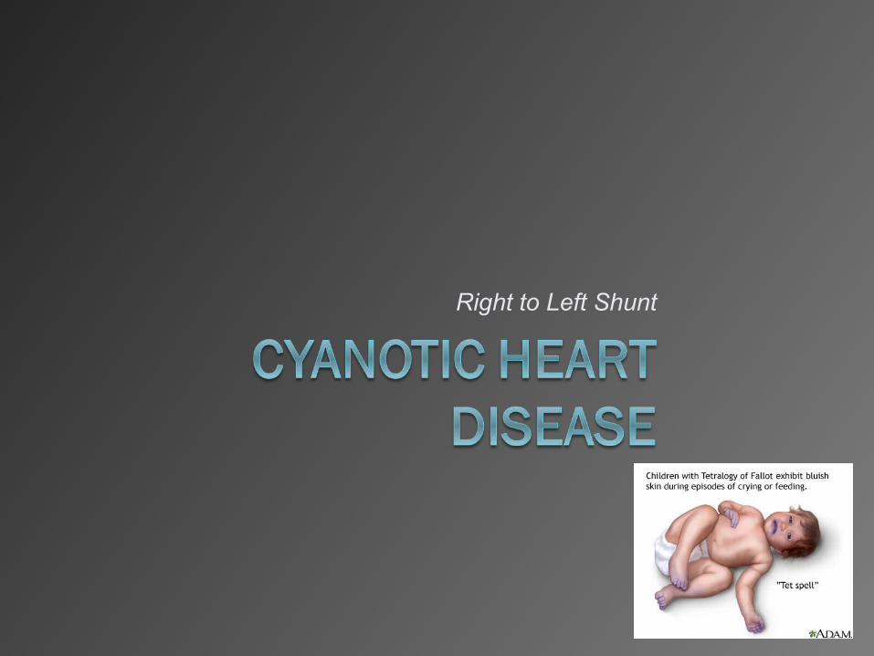
Right to Left Shunt

Who is this guy?

ÉTIENNE-LOUIS ARTHUR FALLOT!
a French physician, 1888 Fallot accurately described in detail the four anatomical characteristics of tetralogy of Fallot.
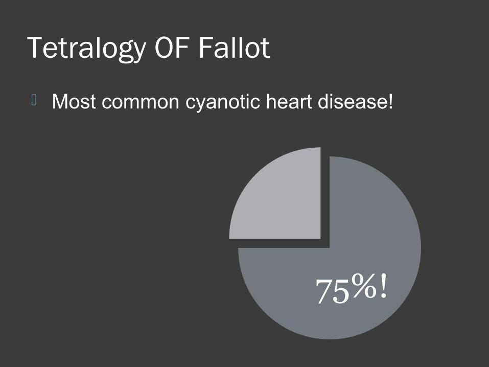
Tetralogy OF Fallot
Most common cyanotic heart disease!
75%!


TOF
4 component!
Imagine this is a HEART!

TOF
1) Vetricular Septal Defect

TOF
1) Vetricular Septal Defect
2) Pulmonic Stenosis

TOF
1) Vetricular Septal Defect
2) Pulmonic Stenosis
3) Overriding of dextroposed aorta

TOF
1) Vetricular Septal Defect
2) Pulmonic Stenosis
3) Overriding of dextroposed aorta
4) Right Ventricular hypertrophy

TOF
1) Vetricular Septal Defect
2) Pulmonic Stenosis
3) Overriding of dextroposed aorta
4) Right Ventricular hypertrophy
Concentric R ventricular hypertrophy without cardiac enlargement

TOF
1) Vetricular Septal Defect
2) Pulmonic Stenosis
3) Overriding of dextroposed aorta
4) Right Ventricular hypertrophy
Concentric R ventricular hypertrophy without cardiac enlargement
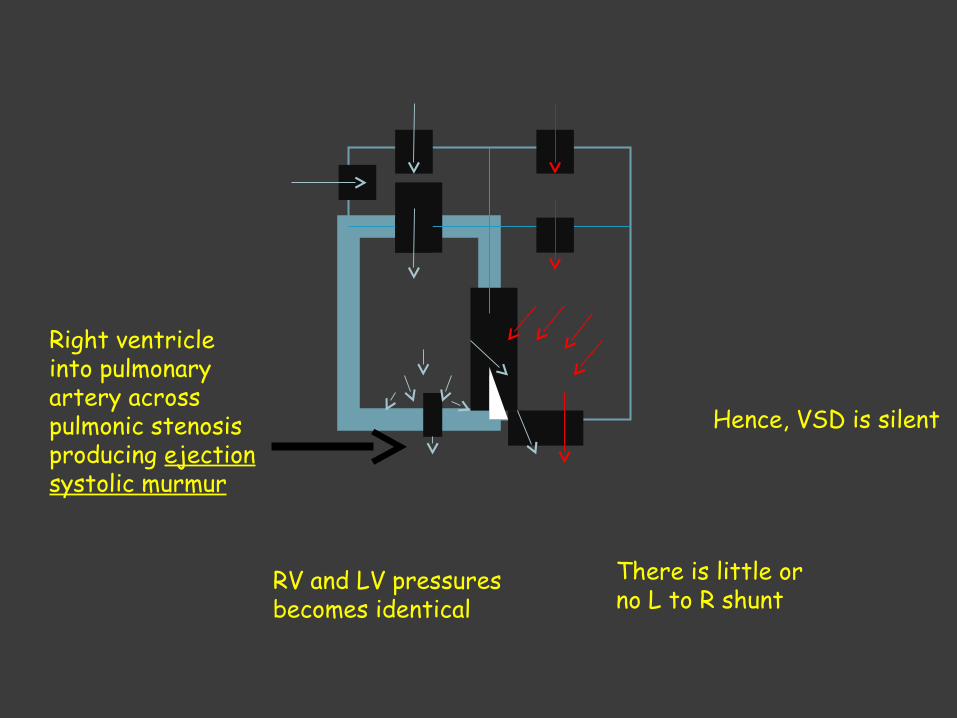
Increase in right ventricular pressure*


RV and LV pressures becomes identical
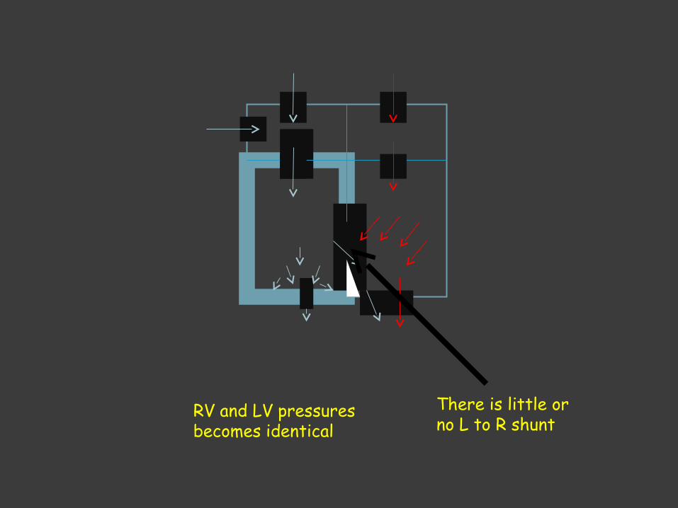
RV and LV pressures becomes identical
There is little or no L to R shunt
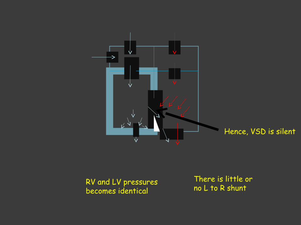
RV and LV pressures becomes identical
There is little or no L to R shunt
Hence, VSD is silent
RV and LV pressures becomes identical
There is little or no L to R shunt
Hence, VSD is silent
Right ventricle into pulmonary artery across pulmonic stenosis producing ejection systolic murmur
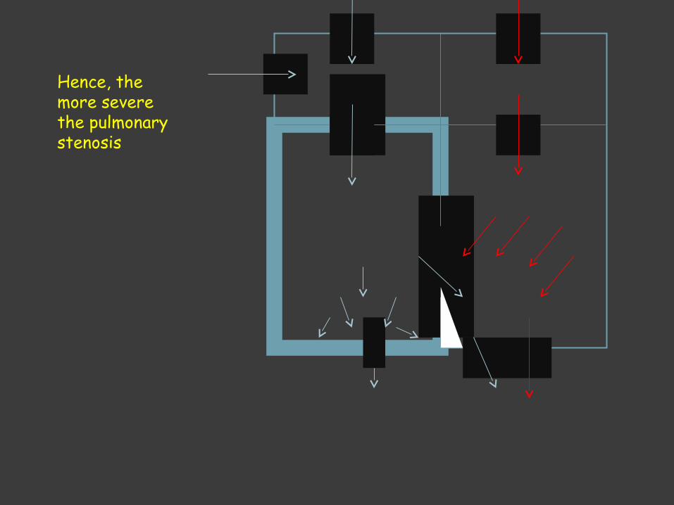
Hence, the more severe the pulmonary stenosis

Hence, the more severe the pulmonary stenosis
The BIGGER the Left to RIGHT shunt

Hence, the more severe the pulmonary stenosis
The BIGGER the Left to RIGHT shunt
Less flow into the pulmonary artery

Hence, the more severe the pulmonary stenosis
The BIGGER the Left to RIGHT shunt
Less flow into the pulmonary artery
Shorter the ejection systolic murmur

Hence, the more severe the pulmonary stenosis
The BIGGER the Left to RIGHT shunt
Less flow into the pulmonary artery
Shorter the ejection systolic murmur
More cynosis because of less flow to the lung!

Hence,
Severity of cyanosis is directly proportional to the severity of pulmonic stenosis
Intensity of the systolic murmur is inversely related to the severity of pulmonic stenosis
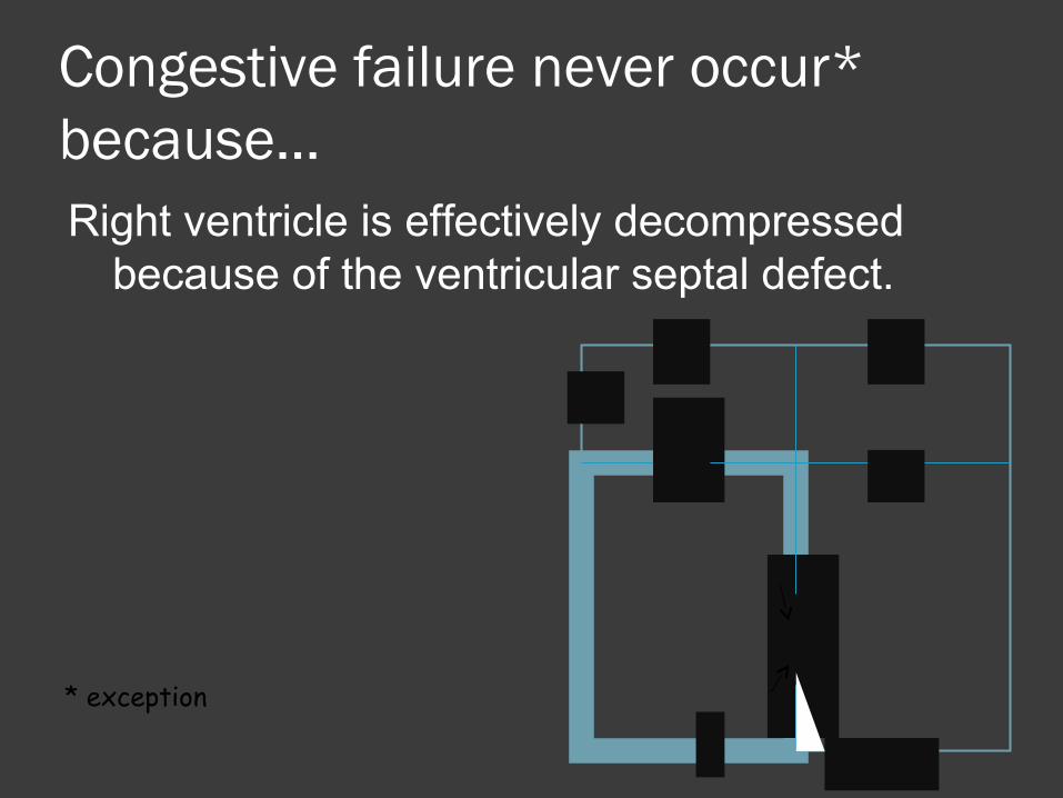
Congestive failure never occur* because…Right ventricle is effectively decompressed
because of the ventricular septal defect.
* exception

Congestive failure never occur* because…Right ventricle is effectively decompressed
because of the ventricular septal defect.
* exception
1)Anemia2)Infective Endocarditis3)Systemic hypertension4)Unrelated myocarditis
complicating TOF5)Aortic or pulmonary valve
regurgitation

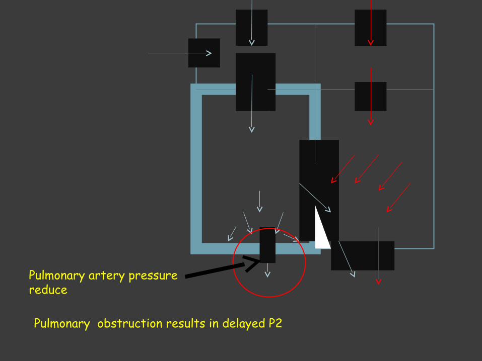
Pulmonary obstruction results in delayed P2
Pulmonary obstruction results in delayed P2
Pulmonary artery pressure reduce
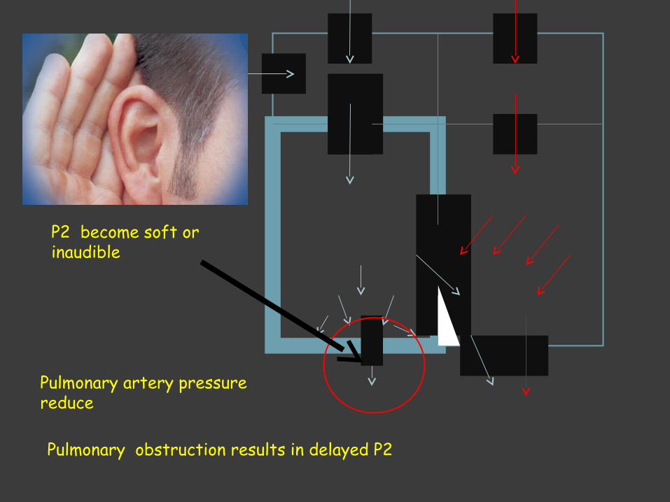
Pulmonary obstruction results in delayed P2
Pulmonary artery pressure reduce
P2 become soft or inaudible

Pulmonary obstruction results in delayed P2
Pulmonary artery pressure reduce
P2 become soft or inaudible

Ascending aorta in TOF is large, results aortic ejection click

Diastolic interval is clear No S3 No S4

Concentric right ventricular hypertrophy reduce the distensibility of the right ventricle during diastole
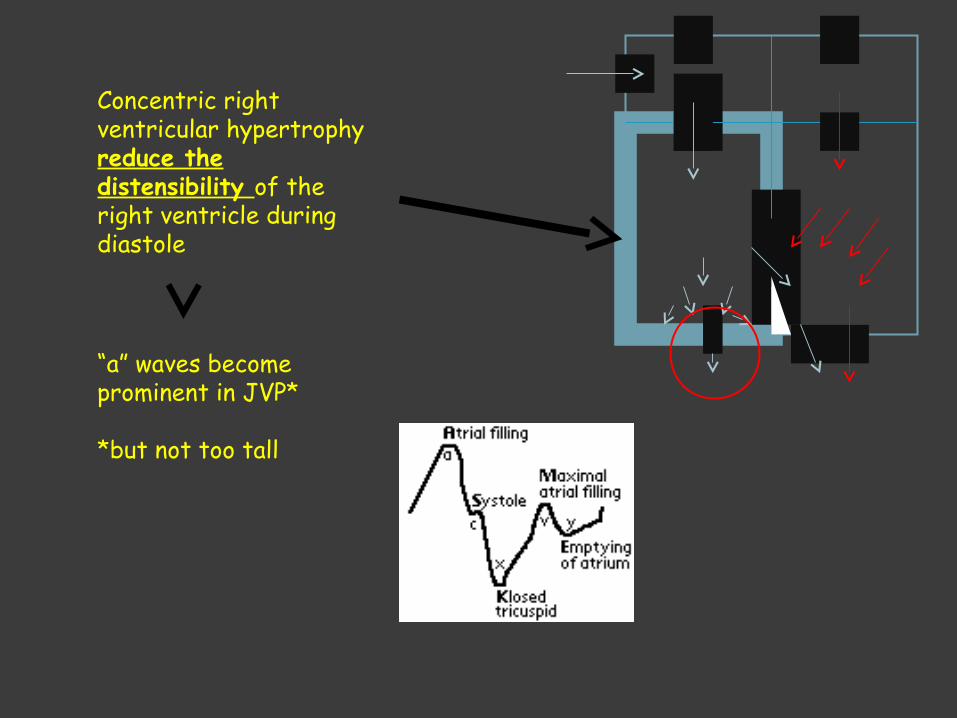
Concentric right ventricular hypertrophy reduce the distensibility of the right ventricle during diastole
“a” waves become prominent in JVP*
*but not too tall

Clinical Picture Symptomatic any time after birth Paroxysmal attacks of dyspnea
Anoxic spellsPredominantly after waking upChild cryDyspneaBlueLose consciousConvulsionFrequency varies from
once a few days to many attack everyday
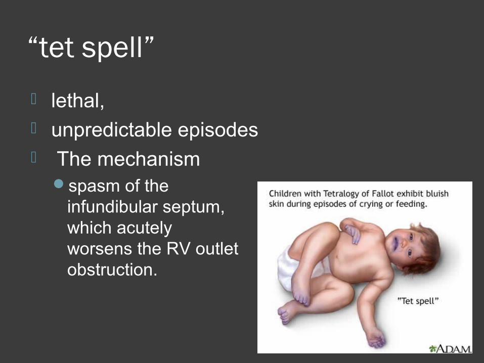
“tet spell”
lethal, unpredictable episodes The mechanism
spasm of the infundibular septum,which acutely worsens the RV outlet obstruction.
Dyspnea on exertion Exercise intolerance

Sitting posture – squattingCompensatory mechanismSquatting increases the peripheral vascular
resistance, which diminishes the
right-to-left shunt increases pulmonary
blood flow.

Cyanosis during feedingPoor feedingfussiness, tachypnea, and agitation.Birth weight is low.Growth is retarded.Development and puberty may be delayed.

Rarely, patient remain asymptomatic into adult life.
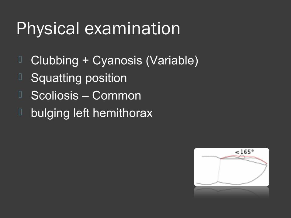
Physical examination
Clubbing + Cyanosis (Variable) Squatting position Scoliosis – Common bulging left hemithorax

Prominent “a” waves JVP Normal heart size
Mild parasternal impulse
Systolic trill (30%)

S1 normal S2 single
only A2 heardP2 soft & delayed: INAUDIBLE
MurmurShunt murmur (VSD) absentFlow murmur: Ejection systolic,
the smaller the flow the shorter the murmur
Ejection aortic click

Retinal engorgement Hemoptysis

ECG
ECG

ECG
ECG
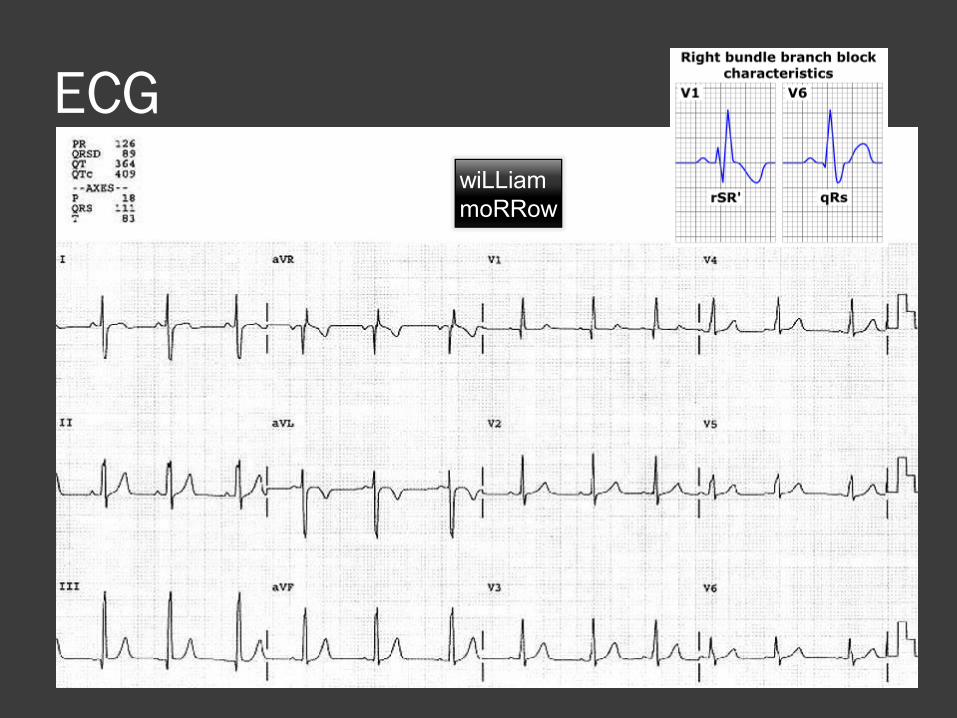
ECG
ECGwiLLiammoRRow

ECG
Right axis deviation (+120° to +150°) Right or combined ventricular
hypertrophy Right atrial hypertrophy Partial or complete right bundle branch
block (especially true of patients after surgical repair)
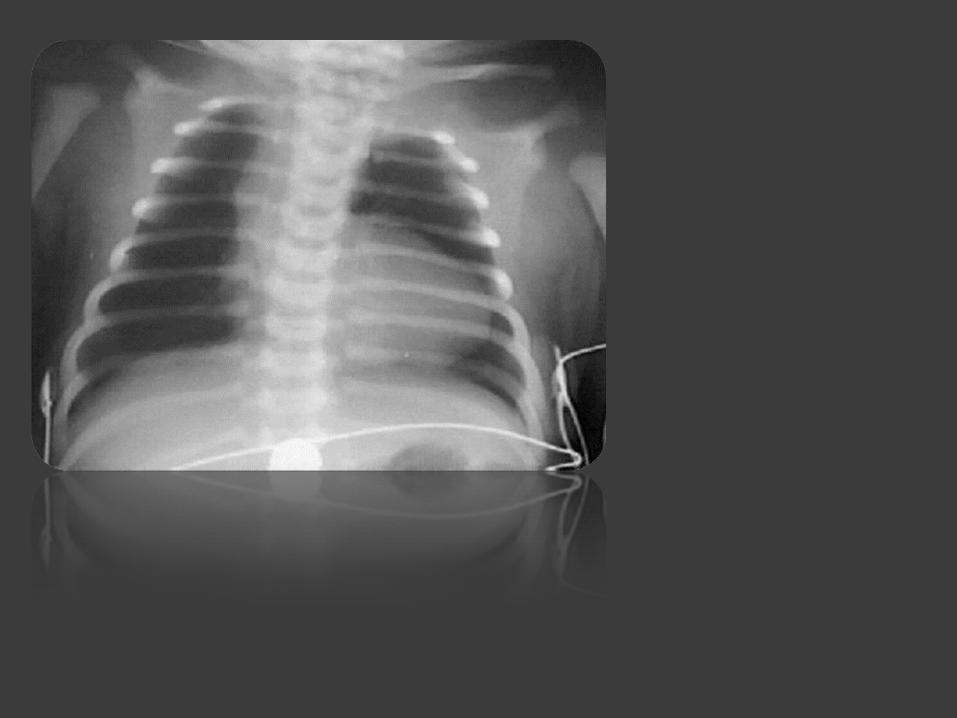

Coeur en sabot (boot-shaped heart)
secondary to uplifting of the cardiac apex from RVH
and the absence of a normal main pulmonary artery segment
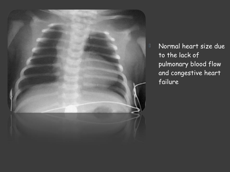
Normal heart size due to the lack of pulmonary blood flow and congestive heart failure

Decreased pulmonary vascularity

Right atrial enlargement
Right-sided aortic arch (20-25% of patients) with indentation of leftward-positioned tracheobronchial shadow

Echocardiography
Reveals a large VSD overriding aorta variable degrees of right ventricular
outflow tract (RVOT) obstruction

Course and Complication
1) Each anoxic spell is potentially fatal
2) Polycytemia1) Cerebrovascular thrombosis
3) Anoxic infaction of CNS1) Neurological complication

4) LUNG is an awesome filter. 1) Bypassing it may not be a good idea!2) TOF, venous blood from gut, peripheral
system by pass the lung and re-enter circulation
3) Hence TOF can cause:1) Brain Abcess2) Infective endocarditis3) Paradoxical embolism

Management of anoxic spell
1) Knee chest position
2) Humified O2
3) Be careful not to provoke the child 1) Especially you are bad at gaining IV access
2) Ask for help from someone more experience
3) Permit the baby to remain with mother
4) Morphine 0.1 -0.2 mg/Kg Subcutaneous
5) Correct acidosis – Sodium Bicarb IV

6) Propanolol1) 0.1mg/kg/IV during spells
2) 0.5 to 1.0 mg/kg/ 4-6hourly orally
7) Vasopressors: Methoxamine IM or IV drip
8) Correct anemia
9) GA is the last resort

Palliative Surgery
Blalock-Taussig shunt Pott procedure Waterston shunt
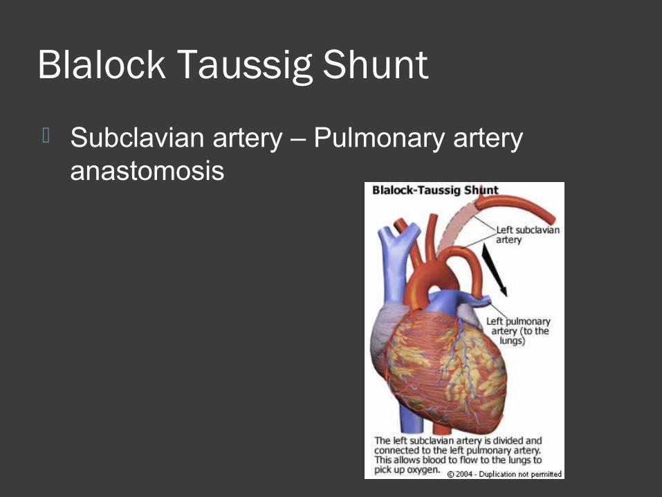
Blalock Taussig Shunt
Subclavian artery – Pulmonary artery anastomosis

Modified Blalock Taussig Shunt Goretex graft


Surgical Palliation

Palliative operation prolong life Increase exercise tolerance
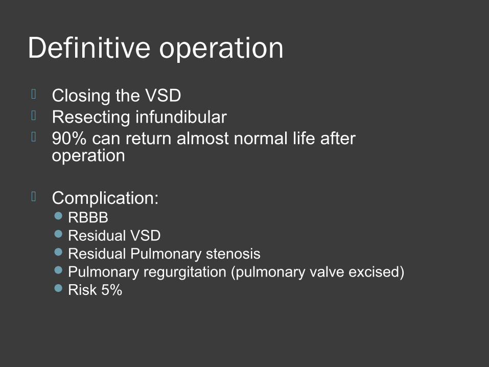
Definitive operation Closing the VSD Resecting infundibular 90% can return almost normal life after
operation
Complication:RBBBResidual VSDResidual Pulmonary stenosisPulmonary regurgitation (pulmonary valve excised)Risk 5%

Transposition of Great Areries (TGA)
Aorta originating from the right ventricle, and pulmonary artery originating from the left ventricle
Accounts for 5-7% of all congenital heart disease

TGA Survival is dependent on the presence of
mixing between the pulmonary and systemic circulation
Atrial septal defect is essential for survival 50% of patients have a VSD Usually presents in the first day of life with
profound cyanosis More common in boys

TGA• Exam :
• cyanosis in an otherwise healthy looking baby
• Loud S2 ( aorta is anterior )
• CXR : • Egg on side• Narrow
mediastinum

TGA .. Acute Management
PGE-1 with no supplemental O2 Maintain ductus arteriosus patency, this will
increase the effective pulmonary blood flow, and thence increase the left atrial pressure, therefore inhance the left to right shunt at the atrial level
Balloon atrial septostomyLife saving procedure in the presence of
inadequate atrial septal defect
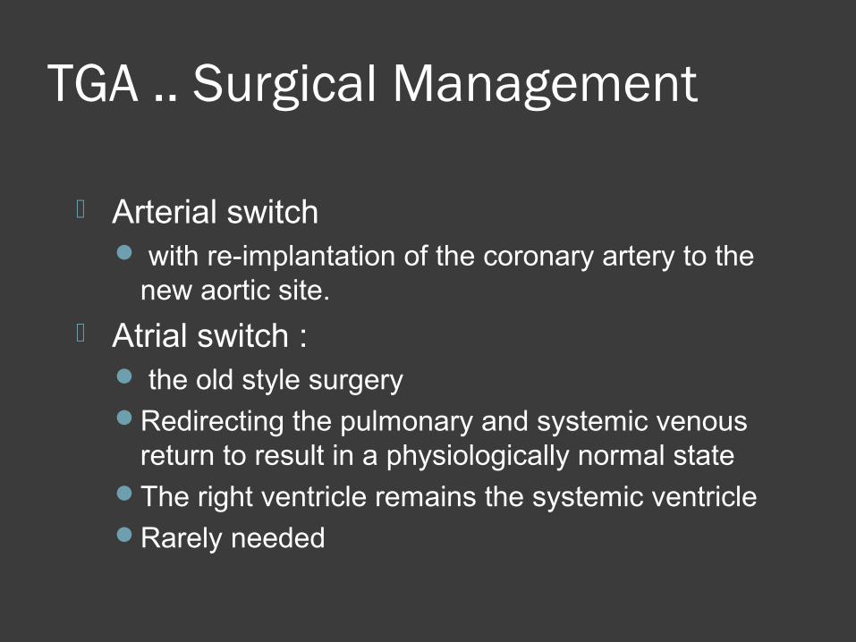
TGA .. Surgical Management
Arterial switch with re-implantation of the coronary artery to the
new aortic site.
Atrial switch : the old style surgeryRedirecting the pulmonary and systemic venous
return to result in a physiologically normal stateThe right ventricle remains the systemic ventricleRarely needed

Truncus Arteriosus
The presence of a common trunk that supply the systemic, pulmonary and coronary circulation
Almost always associated with VSD
1.2-2.5% of all congenital heart disease
Truncus Arteriosus
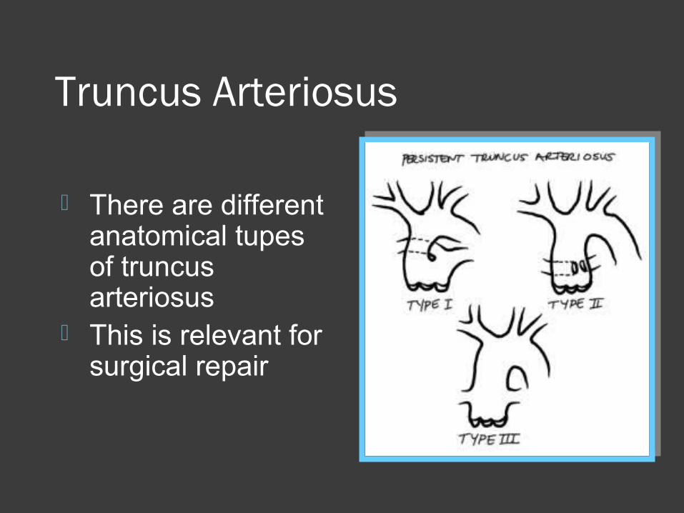
There are different anatomical tupes of truncus arteriosus
This is relevant for surgical repair

Truncus Arteriosus Generally patients have increased
pulmonary blood flow Degree of cyanosis is mild and may not
be evident clinically until late stage with pulmonary vascular disease
Presenting feature is congestive heart failure (tachypnia, hepatomegally)
Truncus Arteriosus Exam is significant for
Single S2Ejection click of the abnormal truncal valve Systolic murmur of truncal valve stenosis if
presentDiaastolic murmur of truncal valve
insufficiencyGallop
CXR : Cardiomegally , increased pulmonary circulation

Managment Acute management
No O2 to minimize pulmonary blood flow DiureticsAfterload reduction to inhance systemic blood flow
•Surgical management: complete repair with VSD closure and conduit placement between the right ventricle and pulmonary arteries•Long term problems :
–truncal valve dysfunction–RV conduit obstruction
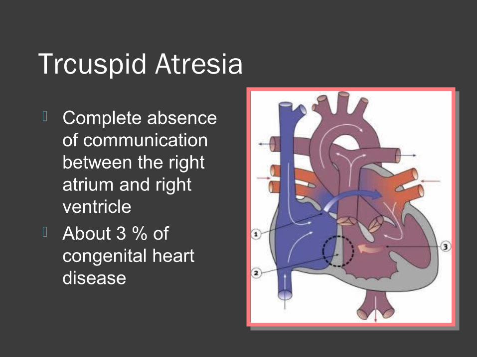
Trcuspid Atresia
Complete absence of communication between the right atrium and right ventricle
About 3 % of congenital heart disease

Tricuspid Atresia There is an obligate interatrial
communication Usually associated with VSD The pulmonary blood flow is dependent on
the size of the VSD Pulmonary blood flow can be increased or
decreased causing variable presenting symptoms
If there is no VSD ( also called Hypoplastic right ventricle) the pulmonary blood flow is dependent on the PDA

Tricuspid Atresia- presentation The presentation will depend on the
amount of pulmonary blood flowIf the PBF is decreased, the main presenting
symptom is cyanosisIf the PBF is increased the presentation is
that of congestive heart failure
CXR will also reflect the amount of pulmonary blood flow
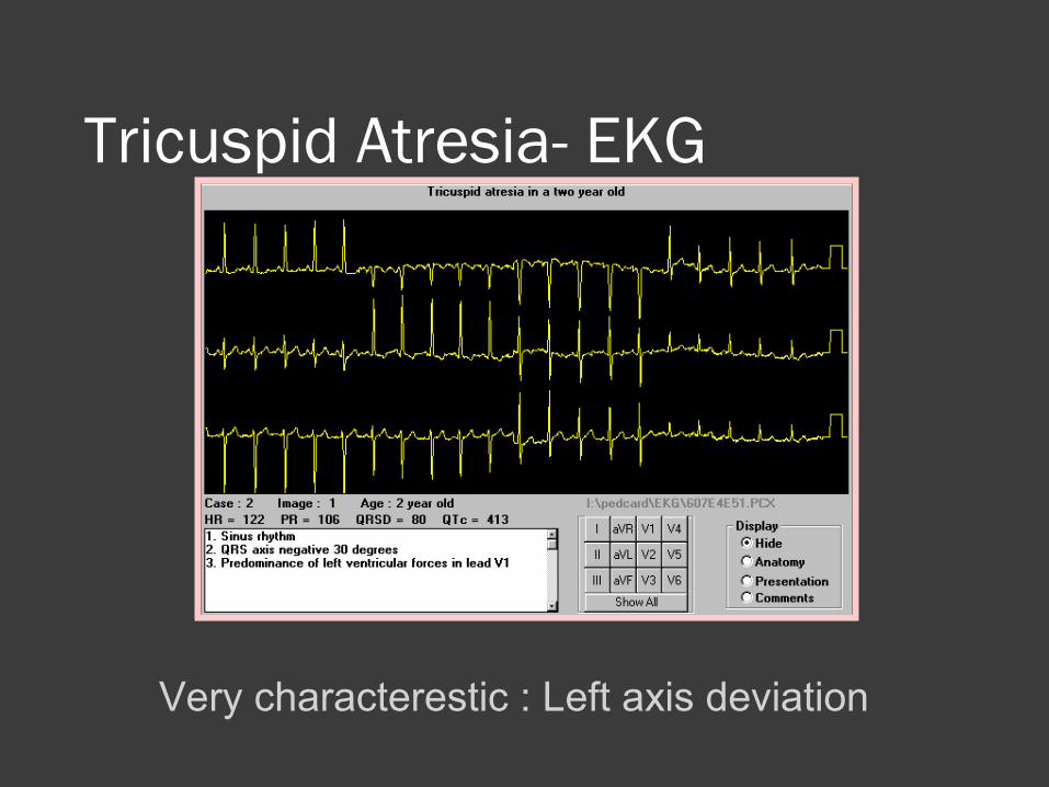
Tricuspid Atresia- EKG
Very characterestic : Left axis deviation
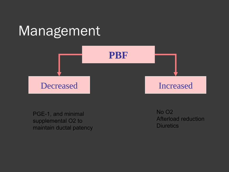
Management
PBF
Decreased Increased
PGE-1, and minimal supplemental O2 to maintain ductal patency
No O2Afterload reductionDiuretics
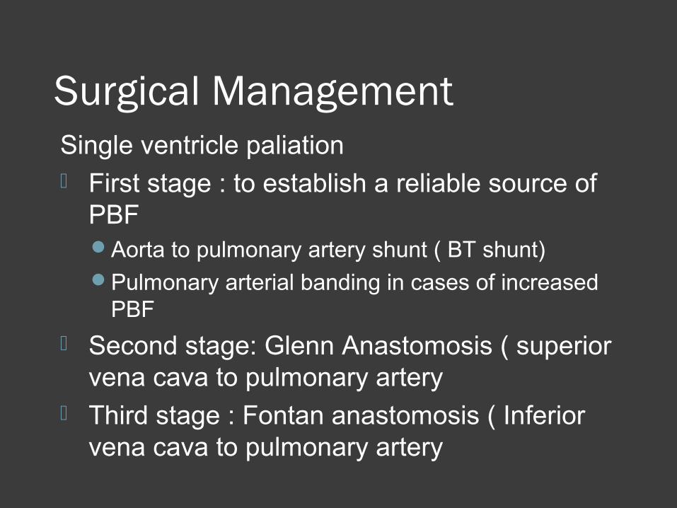
Surgical ManagementSingle ventricle paliation First stage : to establish a reliable source of
PBFAorta to pulmonary artery shunt ( BT shunt)Pulmonary arterial banding in cases of increased
PBF
Second stage: Glenn Anastomosis ( superior vena cava to pulmonary artery
Third stage : Fontan anastomosis ( Inferior vena cava to pulmonary artery

Total Anomalous Pulmonary Venous Return (TAPVR)

TAPVR- Infracardiac

Radiography

Infracardiac type


Thank You