Congenital Choroid Plexus Papilloma of the Third Ventricle ... · Congenital Choroid Plexus...
2
797 Congenital Choroid Plexus Papilloma of the Third Ventricle: Diagnosis with Real-Time Sonography and MR Imaging Kurt P. Schellhas,1 Richard C. Siebert,2 Kenneth B. Heithoff,1 and Ralph A. Franciosi 3 Congenital neoplasms of the choroid plexus are uncom- mon. A case of a surgically resected congenital third-ventric- ular choroid plexus papilloma is presented to demonstrate the appearance of this tumor using real-time sonography and MR imaging. The sonographic features of this tumor are reviewed and compared with previous reports. The MR char- acteristics are correlated with sonographic findings, with gross pathologic features, and with histology. A review of choroid plexus papilloma presenting within the neonatal period is also provided. Case Report A 3920-g male infant was born at 41 .5 weeks gestation by spon- taneous vaginal delivery. Physical examination was unremarkable except for moderate enlargement of the head circumference. Neuro- logic evaluation was normal. Real-time sonography revealed marked enlargement of the lateral ventricles, with a homogeneous, echogenic mass occupying the enlarged third ventricle and obstructing the cerebral aqueduct (Figs. 1A and 18). An unenhanced CT scan re- vealed a homogeneously dense mass within the third ventricle (im- ages were degraded by motion). The diagnosis of choroid plexus papilloma was suggested from the combined sonographic and CT findings. MR , performed with the baby under general anesthesia, sharply defined the third-ventricular neoplasm and allowed a confident preoperative diagnosis of choroid plexus papilloma (Figs. 1 C and 10). A right frontal craniotomy was performed. Right frontal corticotomy and lateral ventricular approach, using 6x loupes, revealed a pink , soft, friable, papillary neoplasm filling the enlarged third ventricle and bulging into the foramina of Monro. The tumor was easily removed from the third ventricle and appeared to be totally removed from its choroidal origin at the venous angle. Histologic evaluation revealed a typical choroid plexus papilloma (Fig . 1 E) . Postoperatively, the patient has been neurologically normal; how- ever, a left frontal ventriculoperitoneal shunt was required 6 weeks after initial surgery because of communicating hydrocephalus, dem- onstrated by sonography. Follow-up MR at 9 weeks, performed with intramuscular Nembutal sedation, demonstrated complete absence of residual tumor and some ventricular decompression (Fig . 1 F) . Discussion Choroid plexus papilloma is an uncommon epithelial neo- plasm of the CNS, accounting for 3-5% of brain tumors in Received December 11 , 1986; accepted after revision January 27, 1987. children [1-9]. Twenty percent of all choroid ple xus papillo- mas occur in patients under the age of 1 year. In a large series, these tumors represented 12 .5% of verified neoplasms of the CNS [2]. Previous reports of sonographic findings of choroid plexus papilloma describe highly echogenic intraventricular mass le- sions with irregular borders and associated hydrocephalus [10, 11]. Doppler findings have been described and may contribute significantly to the knowledge of tumor vascularity [10]. Although Doppler was not performed in our case, pul- satile vascular channels were observed within the mass dur- ing real-time sonography. At the time this paper was submitted for publication, there were no published reports of the MR appearance of choroid plexus papilloma in the neonate. The MR signal intensities in this case are entirely nonspecific, showing intermediate T1 and long T2 signal characteristics. The signal intensity f rom the neoplasm is quite uniform (Figs. 1C and 1 D) . Prominent vessels are seen within the tumor mass, proved at su rge ry to be venous channels draining posteriorly into the internal cer- ebral veins . The presence of marked third-ventricular expansion around the intraventricular neoplasm provides a major clue toward the diagnosis of choroid plexus papilloma ( Fig s. 18- 1D). The differenti al diagnosis of intraventricular masses in the neonate must include hematomas and other less common lesions, such as astrocytoma, ependymoma, colloid cyst, and meningioma [2, 4]. MR will subst an tially enhance ou r abi li ty to differentiate these specific lesions through a combinat ion of morphology and signal inte nsi ty. Certai nly, hematomas and colloid cysts will have strikin gl y different signal characteristics and appearances compared with other solid lesions. Cerebral arteriography may provide important di agnostic clues in diffi- cult cases [4] . Postoperative follow-up sonography is a valu abl e exami- nation that can be performed wi thout sedati on , with a mini- mum of patient discomfort and expense . In light of cost factors and risks to t he pati en t , sonography is cl early the examination of choice for ini t ial screeni ng of the neonate and for routine follow-u p. MR may be used whenever a mass is identified, in cases of occult hydrocephalus, or whenever more detailed , Center for Diagnostic Imaging, 5775 Wayzata Blvd ., Sui te 190, St. Louis Park, MN 5541 6. Address reprint requests to K. P. Schell has . 2 Medi cal Arts Bldg. , Suite 91 1, Nicoll ete and Ninth , Minneapolis , MN 55402. 3 Department of Pathology, Chi ldren's Medical Center, 2525 Chicago Ave., South, Mi nneapoli s, MN 55404. AJNR 9:797-798, July/August 19880195- 61 08/88/0905-0797 10 Ameri can Society of Neuroradiology
Transcript of Congenital Choroid Plexus Papilloma of the Third Ventricle ... · Congenital Choroid Plexus...
797
Congenital Choroid Plexus Papilloma of the Third Ventricle: Diagnosis with Real-Time Sonography and MR Imaging Kurt P. Schellhas,1 Richard C. Siebert,2 Kenneth B. Heithoff,1 and Ralph A. Franciosi3
Congenital neoplasms of the choroid plexus are uncommon. A case of a surgically resected congenital third-ventricular choroid plexus papilloma is presented to demonstrate the appearance of this tumor using real-time sonography and MR imaging. The sonographic features of this tumor are reviewed and compared with previous reports . The MR characteristics are correlated with sonographic findings, with gross pathologic features, and with histology. A review of choroid plexus papilloma presenting within the neonatal period is also provided.
Case Report
A 3920-g male infant was born at 41 .5 weeks gestation by spontaneous vaginal delivery. Physical examination was unremarkable except for moderate enlargement of the head circumference. Neurologic evaluation was normal. Real-time sonography revealed marked enlargement of the lateral ventricles, with a homogeneous, echogenic mass occupying the enlarged third ventricle and obstructing the cerebral aqueduct (Figs . 1 A and 18). An unenhanced CT scan revealed a homogeneously dense mass within the third ventricle (images were degraded by motion). The diagnosis of choroid plexus papilloma was suggested from the combined sonographic and CT findings. MR, performed with the baby under general anesthesia, sharply defined the third-ventricular neoplasm and allowed a confident preoperative diagnosis of choroid plexus papilloma (Figs. 1 C and 10).
A right frontal craniotomy was performed. Right frontal corticotomy and lateral ventricular approach, using 6x loupes, revealed a pink , soft, friable, papillary neoplasm filling the enlarged third ventricle and bulging into the foramina of Monro. The tumor was easily removed from the third ventricle and appeared to be totally removed from its choroidal origin at the venous angle. Histologic evaluation revealed a typical choroid plexus papilloma (Fig . 1 E).
Postoperatively, the patient has been neurologically normal; however, a left frontal ventriculoperitoneal shunt was required 6 weeks after initial surgery because of communicating hydrocephalus, demonstrated by sonography. Follow-up MR at 9 weeks, performed with intramuscular Nembutal sedation , demonstrated complete absence of residual tumor and some ventricular decompression (Fig . 1 F).
Discussion
Choroid plexus papilloma is an uncommon epithelial neoplasm of the CNS, accounting for 3-5% of brain tumors in
Received December 11 , 1986; accepted after revision January 27, 1987.
children [1-9]. Twenty percent of all choroid plexus papillomas occur in patients under the age of 1 year. In a large series, these tumors represented 12.5% of verified neoplasms of the CNS [2].
Previous reports of sonographic findings of choroid plexus papilloma describe highly echogenic intraventricular mass lesions with irregular borders and associated hydrocephalus [10, 11]. Doppler findings have been described and may contribute significantly to the knowledge of tumor vascularity [10]. Although Doppler was not performed in our case, pulsatile vascular channels were observed within the mass during real-time sonography.
At the time this paper was submitted for publication, there were no published reports of the MR appearance of choroid plexus papilloma in the neonate. The MR signal intensities in this case are entirely nonspecific, showing intermediate T1 and long T2 signal characteristics. The signal intensity from the neoplasm is quite uniform (Figs. 1 C and 1 D). Prominent vessels are seen within the tumor mass, proved at surgery to be venous channels draining posteriorly into the internal cerebral veins .
The presence of marked third-ventricular expansion around the intraventricular neoplasm provides a major clue toward the diagnosis of choroid plexus papilloma (Figs. 18- 1 D).
The differential diagnosis of intraventricular masses in the neonate must include hematomas and other less common lesions, such as astrocytoma, ependymoma, colloid cyst , and meningioma [2, 4] . MR will substantially enhance our ability to differentiate these specific lesions through a combination of morphology and signal intensity. Certainly, hematomas and colloid cysts will have strikingly different signal characteristics and appearances compared with other solid lesions. Cerebral arteriography may provide important diagnostic clues in difficult cases [4] .
Postoperative follow-up sonography is a valuable examination that can be performed without sedation , with a minimum of patient discomfort and expense. In light of cost factors and risks to the patient , sonography is clearly the examination of choice for initial screening of the neonate and for routine follow-up. MR may be used whenever a mass is identified, in cases of occult hydrocephalus, or whenever more detailed
, Center for Diagnostic Imaging, 5775 Wayzata Blvd ., Suite 190, St. Louis Park, MN 5541 6. Address reprint requests to K. P. Schellhas. 2 Medical Arts Bldg. , Suite 91 1, Nicollete and Ninth, Minneapolis, MN 55402. 3 Department of Pathology, Children's Medical Center, 2525 Chicago Ave., South, Minneapolis, MN 55404.
AJNR 9:797-798, July/August 19880195-61 08/88/0905-0797 10 American Society of Neuroradiology
o E F
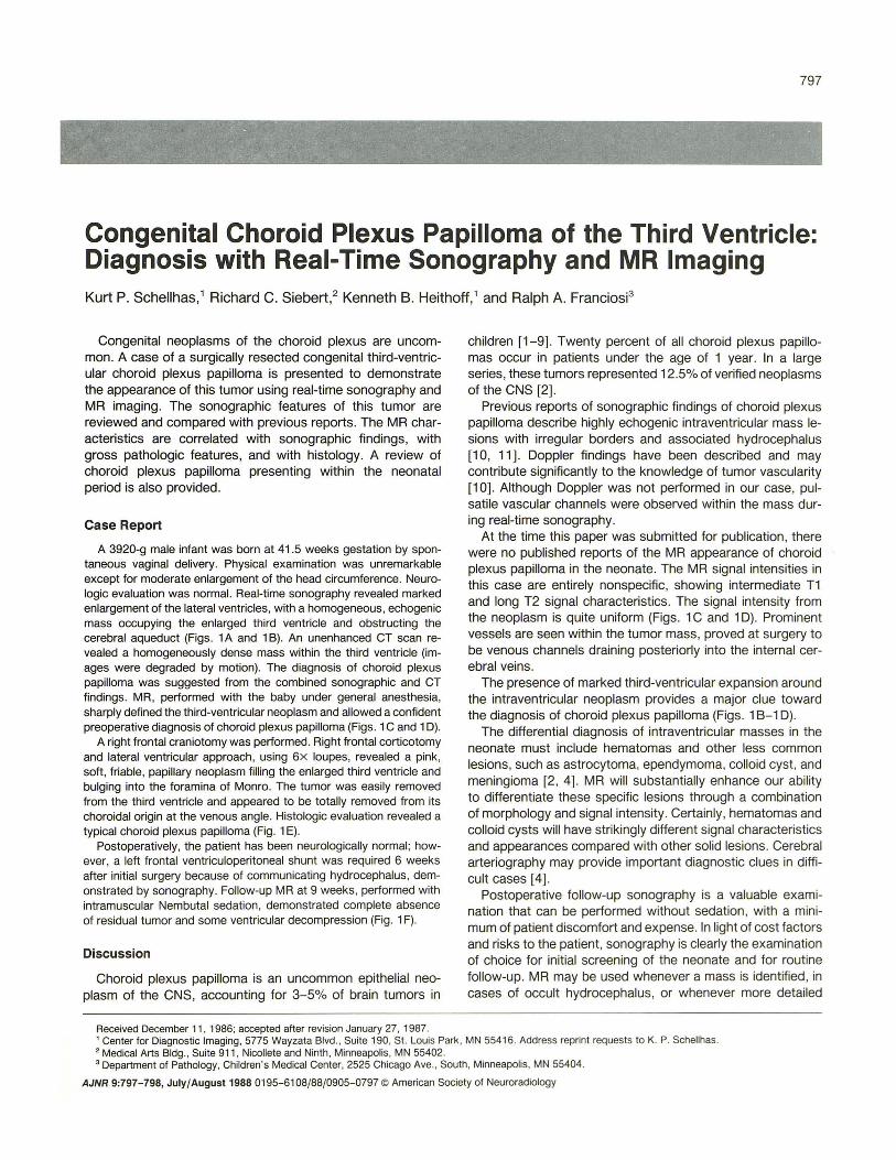
Fig. 1.-A and 8, Coronal (A) and midline sagittal (8) sonographic images obtained with a 5-MHz transducer (Diasonics, Inc., Milpitas, CAl. A homogeneous, echogenic mass (M) lies within enlarged third ventricle. The mass exhibits a similar echo pattern to the lateral ventricular choroid (arrow in A). Note CSF (arrows in 8) that lies above and below mass, further distending the enlarged third ventricle, separating mass from roof and floor of ventricle. Note marked lateral ventricular enlargement (A).
C, Spin-echo 8-mm-thick coronal MR image (2500/28, 90/2), 256 x 256 matrix. Image obtained on a Siemens Magnatom 1.0-T MR unit (Iselin, NJ) demonstrates a multilobulated, homogeneous mass (open arrows in C) within enlarged third ventricle, associated with hydrocephalus. Note sharply defined vascular structure (curved arrow) within lesion, which proved to be a venous channel at surgery. The lesion exhibits isointensity with basal ganglia on first echo and is hyperintense compared with brain on T2-weighted image.
D, T1-weighted (partial saturation technique) 4-mm-thick midline sagittal MR image (900/17/4), 256 x 256 matrix, shows lobulated mass (M, arrow) within dilated third ventricle. Note distention of third ventricle by CSF (white arrows), elevating roof and floor of third ventricle. Lesion deforms upper brainstem (black arrow) near entrance to cerebral aqueduct. Note normal-sized fourth ventricle and superior cerebellar cistern, implying aqueductal patency.
E, Photomicrograph demonstrating typical papillary fronds seen with choroid plexus papilloma (magnification x60). F, Postoperative MR study performed at 9 weeks shows partial ventricular decompression (compare with A-D) and complete absence of residual tumor.
T1-weighted (partial saturation technique) 3-mm-thick coronal (slightly rotated) MR image (800/20/4), 256 x 256 matrix, obtained on a 1.5-T magnet (General Electric, Milwaukee, WI) demonstrates smooth third-ventricular walls and a ventricular shunt (large arrow) over left convexity. Left foramen of Monro (small arrow) remains enlarged.
information than sonography can provide is required. MR can be performed successfully with oral , rectal , or intramuscular sedation in most circumstances.
ACKNOWLEDGMENTS
Sonographic images (Figs. 1 A and 18) were provided by Frank E. Mork, M.D. , Department of Radiology , Methodist Hospital , St. Louis Park, MN.
REFERENCES 1. Abramson N, Raben M, Cavanaugh PJ. Brain tumors in children: analysis
of 136 cases. Radiology 1974;11 2:669-672 2. Jooma R, Hayward RD, Grant ON. Intracranial neoplasms during the first
year of life: analysis of 100 consecutive cases. Neurosurgery 1984;14(1 ):31 - 41
3. Kendall B, Reider-Grosswasser I, Valentine A. Diagnosis of masses presenting within the ventricles on computed tomography. Neuroradiology
1983;25:1 1-22 4. Morrison G, Sobel OF, Kelley WM , Norman D. Intraventricular mass lesions.
Radiology 1984;153:435-442 5. Gradin WC, Taylon C, Fruin AH . Choroid plexus papilloma of the third
ventricle: case report and review of the literature. Neurosurgery 1983;12(2):217-220
6. Pascual-Castroviejo I, Villarejo F, Perez-Higueras A, et al. Childhood choroid plexus neoplasms. Pediatrics 1983;140 :51-56
7. SChijman E. Letter to the editor. Child's Brain 1984;11 :349-352 8. Carter LP, Beggs J, Waggener JD. Ultra-structure of three choroid plexus
papillomas. Cancer 1972;30: 1130-1136 9. Tomasello F, Albanese V, Bemini F, Picozzi P. Choroid plexus papilloma
of the third ventricle. Surg Neuro/1981;16(1) :69-71 10. Chow PT, Horgan JG, Burns PN, Weltin G, Taylor KJW. Choroid plexus
papilloma: detection by real-time and Doppler sonography. AJNR 1986;7: 168-170
11 . Cappe IP. Lam AH. Ultrasound in the diagnosis of choroid plexus papilloma. J Clin Ultrasound 1985;13 :121-123


![First Trimester Anatomy Screening [Read-Only] · First Trimester Diagnosis Spina Bifida Midsagittal view of the fetal face 4tthh ventricle is between the brainstem and the choroid](https://static.fdocuments.net/doc/165x107/5f0569287e708231d412d3f6/first-trimester-anatomy-screening-read-only-first-trimester-diagnosis-spina-bifida.jpg)