Congenital anomalies of the vertebrae in dogsvdt.ugent.be/sites/default/files/ArtinEng07.pdfvolving...
14
Vlaams Diergeneeskundig Tijdschrift, 2017, 86 Continuing education 105 Congenital anomalies of the vertebrae in dogs Aangeboren afwijkingen van de wervels bij de hond L. De Rycke, J.H. Saunders Department of Small Animal Orthopedics and Medical Imaging, Faculty of Veterinary Medicine, Ghent University, Salisburylaan 133, 9820 Merelbeke, Belgium [email protected] CLASSIFICATION Spinal dysraphism Spinal dysraphism is a broad term given to a group of anomalies with malformations in the dorsum of the embryo, including neural tube defects (NTD). NTD occur secondary to abnormal closure of the develop- ing neural tube and result in a defective neural arch through which meninges or neural elements may her- niate (Figure 1). Spinal dysraphism are categorized clinically into open and closed, based on whether the abnormal nervous tissue is exposed to the environ- ment or covered by skin (Rossi et al., 2006, Song et al., 2016). BSTRACT In this review article, a overview is given of the congenital pathologies of vertebrae in the dog. These pathologies are common in dogs and can be divided in two major groups: neural tube defects or spinal dysraphism, such as spina bifida with or without meningocoele and dermal sinus tract, and congenital anomalies of the vertebral column. The latter group can be subdivided in malformations originating in the embryonic period of development, such as butterfly vertebra, mediolateral wedged vertebra and transitional vertebra, or in the foetal period, such as block vertebra and dorsoventral wedged vertebra. Congenital vertebral anomalies can be incidental findings on radiographs or CT, but sometimes they can be the underlying cause of a clinical, mostly neurological problem. Due to pressure on the spinal cord, symptoms, such as pelvic limb ataxia, paresis, loss of spinal reflexes, incontinence and atrophy of the pelvic limbs may occur. SAMENVATTING In dit artikel wordt een overzicht gegeven van aangeboren afwijkingen van de wervels bij honden. Vertebrale anomalieën komen geregeld voor bij deze dieren en kunnen onderverdeeld worden in twee grote groepen, namelijk spinaal dysrafisme zoals spina bifida met of zonder meningocoele en dermale sinus, en anomalieën van de vertebrale kolom. Deze laatste groep kan worden opgesplitst in malformaties die hun ontstaan vinden in de embryonale ontwikkelingsperiode, zoals vlinderwervels, medio-laterale wigwervels en transitionele wervels, of in de foetale periode, zoals blokwervels en dorsoventrale wigwervels. Aangeboren anomalieën van de wervels kunnen toevalsbevindingen zijn op RX of CT, maar soms zijn ze de onderliggende oorzaak van klinische, meestal neurologische problemen. Door druk op het ruggenmerg kunnen symptomen zoals ataxie van de achterste ledematen, parese, verlies van spinale reflexen, kyfose, lordose, scoliose, incontinentie en atrofie van de spieren van de achterste ledematen optreden. A Open spinal dysraphism or spina bifida aperta includes meningocoeles and myelomeningocoeles, whereby the spinal cord fails to fuse dorsally, and is open to the environment because it is not covered by skin. These severe dysraphisms in animal neonates usually result in death or early termination. Such enti- ties have rarely been reported in dogs (Chesney, 1973; Clayton and Boyd, 1983). Closed spinal dysraphism (CSD), also known as spina bifida occulta (SBO), includes various malfor- mations of the spine that are covered by skin. The simplest form of CSD is dorsal spina bifida and is usually clinically insignificant, with no or minor neu- ral anomalies (Figure 2). Occasionally, a subcutane- ous mass may be present in SBO, which consists of a
Transcript of Congenital anomalies of the vertebrae in dogsvdt.ugent.be/sites/default/files/ArtinEng07.pdfvolving...
Vlaams Diergeneeskundig Tijdschrift, 2017, 86 105Vlaams Diergeneeskundig Tijdschrift, 2017, 86 Continuing education 105
Congenital anomalies of the vertebrae in dogs
Aangeboren afwijkingen van de wervels bij de hond
L. De Rycke, J.H. Saunders
Department of Small Animal Orthopedics and Medical Imaging, Faculty of Veterinary Medicine,Ghent University, Salisburylaan 133, 9820 Merelbeke, Belgium
CLASSIFICATION
Spinal dysraphism
Spinal dysraphism is a broad term given to a group of anomalies with malformations in the dorsum of the embryo, including neural tube defects (NTD). NTD occur secondary to abnormal closure of the develop-ing neural tube and result in a defective neural arch through which meninges or neural elements may her-niate (Figure 1). Spinal dysraphism are categorized clinically into open and closed, based on whether the abnormal nervous tissue is exposed to the environ-ment or covered by skin (Rossi et al., 2006, Song et al., 2016).
BSTRACT
In this review article, a overview is given of the congenital pathologies of vertebrae in the dog. These pathologies are common in dogs and can be divided in two major groups: neural tube defects or spinal dysraphism, such as spina bifida with or without meningocoele and dermal sinus tract, and congenital anomalies of the vertebral column. The latter group can be subdivided in malformations originating in the embryonic period of development, such as butterfly vertebra, mediolateral wedged vertebra and transitional vertebra, or in the foetal period, such as block vertebra and dorsoventral wedged vertebra. Congenital vertebral anomalies can be incidental findings on radiographs or CT, but sometimes they can be the underlying cause of a clinical, mostly neurological problem. Due to pressure on the spinal cord, symptoms, such as pelvic limb ataxia, paresis, loss of spinal reflexes, incontinence and atrophy of the pelvic limbs may occur.
SAMENVATTING
In dit artikel wordt een overzicht gegeven van aangeboren afwijkingen van de wervels bij honden. Vertebrale anomalieën komen geregeld voor bij deze dieren en kunnen onderverdeeld worden in twee grote groepen, namelijk spinaal dysrafisme zoals spina bifida met of zonder meningocoele en dermale sinus, en anomalieën van de vertebrale kolom. Deze laatste groep kan worden opgesplitst in malformaties die hun ontstaan vinden in de embryonale ontwikkelingsperiode, zoals vlinderwervels, medio-laterale wigwervels en transitionele wervels, of in de foetale periode, zoals blokwervels en dorsoventrale wigwervels. Aangeboren anomalieën van de wervels kunnen toevalsbevindingen zijn op RX of CT, maar soms zijn ze de onderliggende oorzaak van klinische, meestal neurologische problemen. Door druk op het ruggenmerg kunnen symptomen zoals ataxie van de achterste ledematen, parese, verlies van spinale reflexen, kyfose, lordose, scoliose, incontinentie en atrofie van de spieren van de achterste ledematen optreden.
A
Open spinal dysraphism or spina bifida aperta includes meningocoeles and myelomeningocoeles, whereby the spinal cord fails to fuse dorsally, and is open to the environment because it is not covered by skin. These severe dysraphisms in animal neonates usually result in death or early termination. Such enti-ties have rarely been reported in dogs (Chesney, 1973; Clayton and Boyd, 1983).
Closed spinal dysraphism (CSD), also known as spina bifida occulta (SBO), includes various malfor-mations of the spine that are covered by skin. The simplest form of CSD is dorsal spina bifida and is usually clinically insignificant, with no or minor neu-ral anomalies (Figure 2). Occasionally, a subcutane-ous mass may be present in SBO, which consists of a
106 Vlaams Diergeneeskundig Tijdschrift, 2017, 86
meningocele (Parker et al., 1973; Wilson et al., 1979; Gómez et al., 2012; Song et al., 2014). Another form of SBO are dermal sinus tracts (Miller and Tobias, 2003, Kiviranta et al., 2011; Song et al., 2016).
SBO with meningocoele (MC), meningomyelocoele (MMC)
Moderate to severe forms of NTD, such as SBO with MMC or MC, are typically diagnosed in young animals. Although a congenital anomaly may be seen in any breed, certain breeds have been overrepresented with spina bifida occulta such as the English bulldogs (Parker and Byerly, 1973; Parker et al., 1973; Wilson et al., 1979; Fingeroth et al., 1989, Brunetti et al., 1993; Ployart et al., 2013) and the German shepherd dogs (Clayton, 1983; Song et al., 2014). A meningo-coele has also been described in a Collie (Wilson et al., 1979) (Figure 3). Bulldogs have been reported with L7 or sacral dorsal vertebral arch defects and subcu-taneous meningocoeles (Parker et al., 1973; Brunetti et al., 1993) midline caudal hair streaming, (Wilson et al., 1979) and apparent caudal tethering of the filum terminale by a fibrous cord (Fingeroth et al., 1989; Shamir et al., 2001). Although reports of attempted surgical repair are rare, three reports of bulldogs that had surgery included a dorsal surgical approach to the caudal spine, opening of the dura, and transection of a fibrous filum terminale, resulting in release of the cau-dal tether (Fingeroth et al., 1989; Shamir et al., 2001, Song et al., 2014). One dog was euthanatized two weeks after surgery, although some sensory improve-ment was evident (Fingeroth et al., 1989). The sec-ond bulldog showed a decreased incidence of urinary incontinence. Physical and neurologic examinations were otherwise unchanged from prior to surgery. Four months postoperatively, only rare incidences of urine dribbling, a tail that was no longer deviated, and no evidence of pain or discomfort at the previous surgical site was seen (Song et al., 2014). In the third report, the dog's gait and hindlimb strength improved gradu-ally over the following four months, by which time they appeared to be normal. Urinary dribbling and fecal incontinence persisted accompanied by hypo- algesia of the perineal area and tail (Shamir et al., 2001).
SBO with dermal sinus tracts
Dermal sinus tracts are a well-recognized form of SBO (Miller and Tobias, 2003). Dermal sinus tracts are tubular sacs extending ventrally from the dorsal midline of the skin into the underlying tissues, occur-ring most commonly in the cervical, cranial thoracic, and lumbosacral regions (Miller and Tobias, 2003). The most common breed affected is the Rhodesian ridgeback (Salmon et al., 2007; Hillbertz, 2005; Hill-bertz and Andersson, 2006). Other reported breeds include the shih tzu, the boxer, the Siberian husky
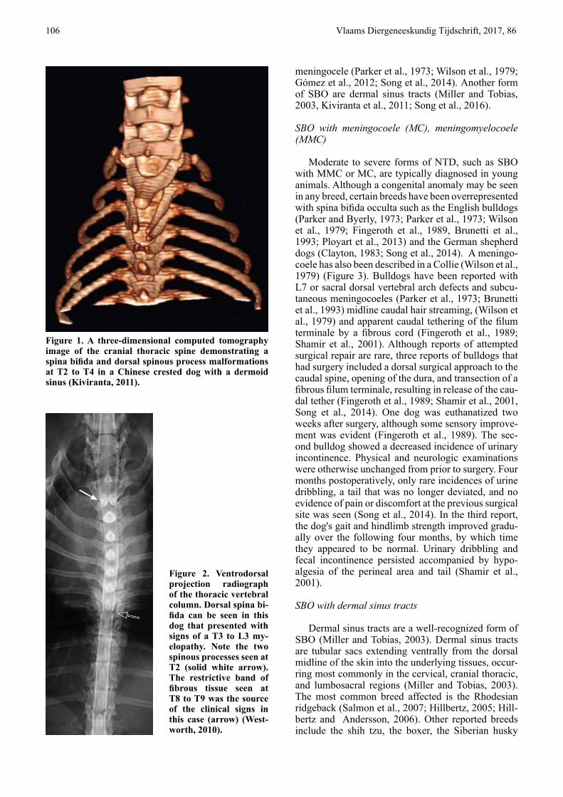
Figure 1. A three-dimensional computed tomography image of the cranial thoracic spine demonstrating a spina bifida and dorsal spinous process malformations at T2 to T4 in a Chinese crested dog with a dermoid sinus (Kiviranta, 2011).
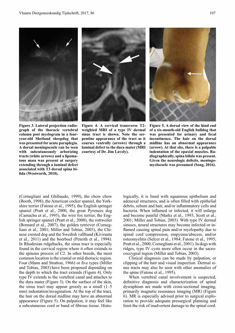
Figure 2. Ventrodorsal projection radiograph of the thoracic vertebral column. Dorsal spina bi-fida can be seen in this dog that presented with signs of a T3 to L3 my-elopathy. Note the two spinous processes seen at T2 (solid white arrow). The restrictive band of fibrous tissue seen at T8 to T9 was the source of the clinical signs in this case (arrow) (West-worth, 2010).
Vlaams Diergeneeskundig Tijdschrift, 2017, 86 107
(Cornegliani and Ghibaudo, 1999), the chow chow (Booth, 1998), the American cocker spaniel, the York-shire terrier (Fatone et al., 1995), the English springer spaniel (Pratt et al., 2000, the great Pyrenees dog (Camacho et al., 1995), the wire fox terrier, the Eng-lish springer spaniel (Pratt et al., 2000), the rottweiler (Bornard et al., 2007), the golden retriever (Corneg-liani et al., 2001; Miller and Tobias, 2003), the Chi-nese crested dog and the Swedish vallhund (Kiviranta et al., 2011) and the boerboel (Penrith et al., 1994). In Rhodesian ridgebacks, the sinus tract is especially found in the cervical region where it often extends to the spinous process of C2. In other breeds, the most common location is the cranial or mid-thoracic region. Four (Mann and Stratton, 1966) or five types (Miller and Tobias, 2003) have been proposed depending on the depth to which the tract extends (Figure 4). Only type IV extends to the vertebral canal and attaches to the dura mater (Figure 3). On the surface of the skin, the sinus tract may appear grossly as a small (1–5 mm) indentation/invagination. At the top of the tract, the hair on the dorsal midline may have an abnormal appearance (Figure 5). On palpation, it may feel like a subcutaneous cord or band of fibrous tissue. Histo-
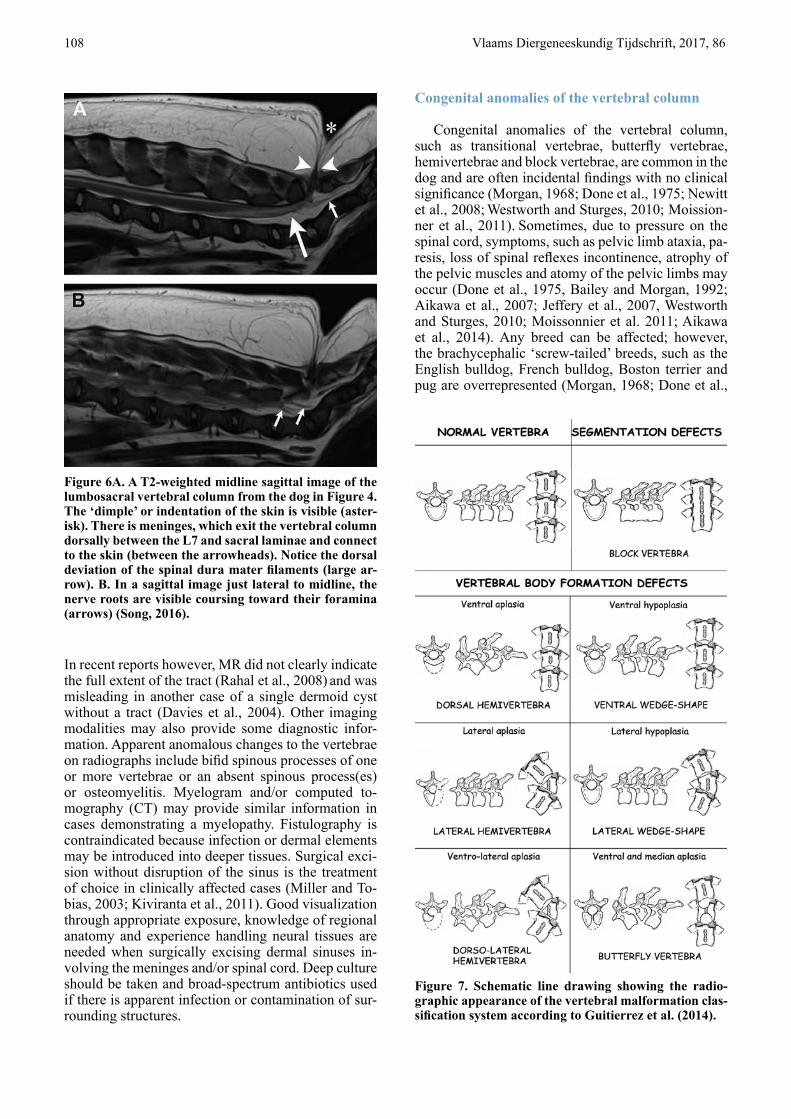
Figure 3. Lateral projection radio-graph of the thoracic vertebral column post myelogram in a four-year-old Shetland sheepdog that was presented for acute paraplegia. A dorsal meningocoele can be seen with subcutaneously arborizing tracts (white arrows) and a lipoma-tous mass was present at surgery extending through a laminal defect associated with T3 dorsal spina bi-fida (Westworth, 2010).
Figure 4. A cervical transverse T2-weighted MRI of a type IV dermal sinus tract is shown. Note the ser-pentine appearance of the tract as it courses ventrally (arrows) through a laminal defect to the dura mater (MRI courtesy of Dr. Jim Lavely).
Figure 5. A dorsal view of the hind end of a six-month-old English bulldog that was presented for urinary and fecal incontinence. The hair on the dorsal midline has an abnormal appearance (arrow). At that site, there is a palpable indentation of the epaxial muscles. Ra-diographically, spina bifida was present. Given the neurologic deficits, meningo-myelocoele was presumed (Song, 2016).
logically, it is lined with squamous epithelium and adenexal structures, and is often filled with epithelial debris, sebum and hair, and/or inflammatory cells and bacteria. When inflamed or infected, it will enlarge and become painful (Marks et al., 1993, Scott et al., 2001; Miller and Tobias, 2003). With type IV dermal sinuses, neural structures may become infected or in-flamed causing spinal pain and/or myelopathy due to spinal cord compression, empyema/abscess, and/or osteomyelitis (Selcer et al., 1984; Fatone et al., 1995; Pratt et al., 2000; Cornegliani et al., 2001). In dogs with ridges, type IV cysts more often occur in the sacro- coccygeal region (Miller and Tobias, 2003).
Clinical diagnosis can be made by palpation, or clipping of the hair and visual inspection. Dermal si-nus tracts may also be seen with other anomalies of the spine (Fatone et al., 1995).
When vertebral canal involvement is suspected, definitive diagnosis and characterization of spinal dysraphism are made with cross-sectional imaging, primarily magnetic resonance imaging (MR) (Figure 6). MR is especially advised prior to surgical explo-ration to provide adequate presurgical planning and limit the risk of inadvertent damage to the spinal cord.
108 Vlaams Diergeneeskundig Tijdschrift, 2017, 86
In recent reports however, MR did not clearly indicate the full extent of the tract (Rahal et al., 2008) and was misleading in another case of a single dermoid cyst without a tract (Davies et al., 2004). Other imaging modalities may also provide some diagnostic infor-mation. Apparent anomalous changes to the vertebrae on radiographs include bifid spinous processes of one or more vertebrae or an absent spinous process(es) or osteomyelitis. Myelogram and/or computed to-mography (CT) may provide similar information in cases demonstrating a myelopathy. Fistulography is contraindicated because infection or dermal elements may be introduced into deeper tissues. Surgical exci-sion without disruption of the sinus is the treatment of choice in clinically affected cases (Miller and To-bias, 2003; Kiviranta et al., 2011). Good visualization through appropriate exposure, knowledge of regional anatomy and experience handling neural tissues are needed when surgically excising dermal sinuses in-volving the meninges and/or spinal cord. Deep culture should be taken and broad-spectrum antibiotics used if there is apparent infection or contamination of sur-rounding structures.
Congenital anomalies of the vertebral column
Congenital anomalies of the vertebral column, such as transitional vertebrae, butterfly vertebrae, hemivertebrae and block vertebrae, are common in the dog and are often incidental findings with no clinical significance (Morgan, 1968; Done et al., 1975; Newitt et al., 2008; Westworth and Sturges, 2010; Moission-ner et al., 2011). Sometimes, due to pressure on the spinal cord, symptoms, such as pelvic limb ataxia, pa-resis, loss of spinal reflexes incontinence, atrophy of the pelvic muscles and atomy of the pelvic limbs may occur (Done et al., 1975, Bailey and Morgan, 1992; Aikawa et al., 2007; Jeffery et al., 2007, Westworth and Sturges, 2010; Moissonnier et al. 2011; Aikawa et al., 2014). Any breed can be affected; however, the brachycephalic ‘screw-tailed’ breeds, such as the English bulldog, French bulldog, Boston terrier and pug are overrepresented (Morgan, 1968; Done et al.,
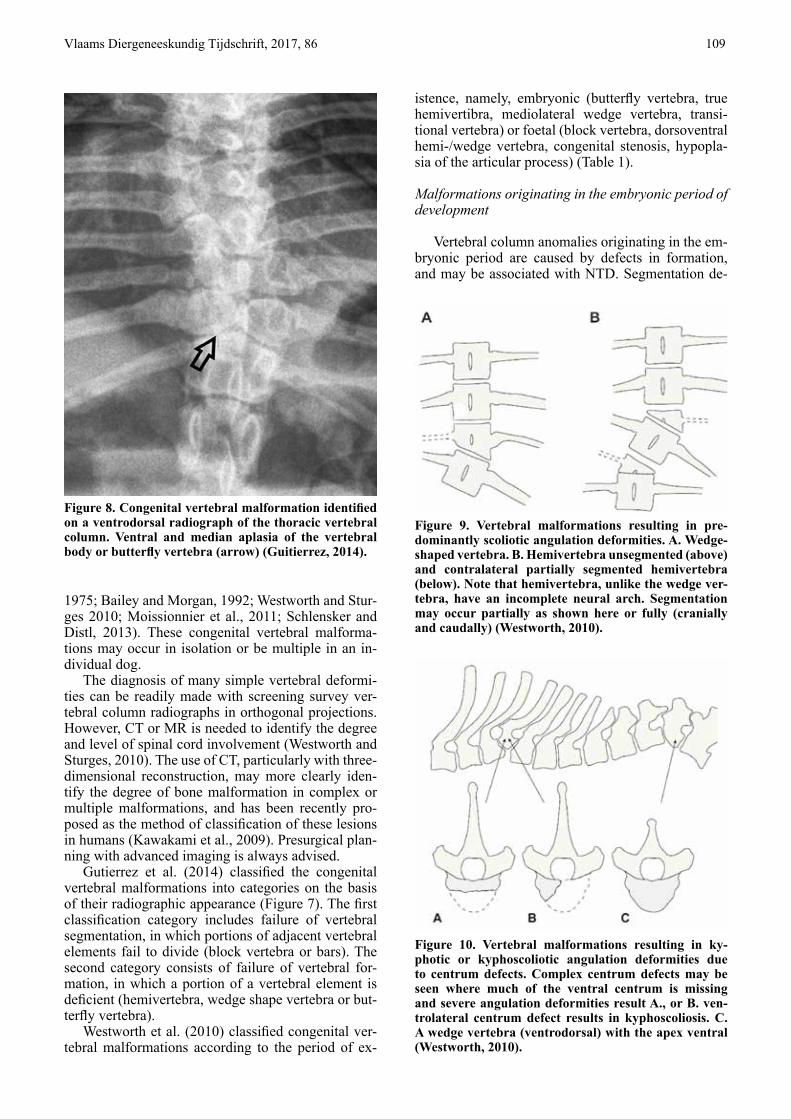
Figure 6A. A T2-weighted midline sagittal image of the lumbosacral vertebral column from the dog in Figure 4. The ‘dimple’ or indentation of the skin is visible (aster-isk). There is meninges, which exit the vertebral column dorsally between the L7 and sacral laminae and connect to the skin (between the arrowheads). Notice the dorsal deviation of the spinal dura mater filaments (large ar-row). B. In a sagittal image just lateral to midline, the nerve roots are visible coursing toward their foramina (arrows) (Song, 2016).
Figure 7. Schematic line drawing showing the radio-graphic appearance of the vertebral malformation clas-sification system according to Guitierrez et al. (2014).
Vlaams Diergeneeskundig Tijdschrift, 2017, 86 109
Figure 8. Congenital vertebral malformation identified on a ventrodorsal radiograph of the thoracic vertebral column. Ventral and median aplasia of the vertebral body or butterfly vertebra (arrow) (Guitierrez, 2014).
Figure 9. Vertebral malformations resulting in pre-dominantly scoliotic angulation deformities. A. Wedge-shaped vertebra. B. Hemivertebra unsegmented (above) and contralateral partially segmented hemivertebra (below). Note that hemivertebra, unlike the wedge ver-tebra, have an incomplete neural arch. Segmentation may occur partially as shown here or fully (cranially and caudally) (Westworth, 2010).
Figure 10. Vertebral malformations resulting in ky-photic or kyphoscoliotic angulation deformities due to centrum defects. Complex centrum defects may be seen where much of the ventral centrum is missing and severe angulation deformities result A., or B. ven-trolateral centrum defect results in kyphoscoliosis. C. A wedge vertebra (ventrodorsal) with the apex ventral (Westworth, 2010).
1975; Bailey and Morgan, 1992; Westworth and Stur-ges 2010; Moissionnier et al., 2011; Schlensker and Distl, 2013). These congenital vertebral malforma-tions may occur in isolation or be multiple in an in-dividual dog.
The diagnosis of many simple vertebral deformi-ties can be readily made with screening survey ver-tebral column radiographs in orthogonal projections. However, CT or MR is needed to identify the degree and level of spinal cord involvement (Westworth and Sturges, 2010). The use of CT, particularly with three-dimensional reconstruction, may more clearly iden-tify the degree of bone malformation in complex or multiple malformations, and has been recently pro-posed as the method of classification of these lesions in humans (Kawakami et al., 2009). Presurgical plan-ning with advanced imaging is always advised.
Gutierrez et al. (2014) classified the congenital vertebral malformations into categories on the basis of their radiographic appearance (Figure 7). The first classification category includes failure of vertebral segmentation, in which portions of adjacent vertebral elements fail to divide (block vertebra or bars). The second category consists of failure of vertebral for-mation, in which a portion of a vertebral element is deficient (hemivertebra, wedge shape vertebra or but-terfly vertebra).
Westworth et al. (2010) classified congenital ver-tebral malformations according to the period of ex-
istence, namely, embryonic (butterfly vertebra, true hemivertibra, mediolateral wedge vertebra, transi-tional vertebra) or foetal (block vertebra, dorsoventral hemi-/wedge vertebra, congenital stenosis, hypopla-sia of the articular process) (Table 1).
Malformations originating in the embryonic period of development
Vertebral column anomalies originating in the em-bryonic period are caused by defects in formation, and may be associated with NTD. Segmentation de-
110 Vlaams Diergeneeskundig Tijdschrift, 2017, 86
fects may occur secondarily. Those that involve only the vertebrae include butterfly vertebra and various forms of true or classic hemivertebra (Westworth and Sturges, 2010).
Butterfly vertebra
The term butterfly vertebra comes from the appear-ance of a sagittal cleft in the vertebral body on view-ing a ventrodorsal radiographic projection (Figure 8). There is partial or complete failure of formation of the ventral and central portions of the vertebral body, leaving two dorsolateral fragments of bone attached to the neural arch (Westworth and Sturges, 2010). If the vertebral body is very diminutive, it may result in kyphotic angulation, especially if associated with cen-trum hypoplasia. This anomaly is most often seen in brachycephalic, screw-tailed breeds and is often not clinically significant (Morgan, 1968; Gutierrez et al., 2014).
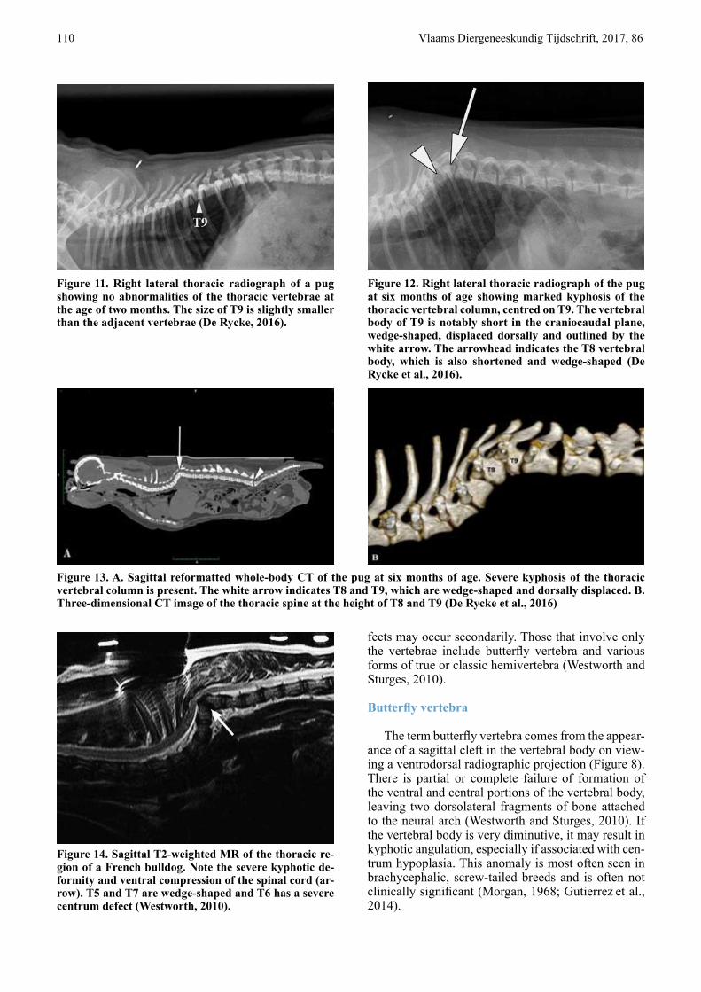
Figure 11. Right lateral thoracic radiograph of a pug showing no abnormalities of the thoracic vertebrae at the age of two months. The size of T9 is slightly smaller than the adjacent vertebrae (De Rycke, 2016).
Figure 12. Right lateral thoracic radiograph of the pug at six months of age showing marked kyphosis of the thoracic vertebral column, centred on T9. The vertebral body of T9 is notably short in the craniocaudal plane, wedge-shaped, displaced dorsally and outlined by the white arrow. The arrowhead indicates the T8 vertebral body, which is also shortened and wedge-shaped (De Rycke et al., 2016).
Figure 13. A. Sagittal reformatted whole-body CT of the pug at six months of age. Severe kyphosis of the thoracic vertebral column is present. The white arrow indicates T8 and T9, which are wedge-shaped and dorsally displaced. B. Three-dimensional CT image of the thoracic spine at the height of T8 and T9 (De Rycke et al., 2016)
Figure 14. Sagittal T2-weighted MR of the thoracic re-gion of a French bulldog. Note the severe kyphotic de-formity and ventral compression of the spinal cord (ar-row). T5 and T7 are wedge-shaped and T6 has a severe centrum defect (Westworth, 2010).
Vlaams Diergeneeskundig Tijdschrift, 2017, 86 111
Table 1. Classification of common congenital vertebral anomalies in dogs and cats (Tsou P.M., Yau A., Hodgson A.R., 1980).
Embryonic period
Diastematomyelia and centrum median cleft Centrum median cleft only (butterfly vertebrae) True hemivertebrae with or without segmentation deformity Wedge vertebra (mediolateral wedged vertebrae) with or without segmentation deformity Transitional vertebra (embryonic or fetal period depending on anomaly)
Fetal period
Failure of segmentation and or late formation Block vertebrae (partial or complete) Hypoplasia of articular processes Costovertebral joint failure of segmentation Centrum hypoplasia or aplasia (dorsoventral wedged vertebrae)
Complex anomalies are those not readily classifiable or unclassifiable, and may involve anymultiple defects of formation and segmentation.
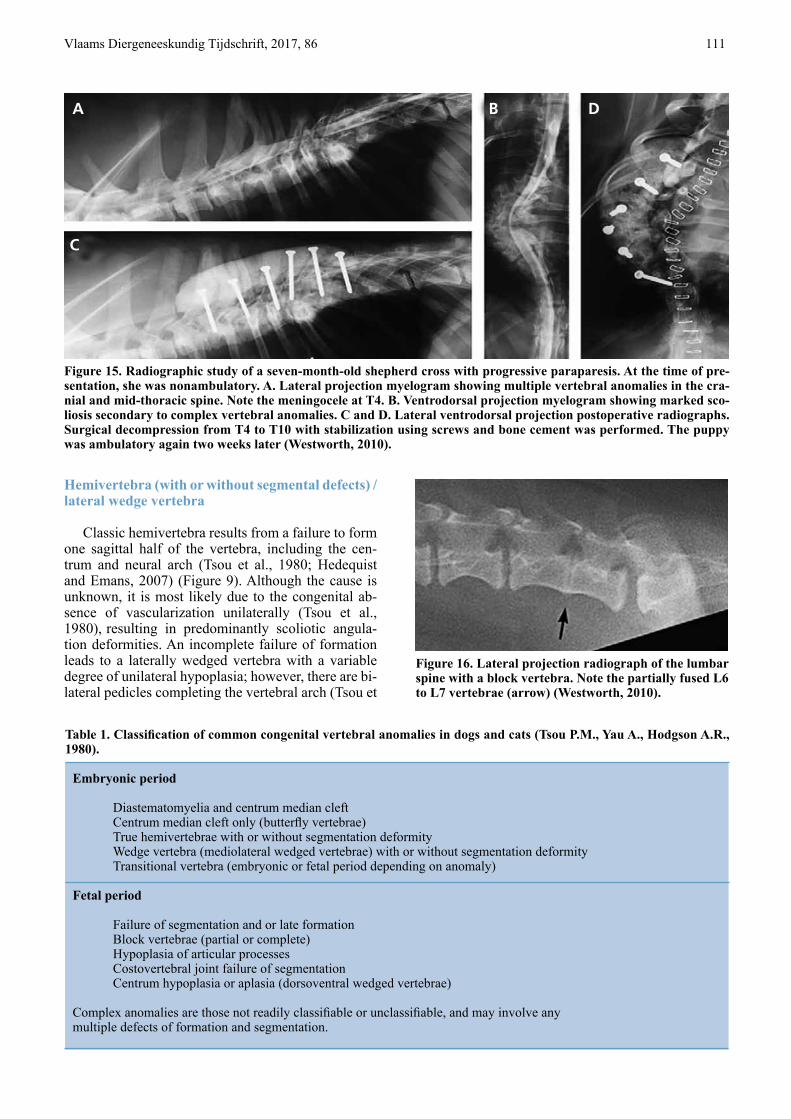
Figure 15. Radiographic study of a seven-month-old shepherd cross with progressive paraparesis. At the time of pre-sentation, she was nonambulatory. A. Lateral projection myelogram showing multiple vertebral anomalies in the cra-nial and mid-thoracic spine. Note the meningocele at T4. B. Ventrodorsal projection myelogram showing marked sco-liosis secondary to complex vertebral anomalies. C and D. Lateral ventrodorsal projection postoperative radiographs. Surgical decompression from T4 to T10 with stabilization using screws and bone cement was performed. The puppy was ambulatory again two weeks later (Westworth, 2010).
Figure 16. Lateral projection radiograph of the lumbar spine with a block vertebra. Note the partially fused L6 to L7 vertebrae (arrow) (Westworth, 2010).
Hemivertebra (with or without segmental defects) / lateral wedge vertebra
Classic hemivertebra results from a failure to form one sagittal half of the vertebra, including the cen-trum and neural arch (Tsou et al., 1980; Hedequist and Emans, 2007) (Figure 9). Although the cause is unknown, it is most likely due to the congenital ab-sence of vascularization unilaterally (Tsou et al., 1980), resulting in predominantly scoliotic angula-tion deformities. An incomplete failure of formation leads to a laterally wedged vertebra with a variable degree of unilateral hypoplasia; however, there are bi-lateral pedicles completing the vertebral arch (Tsou et
A B D
C
112 Vlaams Diergeneeskundig Tijdschrift, 2017, 86
al., 1980; Hedequist and Emans, 2007) (Figure 9A). Further classification of these anomalies is based on the degree of segmentation failure during the fetal pe-riod, resulting in the presence or absence of fusion to vertebral bodies cranially or caudally (Westworth and Sturges, 2010) (Figure 9B).
Defects of segmentation result in a concave angu-lation on the side of the lateral block with restricted growth, due to loss of growth plates on the affected side (Hedequist and Emans, 2007). Single or multiple ipsilateral anomalies may result in severe lateral an-gulation and hence in scoliosis. A hemi-metameric shift occurs if the angulation is counterbalanced with a contralateral hemivertebrae, with the resulting de-gree of scoliosis being less pronounced (Tsou et al., 1980). Mixed malformations may result in complex and not readily classifiable anomalies.
Malformations originating in the fetal period of de-velopment
Vertebral column anomalies originating in the fetal period are associated with defects in formation and particularly segmentation. Such deformities are well-differentiated and occur late in the chrondrification and ossification stages, and are much less commonly associated with other defects and spinal cord anoma-lies because they occur after the formation of those structures (Tsou et al., 1980). Anomalies originating in this period of development include dorsoventral wedged vertebrae (centrum hypoplasia or aplasia), blockvertebrae, articular facet aplasia/dysplasia, tran-sitional vertebrae and spinal stenosis (Tsou et al., 1980).
Vertebral anomalies of the centrum/ventral hemi- or wedge vertebra
Vertebral anomalies of the centrum result in pre-dominantly kyphotic angulation deformities caused by diminished vertebral body longitudinal growth. These anomalies affect the vertebral body only (un-less a mixed deformity), with normal or near nor-mal neural arch development (McMaster and Singh, 1999), and are caused by a defect in formation or seg-mentation (Tsou et al., 1980).
Centrum hypoplasia or aplasia results in variable loss of the body, and bilateral or unilateral defects may occur (Figure 10). Unilateral centrum defects may re-sult in a degree of scoliosis (kyphoscoliosis) (West-worth and Sturges, 2010) (Figure 10 B). The cause is unknown but severe forms may be due to congenital absence of vascularization or may be caused by any teratogenic insult to the very active cartilaginous pro-liferation in the ventral rim (Westworth and Sturges 2010; Moissionnier et al., 2011; Schlensker and Distl, 2013). The term hemivertebra is inappropriate for such malformations (Tsou, 1977). The resulting de-gree of kyphosis is related to the number of vertebrae
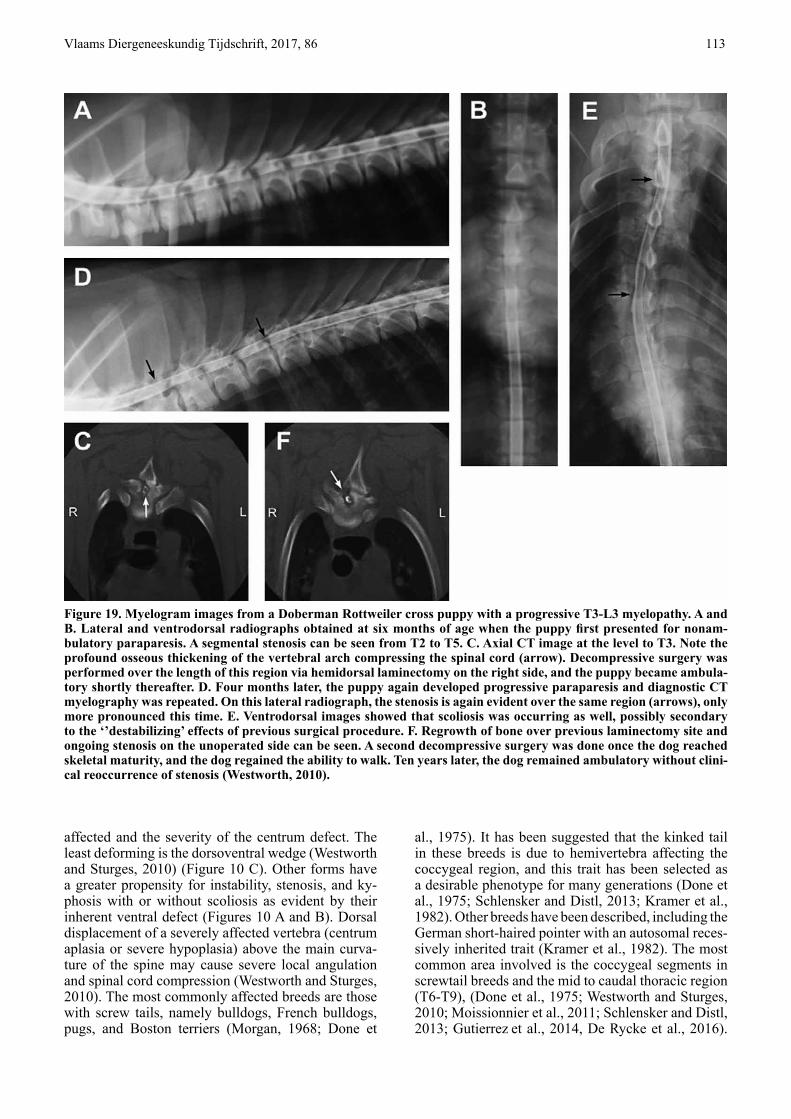
Figure 17. Ventrodorsal pro-jection radiograph of the thoracolumbar vertebral col-umn with a transitional ver-tebra. Note the missing costa and small perpendicular de-formed transverse process on the right side of T13 (arrow) (Westworth, 2010).
Figure 18. Ventrodorsal radiograph of a German shep-herd dog with lumbosacral transitional vertebra of type 2 on the right side (white arrow) and type 3 on the left side (black arrow). On the right side, the tip of the transverse process is visible; on the left side it is not (Lappalainen, 2012).
Vlaams Diergeneeskundig Tijdschrift, 2017, 86 113
affected and the severity of the centrum defect. The least deforming is the dorsoventral wedge (Westworth and Sturges, 2010) (Figure 10 C). Other forms have a greater propensity for instability, stenosis, and ky-phosis with or without scoliosis as evident by their inherent ventral defect (Figures 10 A and B). Dorsal displacement of a severely affected vertebra (centrum aplasia or severe hypoplasia) above the main curva-ture of the spine may cause severe local angulation and spinal cord compression (Westworth and Sturges, 2010). The most commonly affected breeds are those with screw tails, namely bulldogs, French bulldogs, pugs, and Boston terriers (Morgan, 1968; Done et
al., 1975). It has been suggested that the kinked tail in these breeds is due to hemivertebra affecting the coccygeal region, and this trait has been selected as a desirable phenotype for many generations (Done et al., 1975; Schlensker and Distl, 2013; Kramer et al., 1982). Other breeds have been described, including the German short-haired pointer with an autosomal reces-sively inherited trait (Kramer et al., 1982). The most common area involved is the coccygeal segments in screwtail breeds and the mid to caudal thoracic region (T6-T9), (Done et al., 1975; Westworth and Sturges, 2010; Moissionnier et al., 2011; Schlensker and Distl, 2013; Gutierrez et al., 2014, De Rycke et al., 2016).
Figure 19. Myelogram images from a Doberman Rottweiler cross puppy with a progressive T3-L3 myelopathy. A and B. Lateral and ventrodorsal radiographs obtained at six months of age when the puppy first presented for nonam-bulatory paraparesis. A segmental stenosis can be seen from T2 to T5. C. Axial CT image at the level to T3. Note the profound osseous thickening of the vertebral arch compressing the spinal cord (arrow). Decompressive surgery was performed over the length of this region via hemidorsal laminectomy on the right side, and the puppy became ambula-tory shortly thereafter. D. Four months later, the puppy again developed progressive paraparesis and diagnostic CT myelography was repeated. On this lateral radiograph, the stenosis is again evident over the same region (arrows), only more pronounced this time. E. Ventrodorsal images showed that scoliosis was occurring as well, possibly secondary to the ‘’destabilizing’ effects of previous surgical procedure. F. Regrowth of bone over previous laminectomy site and ongoing stenosis on the unoperated side can be seen. A second decompressive surgery was done once the dog reached skeletal maturity, and the dog regained the ability to walk. Ten years later, the dog remained ambulatory without clini-cal reoccurrence of stenosis (Westworth, 2010).
114 Vlaams Diergeneeskundig Tijdschrift, 2017, 86
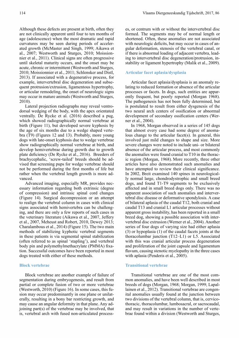
Although these defects are present at birth, often they are not clinically apparent until four to ten months of age (adolescence) when the most dramatic and rapid curvatures may be seen during periods of acceler-ated growth (McMaster and Singh, 1999; Aikawa et al., 2007; Westworth and Sturges, 2010; Moission-nier et al., 2011). Clinical signs are often progressive until skeletal maturity occurs, and the onset may be acute, chronic or intermittent (Westworth and Sturges, 2010; Moissionnier et al., 2011; Schlensker and Distl, 2013). If associated with a degenerative process, for example, intervertebral disc degeneration and subse-quent protrusion/extrusion, ligamentous hypertrophy, or articular remodeling, the onset of neurologic signs may occur in mature animals (Westworth and Sturges, 2010).
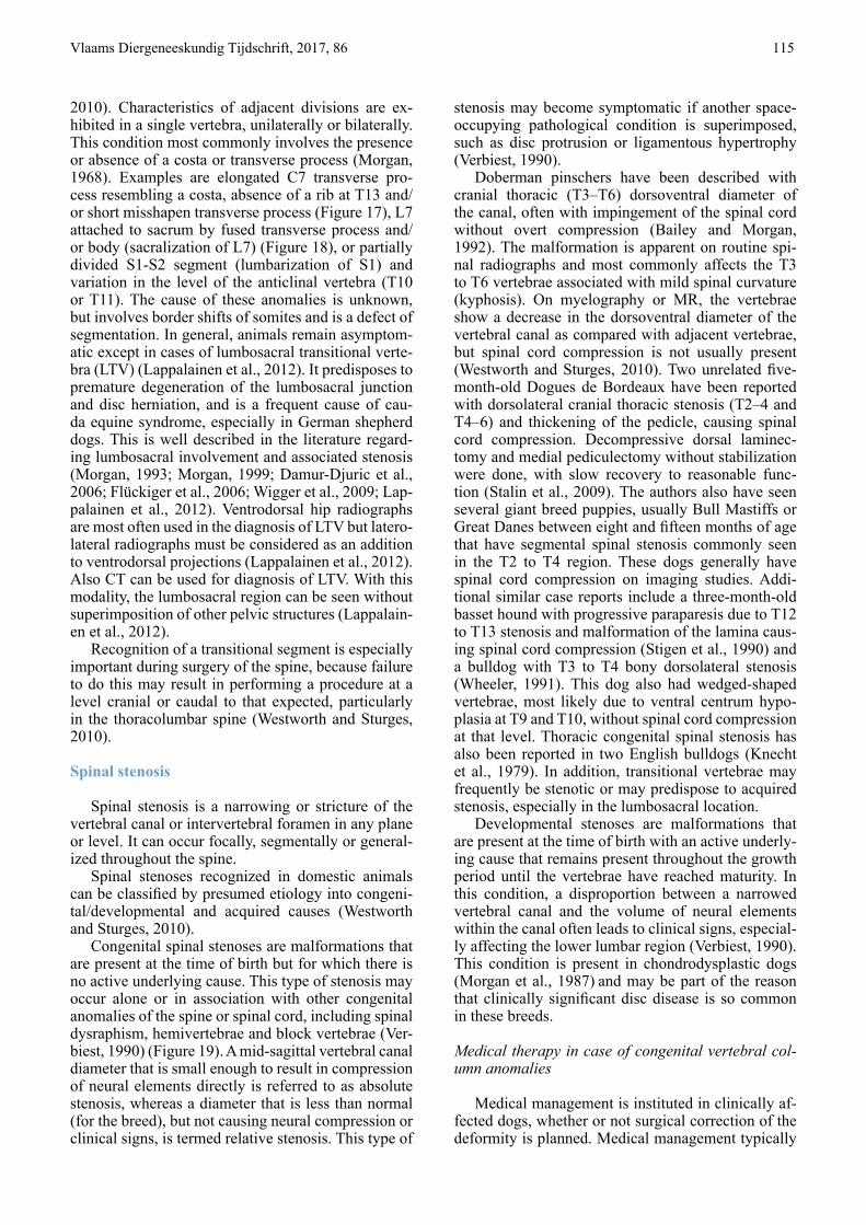
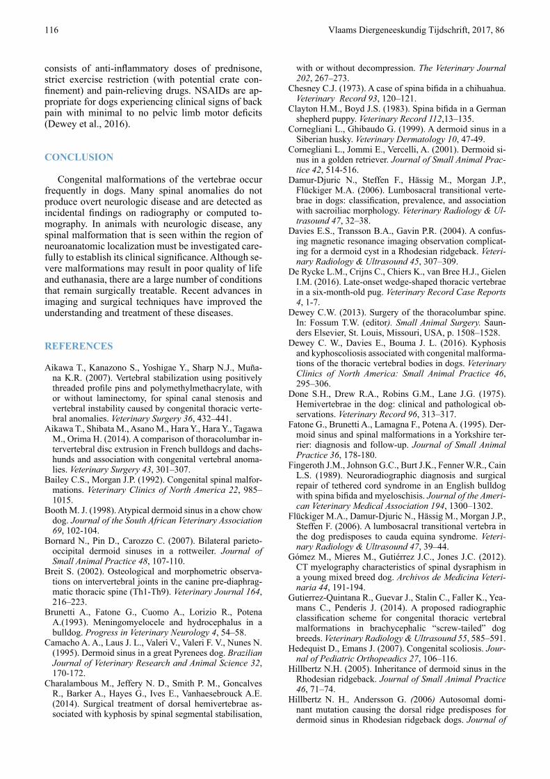
Lateral projection radiographs may reveal ventro-dorsal wedging of the body, with the apex orientated ventrally. De Rycke et al. (2016) described a pug, which showed radiographically normal vertebrae at birth (Figure 11), but developed severe kyphosis by the age of six months due to a wedge shaped verte-bra (T9) (Figures 12 and 13). Probably, more young dogs with late-onset kyphosis due to wedge vertebrae show radiographically normal vertebrae at birth, and develop hemivertebrae during growth due to growth plate deficiency (De Rycke et al., 2016). Breeders of brachycephalic, ‘screw-tailed’ breeds should be ad-vised that screening pups for wedge vertebrae should not be performed during the first months of life but rather when the vertebral length growth is more ad-vanced.
Advanced imaging, especially MR, provides nec-essary information regarding both extrinsic (degree of compression) and intrinsic spinal cord changes (Figure 14). Surgical decompression or an attempt to realign the vertebral column in cases with clinical signs associated with hemivertebra can be challeng-ing, and there are only a few reports of such cases in the veterinary literature (Aikawa et al., 2007, Jeffery et al., 2007, Meheust and Robert, 2010, Dewey 2013; Charalambous et al., 2014) (Figure 15). The two main methods of stabilizing kyphotic vertebral segments in these patients is via segmental spinal stabilization (often referred to as spinal ‘stapling’), and vertebral body pin and polymethylmethacrylate (PMMA) fixa-tion. Successful outcomes have been reported in most dogs treated with either of these methods.
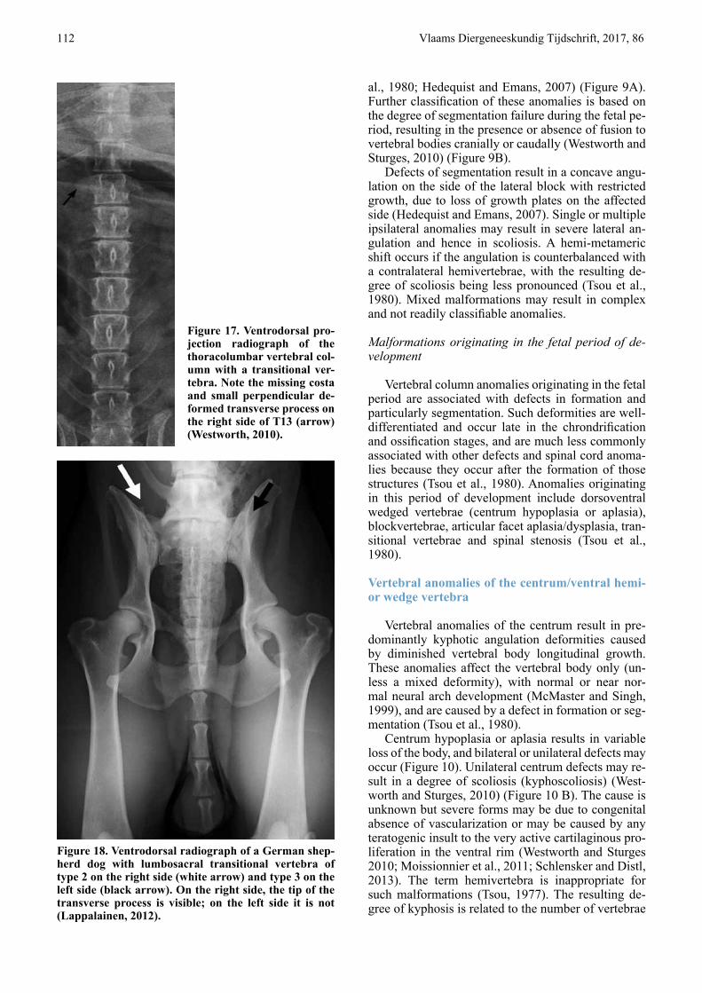
Block vertebrae
Block vertebrae are another example of failure of segmentation during embryogenesis, and result from partial or complete fusion of two or more vertebrae (Westworth, 2010) (Figure 16). In some cases, this fu-sion may occur predominantly in one plane or unilat-erally, resulting in a bony bar restricting growth, and may cause an angular deformity in that plane. Any ad-joining part(s) of the vertebrae may be involved, that is, vertebral arch with fused non-articulated process-
es, or centrum with or without the intervertebral disc formed. The segments may be of normal length or shortened. Often, these anomalies are not associated with neurologic deficits, but may occur in cases of an-gular deformation, stenosis of the vertebral canal, or if there is abnormal loading of adjacent vertebra, lead-ing to intervertebral disc degeneration/protrusion, in-stability or ligament hypertrophy (Malik et al., 2009).
Articular facet aplasia/dysplasia
Articular facet aplasia/dysplasia is an anomaly re-lating to reduced formation or absence of the articular processes or facets. In dogs, such entities are appar-ently frequent, but poorly reported (Morgan 1968). The pathogenesis has not been fully determined, but is postulated to result from either dysgenesis of the two neural arch centers of ossification or abnormal development of secondary ossification centers (Wer-ner et al., 2004).
In 1968, Morgan observed in a series of 145 dogs that almost every case had some degree of anoma-lous change to the articular facet(s). In general, this involved just mild changes to shape and size. More severe changes were noted to include uni- or bilateral absence of the articular process, and most commonly the anomalies were found cranial to T10 in the thorac-ic region (Morgan, 1968). More recently, three other articles have also demonstrated such anomalies and have attempted to review their clinical significance. In 2002, Breit examined 140 spines in neurological-ly normal large, chondrodystrophic and small breed dogs, and found T1-T9 segments to be exclusively affected and in small breed dogs only. There was no apparent association of such anomalies and interver-tebral disc disease or deformative spondylosis. A case of bilateral aplasia of the caudal T12, both cranial and caudal T13 and cranial L1 articular processes without apparent gross instability, has been reported in a small breed dog, showing a possible association with inter-vertebral disc extrusion (Werner et al., 2004). Another series of four dogs of varying size had either aplasia (3) or hypoplasia (1) of the caudal facets joints at the thoracolumbar junction (T12–L1) or L5. Associated with this was cranial articular process degeneration and proliferation of the joint capsule and ligamentum flavum, causing stenotic myelopathy in the three cases with aplasia (Penderis et al., 2005).
Transitional vertebrae
Transitional vertebrae are one of the most com-mon anomalies, and have been well described in most breeds of dogs (Morgan, 1968; Morgan, 1999; Lapal-lainen et al., 2012). Transitional vertebrae are congen-ital anomalies usually found at the junction between two divisions of the vertebral column, that is, cervico-thoracic, thoracolumbar, lumbosacral, or sacrocaudal, and may result in variations in the number of verte-brae found within a division (Westworth and Sturges,
Vlaams Diergeneeskundig Tijdschrift, 2017, 86 115
2010). Characteristics of adjacent divisions are ex-hibited in a single vertebra, unilaterally or bilaterally. This condition most commonly involves the presence or absence of a costa or transverse process (Morgan, 1968). Examples are elongated C7 transverse pro-cess resembling a costa, absence of a rib at T13 and/or short misshapen transverse process (Figure 17), L7 attached to sacrum by fused transverse process and/or body (sacralization of L7) (Figure 18), or partially divided S1-S2 segment (lumbarization of S1) and variation in the level of the anticlinal vertebra (T10 or T11). The cause of these anomalies is unknown, but involves border shifts of somites and is a defect of segmentation. In general, animals remain asymptom-atic except in cases of lumbosacral transitional verte-bra (LTV) (Lappalainen et al., 2012). It predisposes to premature degeneration of the lumbosacral junction and disc herniation, and is a frequent cause of cau-da equine syndrome, especially in German shepherd dogs. This is well described in the literature regard-ing lumbosacral involvement and associated stenosis (Morgan, 1993; Morgan, 1999; Damur-Djuric et al., 2006; Flückiger et al., 2006; Wigger et al., 2009; Lap-palainen et al., 2012). Ventrodorsal hip radiographs are most often used in the diagnosis of LTV but latero-lateral radiographs must be considered as an addition to ventrodorsal projections (Lappalainen et al., 2012). Also CT can be used for diagnosis of LTV. With this modality, the lumbosacral region can be seen without superimposition of other pelvic structures (Lappalain-en et al., 2012).
Recognition of a transitional segment is especially important during surgery of the spine, because failure to do this may result in performing a procedure at a level cranial or caudal to that expected, particularly in the thoracolumbar spine (Westworth and Sturges, 2010).
Spinal stenosis
Spinal stenosis is a narrowing or stricture of the vertebral canal or intervertebral foramen in any plane or level. It can occur focally, segmentally or general-ized throughout the spine.
Spinal stenoses recognized in domestic animals can be classified by presumed etiology into congeni-tal/developmental and acquired causes (Westworth and Sturges, 2010).
Congenital spinal stenoses are malformations that are present at the time of birth but for which there is no active underlying cause. This type of stenosis may occur alone or in association with other congenital anomalies of the spine or spinal cord, including spinal dysraphism, hemivertebrae and block vertebrae (Ver-biest, 1990) (Figure 19). A mid-sagittal vertebral canal diameter that is small enough to result in compression of neural elements directly is referred to as absolute stenosis, whereas a diameter that is less than normal (for the breed), but not causing neural compression or clinical signs, is termed relative stenosis. This type of
stenosis may become symptomatic if another space-occupying pathological condition is superimposed, such as disc protrusion or ligamentous hypertrophy (Verbiest, 1990).
Doberman pinschers have been described with cranial thoracic (T3–T6) dorsoventral diameter of the canal, often with impingement of the spinal cord without overt compression (Bailey and Morgan, 1992). The malformation is apparent on routine spi-nal radiographs and most commonly affects the T3 to T6 vertebrae associated with mild spinal curvature (kyphosis). On myelography or MR, the vertebrae show a decrease in the dorsoventral diameter of the vertebral canal as compared with adjacent vertebrae, but spinal cord compression is not usually present (Westworth and Sturges, 2010). Two unrelated five-month-old Dogues de Bordeaux have been reported with dorsolateral cranial thoracic stenosis (T2–4 and T4–6) and thickening of the pedicle, causing spinal cord compression. Decompressive dorsal laminec-tomy and medial pediculectomy without stabilization were done, with slow recovery to reasonable func-tion (Stalin et al., 2009). The authors also have seen several giant breed puppies, usually Bull Mastiffs or Great Danes between eight and fifteen months of age that have segmental spinal stenosis commonly seen in the T2 to T4 region. These dogs generally have spinal cord compression on imaging studies. Addi-tional similar case reports include a three-month-old basset hound with progressive paraparesis due to T12 to T13 stenosis and malformation of the lamina caus-ing spinal cord compression (Stigen et al., 1990) and a bulldog with T3 to T4 bony dorsolateral stenosis (Wheeler, 1991). This dog also had wedged-shaped vertebrae, most likely due to ventral centrum hypo-plasia at T9 and T10, without spinal cord compression at that level. Thoracic congenital spinal stenosis has also been reported in two English bulldogs (Knecht et al., 1979). In addition, transitional vertebrae may frequently be stenotic or may predispose to acquired stenosis, especially in the lumbosacral location.
Developmental stenoses are malformations that are present at the time of birth with an active underly-ing cause that remains present throughout the growth period until the vertebrae have reached maturity. In this condition, a disproportion between a narrowed vertebral canal and the volume of neural elements within the canal often leads to clinical signs, especial-ly affecting the lower lumbar region (Verbiest, 1990).
This condition is present in chondrodysplastic dogs (Morgan et al., 1987) and may be part of the reason that clinically significant disc disease is so common in these breeds.
Medical therapy in case of congenital vertebral col-umn anomalies
Medical management is instituted in clinically af-fected dogs, whether or not surgical correction of the deformity is planned. Medical management typically
116 Vlaams Diergeneeskundig Tijdschrift, 2017, 86
consists of anti-inflammatory doses of prednisone, strict exercise restriction (with potential crate con-finement) and pain-relieving drugs. NSAIDs are ap-propriate for dogs experiencing clinical signs of back pain with minimal to no pelvic limb motor deficits (Dewey et al., 2016).
CONCLUSION
Congenital malformations of the vertebrae occur frequently in dogs. Many spinal anomalies do not produce overt neurologic disease and are detected as incidental findings on radiography or computed to-mography. In animals with neurologic disease, any spinal malformation that is seen within the region of neuroanatomic localization must be investigated care-fully to establish its clinical significance. Although se-vere malformations may result in poor quality of life and euthanasia, there are a large number of conditions that remain surgically treatable. Recent advances in imaging and surgical techniques have improved the understanding and treatment of these diseases.
REFERENCES
Aikawa T., Kanazono S., Yoshigae Y., Sharp N.J., Muña-na K.R. (2007). Vertebral stabilization using positively threaded profile pins and polymethylmethacrylate, with or without laminectomy, for spinal canal stenosis and vertebral instability caused by congenital thoracic verte-bral anomalies. Veterinary Surgery 36, 432–441.
Aikawa T., Shibata M., Asano M., Hara Y., Hara Y., Tagawa M., Orima H. (2014). A comparison of thoracolumbar in-tervertebral disc extrusion in French bulldogs and dachs-hunds and association with congenital vertebral anoma-lies. Veterinary Surgery 43, 301–307.
Bailey C.S., Morgan J.P. (1992). Congenital spinal malfor-mations. Veterinary Clinics of North America 22, 985–1015.
Booth M. J. (1998). Atypical dermoid sinus in a chow chow dog. Journal of the South African Veterinary Association 69, 102-104.
Bornard N., Pin D., Carozzo C. (2007). Bilateral parieto-occipital dermoid sinuses in a rottweiler. Journal of Small Animal Practice 48, 107-110.
Breit S. (2002). Osteological and morphometric observa-tions on intervertebral joints in the canine pre-diaphrag-matic thoracic spine (Th1-Th9). Veterinary Journal 164, 216–223.
Brunetti A., Fatone G., Cuomo A., Lorizio R., Potena A.(1993). Meningomyelocele and hydrocephalus in a bulldog. Progress in Veterinary Neurology 4, 54–58.
Camacho A. A., Laus J. L., Valeri V., Valeri F. V., Nunes N. (1995). Dermoid sinus in a great Pyrenees dog. Brazilian Journal of Veterinary Research and Animal Science 32, 170-172.
Charalambous M., Jeffery N. D., Smith P. M., Goncalves R., Barker A., Hayes G., Ives E., Vanhaesebrouck A.E. (2014). Surgical treatment of dorsal hemivertebrae as-sociated with kyphosis by spinal segmental stabilisation,
with or without decompression. The Veterinary Journal 202, 267–273.
Chesney C.J. (1973). A case of spina bifida in a chihuahua. Veterinary Record 93, 120–121.
Clayton H.M., Boyd J.S. (1983). Spina bifida in a German shepherd puppy. Veterinary Record 112,13–135.
Cornegliani L., Ghibaudo G. (1999). A dermoid sinus in a Siberian husky. Veterinary Dermatology 10, 47-49.
Cornegliani L., Jommi E., Vercelli, A. (2001). Dermoid si-nus in a golden retriever. Journal of Small Animal Prac-tice 42, 514-516.
Damur-Djuric N., Steffen F., Hässig M., Morgan J.P., Flückiger M.A. (2006). Lumbosacral transitional verte-brae in dogs: classification, prevalence, and association with sacroiliac morphology. Veterinary Radiology & Ul-trasound 47, 32–38.
Davies E.S., Transson B.A., Gavin P.R. (2004). A confus-ing magnetic resonance imaging observation complicat-ing for a dermoid cyst in a Rhodesian ridgeback. Veteri-nary Radiology & Ultrasound 45, 307–309.
De Rycke L.M., Crijns C., Chiers K., van Bree H.J., Gielen I.M. (2016). Late-onset wedge-shaped thoracic vertebrae in a six-month-old pug. Veterinary Record Case Reports 4, 1-7.
Dewey C.W. (2013). Surgery of the thoracolumbar spine. In: Fossum T.W. (editor). Small Animal Surgery. Saun-ders Elsevier, St. Louis, Missouri, USA, p. 1508–1528.
Dewey C. W., Davies E., Bouma J. L. (2016). Kyphosis and kyphoscoliosis associated with congenital malforma-tions of the thoracic vertebral bodies in dogs. Veterinary Clinics of North America: Small Animal Practice 46, 295–306.
Done S.H., Drew R.A., Robins G.M., Lane J.G. (1975). Hemivertebrae in the dog: clinical and pathological ob-servations. Veterinary Record 96, 313–317.
Fatone G., Brunetti A., Lamagna F., Potena A. (1995). Der-moid sinus and spinal malformations in a Yorkshire ter-rier: diagnosis and follow-up. Journal of Small Animal Practice 36, 178-180.
Fingeroth J.M., Johnson G.C., Burt J.K., Fenner W.R., Cain L.S. (1989). Neuroradiographic diagnosis and surgical repair of tethered cord syndrome in an English bulldog with spina bifida and myeloschisis. Journal of the Ameri-can Veterinary Medical Association 194, 1300–1302.
Flückiger M.A., Damur-Djuric N., Hässig M., Morgan J.P., Steffen F. (2006). A lumbosacral transitional vertebra in the dog predisposes to cauda equina syndrome. Veteri-nary Radiology & Ultrasound 47, 39–44.
Gómez M., Mieres M., Gutiérrez J.C., Jones J.C. (2012). CT myelography characteristics of spinal dysraphism in a young mixed breed dog. Archivos de Medicina Veteri-naria 44, 191-194.
Gutierrez-Quintana R., Guevar J., Stalin C., Faller K., Yea-mans C., Penderis J. (2014). A proposed radiographic classification scheme for congenital thoracic vertebral malformations in brachycephalic “screw-tailed” dog breeds. Veterinary Radiology & Ultrasound 55, 585–591.
Hedequist D., Emans J. (2007). Congenital scoliosis. Jour-nal of Pediatric Orthopeadics 27, 106–116.
Hillbertz N.H. (2005). Inheritance of dermoid sinus in the Rhodesian ridgeback. Journal of Small Animal Practice 46, 71–74.
Hillbertz N. H., Andersson G. (2006) Autosomal domi-nant mutation causing the dorsal ridge predisposes for dermoid sinus in Rhodesian ridgeback dogs. Journal of
Vlaams Diergeneeskundig Tijdschrift, 2017, 86 117
Small Animal Practice 47, 184–188.Hillbertz N.H., Isaksson M., Karlsson E.K. (2007). Dupli-
cation of FGF3, FGF4, FGF19 and ORAOV1 causes hair ridge and predisposition to dermoid sinus in ridgeback dogs. Nature Genetics 19, 1318–1320.
Jeffery N.D., Smith P.M., Talbot C.E. (2007). Imaging find-ings and surgical treatment of hemivertebrae in 3 dogs. Journal of the American Veterinary Medical Association 230, 532–536.
Kawakami N., Tsuji T., Imagama S. (2009). Classification of congenital scoliosis and kyphosis: a new approach to the three-dimensional classification for progressive ver-tebral anomalies requiring operative treatment. Spine 41, 1756–1765.
Kiviranta A-M., Lappalainen A.K., Hagner K., Jokinen T. (2011). Dermoid sinus and spina bifida in three dogs and a cat. Journal of Small Animal Practice 52, 319–324.
Knecht C.D., Blevins W.E., Raffe M.R. (1979). Stenosis of the thoracic spinal canal in English bulldogs. Journal of the American Animal Hospital Association 15, 181–183.
Kramer J. W., Schiffer S. P., Sande R. D., Rantanen N. W., Whitener E. K. (1982). Characterization of heritable tho-racic hemivertebra of the German shorthaired pointer. Journal of the American Veterinary Medical Association 181, 814–815.
Lappalainen A.K. , Salomaa R., Junnila J., Snellman M., Laitinen-Vapaavuori O. (2012). Alternative classification and screening protocol for transitional lumbosacral ver-tebra in German shepherd dogs. Acta Veterinaria Scan-dinavica 54, 1-10.
Malik Y., Konar M., Wernick M., Howart J., Forterre F. (2009). Chronic intervertebral disk herniation associated with fused vertebrae treated by lateral corpectomy in a cat. Veterinary Comparative Orthopaedics and Trauma-tology 22, 170–173.
Mann G.E., Stratton J. (1966). Dermoid sinus in the Rho-desian ridgeback. Journal of Small Animal Practice 7, 631–642.
Marks S. L., Harari J., Dernell, W.S. (1993). Dermoid sinus in a Rhodesian ridgeback. Journal of Small Animal Prac-tice 34, 356-358.
McMaster M.J., Singh H. (1999). Natural history of con-genital kyphosis and kyphoscoliosis. The Journal of Bone and Joint Surgery. American Volume 81, 1367–1383.
Meheust P., Robert R. (2010). Surgical treatment of a hemi-vertebra by partial ventral corpectomy and fusion in a labrador puppy. Veterinary and Comparative Orthopae-dics and Traumatology 23, 262–265.
Miller L, Tobias K. (2003). Dermoid sinuses: description, diagnosis and treatment. Compendium on Continuing Education for the Practicing Veterinarian 25, 295–299.
Moissonnier P., Gossot P., Scotti S. (2011). Thoracic ky-phosis associated with hemivertebra. Veterinary Surgery 40, 1029–1032.
Morgan J.P. (1968). Congenital anomalies of the vertebral column of the dog: a study of the incidence and signifi-cance based on a radiographic and morphologic study. Journal of the American Veterinary Radiology Society 9, 21–29.
Morgan J.P., Atilola M., Bailey C.S. (1987). Vertebral ca-nal and spinal cord mensuration: a comparative study of its effect on lumbosacral myelography in the Dachshund and German shepherd dog. Journal of the American Vet-erinary Medical Association 191, 951–957.
Morgan J.P. , Bahr A., Franti C.E., Bailey C.S. (1993).
Lumbosacral transitional vertebrae as a predisposing cause of cauda equina syndrome in German shepherd dogs: 161 cases (1987-1990). Journal of the American Veterinary Medical Association 202, 1877-1882.
Morgan J.P. (1999). Transitional lumbosacral vertebral anomaly in the dog: A radiographic study. Journal of Small Animal Practice 40, 167–172.
Newitt A., German A.J., Barr F.J. (2008). Congenital ab-normalities of the feline vertebral column. Veterinary Radiology & Ultrasound 49, 35–41.
Parker A.J., Byerly C.S. (1973). Meningomyelocele in a dog. Veterinary Pathology 10, 266–273.
Parker A.J., Park R.D., Byerly C.S., Stowater J.L. (1973). Spina bifida with protrusion of spinal cord issue in a dog. Journal of the American Veterinary Medical Association 163, 158–160.
Penderis J., Schwarz T., McConnell J.F., Garosi L.S., Thomson C.E., Dennis R. (2005). Dysplasia of the cau-dal articular facets in four dogs: results of radiographic, myelographic and magnetic resonance investigations. Veterinary Record 156, 601–605.
Penrith M. L., Van Schouwenburg S. (1994). Dermoid si-nus in a boerboel bitch. Journal of the South African Vet-erinary Association 65, 38-39.
Ployart S., Doran I., Bomassi E., Bille C., Libermann S. (2013). Myelomeningocoele and a dermoid sinus-like le-sion in a French bulldog. Canadian Veterinary Journal 54,1133–1136.
Pratt J. N.J., Knottenbelt C.M., Welsh, E.M. (2000). Der-moid sinus at the lumbosacral junction in an English springer spaniel. Journal of Small Animal Practice 41, 24-26.
Rahal S., Mortari A.C., Yamashita S. (2008). Magnetic res-onance imaging in the diagnosis of type 1 dermoid sinus in two Rhodesian ridgeback dogs. Canadian Veterinary Journal 49, 871–876.
Rossi A., Gandolfo C., Morana G., Piatelli G., Ravegnani M., Consales A., Pavanello M., Cama A., Tortori-Donati P. (2006). Current classification and imaging of con-genital spinal anomalies. Seminars in Roentgenology 41, 250–273.
Scott D. W., Miller H. M., Griffin C. E. (2001). Congenital and hereditary defects. In: Scott D.W., Miller W.H., Grif-fin C.E. (editors). Muller & Kirk’s Small Animal Derma-tology. Sixth edition, W.B. Saunders, Philadelphia, PA, USA, p. 936-937.
Selcer E.A., Helman R.G., Selcer R.R. (1984). Dermoid sinus in a shih tzu and a boxer. Journal of the American Animal Hospital Association 20, 634-636.
Schlensker E., Distl O. (2013). Prevalence, grading and genetics of hemivertebrae in dogs. European Journal of Companion Animal Practice 23, 119–123.
Shamir M., Rochkind S., Johnston D. (2001). Surgical treatment of tethered spinal cord syndrome in a dog with myelomeningocele. Veterinary Record 48, 755–756.
Song R.B., Glass E.N., Kent M., Sánchez M.D., Smith D.M., de Lahunta A. (2014). Surgical correction of a sacral meningomyelocele in a dog. Journal of the Ameri-can Animal Hospital Association 50, 436–443.
Song R.B., Glass E.N., Kent M. (2016). Spina bifida, me-ningomyelocele, and meningocele. Veterinary Clinics of North America; Small Animals 46, 327–345.
Stalin C.E., Pratt J.N., Smith P.M., Jeffery N.D. (2009). Thoracic stenosis causing lateral compression of the spi-nal cord in two immature Dogues de Bordeaux. Veteri-
118 Vlaams Diergeneeskundig Tijdschrift, 2017, 86
nary Comparative Orthopaedics and Traumatology 22, 59–62.
Stigen O., Hagen G., Kolbjornsen O. (1990). Stenosis of the thoraco-lumbar vertebral canal in a basset hound. Journal of Small Animal Practice 31, 621–623.
Tsou P.M. (1977). Embryology of congenital kyphosis. Clinical Orthopaedics and Related Research 128, 18–25.
Tsou P.M., Yau A., Hodgson A.R.(1980). Embryogenesis and prenatal development of congenital vertebral anoma-lies and their classification. Clinical Orthopaedics and Related Research 152, 211–231.
Verbiest H. (1990) Lumbar spinal stenosis. In: Youmans J.R. (editor). Neurological Surgery. Third edition, Vol-ume 4, WB Saunders, Philadelphia, USA, p. 2805–2855.
Werner T., McNicholas T.W., Kim J., Baird D.K., Breur G.J. (2004). Aplastic articular facets in a dog with inter-vertebral disc rupture of the 12th to 13th thoracic ver-
tebral space. Journal of the American Animal Hospital Association 40, 490–494.
Westworth D.R., Sturges B.K. (2010). Congenital spinal malformations in small animals. Veterinary Clinics of North America: Small Animal Practice 40, 951–981.
Wheeler S.J. (1991). Vertebral abnormalities in dogs. Jour-nal of Small Animal Practice 32, 149–150.
Wigger A., Julier-Franz C., Tellhelm B.; Kramer M. (2009). Lumbosakraler Übergangswirbel beim Deutschen Schäfer- hund: Häufigkeit, Formen, Genetik und Korrelation zur Hüftgelenksdysplasie. Tierärztlicher Praxis 37, 7–13.
Wilson J.W., Kurtz H.J., Leipold H.W., Lees G.E. (1979). Spina bifida in the dog. Veterinary Pathology 16, 165–179.
Uit het verleden
HONDEN BIJ ARME STEDELINGEN
‘Hun huis is doorgaans te klein voor het getal mensen dat het bevat. De kinderen zijn onrein en zwak, het ontbreekt hen aan verschoon van linnen en beddegoed; en daarbij nog honden houden! Dat alles geeft een eigenaardige walgelijken geur aan die woningen. Maar indien gij u veroorlooft hier eene opmerking over te maken, dan begint de man of de vrouw met de lof van den hond te zingen: “Het is zulk een braaf beestje, een zacht en trouw dier, enz.”
Dat de hond een trouw en vernuftig dier is, willen wij niet betwisten, maar men vergete ook niet dat de kinderen meer waard zijn dan de honden.
Een hond neemt dagelijks het aandeel voedsel van een kind weg. Indien gij geenen overvloed bezit, waarom wilt ge dan immers nog honden voeden? Nu hoort gij de vrouw die u opnieuw in de rede valt: “Maar Mijnheer, men kan toch zonder beesten niet zijn.”
Er bestaat een algemeen vooroordeel onder de werkende klas, namelijk dat men huisdieren moet houden ten einde de menschen van den huize tegen ziekten en plagen te bevrijden. De plagen die anders de mensen zouden aanranden, vallen op de dieren, zeggen zij. Indien gij naar bewijzen van dat gezegde vraagt, zal de vrouw u antwoorden dat zij nooit anders gehoord heeft; hare ouders hebben het haar zo geleerd, en nu leert zij dat aan hare kinderen. En op deze wijze worden dwaling en bijgeloof zonder onderzoek van hand tot hand overgeleverd en voortgezet.
In de steden zijn de huisdieren bij de werkende klas eene groote oorzaak van onreinheid; en verre van de plagen van de mensch af te weren, zetten zij integendeel soms wel ongedierte, plagen en ziekten aan den mensch over.’
Uit: Frederick C.A. (1867). Handboek der Gezondheidsleer voor Alle Standen. Gent, Annoot - Braeckman, p. 13-14.
Luc Devriese