Confined Catalytic Janus Swimmers in a Crowded Channel...
9
www.MaterialsViews.com 1 © 2016 Wiley-VCH Verlag GmbH & Co. KGaA, Weinheim www.small-journal.com Confined Catalytic Janus Swimmers in a Crowded Channel: Geometry-Driven Rectification Transients and Directional Locking Hailing Yu, Andrii Kopach, Vyacheslav R. Misko, Anna A. Vasylenko, Denys Makarov, Fabio Marchesoni, Franco Nori, Larysa Baraban,* and Gianaurelio Cuniberti at large. Some of the promising research directions include understanding the motion of living objects like ciliated pro- tozoa, [1] performing microsurgery, precise delivery of drugs, eliminating cancer cells, [9,19] and enhancement of the bioana- lyte detection. [7] Apart from the life science inspired appli- cations, well controllable autonomously moving objects can make a strong input into the devices and systems relying on the chemical computing and information processing, acting as DOI: 10.1002/smll.201602039 Self-propelled Janus particles, acting as microscopic vehicles, have the potential to perform complex tasks on a microscopic scale, suitable, e.g., for environmental applications, on-chip chemical information processing, or in vivo drug delivery. Development of these smart nanodevices requires a better understanding of how synthetic swimmers move in crowded and confined environments that mimic actual biosystems, e.g., network of blood vessels. Here, the dynamics of self-propelled Janus particles interacting with catalytically passive silica beads in a narrow channel is studied both experimentally and through numerical simulations. Upon varying the area density of the silica beads and the width of the channel, active transport reveals a number of intriguing properties, which range from distinct bulk and boundary-free diffusivity at low densities, to directional “locking” and channel “unclogging” at higher densities, whereby a Janus swimmer is capable of transporting large clusters of passive particles. Microswimmers H. Yu, A. Kopach, Dr. L. Baraban, Prof. G. Cuniberti Institute for Materials Science and Max Bergmann Center of Biomaterials Dresden University of Technology 01062 Dresden, Germany E-mail: [email protected] Dr. V. R. Misko, Dr. A. A. Vasylenko Departement Fysica Universiteit Antwerpen B-2020 Antwerpen, Belgium Dr. V. R. Misko, Prof. F. Marchesoni, Prof. F. Nori CEMS RIKEN Saitama 351-0198, Japan 1. Introduction Artificial machines, or “microswimmers,” mimicking the motion of living objects, [1] are extensively studied both experimentally and theoretically. [2–20] The effort of both experimentalists and theoreticians has been intensifying in the anticipation of ground-breaking applications to envi- ronmental [21] and medical sciences [22] and nanotechnology Dr. D. Makarov Helmholtz-Zentrum Dresden-Rossendorf e.V. 01314 Dresden, Germany Prof. F. Marchesoni Center for Phononics and Thermal Energy Science School of Physics Science and Engineering Tongji University Shanghai 200092, China Prof. F. Nori Physics Department University of Michigan Ann Arbor, MI 48109-1040, USA Dr. L. Baraban, Prof. G. Cuniberti Center of Advancing Electronics Dresden cfaed Dresden, Germany small 2016, DOI: 10.1002/smll.201602039
Transcript of Confined Catalytic Janus Swimmers in a Crowded Channel...

www.MaterialsViews.com
1© 2016 Wiley-VCH Verlag GmbH & Co. KGaA, Weinheim www.small-journal.com
Confined Catalytic Janus Swimmers in a Crowded Channel: Geometry-Driven Rectification Transients and Directional Locking
Hailing Yu, Andrii Kopach, Vyacheslav R. Misko, Anna A. Vasylenko, Denys Makarov, Fabio Marchesoni, Franco Nori, Larysa Baraban,* and Gianaurelio Cuniberti
at large. Some of the promising research directions include understanding the motion of living objects like ciliated pro-tozoa,[1] performing microsurgery, precise delivery of drugs, eliminating cancer cells,[9,19] and enhancement of the bioana-lyte detection.[7] Apart from the life science inspired appli-cations, well controllable autonomously moving objects can make a strong input into the devices and systems relying on the chemical computing and information processing, acting as
DOI: 10.1002/smll.201602039
Self-propelled Janus particles, acting as microscopic vehicles, have the potential to perform complex tasks on a microscopic scale, suitable, e.g., for environmental applications, on-chip chemical information processing, or in vivo drug delivery. Development of these smart nanodevices requires a better understanding of how synthetic swimmers move in crowded and confined environments that mimic actual biosystems, e.g., network of blood vessels. Here, the dynamics of self-propelled Janus particles interacting with catalytically passive silica beads in a narrow channel is studied both experimentally and through numerical simulations. Upon varying the area density of the silica beads and the width of the channel, active transport reveals a number of intriguing properties, which range from distinct bulk and boundary-free diffusivity at low densities, to directional “locking” and channel “unclogging” at higher densities, whereby a Janus swimmer is capable of transporting large clusters of passive particles.
Microswimmers
H. Yu, A. Kopach, Dr. L. Baraban, Prof. G. CunibertiInstitute for Materials Science and Max Bergmann Center of BiomaterialsDresden University of Technology01062 Dresden, GermanyE-mail: [email protected]
Dr. V. R. Misko, Dr. A. A. VasylenkoDepartement FysicaUniversiteit AntwerpenB-2020 Antwerpen, Belgium
Dr. V. R. Misko, Prof. F. Marchesoni, Prof. F. NoriCEMSRIKENSaitama 351-0198, Japan
1. Introduction
Artificial machines, or “microswimmers,” mimicking the motion of living objects,[1] are extensively studied both experimentally and theoretically.[2–20] The effort of both experimentalists and theoreticians has been intensifying in the anticipation of ground-breaking applications to envi-ronmental[21] and medical sciences[22] and nanotechnology
Dr. D. MakarovHelmholtz-Zentrum Dresden-Rossendorf e.V.01314 Dresden, Germany
Prof. F. MarchesoniCenter for Phononics and Thermal Energy ScienceSchool of Physics Science and EngineeringTongji UniversityShanghai 200092, China
Prof. F. NoriPhysics DepartmentUniversity of MichiganAnn Arbor, MI 48109-1040, USA
Dr. L. Baraban, Prof. G. CunibertiCenter of Advancing Electronics Dresden cfaedDresden, Germany
small 2016, DOI: 10.1002/smll.201602039
full paperswww.MaterialsViews.com
2 www.small-journal.com © 2016 Wiley-VCH Verlag GmbH & Co. KGaA, Weinheim
a thetherless carrier of the chemical species in a channel or as a trigger of some logical operations. The large selection of micro- and nanoswimmers already available include catalyti-cally driven microscopic rods,[4,5] microtubes,[7,11,23] biofunc-tionalized composites of polymers and carbon nanotubes,[24] as well as spherical Janus particles.[6,8,13–16,20] In addition to the typically used catalytic propulsion due to the reaction of decomposition of hydrogen peroxide, other stimuli, e.g., light,[16] ultrasound,[25] magnetic field,[15,26,27] and photochem-ical reactions are utilized to efficiently generate the motion of microobjects.
A property of self-propelled particles, important in view of the aforementioned applications, is their ability of transporting a “cargo,” e.g., organic and inorganic colloidal beads,[8,28–30] or even living cells.[9,31] Apart from classical upload-transpor-tation process, a number of exciting effects, like autonomous pumping of inert particles by self-rectified Janus swimmers in asymmetric channels,[30,32,33] can be observed. While there is a solid background available to describe the behavior of micro-swimmers with and without a cargo in an extended liquid environment,[13,32,34–37] the dynamics of self-propelled motion in complex environments, e.g., under confinement[16] or in crowded space,[14] poses new fundamental questions as well as challenges for their practical realization.
Here, we address experimentally and through numerical simulations the behavior of catalytically driven Janus particles (swimmers) confined in straight microfluidic channels of dif-ferent dimensions containing catalytically passive silica beads. The dynamics of Janus particles is studied as a function of the channel width and the density of the silica beads, by analyzing their spatial diffusion properties, i.e., trajectories and mean squared displacements (MSD). Remarkably, despite the sym-metric geometry of the system under investigation, long-lived transient dynamical states may set in, where spatial symmetry is spontaneously broken. The lifetime of such states can be comparable or even larger than our observation times, namely the typical time a Janus swimmer takes to exit a finite-length channel. This can be explained in terms of directional locking of a swimmer particle by the passive ones, which leads to a much larger rotational diffusion time (and persistence length) of the swimmer. As a result, just a few or even a single Janus particle can efficiently clear up a channel of passive particles immediately suggesting a number of potential applications, e.g., for environmental wastewater cleaning or biomolecules detection with enhanced sensitivity and specificity. On the other hand, this ability to push large aggregates through narrow constrictions can bring an added value to the under-standing of drug delivery processes through tissue barriers, in the human body,[38–41] or cancer cell diffusion through the vas-cular system, leading to metastasis formation.[39]
2. Results and Discussions
2.1. Experimental Setup
We fabricated a device with parallel straight narrow channels of different widths using a soft lithography technique based on the polydimethylsiloxan (PDMS) replica molding process
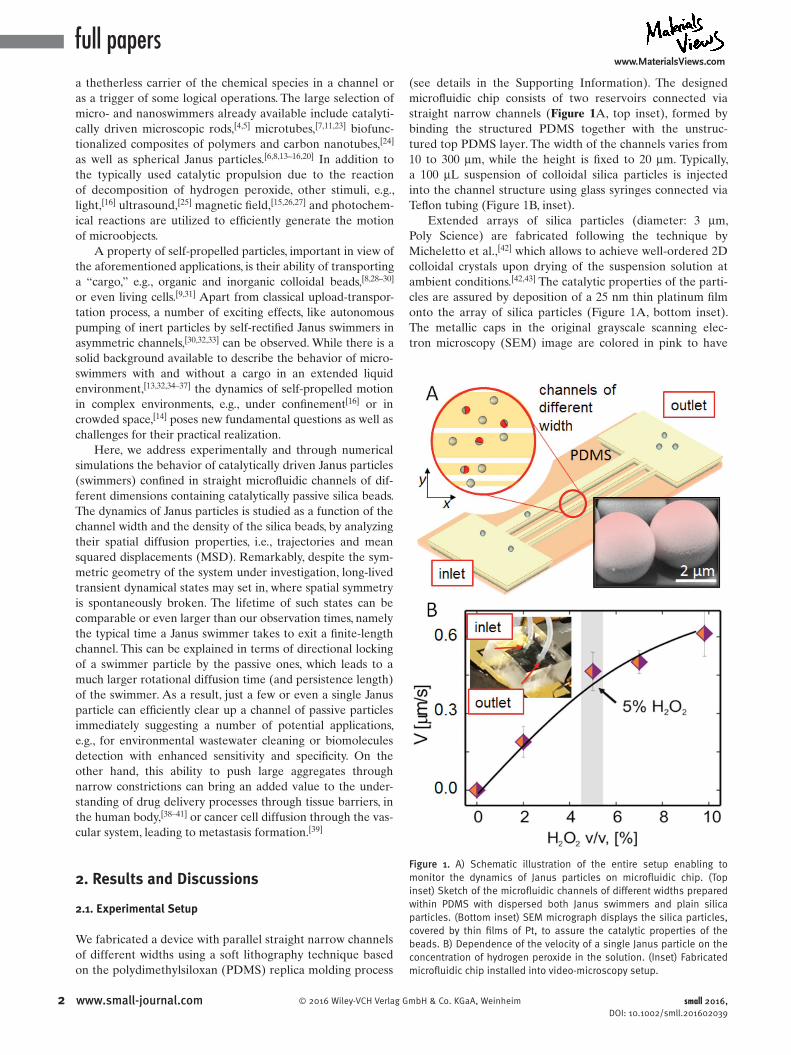
(see details in the Supporting Information). The designed microfluidic chip consists of two reservoirs connected via straight narrow channels (Figure 1A, top inset), formed by binding the structured PDMS together with the unstruc-tured top PDMS layer. The width of the channels varies from 10 to 300 μm, while the height is fixed to 20 μm. Typically, a 100 μL suspension of colloidal silica particles is injected into the channel structure using glass syringes connected via Teflon tubing (Figure 1B, inset).
Extended arrays of silica particles (diameter: 3 μm, Poly Science) are fabricated following the technique by Micheletto et al.,[42] which allows to achieve well-ordered 2D colloidal crystals upon drying of the suspension solution at ambient conditions.[42,43] The catalytic properties of the parti-cles are assured by deposition of a 25 nm thin platinum film onto the array of silica particles (Figure 1A, bottom inset). The metallic caps in the original grayscale scanning elec-tron microscopy (SEM) image are colored in pink to have
small 2016, DOI: 10.1002/smll.201602039
Figure 1. A) Schematic illustration of the entire setup enabling to monitor the dynamics of Janus particles on microfluidic chip. (Top inset) Sketch of the microfluidic channels of different widths prepared within PDMS with dispersed both Janus swimmers and plain silica particles. (Bottom inset) SEM micrograph displays the silica particles, covered by thin films of Pt, to assure the catalytic properties of the beads. B) Dependence of the velocity of a single Janus particle on the concentration of hydrogen peroxide in the solution. (Inset) Fabricated microfluidic chip installed into video-microscopy setup.

www.MaterialsViews.com
3© 2016 Wiley-VCH Verlag GmbH & Co. KGaA, Weinheim www.small-journal.com
it consistent with the schematic representation of the Janus particles in the following graphs. The deposition is carried out at room temperature using magnetron sputtering in a high vacuum chamber (base pressure: 10−7 mbar; argon (Ar) sputter pressure: 10−3 mbar). Fabricated Janus particles are detached from the substrate by sonication and suspended in an aqueous solution of hydrogen peroxide. In the presence of hydrogen peroxide (H2O2), the particles are set into motion, driven by self-diffusiophoresis, with speed determined by the concentration of H2O2 (v/v, serial dilution) in the solu-tion.[13,28] Note that the bubble propulsion mechanism is not dominant in the system of spherical Janus particles, since the geometry of the objects does not provide the necessary conditions for the facile nucleation of the bubbles, such as presence of the cavities or nonhomogeneous surface of the catalyst.[20] The Janus particle is propelled in the direction, opposite to the Pt cap. Catalytic Janus particles can be easily distinguished from plain silica beads by optical contrast, i.e., the difference in transmission/absorption due to the presence of the metal-covered part of silica beads. In the following, we present the quantitative analysis of their motility, caused by the decomposition of H2O2 on the Pt caps.[44]
During the experiment, Janus particles tend to settle onto the plasma treated PDMS substrate due to gravity and then diffuse on an effectively 2D surface of the substrate. This is realized via selection of the silica as a material for the particles (amorphous silicon dioxide, density of about 2.12 g cm−³) with its density almost 2× exceeding the water density. This assures their efficient sedimentation and preferable motion at the bottom wall. The distance between the particle and the sub-strate, determined by the Debye screening length in deion-ized water, is about 100 nm.[45] Accordingly, we recorded the motion of the particles in the xOy plane, the channel being directed along the x-axis (Figure 1A).
For future reference, we characterized the dynamics of the free catalytic Janus particles far from the confining walls. The propulsion velocity Vf of a single Janus particle as a func-tion of the concentration of H2O2 is presented in Figure 1B. The reported values of Vf have been averaged over the speed of at least ten distinct Janus particles. In agreement with pre-vious reports,[13,20,28] the velocity rises with the increase of the H2O2 concentration, revealing a clear trend to saturate at high concentrations. In the following, we fix the concen-tration of H2O2 at 5% in order to avoid the formation and accumulation of air bubbles inside the microfluidic chip. At the chosen H2O2 concentration, the velocity of the Janus par-ticles is Vf = 0.41 ± 0.1 μm s−1.
2.2. Janus Particles in Empty Channel
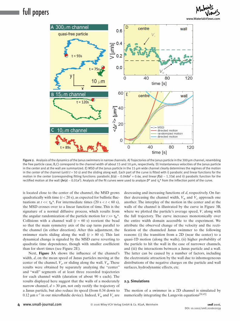
After the Janus particles are placed into the micro fluidic channels, they reveal two distinct regimes of motion, depending on their location with respect to the channel wall: (i) diffusion in the center of the channel and (ii) sliding along the walls, as summarized in Figure 2 (see also Video S1 in the Supporting Information).
According to the Stokes-Einstein equation,[13] the free diffusion coefficient, D, for particles with a diameter of
3 μm, placed in an aqueous environment with viscosity η = 8.9 × 10⁻⁴ Pa s at room temperature is predicted to be D = 0.16 μm2 s−1. Furthermore, the corresponding rotational diffusion time is estimated to be τR = 19 s. In the presence of a constant H2O2 concentration, the ongoing decomposi-tion reaction catalyzed by the Janus particle, boosts its dif-fusivity, hence the enhanced diffusion coefficient, D*, and the reduced diffusion time, τR*, respectively.[13]
Trajectories of both a Janus and a passive particle of the same size are displayed in Figure 2A for comparison. While the passive bead experiences thermal fluctuations, the Janus particle moves ballistically at short times (see arrows), but executes Brownian motion with diffusion coefficient D* = 0.23 μm2 s−1 (D* > D) and characteristic τR* = 16 s (τR* < τR) at longer times (see the fit of the MSD below in Figure 2E). The typical trajectory of a Janus swimmer moving far from the walls in a channel of width larger than 150 μm resembles that in the bulk. However, we observe that upon colliding with a channel wall, the swimmer slides along it and keeps doing so without inverting direction for a rather long time. Similar behavior of the active particles under confine-ment has been predicted by Wensink and Loewen[17] and recently shown experimentally near single straight[32] and cir-cularly shaped wall.[33]
For channel widths smaller than 100 μm, the behavior of the Janus particles grows complex, as the interplay between the two motility regimes in the center and at the channel wall becomes more pronounced (see Video S1 in the Supporting Information). Further shrinking the channel below the width of 30 μm, leads to the apparent prevalence of the wall sliding regime (Figure 2B,C for channels, respectively, 15 and 10 μm wide). The rectified trajectories shown in Figure 2B,C are char-acterized by persistence times much longer than the estimated swimmers’ rotational diffusion time in the bulk, τR*. Conse-quently, the directed motion of the channeled Janus swimmers reported here is attributable to a geometric suppression of their rotational diffusion. In conclusion, rectification of Janus swim-mers in a microfluidic channel can be maintained for increas-ingly long time intervals by lowering the channel width, even without the need of, e.g., external fields to guide them.[8,45,46]
Next, we sampled more trajectories and quantitatively analyzed the velocities and MSDs of catalytically driven Janus particles in a narrow channel. Interestingly, the self-propulsion velocity, V, is found to depend on the particle location in the channel. This claim is supported by the anal-ysis of the instantaneous and mean velocities of the swim-mers sliding along the walls or moving in the center of the channel. Both regimes of motion are clearly visible in Figure 2D, where the instantaneous velocity of a particle in a 15 μm wide channel is plotted versus time (the corresponding trajectory is portrayed in Figure 2B).
In this experiment, a single catalytic Janus par-ticle suspended in 5% (v/v) of H2O2 moves with speed Vc = 0.34 μm s−1, which is close to the value measured for a free Janus particle, Vf (0.41 μm). Upon hitting a wall, it then switches to the sliding regime by lowering its speed to Vw = 0.11 μm s−1. This change of motility regime is reflected in the time dependence of the corresponding MSD curve, ⟨Δr2⟩, plotted in Figure 2E. Initially, when the Janus swimmer
small 2016, DOI: 10.1002/smll.201602039
full paperswww.MaterialsViews.com
4 www.small-journal.com © 2016 Wiley-VCH Verlag GmbH & Co. KGaA, Weinheim
is located close to the center of the channel, the MSD grows quadratically with time (t < 20 s), as expected for ballistic fluc-tuations at t << τR*. For intermediate times (20 s < t < 60 s), the MSD crosses over to a linear function of time. This is the signature of a normal diffusive process, which results from the angular randomization of the particle motion for t >> τR*. Collisions with a channel wall (t ≈ 60 s) reorient the bead so that the main symmetry axis of the cap turns parallel to the channel (in either direction). After this adjustment, the swimmer starts sliding along the wall (t > 80 s). This last dynamical change is signaled by the MSD curve reverting to quadratic time dependence, though with smaller coefficient than for short times (see Figure 2E).
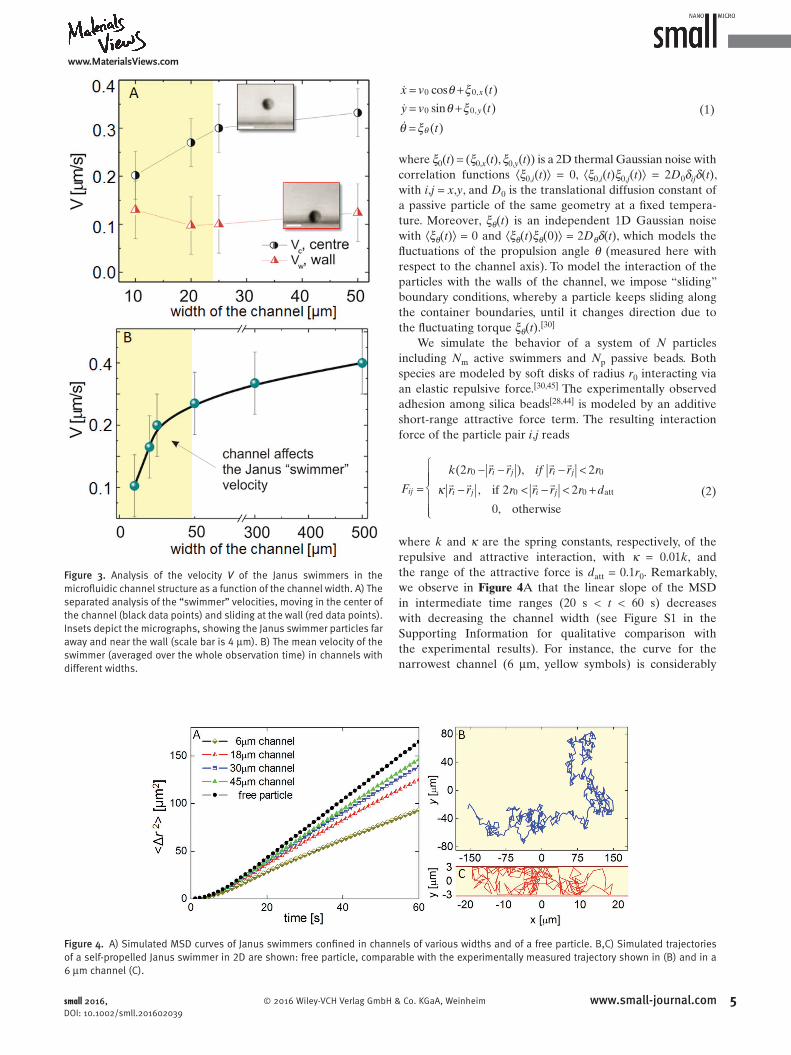
Next, Figure 3A shows the influence of the channel’s width, d, on the mean speed of Janus particles moving at the center of the channel, Vc, or sliding along the wall, Vw. These results were obtained by separately analyzing the “center” and “wall” segments of at least three recorded trajectories for each channel width (duration of about 90 s each). The results displayed here suggest that the walls of a moderately narrow channel, d > 30 μm, not only rectify the trajectory of a Janus particle, but also reduce its speed (from 0.34 down to 0.12 μm s−1 in our microfluidic device). Indeed, Vw and Vc are
decreasing and increasing functions of d, respectively. On fur-ther decreasing the channel width, Vw and Vc approach one another. The interplay of the motion in the center and at the walls of the channel is illustrated by the curve in Figure 3B, where we plotted the particle’s average speed, V, along with the full trajectory. The curve increases monotonically over the entire width domain accessible to the experiment. We attribute the observed change of the velocity and the recti-fication of the channeled Janus swimmer to the following reasons: (i) the transition from a 2D (near the center) to a quasi-1D motion (along the walls), (ii) higher probability of the particle to hit the wall in the case of narrower channels, and (iii) the interactions between a Janus particle and a wall. The latter can be caused by a number of factors, including the electrostatic attraction by the wall due to inhomogeneous distributions of the negative charges on the particle and wall surfaces, hydrodynamic effects, etc.
2.3. Simulations
The motion of a swimmer in a 2D channel is simulated by numerically integrating the Langevin equations[30,45]
small 2016, DOI: 10.1002/smll.201602039
Figure 2. Analysis of the dynamics of the Janus swimmers in narrow channels. A) Trajectories of the Janus particle in the 300 μm channel, resembling the free particle case; B,C) correspond to the channel width of about 15 and 10 μm, respectively. D) Instantaneous velocities of the Janus particle in the center and at the wall are summarized. E) MSD of the Janus particle in the 15 μm wide channel clearly determines the regimes of the motion in the center of the channel (until t = 50 s) and the sliding along wall. Each part of the curve is fitted with i) parabolic and linear functions for the motion in the center (corresponding fitting functions: parabolic fc(x) ∼ 0.048x2 + 0.6x, and linear fl(x) ∼ 1.33x) and ii) parabolic function for the rectified motion at the wall (fw(x) ∼ 0.01x2). Analysis of the fit curves were used to analyze D* and τR* from the inflection point of the curve.
www.MaterialsViews.com
5© 2016 Wiley-VCH Verlag GmbH & Co. KGaA, Weinheim www.small-journal.com
���
cos ( )
sin ( )
( )
0 0,
0 0,
x v t
y v t
t
x
y
θ ξθ ξ
θ ξ
= += +
= θ
(1)
where ξ0(t) = (ξ0,x(t), ξ0,y(t)) is a 2D thermal Gaussian noise with correlation functions ⟨ξ0,i(t)⟩ = 0, ⟨ξ0,i(t)ξ0,j(t)⟩ = 2D0δijδ(t), with i,j = x,y, and D0 is the translational diffusion constant of a passive particle of the same geometry at a fixed tempera-ture. Moreover, ξθ(t) is an independent 1D Gaussian noise with ⟨ξθ(t)⟩ = 0 and ⟨ξθ(t)ξθ(0)⟩ = 2Dθδ(t), which models the fluctuations of the propulsion angle θ (measured here with respect to the channel axis). To model the interaction of the particles with the walls of the channel, we impose “sliding” boundary conditions, whereby a particle keeps sliding along the container boundaries, until it changes direction due to the fluctuating torque ξθ(t).[30]
We simulate the behavior of a system of N particles including Nm active swimmers and Np passive beads. Both species are modeled by soft disks of radius r0 interacting via an elastic repulsive force.[30,45] The experimentally observed adhesion among silica beads[28,44] is modeled by an additive short-range attractive force term. The resulting interaction force of the particle pair i,j reads
� � � �
� � � �(2 ), 2
, if 2 2
0, otherwise
0 0
0 0 attF
k r r r if r r r
r r r r r r dij
i j i j
i j i jκ=− − − <
− < − < +
(2)
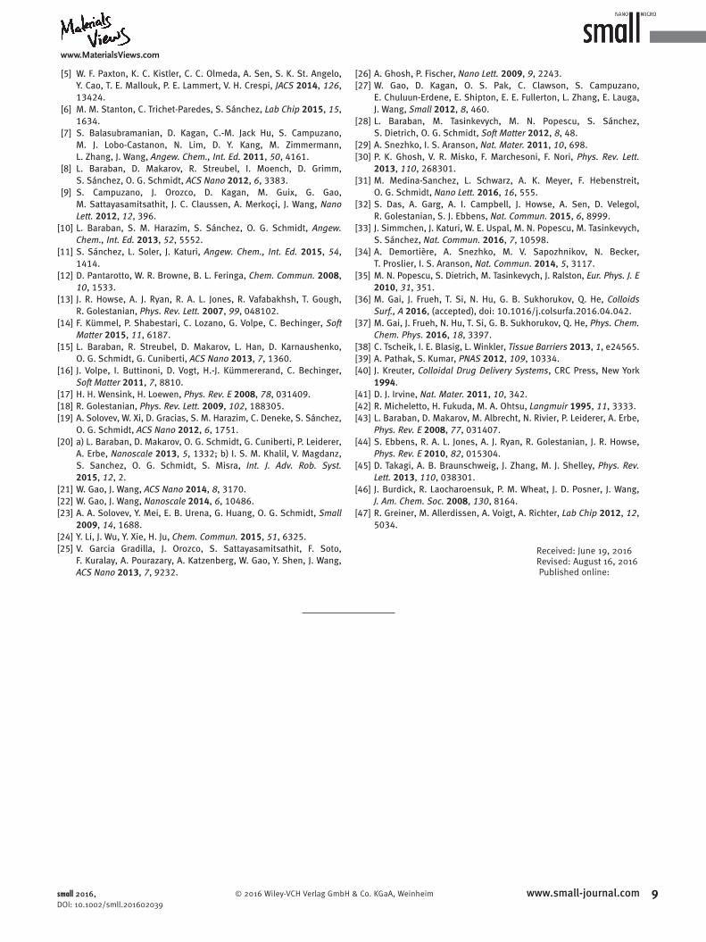
where k and κ are the spring constants, respectively, of the repulsive and attractive interaction, with κ = 0.01k, and the range of the attractive force is datt = 0.1r0. Remarkably, we observe in Figure 4A that the linear slope of the MSD in intermediate time ranges (20 s < t < 60 s) decreases with decreasing the channel width (see Figure S1 in the Supporting Information for qualitative comparison with the experimental results). For instance, the curve for the narrowest channel (6 μm, yellow symbols) is considerably
small 2016, DOI: 10.1002/smll.201602039
Figure 3. Analysis of the velocity V of the Janus swimmers in the microfluidic channel structure as a function of the channel width. A) The separated analysis of the “swimmer” velocities, moving in the center of the channel (black data points) and sliding at the wall (red data points). Insets depict the micrographs, showing the Janus swimmer particles far away and near the wall (scale bar is 4 μm). B) The mean velocity of the swimmer (averaged over the whole observation time) in channels with different widths.
Figure 4. A) Simulated MSD curves of Janus swimmers confined in channels of various widths and of a free particle. B,C) Simulated trajectories of a self-propelled Janus swimmer in 2D are shown: free particle, comparable with the experimentally measured trajectory shown in (B) and in a 6 μm channel (C).

full paperswww.MaterialsViews.com
6 www.small-journal.com © 2016 Wiley-VCH Verlag GmbH & Co. KGaA, Weinheim
lower than those for the wider ones. This effect is the signa-ture of the transition from a fully 2D to a quasi-1D diffusion mechanism in the channel. These results correlate with the dependence of the averaged velocity V, presented in Figure 3 as a function of the channel width d. Generally, such a transi-tion is anticipated in time by narrowing the channel, whereas for asymptotically long times, all MSD curves attain the same slope regardless of the channel width. Examples of simulated trajectories of self-propelled particles are shown in Figure 4B for a particle in the bulk and in a 6 μm channel (Figure 4C). In the latter case, the swimmer undergoes multiple collisions with the walls, which results in a Knudsen’s diffusion pro-cess along the channel. Note that the persistence length of the self-propelled Janus particle considerably increases while sliding along the channel walls, consistently with the experi-mental observations (see Figure 2C).
2.4. Janus Particles in Crowded Channel
To investigate the dynamics of Janus swimmers simultane-ously in a crowded environment and under geometric con-finement, a suspension containing a mixture of catalytic Janus beads and different concentrations of plain silica particles
with a diameter of about 3 μm was injected into the 25 μm wide channel. In the following, the trajectories and instanta-neous velocities of the swimmers and plain beads have been analyzed as a function of the density of the passive particles in the channel (see packing fraction in the Supporting Infor-mation). Note that, in view of the low number of catalytic Janus colloids in the channel, we neglect any effect due to their density fluctuations on the packing fractions.
Depending on the density of the passive particles, we dis-tinguish three main dynamical regimes of the active swim-mers: (i) A low density regime, when the Janus beads move through the passive particles experiencing rare collisions, i.e., freely percolate between the two sides of a channel of finite length; (ii) An intermediate density regime, when a Janus particle can move through the array of passive particles or bind a passive particle cargo and drag it along, thus acting as a carrier (see Movies S2 and S3 in the Supporting Infor-mation); (iii) Finally, a high density regime, when the passive particles clog the channel and the swimmers get locked inside (Movie S4, Supporting Information). Micrographs illustrating the transition from the low to the high density regime are shown in Figure 5A–C.
We analyze now in detail the active colloidal transport in the first two regimes.
small 2016, DOI: 10.1002/smll.201602039
Figure 5. Dynamics of Janus swimmers in a crowded environment and under geometric confinement at the same time. A–C) Microscopy images of the microfluidic channels filled with both, Janus particles and passive beads for the cases of different packing densities of particles, low (a), intermediate (b), and finally high (c). D,E) Instantaneous velocities of Janus swimmers (black circles), and passive colloidal particles (red circles) for the low density of particles in the channel (d) and high density (e), respectively.

www.MaterialsViews.com
7© 2016 Wiley-VCH Verlag GmbH & Co. KGaA, Weinheim www.small-journal.com
2.4.1. Low Density Regime
In the low density case (packing fraction of about 0.2), the behavior of the Janus swimmers is hardly modified by the presence of the passive species in the channel (Figure 5A). In particular, the long-range repulsion due to the presence of silanol groups on the surface of particles prevents the formation of the stable swimmer–cargo composites. Indeed, colloidal pairs do not approach each other close enough to perceive the stronger short-range attraction exerted by the van der Waals forces and capable of binding them.
Nevertheless, we observed the appearance of transient pairs involving both types of particles, which randomly form and dissolve, without ever accruing into stable aggre-gates (Figure 5A, see both cases, shown by the arrows). This is reflected in the corresponding analysis of the instanta-neous velocities of both types of particles (Figure 5D). To this regard, we emphasize that the fabrication process of our Janus particles and the contamination present in the liquid may lead to a partial degradation of the negative charges on the surface of the colloids[28] and thus be responsible for the occasional formation of the carrier–cargo composites,[28,44] shown in Figure 5A.
2.4.2. Intermediate and High Density Regimes
In contrast to the previous situation, a higher density of the passive species in the channel, namely, with packing fraction larger than 0.3, increases the probability of formation of car-rier–cargo complexes. In this case, a Janus particle is able to propel through the matrix of plain silica beads (Figure 5B) or to form clusters bridging the channel’s walls and transport them through the channel (Figure 5C and Videos S2 and S3 (Supporting Information)). In the former case (Figure 5B), a Janus particle can propel itself through the mixture of passive beads across the channel (along the y-axis). Such a transverse motion is favored by the empty room left behind by the colloidal particles as they diffuse in the longitudinal direction (along the x-axis). The latter case which involves the formation and the transport of clusters of passive beads (Figure 5C) is the most unusual. The mechanism is initiated by some active Janus swimmer that happens to rake up a bunch of surrounding passive particles upon moving along the channel (x-axis). Under the push of the swimmer and the action of the short-range pair attraction, these colloidal particles collapse into a seemingly stable cluster. As a conse-quence, a key role in the cluster formation is played by the combination of geometric confinement and particle–particle and particle–wall interactions (both interactions being gen-erally of the same nature). The motion of an active Janus motor in this crowded environment is thus rather complex (see Figure 5C,E and Video S4 (Supporting Information)). The free space across the channel is very limited, and the swimmer spends most of the time either near the wall or embedded into the cluster of passive colloids. When a self-propelling swimmer collides with the particles, it becomes embedded into the colloidal ensemble by forming a number of “bonds” with the surrounding nearest silica beads. This constraint of the rotational degree of freedom considerably
increases the effective rotational diffusion time, or engage-ment time, τen >> τR*. As a rule of thumb, we expect τen to be proportional to the number of bonds between the Janus motor and passive particles. Thus, a collision with a clogging cluster implies a nonvanishing component of the swimmer’s velocity orthogonal to the surface of the cluster and, as a consequence, along the axis of the channel. The engagement mechanism locks the direction of motion for a time of the order of τen, thus resulting in a much larger effective persis-tence length than for a free swimmer. In the regime of low Reynolds numbers, the dynamics of the locked system formed by the swimmer and the cluster is overdamped, which means that it drifts regardless of its total mass. Accordingly, the car-rier–cargo system advances steadily as a whole, with velocity given by the longitudinal component of the propulsion speed of the locked carrier, possibly reduced by frictional forces against the channel walls. In conclusion, a carrier can trans-port a payload without direction inversions an effective dis-tance much larger than its persistence length in the channel. The velocity plots of Figure 5E corroborate the proposed mechanism of carrier–cargo engagement and drift.
2.4.3. Bioinspired System: Unclogging the Narrow Channel
The striking ability of confined Janus swimmers to carry along large clusters of passive particles for surprisingly long time intervals due to the locking mechanism, can prove useful for diverse environmental or biomedical applications, e.g., to unclog microchannels filled by diverse biochemical species or to model the transport processes within the vascular system. Note that human blood capillaries have dimensions of about 10–15 μm, similar to the channels in our experiments.
To demonstrate the effect of channel unclogging, we numerically modeled the experimental situation reported above, when an active Janus motor is embedded in a cluster of passive particles and moves together with the cargo. In Figure 6A (top panel), a single swimmer shown by red circle pushes a cluster of sixteen passive particles (shown by black circles) from the left to the right, while three passive parti-cles do not move. The corresponding velocities of the moving cluster (black dots) and the passive particles (red dots) are shown in stage I of Figure 6B. Two vertical dashed segments separate three distinct temporal regimes. As the colloidal aggregate approaches the isolated passive particles (t ≈ 66 s), it incorporates them one by one (middle and bottom panels of Figure 6A). Correspondingly, during the collisional tran-sient, stage II of Figure 6B, the velocity of the passive parti-cles rapidly increases, showing oscillations or even prominent dip at t ≈ 108 s. This behavior is due to the rearrangement of the particles before these eventually settle into the new cluster. After having rearranged themselves into a close-packed quasicrystalline structure for t > 108 s, all the parti-cles move together (Figure 6A, bottom panel) with the same velocity (Figure 6B, stage III) of the initial cluster. The col-lective motion of the Janus motor and the passive particles reproduces closely the active colloidal transport observed experimentally. In view of the mechanism illustrated above, we conclude that a small fraction of active motors (or even a single active motor) suffices to unclog a narrow, finite length
small 2016, DOI: 10.1002/smll.201602039

full paperswww.MaterialsViews.com
8 www.small-journal.com © 2016 Wiley-VCH Verlag GmbH & Co. KGaA, Weinheim small 2016, DOI: 10.1002/smll.201602039
constriction in an otherwise inaccessible region. Moreover, since such motors are autonomous, i.e., are not guided by externally applied fields, active transport of microscopic objects through narrow channels can be implemented in a fairly noninvasive manner.
3. Conclusion
We addressed both experimentally and numerically the behavior of confined self-propelled catalytically driven Janus particles placed in straight microfluidic channels together with passive microscopic objects. Detailed investigation of the con-figurations, involving the artificial Janus swimmers acting in crowded and confined environments reveal intriguing aspects of their behavior affected by the surrounding matrix, i.e., directed motion of the swimmers and the rectifying role of the narrow channels (of widths below ten particle diameters). Furthermore, the presence of the channel walls substantially influences the speed of Janus particles, leading to a decrease of the velocity at the channel walls (almost by a factor of three).
Upon combining the geometrical confinement of the swimmers with the passive microparticles filling the narrow
channels, new effects emerge. Namely, Janus beads par-ticipate in the formation and transport of large clusters of passive particles, being “built-in” into this colloidal suspen-sion. This striking ability of Janus swimmers to transport large colloidal clusters, together with the observed mecha-nism of persistent directed motion, lends itself to diverse environmental or biomedical applications, i.e., to unclog or clear channels of unwanted biochemical species.
It is expected that artificial self-propelled particles will find applications to diverse emerging areas of nanotech-nology, from micro- and nanorobotics, e.g., for microsurgery purposes, high sensitivity biodetection, and drug delivery, to environmental water cleaning and bio (chemo)electronics. In particular, the demonstrated experimental setup involving essentially both active and passive species, provide key ingre-dients for the design of novel type nanodevices. Such devices could, for instance, realize the passive transportation and detection of reagents, triggering of the reaction steps, rel-evant for the chemical computing, e.g., in design of chemical central processing units [47] Taking into account the internal complexity of the natural systems, investigation of the spe-cific aspects of the dynamics of self-driven micromachines placed into the bioinspired artificially designed spatial con-figurations, e.g., crowded and confined environments, is of high interest.
Supporting Information
Supporting Information is available from the Wiley Online Library or from the author.
Acknowledgements
H.Y., A.K., and L.B. contributed equally to this work. This work was funded in part by the European Union (ERDF) and the Free State of Saxony via the ESF project InnoMedTec, the DFG cluster for Excel-lence, the Center for Advancing Electronics Dresden (CfAED), and via the European Research Council under the European Union’s Seventh Framework program (FP7/2007–2013)/ERC grant agree-ment no. 306277. V.R.M. and A.A.V. acknowledge support from the Odysseus Program of the Flemish Government and the FWO-VI. F.N. is partially supported by the RIKEN iTHES Project, the MURI Center for Dynamic Magneto-Optics via the AFOSR Grant No. FA9550-14-1-0040, the IMPACT program of the JST, and a Grant-in-Aid for the Scientific Research (A).
[1] N. Silvestry-Rodriguez, K. R. Bright, D. C. Slack, D. R. Uhlmann, C. P. Gerba, Appl. Environ. Microbiol. 2008, 74, 1639.
[2] G. A. Ozin, I. Manners, S. Fournier-Bidoz, A. Arsenault, Adv. Mater. 2005, 17, 3011.
[3] R. Dreyfus, J. Baudry, M. L. Roper, M. Fermigier, H. A. Stone, J. Bibette, Nature 2005, 437, 862.
[4] W. F. Paxton, S. Sundararajan, T. E. Mallouk, A. Sen, Angew. Chem., Int. Ed. 2006, 45, 5420.
Figure 6. Unclogging channel filled with passive particles by an active Janus swimmer. A) Particle distribution snapshots before the collision of the moving aggregate of the Janus swimmer (red circle) and 16 passive particles (grey circles) with three unmoving particles (t = 36 s), at the moment of the collision (t = 66 s), and when moving as a whole cluster (t = 108 s). B) The corresponding velocities of the moving aggregate driven by the Janus swimmer (black diamonds) and passive particles (red open circles).
www.MaterialsViews.com
9© 2016 Wiley-VCH Verlag GmbH & Co. KGaA, Weinheim www.small-journal.comsmall 2016, DOI: 10.1002/smll.201602039
[5] W. F. Paxton, K. C. Kistler, C. C. Olmeda, A. Sen, S. K. St. Angelo, Y. Cao, T. E. Mallouk, P. E. Lammert, V. H. Crespi, JACS 2014, 126, 13424.
[6] M. M. Stanton, C. Trichet-Paredes, S. Sánchez, Lab Chip 2015, 15, 1634.
[7] S. Balasubramanian, D. Kagan, C.-M. Jack Hu, S. Campuzano, M. J. Lobo-Castanon, N. Lim, D. Y. Kang, M. Zimmermann, L. Zhang, J. Wang, Angew. Chem., Int. Ed. 2011, 50, 4161.
[8] L. Baraban, D. Makarov, R. Streubel, I. Moench, D. Grimm, S. Sánchez, O. G. Schmidt, ACS Nano 2012, 6, 3383.
[9] S. Campuzano, J. Orozco, D. Kagan, M. Guix, G. Gao, M. Sattayasamitsathit, J. C. Claussen, A. Merkoçi, J. Wang, Nano Lett. 2012, 12, 396.
[10] L. Baraban, S. M. Harazim, S. Sánchez, O. G. Schmidt, Angew. Chem., Int. Ed. 2013, 52, 5552.
[11] S. Sánchez, L. Soler, J. Katuri, Angew. Chem., Int. Ed. 2015, 54, 1414.
[12] D. Pantarotto, W. R. Browne, B. L. Feringa, Chem. Commun. 2008, 10, 1533.
[13] J. R. Howse, A. J. Ryan, R. A. L. Jones, R. Vafabakhsh, T. Gough, R. Golestanian, Phys. Rev. Lett. 2007, 99, 048102.
[14] F. Kümmel, P. Shabestari, C. Lozano, G. Volpe, C. Bechinger, Soft Matter 2015, 11, 6187.
[15] L. Baraban, R. Streubel, D. Makarov, L. Han, D. Karnaushenko, O. G. Schmidt, G. Cuniberti, ACS Nano 2013, 7, 1360.
[16] J. Volpe, I. Buttinoni, D. Vogt, H.-J. Kümmererand, C. Bechinger, Soft Matter 2011, 7, 8810.
[17] H. H. Wensink, H. Loewen, Phys. Rev. E 2008, 78, 031409.[18] R. Golestanian, Phys. Rev. Lett. 2009, 102, 188305.[19] A. Solovev, W. Xi, D. Gracias, S. M. Harazim, C. Deneke, S. Sánchez,
O. G. Schmidt, ACS Nano 2012, 6, 1751.[20] a) L. Baraban, D. Makarov, O. G. Schmidt, G. Cuniberti, P. Leiderer,
A. Erbe, Nanoscale 2013, 5, 1332; b) I. S. M. Khalil, V. Magdanz, S. Sanchez, O. G. Schmidt, S. Misra, Int. J. Adv. Rob. Syst. 2015, 12, 2.
[21] W. Gao, J. Wang, ACS Nano 2014, 8, 3170.[22] W. Gao, J. Wang, Nanoscale 2014, 6, 10486.[23] A. A. Solovev, Y. Mei, E. B. Urena, G. Huang, O. G. Schmidt, Small
2009, 14, 1688.[24] Y. Li, J. Wu, Y. Xie, H. Ju, Chem. Commun. 2015, 51, 6325.[25] V. Garcia Gradilla, J. Orozco, S. Sattayasamitsathit, F. Soto,
F. Kuralay, A. Pourazary, A. Katzenberg, W. Gao, Y. Shen, J. Wang, ACS Nano 2013, 7, 9232.
[26] A. Ghosh, P. Fischer, Nano Lett. 2009, 9, 2243.[27] W. Gao, D. Kagan, O. S. Pak, C. Clawson, S. Campuzano,
E. Chuluun-Erdene, E. Shipton, E. E. Fullerton, L. Zhang, E. Lauga, J. Wang, Small 2012, 8, 460.
[28] L. Baraban, M. Tasinkevych, M. N. Popescu, S. Sánchez, S. Dietrich, O. G. Schmidt, Soft Matter 2012, 8, 48.
[29] A. Snezhko, I. S. Aranson, Nat. Mater. 2011, 10, 698.[30] P. K. Ghosh, V. R. Misko, F. Marchesoni, F. Nori, Phys. Rev. Lett.
2013, 110, 268301.[31] M. Medina-Sanchez, L. Schwarz, A. K. Meyer, F. Hebenstreit,
O. G. Schmidt, Nano Lett. 2016, 16, 555.[32] S. Das, A. Garg, A. I. Campbell, J. Howse, A. Sen, D. Velegol,
R. Golestanian, S. J. Ebbens, Nat. Commun. 2015, 6, 8999.[33] J. Simmchen, J. Katuri, W. E. Uspal, M. N. Popescu, M. Tasinkevych,
S. Sánchez, Nat. Commun. 2016, 7, 10598.[34] A. Demortière, A. Snezhko, M. V. Sapozhnikov, N. Becker,
T. Proslier, I. S. Aranson, Nat. Commun. 2014, 5, 3117.[35] M. N. Popescu, S. Dietrich, M. Tasinkevych, J. Ralston, Eur. Phys. J. E
2010, 31, 351.[36] M. Gai, J. Frueh, T. Si, N. Hu, G. B. Sukhorukov, Q. He, Colloids
Surf., A 2016, (accepted), doi: 10.1016/j.colsurfa.2016.04.042.[37] M. Gai, J. Frueh, N. Hu, T. Si, G. B. Sukhorukov, Q. He, Phys. Chem.
Chem. Phys. 2016, 18, 3397.[38] C. Tscheik, I. E. Blasig, L. Winkler, Tissue Barriers 2013, 1, e24565.[39] A. Pathak, S. Kumar, PNAS 2012, 109, 10334.[40] J. Kreuter, Colloidal Drug Delivery Systems, CRC Press, New York
1994.[41] D. J. Irvine, Nat. Mater. 2011, 10, 342.[42] R. Micheletto, H. Fukuda, M. A. Ohtsu, Langmuir 1995, 11, 3333.[43] L. Baraban, D. Makarov, M. Albrecht, N. Rivier, P. Leiderer, A. Erbe,
Phys. Rev. E 2008, 77, 031407.[44] S. Ebbens, R. A. L. Jones, A. J. Ryan, R. Golestanian, J. R. Howse,
Phys. Rev. E 2010, 82, 015304.[45] D. Takagi, A. B. Braunschweig, J. Zhang, M. J. Shelley, Phys. Rev.
Lett. 2013, 110, 038301.[46] J. Burdick, R. Laocharoensuk, P. M. Wheat, J. D. Posner, J. Wang,
J. Am. Chem. Soc. 2008, 130, 8164.[47] R. Greiner, M. Allerdissen, A. Voigt, A. Richter, Lab Chip 2012, 12,
5034.
Received: June 19, 2016Revised: August 16, 2016Published online: