Conditional inhibition of Plasmodium falciparum …...Conditional inhibition of Plasmodium...
20
Conditional inhibition of Plasmodium falciparum CDPK1 1 Inhibition of Plasmodium falciparum CDPK1 by conditional expression of its J-domain demonstrates a key role in schizont development Mauro F. Azevedo *† , Paul R. Sanders * , Efrosinia Krejany * , Catherine Q. Nie * , Ping Fu ‡ , Leon A. Bach ‡ , Gerhard Wunderlich † , Brendan S. Crabb *‡§1 and Paul R. Gilson *‡1 * Macfarlane Burnet Institute of Medical Research and Public Health, Melbourne, Victoria, Australia; † Departamento de Parasitologia, Instituto de Ciências Biomédicas, Universidade de São Paulo, São Paulo, Brasil; ‡ Monash University, Melbourne, Australia; § University of Melbourne, Melbourne, Australia. 1 Address correspondence to: Paul Gilson, Brendan Crabb, Macfarlane Burnet Institute for Medical Research & Public Health, 85 Commercial Road, Melbourne, Victoria 3004, Australia. Ph. +61 03 8506 2481 Fax. +61 03 9282 2100 Email: [email protected], [email protected] Plasmodium falciparum calcium-dependent protein kinase 1 (PfCDPK1) is highly expressed in parasite asexual blood and mosquito stages. Its role is still poorly understood, but unsuccessful gene knockout attempts suggest it is essential for parasite replication and/or RBC invasion. Here, by tagging endogenous CDPK1 with GFP, we demonstrate that CDPK1 localises to the parasite plasma membrane of replicating and invasive forms as well as very young intracellular parasites and does not appear to be exported into the erythrocyte. While a knockdown of endogenous CDPK1 was achieved using a destabilization domain, parasites tolerated reduced expression without displaying a phenotype. Because of this, the PfCDPK1 auto-inhibitory junction region (J) was explored as a means of achieving inducible and specific inhibition. Under in vitro conditions, a fusion protein comprising a J-GFP fusion specifically bound to PfCDPK1 and inhibited its activity. This fusion protein was conditionally expressed in P. falciparum asexual blood stages under the regulation of a destabilization domain (J-GFP-DD). We demonstrate that J-GFP-DD binds to CDPK1 and that this results in the arrest of parasite development late in the cell cycle during early schizogony. These data point to an early schizont function for PfCDPK1 and demonstrates that conditionally expressing auto-inhibitory regions can be an effective way to address the function of Plasmodium enzymes. Short title: Conditional inhibition of Plasmodium falciparum CDPK1 Keywords: Plasmodium, kinase, calcium ABBREVIATIONS FOOTNOTE Abbreviations used: CDPK, calcium-dependent protein kinase; DD, destabilization domain; J, CDPK junction domain. INTRODUCTION Intracellular signalling events are critically involved in all stages of malarial parasite development. Signalling in the asexual blood stage has received particular attention since it is this stage that produces symptomatic disease. Asexual blood stage parasites go through four distinct phases in their cell cycle. They begin as small invasive merozoites that are released from mature infected erythrocytes. The merozoite is the only blood stage form that is extracellular and it rapidly invades a new erythrocyte before differentiating into a small amoeboid, intracellular ring stage parasite. The ring stage lasts for about first half of the ~48 hour cell cycle after which it becomes a trophozoite stage parasite. This stage grows rapidly until about 32-36 hours post invasion when it undergoes several rounds of nuclear division in preparation for producing new merozoites. Each of these replicative schizont stage parasites will produce Biochemical Journal Immediate Publication. Published on 03 Apr 2013 as manuscript BJ20130124 THIS IS NOT THE VERSION OF RECORD - see doi:10.1042/BJ20130124 Accepted Manuscript Licenced copy. Copying is not permitted, except with prior permission and as allowed by law. © 2013 The Authors Journal compilation © 2013 Biochemical Society
Transcript of Conditional inhibition of Plasmodium falciparum …...Conditional inhibition of Plasmodium...

Conditional inhibition of Plasmodium falciparum CDPK1
1
Inhibition of Plasmodium falciparum CDPK1 by conditional expression of its J-domain demonstrates a key role in schizont development
Mauro F. Azevedo*†, Paul R. Sanders*, Efrosinia Krejany*, Catherine Q. Nie*, Ping Fu‡, Leon A. Bach‡, Gerhard Wunderlich†, Brendan S. Crabb*‡§1 and Paul R. Gilson*‡1
*Macfarlane Burnet Institute of Medical Research and Public Health, Melbourne, Victoria, Australia; †Departamento de Parasitologia, Instituto de Ciências Biomédicas, Universidade de São Paulo, São Paulo, Brasil; ‡Monash University, Melbourne, Australia; §University of Melbourne, Melbourne, Australia. 1Address correspondence to: Paul Gilson, Brendan Crabb, Macfarlane Burnet Institute for Medical Research & Public Health, 85 Commercial Road, Melbourne, Victoria 3004, Australia. Ph. +61 03 8506 2481 Fax. +61 03 9282 2100 Email: [email protected], [email protected]
Plasmodium falciparum calcium-dependent protein kinase 1 (PfCDPK1) is highly expressed in parasite asexual blood and mosquito stages. Its role is still poorly understood, but unsuccessful gene knockout attempts suggest it is essential for parasite replication and/or RBC invasion. Here, by tagging endogenous CDPK1 with GFP, we demonstrate that CDPK1 localises to the parasite plasma membrane of replicating and invasive forms as well as very young intracellular parasites and does not appear to be exported into the erythrocyte. While a knockdown of endogenous CDPK1 was achieved using a destabilization domain, parasites tolerated reduced expression without displaying a phenotype. Because of this, the PfCDPK1 auto-inhibitory junction region (J) was explored as a means of achieving inducible and specific inhibition. Under in vitro conditions, a fusion protein comprising a J-GFP fusion specifically bound to PfCDPK1 and inhibited its activity. This fusion protein was conditionally expressed in P. falciparum asexual blood stages under the regulation of a destabilization domain (J-GFP-DD). We demonstrate that J-GFP-DD binds to CDPK1 and that this results in the arrest of parasite development late in the cell cycle during early schizogony. These data point to an early schizont function for PfCDPK1 and demonstrates that conditionally expressing auto-inhibitory regions can be an effective way to address the function of Plasmodium enzymes. Short title: Conditional inhibition of Plasmodium falciparum CDPK1 Keywords: Plasmodium, kinase, calcium ABBREVIATIONS FOOTNOTE Abbreviations used: CDPK, calcium-dependent protein kinase; DD, destabilization domain; J, CDPK junction domain. INTRODUCTION Intracellular signalling events are critically involved in all stages of malarial parasite development. Signalling in the asexual blood stage has received particular attention since it is this stage that produces symptomatic disease. Asexual blood stage parasites go through four distinct phases in their cell cycle. They begin as small invasive merozoites that are released from mature infected erythrocytes. The merozoite is the only blood stage form that is extracellular and it rapidly invades a new erythrocyte before differentiating into a small amoeboid, intracellular ring stage parasite. The ring stage lasts for about first half of the ~48 hour cell cycle after which it becomes a trophozoite stage parasite. This stage grows rapidly until about 32-36 hours post invasion when it undergoes several rounds of nuclear division in preparation for producing new merozoites. Each of these replicative schizont stage parasites will produce
Biochemical Journal Immediate Publication. Published on 03 Apr 2013 as manuscript BJ20130124T
HIS
IS N
OT
TH
E V
ER
SIO
N O
F R
EC
OR
D -
see
doi
:10.
1042
/BJ2
0130
124
Acce
pted
Man
uscr
ipt
Licenced copy. Copying is not permitted, except with prior permission and as allowed by law.
© 2013 The Authors Journal compilation © 2013 Biochemical Society

Conditional inhibition of Plasmodium falciparum CDPK1
2
~16 merozoite daughters which are released when the infected erythrocyte ruptures. There is a great need to understand signalling pathways that underpin the blood stage as well as other life cycle stages of the parasite since they may reveal key future targets for anti-malarial drug design. Of particular interest for understanding signalling are the parasite’s protein kinases and the genome of Plasmodium falciparum, the most pathogenic malaria species of humans, is predicted to encode almost one hundred of these enzymes [1], of which 65 group with other eukaryotic kinase families [2]. The application of high-throughput gene knockout strategies suggests that about half of the conserved kinases are essential for malaria parasites during the asexual life cycle, rendering them potential drug targets [3].
Calcium-dependent protein kinases (CDPKs) are found only in plants and in alveolate protozoa [4], and are most closely related to the calmodulin-dependent protein kinases (CaMKs). Their structure typically comprises in the following order a variable N-terminal region, a catalytic kinase domain (KD), a junction region (J) and a calmodulin-like domain (CLD), which usually contains four EF hand motifs that bind Ca2+ [5]. Structural studies of CDPKs of Toxoplasma gondii and Cryptosporidium parvum [6] revealed that J, together with the N-terminal EF hand, forms a long alpha helix, referred to as CH1. Subsequently, J and CLD were reclassified as the CDPK activation domain (CAD) and appear to play a key role in the regulation of CDPKs [6, 7]. In the absence of Ca2+, J interacts with KD to inhibit its catalytic activity.
P. falciparum has 5 CDPKs that feature this canonical domain structure and 2 CDPKs that contain one or more EF hands at the N-terminal region [8]. P. falciparum CDPK1 (PfCDPK1) (PlasmoDB No. PF3D7_0217500) was the first P. falciparum CDPK to be cloned and characterized [9]. A recombinant form of PfCDPK1 expressed in Escherichia coli was found to have calcium dependent kinase activity and bound four Ca2+ ions as expected. However, its affinity for the cation was found to be lower when compared to calmodulin [10]. Plasmodium transcriptomic data suggest that cdpk1 is mostly expressed during the schizont stage [11, 12]. N-terminal acylation is required for membrane targeting of some CDPKs [13], including P. falciparum CDPK1 and CDPK4 [7, 14]. Transfection of P. falciparum with the N-terminal region of CDPK1 fused to green fluorescent protein (GFP) as well as immunoelectron microscopy suggested that CDPK1 may be exported into the parasitophorous vacuole (PV) [14]. A more recent study using immunofluorescence microscopy has challenged this by indicating the protein localizes to the inner surface of the plasma membrane (PM) [15]. This latter finding was also supported by observations of a full length CDPK1-GFP fusion in P. berghei [16].
Thus far attempts to knock out cdpk1 in the asexual blood stage have been unsuccessful [17], implying that CDPK1 may be essential for survival at this stage of the life cycle. A potential role for PfCDPK1 is to phosphorylate in a calcium dependent manner, a number of proteins with which it is co-expressed, involved in motility and invasion. In line with this, recombinant PfCDPK1 is able to phosphorylate two components of the motor complex in merozoites [15, 17], suggesting that it may be a key regulator of the actin myosin motor that drives parasite motility and red blood cell invasion. Recently, a study employing a promoter swap strategy that resulted in knockdown of P. berghei CDPK1 expression in sexual stages showed defects in multiple aspects of parasite sexual development and in transmission to mosquitos [16]. This supports a multifunctional role for CDPK1 in which the enzyme may regulate various signalling pathways across all Plasmodium life cycle stages in addition to its role in actin-myosin motor regulation.
The essentiality of CDPK1 means it is not possible to study its function through traditional gene knockout strategies. Therefore, to resolve the kinase’s function and to evaluate its potential as a drug target various CDPK1 drug inhibitors have been tested in P. falciparum. At high concentrations the general CaMK inhibitor K252a, blocked schizont rupture and at an intermediate concentration merozoite invasion was inhibited [15]. A chemical screen for a more specific inhibitor of CDPK1 lead to the discovery of purfalcamine, a 2,6,9-trisubstituted purine, that blocked schizont development at a pre-cytokinesis stage [17]. The apparent discrepancies between the effects of CDPK1 inhibiting drugs suggest that one or both drugs may have off target effects. To resolve this we have employed two specific genetic approaches to conditionally knockdown CDPK1 function. First we utilized the FKBP based protein destabilization (DD) system to conditionally knockdown levels of the kinase. DD is a mutant form of
Biochemical Journal Immediate Publication. Published on 03 Apr 2013 as manuscript BJ20130124T
HIS
IS N
OT
TH
E V
ER
SIO
N O
F R
EC
OR
D -
see
doi
:10.
1042
/BJ2
0130
124
Acce
pted
Man
uscr
ipt
Licenced copy. Copying is not permitted, except with prior permission and as allowed by law.
© 2013 The Authors Journal compilation © 2013 Biochemical Society

Conditional inhibition of Plasmodium falciparum CDPK1
3
FKBP engineered to be structurally unstable and when fused to another protein will flag the fusion for rapid degradation by the ubiquitin pathway [18]. Protein degradation can however be blocked by the small, cell permeable compound Shld-1, that binds to and stabilizes the DD protein [18]. Here we integrated a GFP-DD coding sequence into the 3’ end of the PfCDPK1 gene to create a PfCDPK1-GFP-DD fusion that leads to about 70% reduction in enzyme levels but no accompanying phenotypic changes.
Secondly, we attempted to inhibit PfCDPK1 in trans, through expression of its auto-inhibitory J region. We have demonstrated in vitro that J retains its ability to bind and inhibit the activity of full-length kinase even when fused to markedly larger polypeptides such as GFP. To inhibit endogenous CDPK1 in P. falciparum we transfected the parasites with J-GFP N-terminally fused to the DD24 FKBP variant, which we have recently shown can routinely produce a four fold change in protein levels +/- Shld-1 in P. falciparum [19, 20]. To reduce the potentially toxic effects of the J-GFP-DD24 protein during the transfection period, parasites were cultured without Shld-1 to decrease levels of the fusion protein. We show that the addition of Shld-1 and subsequent increase in J-GFP-DD24 fusion protein levels arrests schizont development at a pre-cytokinesis stage similar to that caused by purfalcamine [17]. The successful inhibition of CDPK1 in trans via its auto-inhibitory domain opens the possibility of using this approach to specifically target other enzymes with auto-inhibitory domains.
EXPERIMENTAL Parasite Culture and transfection P. falciparum 3D7 strain was cultivated and transfected as previously described [21-23]. Transgenic lines were generated with plasmids pTM2CPKwtGFP and pTM2CPKwtGFP-DD (Supplementary data). Integration of the plasmid DNA over episomal maintenance was selected for by cycling the parasites on and off WR99210 and detected by PCR. Parasites with integrated plasmids were cloned by limiting dilution. Parasitemia and development were monitored by microscopic examination of Giemsa-stained thin smears of the cultures and by flow cytometry of ethidium bromide stained parasites as previously described [24].
In vitro kinase assay Reaction was performed in 20 mM Tris pH 7.4, 0.5 mM DTT, 10 mM MgCl2, 0.1 mM EGTA, 0.1 mM ATP, 0.5 mM MnCl2, 1 mM CaCl2, 0.5 mg/mL dephosphorylated casein, 100 ng rCDPK1 in a final volume of 35 μL at 30°C for 5 minutes. Reaction was initiated adding ATP and stopped with methanol. Phosphorylation was quantified by SDS-page stained with Pro-Q Diamond phosphoprotein gel stain (Invitrogen) and the signal normalised by total protein levels determined by SYPRO-Ruby staining. Monoclonal antibodies A synthetic peptide corresponding to CDPK1 residues 3-18 (SQSSNVKDFKTRRSK) was conjugated to keyhole limpet hemocyanin (Genscript, NJ) and used to produce the mouse monoclonal antibody 3E6, at the Walter and Eliza Hall Institute’s monoclonal antibody facility (see supplementary methods for more detail).
Western blotting P. falciparum infected RBCs were lysed in 0.15% saponin to remove excess RBC protein and the parasite cells pellet were solubilized in reducing SDS sample buffer and after electrophoresis proteins were transferred to PVDF membranes (Millipore). Membranes were probed with CDPK1 mAb 3E6 (1/2000), rabbit polyclonal sera targeting P. falciparum MSP2 (1/500) [25], aldolase (1/500) [26] and Exp2 [27], an anti-GFP mAb (1/1000, Roche) and a chicken polyclonal specific to HA epitope (1/1000, Abcam). Detection was performed with IR-dye conjugated goat anti-mouse or anti-rabbit immunoglobulins (1/5000) (Rockland) visualized by fluorescent scanning (Odyssey, LiCor) and by chemiluminescence via goat HRP-conjugated anti-chicken (Abcam).
Biochemical Journal Immediate Publication. Published on 03 Apr 2013 as manuscript BJ20130124T
HIS
IS N
OT
TH
E V
ER
SIO
N O
F R
EC
OR
D -
see
doi
:10.
1042
/BJ2
0130
124
Acce
pted
Man
uscr
ipt
Licenced copy. Copying is not permitted, except with prior permission and as allowed by law.
© 2013 The Authors Journal compilation © 2013 Biochemical Society

Conditional inhibition of Plasmodium falciparum CDPK1
4
Live cell imaging Live P. falciparum expressing GFP were microscopically observed after staining with DAPI (0.5 mg ml-1, Sigma) and CellMask (1/50,000, Invitrogen) using an Olympus-DeltaVision inverted fluorescence microscope (Applied Precision Inc. Seattle, USA). DeltaVision software was used to deconvolve Z-stacked fluorescent optical images taken through the cells. Expression and purification of recombinant proteins Recombinant proteins were expressed in E. coli BL-21 DE3 cells and purified as described previously [28]. Prior to use in SPR, HisTrap purified rCDPK1, GFP-J, GFP were buffer exchanged in 10 mM HEPES, 150 mM NaCl, pH 7.4 by dialysis. rCDPK1 was further purified by size exclusion chromatography using a Superdex 200 10/300 GL column (GE Healthcare) by FPLC (ÄKTA Purifier, GE Healthcare) in the HEPES buffer used for dialysis. A single A280 nm chromatographic peak corresponding to monomeric rCDPK1 was collected and applied as the analyte in SPR experiments. Surface plasmon resonance Biophysical binding analysis was performed by surface plasmon resonance (SPR) on a Biacore T100 instrument (GE Healthcare). 100nM rCDPK1 in 10 mM HEPES, 150 mM NaCl, 0.05% Surfactant P20 (v/v), pH 7.4 was purified by gel filtration and amine coupled to a CM5 sensor chip (Biacore, GE Healthcare). Refer to supplementary data for conditions used. Kinetic and equilibrium binding data were analysed (BIAevaluation Software) using reference-subtracted sensorgrams (rJ-GFP minus rGFP). 100nM BSA was used as a negative control. Experiments were repeated 5 times on two CM5 chips with ligand and analyte from two independently expressed and purified batches. 200 nM of three J domain-spanning peptides used in CDPK1 J-GFP competition assays were independently incubated with 100 nM rCDPK1 (30 min at RT) prior to injection onto the GFP/J-GFP chip. Refer to supplementary data for further details. CDPK1 alone and CDPK1 + DMSO controls were included and experiments repeated 4 times.
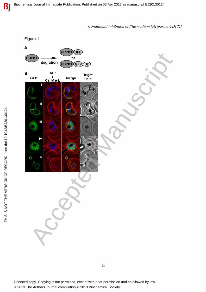
RESULTS AND DISCUSSION CDPK1 localises to the plasma membrane of schizonts, merozoite and early rings. To visualize CDPK1 in live cells, we engineered parasites expressing GFP fused to the C-terminus of CDPK1. To ensure authentic and functional expression of this chimera, gfp coding sequence was appended to the 3' end of the endogenous cdpk1 gene by single-crossover homologous recombination in transfected P. falciparum (3D7 line) parasites (Figure 1A). A single cdpk1-gfp cloned parasite line was isolated and integration into the endogenous locus was confirmed by PCR (Supplementary figure 1).
Fluorescence images were taken of live CDPK1-GFP parasites of different ages. The fusion protein is first seen as a weak signal localised at the periphery of young schizonts, which is indicative of plasma membrane (PM) or parasitophorous vacuole (PV) localization but not inner membrane complex which initially appear as internal punctate structures (Figure 1B, i & ii). In older parasites undergoing cytokinesis, CDPK1-GFP fluorescence increased and remained localized to the periphery of developing merozoites, a pattern that suggests PM, but not PV localisation (Figure 1B, panel iii and iv). This was confirmed following schizont rupture, where CDPK1 was still present at the periphery of free merozoites and even inside the amoeboid-like outer margin of newly invaded RBCs (Figure 1B, panel v). Although previous data using antibodies raised to CDPK1 has assigned localisation to either PM [15, 16] or both PM and PV [14, 29], our data, where we are directly visualising the endogenous kinase, indicates localisation
Biochemical Journal Immediate Publication. Published on 03 Apr 2013 as manuscript BJ20130124T
HIS
IS N
OT
TH
E V
ER
SIO
N O
F R
EC
OR
D -
see
doi
:10.
1042
/BJ2
0130
124
Acce
pted
Man
uscr
ipt
Licenced copy. Copying is not permitted, except with prior permission and as allowed by law.
© 2013 The Authors Journal compilation © 2013 Biochemical Society
Conditional inhibition of Plasmodium falciparum CDPK1
5
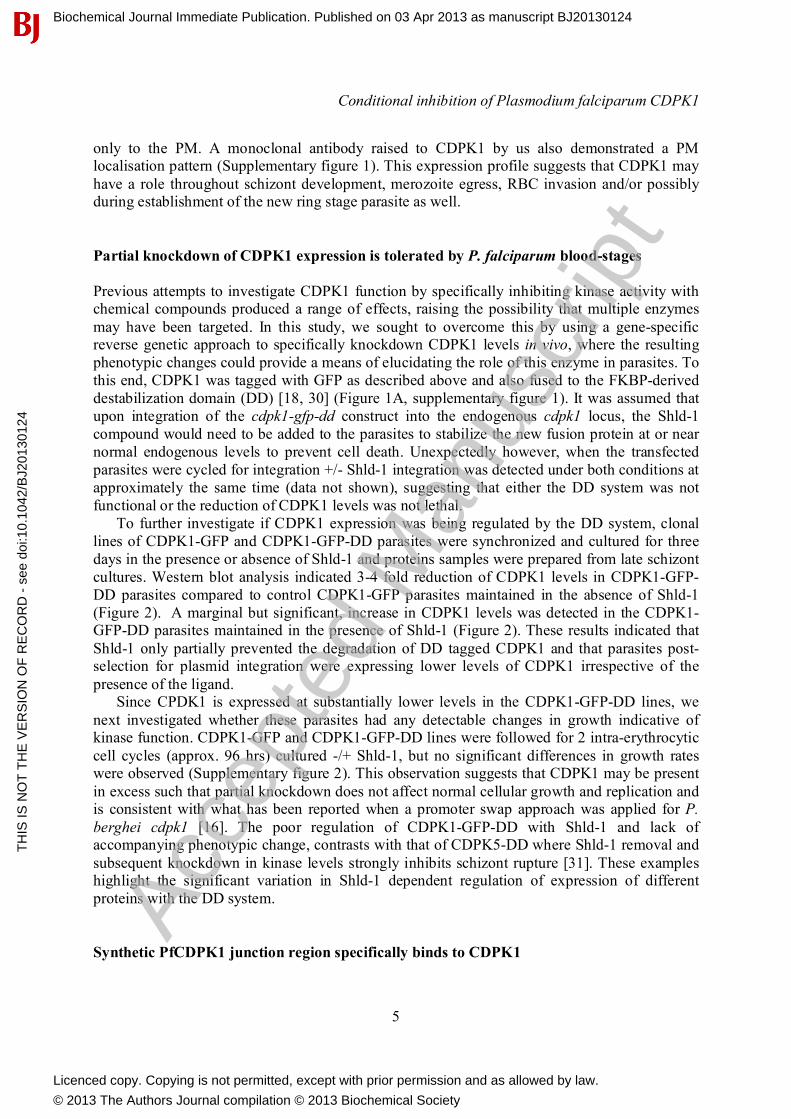
only to the PM. A monoclonal antibody raised to CDPK1 by us also demonstrated a PM localisation pattern (Supplementary figure 1). This expression profile suggests that CDPK1 may have a role throughout schizont development, merozoite egress, RBC invasion and/or possibly during establishment of the new ring stage parasite as well.
Partial knockdown of CDPK1 expression is tolerated by P. falciparum blood-stages Previous attempts to investigate CDPK1 function by specifically inhibiting kinase activity with chemical compounds produced a range of effects, raising the possibility that multiple enzymes may have been targeted. In this study, we sought to overcome this by using a gene-specific reverse genetic approach to specifically knockdown CDPK1 levels in vivo, where the resulting phenotypic changes could provide a means of elucidating the role of this enzyme in parasites. To this end, CDPK1 was tagged with GFP as described above and also fused to the FKBP-derived destabilization domain (DD) [18, 30] (Figure 1A, supplementary figure 1). It was assumed that upon integration of the cdpk1-gfp-dd construct into the endogenous cdpk1 locus, the Shld-1 compound would need to be added to the parasites to stabilize the new fusion protein at or near normal endogenous levels to prevent cell death. Unexpectedly however, when the transfected parasites were cycled for integration +/- Shld-1 integration was detected under both conditions at approximately the same time (data not shown), suggesting that either the DD system was not functional or the reduction of CDPK1 levels was not lethal.
To further investigate if CDPK1 expression was being regulated by the DD system, clonal lines of CDPK1-GFP and CDPK1-GFP-DD parasites were synchronized and cultured for three days in the presence or absence of Shld-1 and proteins samples were prepared from late schizont cultures. Western blot analysis indicated 3-4 fold reduction of CDPK1 levels in CDPK1-GFP-DD parasites compared to control CDPK1-GFP parasites maintained in the absence of Shld-1 (Figure 2). A marginal but significant, increase in CDPK1 levels was detected in the CDPK1-GFP-DD parasites maintained in the presence of Shld-1 (Figure 2). These results indicated that Shld-1 only partially prevented the degradation of DD tagged CDPK1 and that parasites post-selection for plasmid integration were expressing lower levels of CDPK1 irrespective of the presence of the ligand.
Since CPDK1 is expressed at substantially lower levels in the CDPK1-GFP-DD lines, we next investigated whether these parasites had any detectable changes in growth indicative of kinase function. CDPK1-GFP and CDPK1-GFP-DD lines were followed for 2 intra-erythrocytic cell cycles (approx. 96 hrs) cultured -/+ Shld-1, but no significant differences in growth rates were observed (Supplementary figure 2). This observation suggests that CDPK1 may be present in excess such that partial knockdown does not affect normal cellular growth and replication and is consistent with what has been reported when a promoter swap approach was applied for P. berghei cdpk1 [16]. The poor regulation of CDPK1-GFP-DD with Shld-1 and lack of accompanying phenotypic change, contrasts with that of CDPK5-DD where Shld-1 removal and subsequent knockdown in kinase levels strongly inhibits schizont rupture [31]. These examples highlight the significant variation in Shld-1 dependent regulation of expression of different proteins with the DD system.
Synthetic PfCDPK1 junction region specifically binds to CDPK1
Biochemical Journal Immediate Publication. Published on 03 Apr 2013 as manuscript BJ20130124T
HIS
IS N
OT
TH
E V
ER
SIO
N O
F R
EC
OR
D -
see
doi
:10.
1042
/BJ2
0130
124
Acce
pted
Man
uscr
ipt
Licenced copy. Copying is not permitted, except with prior permission and as allowed by law.
© 2013 The Authors Journal compilation © 2013 Biochemical Society

Conditional inhibition of Plasmodium falciparum CDPK1
6
Since DD-tagging of endogenous CDPK1 did not result in sufficient knockdown to produce a phenotype, an alternative inhibition strategy was employed. It has been reported that CDPK activity can be inhibited in trans by their respective junction regions and by peptides corresponding to lengths of this region [32]. Hence, expressing the PfCDPK1 junction region (J, S322–T370) alone in trans could foreseeably cause a dominant negative phenotype, by specifically inhibiting the endogenous kinase.
We first investigated whether a recombinant J could bind full-length recombinant CDPK1 and inhibit its kinase activity in trans under in vitro conditions. Since we planned to express the J domain with a GFP tag in parasites so we could follow its expression, we likewise expressed the J domain as N-terminal fusion to GFP in E. coli (referred to as J-GFP, Figure 3A). Also included in the fusion protein were triple haemagglutinin (HA) and 6-His tags to aid in detection and purification, respectively (Figure 3A). A similarly tagged GFP lacking the J domain was also expressed as a control protein. PfCDPK1 was expressed in E. coli with a N-terminal 6-His tag (rCDPK1) and all three recombinant proteins were purified by HisTrap to >90% purity (data not shown). Following incubation of rCDPK1 with equimolar ratios of either GFP or J-GFP, immunoprecipitations were performed with the CDPK1. Bound and unbound fractions were analysed by SDS-PAGE followed by Western blot (Figure 3B). J-GFP was detected in the CDPK1-bound fraction, but was not co-immunoprecipitated in the absence of rCDPK1, confirming the specificity of the immunoprecipitation. GFP was not co-immunoprecipitated following incubation with rCDPK1, demonstrating that rCDPK1 was interacting with J and not with any other region of the fusion protein. Of note, the co-immunoprecipitated form of J-GFP was consistently full-length (38 kDa), while in the unbound fraction multiple species corresponding to truncated forms were detected in high abundance. To test if the binding would be affected by Ca2+, the assay was repeated in the presence or absence of 10 mM CaCl2 (Supplementary figure 3). A small decrease in the amount of J-GFP co-immunoprecipitated in the presence of Ca2+ was consistently observed. This may reflect a decreased affinity between rCDPK1 and J-GFP in the presence of Ca2+ or reflect lower immunoprecipitation efficiency. The reciprocal immunoprecipitation was performed using GFP-specific antibodies. In this instance bands corresponding to full-length J-GFP were not detected, suggesting J-GFP is either very unstable or full-length J-GFP was poorly immunoprecipitated with the antibody used. J-GFP degradation occurred rapidly (within hours of purification) when not bound to CDPK1 (Figure 3B and Supplementary figure 3, unbound fraction). Together, these results suggest J-GFP is able to bind full-length CDPK1 either in the presence or absence of Ca2+.
To reciprocally confirm the affinity and to assess the binding kinetics of the J-GFP and rCDPK1 interaction, surface plasmon resonance (SPR) was employed. The J-GFP ligand was immobilized to a CM5 chip via amine linking and rCDPK1 was used as the analyte. As a negative control GFP was bound to a control cell as a ligand and no binding of rCDPK1 was detected. Repeated sensorgrams indicated that rCDPK1 binds immobilized J-GFP in a dose-dependent manner (Figure 3C). The specificity of this interaction was further validated by the lack of binding of a BSA analyte control (100 nM) to immobilized J-GFP (data not shown). In each experiment the lowest Chi2 value was achieved when sensorgrams were fitted to a two state reaction (conformational change). The equilibrium dissociation constant (KD) for the complex was 17.7 ± 9.4 nM (mean ± SEM, n = 5 independent experiments).
To determine the minimal region of J required for binding three overlapping peptides spanning the J domain of CDPK1 (J1-J3 from N to C terminus) were assessed for their ability to
Biochemical Journal Immediate Publication. Published on 03 Apr 2013 as manuscript BJ20130124T
HIS
IS N
OT
TH
E V
ER
SIO
N O
F R
EC
OR
D -
see
doi
:10.
1042
/BJ2
0130
124
Acce
pted
Man
uscr
ipt
Licenced copy. Copying is not permitted, except with prior permission and as allowed by law.
© 2013 The Authors Journal compilation © 2013 Biochemical Society
Conditional inhibition of Plasmodium falciparum CDPK1
7
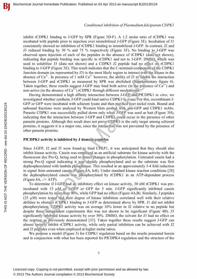
inhibit rCDPK1 binding to J-GFP by SPR (Figure 3D-F). A 1:2 molar ratio of rCDPK1 was incubated with peptide prior to injection over immobilized J-GFP (Figure 3E). Incubation of J1 consistently showed no inhibition of rCDPK1 binding to immobilized J-GFP. In contrast, J2 and J3 reduced binding by 30 % and 75 % respectively (Figure 3F). No binding to J-GFP was observed upon injection of each of the peptides in the absence of rCDPK1 (data not shown), indicating that peptide binding was specific to rCDPK1 and not to J-GFP. DMSO, which was used to solubilize J3 (data not shown) and a CDPK5 J2 peptide had no effect on rCDPK1 binding to J-GFP (Figure 3E). This result indicates that the C-terminal-component of the CDPK1 Junction domain (as represented by J3) is the most likely region to interact with the kinase in the absence of Ca2+. In presence of 1 mM Ca2+ however, the ability of J3 to inhibit the interaction between J-GFP and rCDPK1 as measured by SPR was abolished (Supplementary figure 4). Taken together, these results suggest J-GFP may bind both active (in the presence of Ca2+) and non-active (in the absence of Ca2+) rCDPK1 through different mechanisms.
Having demonstrated a high affinity interaction between J-GFP and PfCDPK1 in vitro, we investigated whether synthetic J-GFP could bind native CDPK1 in trans. For this, recombinant J-GFP or GFP were incubated with schizont lysate and then purified over nickel resin. Bound and unbound fractions were analysed by Western blots probed with anti-GFP and CDPK1 mAbs. Parasite CDPK1 was successfully pulled down only when J-GFP was used as bait (Figure 3H), indicating that the interaction between J-GFP and CDPK1 could occur in the presence of other parasite proteins. Although this result does not prove CDPK1 is the only target among schizont proteins, it suggests it is a major one, since the interaction was not prevented by the presence of other parasite proteins. PfCDPK1 activity is inhibited by J domain peptides Since J-GFP, J2 and J3 were found to bind CPKP1, it was anticipated that they should also inhibit kinase activity. Casein was employed as an artificial substrate for kinase activity with the fluorescent dye Pro-Q, being used to detect changes in phosphorylation. Untreated casein had a strong Pro-Q signal indicating it was already phosphorylated and so the substrate was first dephosphorylated with lambda phosphatase. This resulted in an approximately 3-4 fold reduction in signal from untreated casein (Figure 4A, left). Under standard kinase reaction conditions [28] the dephosphorylated casein was phosphorylated by rCDPK1 in an ATP-dependant process (Figure 4A, -/+ ATP).
To determine if J-GFP had an inhibitory effect on kinase activity, 30 nM rCDPK1 was pre-
incubated with 15 M of J-GFP or GFP for 5 min. J-GFP significantly inhibited casein phosphorylation by more than 90%, while GFP had no effect (Figure 4A,B). Similarly, J peptides (25 M) were tested and their degree of kinase inhibition correlated well with their relative abilities to obstruct rCDPK1 binding to J-GFP as determined above by SPR. J1 did not inhibit phosphorylation. CDPK1 activity was on average 10% lower in J2 relative to no peptide but despite three independent experiments this was not shown to be significant (Figure 4B). J3 significantly inhibited kinase activity by over 50%. DMSO, the solvent for J3 had no effect on the reaction as previously demonstrated [33]. Taken together these results suggest J-GFP can almost entirely inhibit rCDPK1 activity, while only partial inhibition can be achieved with J2 and J3 peptides even when employed at higher molar ratios.
We propose a model (Figure 5) for CDPK1 regulation based on the results presented herein and in conjunction with what has been reported for PfCDPK4 regulation and the structure of the
Biochemical Journal Immediate Publication. Published on 03 Apr 2013 as manuscript BJ20130124T
HIS
IS N
OT
TH
E V
ER
SIO
N O
F R
EC
OR
D -
see
doi
:10.
1042
/BJ2
0130
124
Acce
pted
Man
uscr
ipt
Licenced copy. Copying is not permitted, except with prior permission and as allowed by law.
© 2013 The Authors Journal compilation © 2013 Biochemical Society

Conditional inhibition of Plasmodium falciparum CDPK1
8
CDPK1 orthologue in Toxoplasma gondii, TgCDPK3 [6, 7]. In the absence of Ca2+, the N-terminal region of the CDPK1 junction is bound to the KD, rendering the kinase inactive (Figure 5A, left). In this state the CLD is exposed and can be bound in trans by the C-terminal region of J-GFP (Figure 5B i). Support for this scenario is provided by the SPR experiments in which J3 could substantially reduce binding of J-GFP to the full-length kinase. In the presence of Ca2+ the CLD of CDPK1 develops high affinity for the C-terminal region of its own J-domain to which it binds in cis displacing the N-terminal region of the J-domain from the KD (Figure 5A right). We propose that the reason that J-GFP is able to inhibit CDPK1 activity in the presence of Ca2+ is by occupying in trans the exposed active site of CDPK1 KD (Figure 5B ii). Once bound to the KD active site, J-GFP is no longer susceptible to J3 obstructing its binding to the CLD. J3 however can inhibit kinase activity in Ca2+ buffer so some it’s and J-GFPs’s inhibitory properties might be due to their occupation of the CLD which could obstruct the binding the CDPK1’s auto-inhibitory J-domain to its CLD leaving the kinase less active.
Conditional transgenic expression of J-GFP causes developmental arrest in parasites Having established that the CDPK1 junction domain in the context of the J-GFP fusion is a potent inhibitor of the rCDPK1 kinase activity in vitro, we aimed to inhibit CDPK1 activity in parasites with an endogenously expressed version of this fusion protein. In order to overcome the potential toxicity of constitutive expression of J-GFP in transfected parasites, DD24, a FKBP12 mutant that previously showed optimal regulation of protein levels in NIH3T3 cells [19] and in P. falciparum [20], was fused at the C-terminus of J-GFP to generate the fusion protein J-GFP-HA-DD24. Sequence encoding J-GFP-HA-DD24 was placed under the control of the strong constitutive bi-directional promoter ef1- from P. berghei, which is also used to drive expression of the selectable marker gene human dhfr (Figure 6A). These parasites were cultured without Shld-1 to keep levels of the potentially toxic J-GFP-HA-DD24 low. As a control, a parasite line expressing an otherwise identical GFP-HA-DD24 plasmid without J-domain was also generated.
To determine if the fusion proteins were expressing and being conditionally regulated with Shld-1, ring stage parasites transfected with pEF-J-GFP-HA-DD24 or pEF-GFP-HA-DD24 plasmids were cultured with either 0.5 μM, 1.0 μM Shld-1 or no Shld-1 for 24 hrs and subject to Western blot analysis (Figure 6A). A marked increase in levels of each fusion protein was observed for parasites that had been cultured with both concentrations of Shld-1 (Figure 6A). Without Shld-1 no J-GFP-HA-DD24 could be detected so the fold induction with Shld-1 could not be ascertained. Normalizing the anti-HA antibody signal to that of anti-EXP2 (used as a loading control) indicated that J-GFP-HA-DD24 was expressed at lower levels than the GFP-HA-DD24 fusion protein, suggesting potential J-domain toxicity (Figure 6A).
To further investigate the possible toxic effect of J-GFP-HA-DD24 expression on cultured parasites, growth rates as measured by flow cytometry were compared between Shld-1 stabilized and non-stabilized lines. Ring stage parasite cultures of both the J-GFP-HA-DD24 and GFP-HA-DD24 lines were adjusted to 1% parasitemia and cultured in the presence or absence of 0.5 M Shld-1. Parasitemias were determined daily from days 2 to 5 (Figure 6B). The most obvious trend to emerge from the time course is that when J-GFP-HA-DD24 was Shld-1-stabilised the proliferation of the parasites more or less stalled from day 2 onwards indicating the fusion protein was toxic (Figure 6B). In the absence of Shld-1 J-GFP-HA-DD24 parasites continued to grow with the degree of growth inhibition in the presence of Shld-1, increasing from ~20% on days 2 & 3 to about 60% on days 4 & 5 (Supplementary figure 5). Most but not all growth
Biochemical Journal Immediate Publication. Published on 03 Apr 2013 as manuscript BJ20130124T
HIS
IS N
OT
TH
E V
ER
SIO
N O
F R
EC
OR
D -
see
doi
:10.
1042
/BJ2
0130
124
Acce
pted
Man
uscr
ipt
Licenced copy. Copying is not permitted, except with prior permission and as allowed by law.
© 2013 The Authors Journal compilation © 2013 Biochemical Society

Conditional inhibition of Plasmodium falciparum CDPK1
9
inhibition can be attributed to the J-GFP-HA-DD24 fusion protein because Shld-1 is itself mildly toxic. Relative to the Shld-1 free control, the compound inhibited GFP-HA-DD24 control parasites growth by about 20% at day 5 (Figure 6B, Supplementary figure 5).
To better understand when during the cell cycle J-GFP-HA-DD24 parasites were arresting, their flow cytometry histograms were examined. The histograms represent fluorescence intensity of ethidium bromide stained DNA in cells versus cell number (Figure 6C). It can be most clearly seen from days 2 to 3 before the cells loose synchronicity, that both Shld-1 free parasite lines progressively increased their DNA content, as they progress from rings to schizonts (Figure 6C). GFP-HA-DD24 parasites cultured in 0.5 µM Shld-1 also showed similar increases in DNA content by ethidium bromide staining, whereas J-GFP-HA-DD24 + 0.5 µM Shld-1 appeared to have arrested at a stage earlier than fully mature schizonts, but later than rings or trophozoites based on their DNA content (Figure 6C).
To validate these results a similar but independent experiment was done where parasite morphology was microscopically examined following Giemsa staining (Supplementary figure 6). In agreement with the flow cytometry analyses above, parasites grown without Shld-1 appeared to show normal development, alternating between ring/trophozoites or schizont/rings each consecutive day. This was similarly observed in the GFP-HA-DD24 control line in the presence of Shld-1. The J-GFP-HA-DD24 parasites grown with Shld-1 on the other hand appeared to contain mainly trophozoite and non-segmented schizont stages from day 3 onwards indicating an arrest in early schizogony (Supplementary figure 6). Parasitemia progression of these parasites compared to all other cultures suggests 80-90% did not egress the RBC and initiated a new cycle (Supplementary figure 6, bottom right).
To assess viability of the parasites maintained on Shld-1, mean GFP fluorescence was measured by flow cytometry after 1 and 3 days on Shld-1 (Supplementary figure 7). After 1 day in the presence of Shld-1, both J-GFP-HA-DD24 and control lines had an approximately 4-fold increase in mean GFP fluorescence. Following 3 days on Shld-1 mean GFP fluorescence of control parasites increased further to ~6-fold, in contrast to a 2-fold induction for J-GFP-HA-DD24. Collectively these results suggest J-GFP-HA-DD24 parasites were either mostly dead or dying and were beginning to loose GFP as they were breaking down. Alternatively, these experiments may have selected for parasites expressing significantly lower amounts of J-GFP-HA-DD24 fusion protein that were not toxic. To address this Shld-1 was removed from the J-GFP-HA-DD24 parasites that had been grown for 3 days in Shld-1. This resulted in only a marginal recovery of growth, confirming that the majority of parasites in the J-expressing population on Shld-1 were no longer viable (data not shown). Conclusions Herein, we applied for the first time an inducible system to conditionally express a fusion protein with dominant negative properties in P. falciparum. In using this strategy we present evidence that PfCDPK1 performs a critical function during early schizogony. Our motivation for using a dominant negative genetic approach was to provide complementary functional evidence to CDPK1 drug inhibitor studies and in this respect our phenotype most resembled the effects of purfalcamine. While we cannot be absolutely sure that our J-domain fusion protein was only targeting CDPK1 the fact that our J-domain pulled down the kinase from parasite extract and that its interaction with rCDPK1 failed to be blocked by a CDPK5 J-peptide (the kinase most similar to CDPK1) gives us confidence that our approach was specific. The only downside of our
Biochemical Journal Immediate Publication. Published on 03 Apr 2013 as manuscript BJ20130124T
HIS
IS N
OT
TH
E V
ER
SIO
N O
F R
EC
OR
D -
see
doi
:10.
1042
/BJ2
0130
124
Acce
pted
Man
uscr
ipt
Licenced copy. Copying is not permitted, except with prior permission and as allowed by law.
© 2013 The Authors Journal compilation © 2013 Biochemical Society

Conditional inhibition of Plasmodium falciparum CDPK1
10
method was that if CDPK1 has additional later functions we might have missed them because parasites arrested growth before then. This is supported by accumulating evidence indicating that CDPK1 may have multiple roles in the malaria parasite life cycle, in addition to its putative invasion related role in regulating the motor complex (Supplementary figure 8) [15-17, 29]. Adopting the strategy of conditionally expressing auto-inhibitory domains may enable further investigation of other kinases and possibly other enzymes that are regulated in trans by these domains. AUTHOUR CONTRIBUITION MFA, PRS, EK and PF performed the experiments. MFA, PRS, CQN, LAB, GW, BSC and PRG interpreted the results and wrote the manuscript. ACKNOWLEDGEMENTS The authors gratefully acknowledge the contribution to this work of the Victorian Operational Infrastructure Support Program received by the Burnet Institute. We thank the Australian Red Cross Blood Bank for the provision of human blood and Jacobus Pharmaceuticals for providing WR99210. FUNDING The work is supported by the National Health and Medical Research Council of Australia (NHMRC) through project and/or program grants (NHMRC award 637406 & 603720) and its Independent Research Institute Infrastructure Support Scheme and by São Paulo Research Foundation (FAPESP) and The National Council for Scientific and Technological Development (CNPq). MFA is a former postdoctoral fellow from FAPESP (Process No: 09/51026-4). REFERENCES 1 Anamika, Srinivasan, N. and Krupa, A. (2005) A genomic perspective of protein kinases in Plasmodium falciparum. Proteins. 58, 180-189 2 Ward, P., Equinet, L., Packer, J. and Doerig, C. (2004) Protein kinases of the human malaria parasite Plasmodium falciparum: the kinome of a divergent eukaryote. BMC Genomics. 5, 79 3 Solyakov, L., Halbert, J., Alam, M. M., Semblat, J. P., Dorin-Semblat, D., Reininger, L., Bottrill, A. R., Mistry, S., Abdi, A., Fennell, C., Holland, Z., Demarta, C., Bouza, Y., Sicard, A., Nivez, M. P., Eschenlauer, S., Lama, T., Thomas, D. C., Sharma, P., Agarwal, S., Kern, S., Pradel, G., Graciotti, M., Tobin, A. B. and Doerig, C. (2011) Global kinomic and phospho-proteomic analyses of the human malaria parasite Plasmodium falciparum. Nat. Commun. 2, 565 4 Harper, J. F. and Harmon, A. (2005) Plants, symbiosis and parasites: a calcium signalling connection. Nat. Rev. Mol. Cell Biol. 6, 555-566 5 Holder, A. A., Mohd Ridzuan, M. A. and Green, J. L. (2012) Calcium dependent protein kinase 1 and calcium fluxes in the malaria parasite. Microbes Infect. 14, 825-830
Biochemical Journal Immediate Publication. Published on 03 Apr 2013 as manuscript BJ20130124T
HIS
IS N
OT
TH
E V
ER
SIO
N O
F R
EC
OR
D -
see
doi
:10.
1042
/BJ2
0130
124
Acce
pted
Man
uscr
ipt
Licenced copy. Copying is not permitted, except with prior permission and as allowed by law.
© 2013 The Authors Journal compilation © 2013 Biochemical Society

Conditional inhibition of Plasmodium falciparum CDPK1
11
6 Wernimont, A. K., Artz, J. D., Finerty, P., Jr., Lin, Y. H., Amani, M., Allali-Hassani, A., Senisterra, G., Vedadi, M., Tempel, W., Mackenzie, F., Chau, I., Lourido, S., Sibley, L. D. and Hui, R. (2010) Structures of apicomplexan calcium-dependent protein kinases reveal mechanism of activation by calcium. Nat. Struct. Mol. Biol. 17, 596-601 7 Ranjan, R., Ahmed, A., Gourinath, S. and Sharma, P. (2009) Dissection of mechanisms involved in the regulation of Plasmodium falciparum calcium-dependent protein kinase 4. J. Biol. Chem. 284, 15267-15276 8 Billker, O., Lourido, S. and Sibley, L. D. (2009) Calcium-dependent signaling and kinases in apicomplexan parasites. Cell Host Microbe. 5, 612-622 9 Zhao, Y., Kappes, B. and Franklin, R. M. (1993) Gene structure and expression of an unusual protein kinase from Plasmodium falciparum homologous at its carboxyl terminus with the EF hand calcium-binding proteins. J. Biol. Chem. 268, 4347-4354 10 Zhao, Y., Pokutta, S., Maurer, P., Lindt, M., Franklin, R. M. and Kappes, B. (1994) Calcium-binding properties of a calcium-dependent protein kinase from Plasmodium falciparum and the significance of individual calcium-binding sites for kinase activation. Biochemistry. 33, 3714-3721 11 Bozdech, Z., Llinas, M., Pulliam, B. L., Wong, E. D., Zhu, J. and DeRisi, J. L. (2003) The transcriptome of the intraerythrocytic developmental cycle of Plasmodium falciparum. PLoS Biol. 1, E5 12 Le Roch, K. G., Zhou, Y., Blair, P. L., Grainger, M., Moch, J. K., Haynes, J. D., De La Vega, P., Holder, A. A., Batalov, S., Carucci, D. J. and Winzeler, E. A. (2003) Discovery of gene function by expression profiling of the malaria parasite life cycle. Science. 301, 1503-1508 13 Dammann, C., Ichida, A., Hong, B., Romanowsky, S. M., Hrabak, E. M., Harmon, A. C., Pickard, B. G. and Harper, J. F. (2003) Subcellular targeting of nine calcium-dependent protein kinase isoforms from Arabidopsis. Plant Physiol. 132, 1840-1848 14 Moskes, C., Burghaus, P. A., Wernli, B., Sauder, U., Durrenberger, M. and Kappes, B. (2004) Export of Plasmodium falciparum calcium-dependent protein kinase 1 to the parasitophorous vacuole is dependent on three N-terminal membrane anchor motifs. Mol. Microbiol. 54, 676-691 15 Green, J. L., Rees-Channer, R. R., Howell, S. A., Martin, S. R., Knuepfer, E., Taylor, H. M., Grainger, M. and Holder, A. A. (2008) The motor complex of Plasmodium falciparum: phosphorylation by a calcium-dependent protein kinase. J. Biol. Chem. 283, 30980-30989 16 Sebastian, S., Brochet, M., Collins, M. O., Schwach, F., Jones, M. L., Goulding, D., Rayner, J. C., Choudhary, J. S. and Billker, O. (2012) A Plasmodium calcium-dependent protein kinase controls zygote development and transmission by translationally activating repressed mRNAs. Cell Host Microbe. 12, 9-19 17 Kato, N., Sakata, T., Breton, G., Le Roch, K. G., Nagle, A., Andersen, C., Bursulaya, B., Henson, K., Johnson, J., Kumar, K. A., Marr, F., Mason, D., McNamara, C., Plouffe, D., Ramachandran, V., Spooner, M., Tuntland, T., Zhou, Y., Peters, E. C., Chatterjee, A., Schultz, P. G., Ward, G. E., Gray, N., Harper, J. and Winzeler, E. A. (2008) Gene expression signatures and small-molecule compounds link a protein kinase to Plasmodium falciparum motility. Nat. Chem. Biol. 4, 347-356
Biochemical Journal Immediate Publication. Published on 03 Apr 2013 as manuscript BJ20130124T
HIS
IS N
OT
TH
E V
ER
SIO
N O
F R
EC
OR
D -
see
doi
:10.
1042
/BJ2
0130
124
Acce
pted
Man
uscr
ipt
Licenced copy. Copying is not permitted, except with prior permission and as allowed by law.
© 2013 The Authors Journal compilation © 2013 Biochemical Society

Conditional inhibition of Plasmodium falciparum CDPK1
12
18 Banaszynski, L. A., Chen, L. C., Maynard-Smith, L. A., Ooi, A. G. and Wandless, T. J. (2006) A rapid, reversible, and tunable method to regulate protein function in living cells using synthetic small molecules. Cell. 126, 995-1004 19 Chu, B. W., Banaszynski, L. A., Chen, L. C. and Wandless, T. J. (2008) Recent progress with FKBP-derived destabilizing domains. Bioorg. Med. Chem. Lett. 18, 5941-5944 20 de Azevedo, M. F., Gilson, P. R., Gabriel, H. B., Simoes, R. F., Angrisano, F., Baum, J., Crabb, B. S. and Wunderlich, G. (2012) Systematic analysis of FKBP inducible degradation domain tagging strategies for the human malaria parasite Plasmodium falciparum. PLoS One. 7, e40981 21 Crabb, B. S. and Cowman, A. F. (1996) Characterization of promoters and stable transfection by homologous and nonhomologous recombination in Plasmodium falciparum. Proc. Natl. Acad. Sci. U.S.A. 93, 7289-7294 22 Fidock, D. A. and Wellems, T. E. (1997) Transformation with human dihydrofolate reductase renders malaria parasites insensitive to WR99210 but does not affect the intrinsic activity of proguanil. Proc. Natl. Acad. Sci. U.S.A. 94, 10931-10936 23 Trager, W. and Jensen, J. B. (1976) Human malaria parasites in continuous culture. Science. 193, 673-675 24 Wilson, D. W., Crabb, B. S. and Beeson, J. G. (2010) Development of fluorescent Plasmodium falciparum for in vitro growth inhibition assays. Malar. J. 9, 152 25 Wickham, M. E., Thompson, J. K. and Cowman, A. F. (2003) Characterisation of the merozoite surface protein-2 promoter using stable and transient transfection in Plasmodium falciparum. Mol. Biochem. Parasitol. 129, 147-156 26 Baum, J., Richard, D., Healer, J., Rug, M., Krnajski, Z., Gilberger, T. W., Green, J. L., Holder, A. A. and Cowman, A. F. (2006) A conserved molecular motor drives cell invasion and gliding motility across malaria life cycle stages and other apicomplexan parasites. J. Biol. Chem. 281, 5197-5208 27 Bullen, H. E., Charnaud, S. C., Kalanon, M., Riglar, D. T., Dekiwadia, C., Kangwanrangsan, N., Torii, M., Tsuboi, T., Baum, J., Ralph, S. A., Cowman, A. F., de Koning-Ward, T. F., Crabb, B. S. and Gilson, P. R. (2012) Biosynthesis, localisation and macromolecular arrangement of the Plasmodium falciparum translocon of exported proteins; PTEX. J. Biol. Chem. 287, 7871-7784 28 Zhao, Y., Franklin, R. M. and Kappes, B. (1994) Plasmodium falciparum calcium-dependent protein kinase phosphorylates proteins of the host erythrocytic membrane. Mol. Biochem. Parasitol. 66, 329-343 29 Bansal, A., Singh, S., More, K. R., Hans, D., Nangalia, K., Yogavel, M., Sharma, A. and Chitnis, C. E. (2012) Characterization of Plasmodium falciparum calcium dependent protein kinase 1 (PfCDPK1) and its role in microneme secretion during erythrocyte invasion. J. Biol. Chem. 288, 1590-1602 30 Armstrong, C. M. and Goldberg, D. E. (2007) An FKBP destabilization domain modulates protein levels in Plasmodium falciparum. Nat. Methods. 4, 1007-1009 31 Dvorin, J. D., Martyn, D. C., Patel, S. D., Grimley, J. S., Collins, C. R., Hopp, C. S., Bright, A. T., Westenberger, S., Winzeler, E., Blackman, M. J., Baker, D. A., Wandless, T. J. and Duraisingh, M. T. (2010) A plant-like kinase in Plasmodium falciparum regulates parasite egress from erythrocytes. Science. 328, 910-912 32 Harmon, A. C., Yoo, B. C. and McCaffery, C. (1994) Pseudosubstrate inhibition of CDPK, a protein kinase with a calmodulin-like domain. Biochemistry. 33, 7278-7287
Biochemical Journal Immediate Publication. Published on 03 Apr 2013 as manuscript BJ20130124T
HIS
IS N
OT
TH
E V
ER
SIO
N O
F R
EC
OR
D -
see
doi
:10.
1042
/BJ2
0130
124
Acce
pted
Man
uscr
ipt
Licenced copy. Copying is not permitted, except with prior permission and as allowed by law.
© 2013 The Authors Journal compilation © 2013 Biochemical Society

Conditional inhibition of Plasmodium falciparum CDPK1
13
33 Lemercier, G., Fernandez-Montalvan, A., Shaw, J. P., Kugelstadt, D., Bomke, J., Domostoj, M., Schwarz, M. K., Scheer, A., Kappes, B. and Leroy, D. (2009) Identification and characterization of novel small molecules as potent inhibitors of the plasmodial calcium-dependent protein kinase 1. Biochemistry. 48, 6379-6389
FIGURE LEGENDS FIGURE 1. CDPK1-GFP localises to the plasma membrane of schizonts, merozoites and early rings. (A) Schematic diagram of the GFP and GFP-DD fusion proteins. (B) Live fluorescence images of CDPK1-GFP parasites showing the fusion protein localizing to the plasma membrane. i and ii - early schizonts showing the GFP fluorescence at the parasite periphery, iii and iv - late segmented schizonts with GFP fluorescence surrounding each newly formed merozoite, v – a single free merozoite with strong plasma membrane GFP fluorescence and a newly invaded ring-stage parasite where fluorescence at the ring periphery remains detectable. Membranes have been stained with CellMask Orange. Scale bars = 2 µm. FIGURE 2. Conditional regulation of CDPK1 fusion proteins levels. Synchronous CDPK1-GFP and CDPK1-GFP-DD ring stage parasites were split and maintained +/- of Shld-1 for 3 days. Proteins were extracted at schizont stage and CDPK1 fusion expression levels were assessed by probing Western blots with antibodies for GFP and a loading control, MSP2. Expression of the CDPK1-GFP and CDPK1-GFP-DD in parasites maintained +/- Shld-1 was quantified by densitometry normalized to MSP2 levels. Bars represent means of 5 western blots standard deviation (SD). Data was analysed by t test. *Statistically different (p < 0.05). **Not statistically different (p > 0.05). FIGURE 3. CDPK1 junction region fused to GFP at its N-terminus binds to the full-length kinase. (A) Diagram of CDPK1 showing its N-terminal domain (N), kinase domain (KD), junction region (J) and calmodulin-like domain (CLD) as well as the J-GFP and GFP fusion proteins. (B) Recombinant CDPK1 was incubated with either GFP or J-GFP and the kinase immunoprecipitated with anti-CDPK1 mAb. Western blots (WB) of the bound (precipitated) and unbound fractions were probed with CDPK1 mAb (CDPK1) or anti-GFP rabbit polyclonal IgG ( . (C) Biochemical analysis of the recombinant J-GFP - CDPK1 interaction by surface plasmon resonance. Serial two-fold dilutions of CDPK1 (0-100 nM) were injected through flow cells containing GFP (as a reference) and J-GFP captured on a CM5 sensor chip for 180 s. Kinetic data for the J-GFP - CDPK1 interaction is represented and these sensorgrams best fitted to a two-state (conformational change) binding model. (D) Synthetic peptides spanning the CDPK1 and CDPK5 junction regions were designed. (E) SPR sensorgrams showing J-GFP binding to CDPK1 that was pre-incubated with J-peptides in a 2:1 molar ratio. The interactions of J-peptide treated CDPK1 (relative to a no peptide rCDPK1 positive control) with immobilized J-GFP was analysed with immobilized GFP as a reference. (F) The percentage of CDPK1 incubated with the indicated J-peptide binding to J-GFP relative to CDPK1 alone was plotted. Bars represent the mean ± SD of 4 independent experiments. (G) P. falciparum schizont lysate was incubated with either J-GFP or GFP and the recombinant proteins were captured by His-trap.
Biochemical Journal Immediate Publication. Published on 03 Apr 2013 as manuscript BJ20130124T
HIS
IS N
OT
TH
E V
ER
SIO
N O
F R
EC
OR
D -
see
doi
:10.
1042
/BJ2
0130
124
Acce
pted
Man
uscr
ipt
Licenced copy. Copying is not permitted, except with prior permission and as allowed by law.
© 2013 The Authors Journal compilation © 2013 Biochemical Society

Conditional inhibition of Plasmodium falciparum CDPK1
14
Bound and unbound fractions were analysed by western blots probed with anti-GFP or anti-CDPK1 mAb. FIGURE 4. Inhibition of CDPK1 phosphorylation activity. (A) Casein was dephosphorylated with λ phosphatase to be used as a substrate for recombinant CDPK1 phosphorylation assays. The phosphorylation reaction was analysed by SDS-PAGE. Total protein and phosphorylated protein was stained with Sypro Ruby and Pro-Q stains respectively. Kinase reactions performed in the absence (-) or presence (+) of ATP were used as negative and positive controls respectively. Recombinant CDPK1 was pre-incubated with 15 μM GFP or J-GFP, 25 μM J1, J2 or J3 or 2% DMSO. A representative gel from 3 independent experiments is shown. (B) After normalizing the Pro-Q signal to that of Sypro Ruby for individual lanes and subtracting the negative control signal, the level of phosphorylation in each of the reaction conditions is represented relative to the positive control. Results represent the mean ± SD of 3 independent experiments. Data was analysed by one-way ANOVA. *Significantly different from control (p <0.05). FIGURE 5. A model for PfCDPK1 inhibition by J-GFP. (A) Without calcium ions (Ca2+), CDPK1 is maintained in an inactive state (purple) via the auto-inhibitory in cis interaction of the N-terminal part of its junction region (JN) with the kinase domain (KD). The binding of Ca2+ to the calmodulin-like domain (CLD) of CDPK1 triggers a conformational change in the CLD that results in binding to the C-terminal region of J (JC) displacing JN from the KD thereby activating the kinase (green). (B) i -Without Ca2+
, J-GFP binds via JC to the CLD of CDPK1 in cis ii – In the presence of Ca2+, JN of J-GFP binds to the KD of CDPK1 inhibiting its activity. FIGURE 6. Expression of the J-domain of CDPK1 as a J-GFP-HA-DD24 fusion protein arrests parasite growth. (A) Diagrammatic representation of gene construct and conditional expression of CDPK1 junction as a fusion protein under regulation of the DD24 system. Total protein extracts from transgenic parasites cultured with Shld-1 (concentrations indicated) for 24 hours were analysed for fusion protein expression. Junction and control proteins were detected by Western blots probed with anti-HA (αHA). Blots were probed with αEXP2 as a loading control.
(B) Ring stage parasites were incubated in the presence or absence of 0.5 M Shld-1 and parasitemias determined over five days by flow cytometry. (C) Flow cytometry histograms showing cell DNA content (x axis log scale) versus cell number (y axis) in parasites +/- Shld-1. Ring stage J-GFP-HA-DD24 or GFP-HA-DD24 control parasites were treated +/- Shld-1 and parasite progression through the blood-stage cycle was monitored by ethidium bromide staining and flow cytometry between days 2 to 5.
Biochemical Journal Immediate Publication. Published on 03 Apr 2013 as manuscript BJ20130124T
HIS
IS N
OT
TH
E V
ER
SIO
N O
F R
EC
OR
D -
see
doi
:10.
1042
/BJ2
0130
124
Acce
pted
Man
uscr
ipt
Licenced copy. Copying is not permitted, except with prior permission and as allowed by law.
© 2013 The Authors Journal compilation © 2013 Biochemical Society
Conditional inhibition of Plasmodium falciparum CDPK1
15
Figure 1
Biochemical Journal Immediate Publication. Published on 03 Apr 2013 as manuscript BJ20130124T
HIS
IS N
OT
TH
E V
ER
SIO
N O
F R
EC
OR
D -
see
doi
:10.
1042
/BJ2
0130
124
Acce
pted
Man
uscr
ipt
Licenced copy. Copying is not permitted, except with prior permission and as allowed by law.
© 2013 The Authors Journal compilation © 2013 Biochemical Society

Conditional inhibition of Plasmodium falciparum CDPK1
16
Figure 2
Biochemical Journal Immediate Publication. Published on 03 Apr 2013 as manuscript BJ20130124T
HIS
IS N
OT
TH
E V
ER
SIO
N O
F R
EC
OR
D -
see
doi
:10.
1042
/BJ2
0130
124
Acce
pted
Man
uscr
ipt
Licenced copy. Copying is not permitted, except with prior permission and as allowed by law.
© 2013 The Authors Journal compilation © 2013 Biochemical Society

Conditional inhibition of Plasmodium falciparum CDPK1
17
Figure 3
Biochemical Journal Immediate Publication. Published on 03 Apr 2013 as manuscript BJ20130124T
HIS
IS N
OT
TH
E V
ER
SIO
N O
F R
EC
OR
D -
see
doi
:10.
1042
/BJ2
0130
124
Acce
pted
Man
uscr
ipt
Licenced copy. Copying is not permitted, except with prior permission and as allowed by law.
© 2013 The Authors Journal compilation © 2013 Biochemical Society

Conditional inhibition of Plasmodium falciparum CDPK1
18
Figure 4
Biochemical Journal Immediate Publication. Published on 03 Apr 2013 as manuscript BJ20130124T
HIS
IS N
OT
TH
E V
ER
SIO
N O
F R
EC
OR
D -
see
doi
:10.
1042
/BJ2
0130
124
Acce
pted
Man
uscr
ipt
Licenced copy. Copying is not permitted, except with prior permission and as allowed by law.
© 2013 The Authors Journal compilation © 2013 Biochemical Society

Conditional inhibition of Plasmodium falciparum CDPK1
19
Figure 5
Biochemical Journal Immediate Publication. Published on 03 Apr 2013 as manuscript BJ20130124T
HIS
IS N
OT
TH
E V
ER
SIO
N O
F R
EC
OR
D -
see
doi
:10.
1042
/BJ2
0130
124
Acce
pted
Man
uscr
ipt
Licenced copy. Copying is not permitted, except with prior permission and as allowed by law.
© 2013 The Authors Journal compilation © 2013 Biochemical Society

Conditional inhibition of Plasmodium falciparum CDPK1
20
Figure 6
Biochemical Journal Immediate Publication. Published on 03 Apr 2013 as manuscript BJ20130124T
HIS
IS N
OT
TH
E V
ER
SIO
N O
F R
EC
OR
D -
see
doi
:10.
1042
/BJ2
0130
124
Acce
pted
Man
uscr
ipt
Licenced copy. Copying is not permitted, except with prior permission and as allowed by law.
© 2013 The Authors Journal compilation © 2013 Biochemical Society