Concussion and Chronic Traumatic Encephalopathy … · Biomechanics How much is enough ... Boxing...
61
0 Blast Injury Institute Concussion and Chronic Traumatic Encephalopathy (CTE) James P. Kelly, MA, MD Director National Intrepid Center of Excellence Clinical Professor of Neurosurgery University of Colorado School of Medicine
-
Upload
duongnguyet -
Category
Documents
-
view
218 -
download
0
Transcript of Concussion and Chronic Traumatic Encephalopathy … · Biomechanics How much is enough ... Boxing...

0
Blast Injury Institute
Concussion and Chronic Traumatic
Encephalopathy (CTE)
James P. Kelly, MA, MD
Director
National Intrepid Center of Excellence
Clinical Professor of Neurosurgery
University of Colorado School of Medicine

1
Primary: Direct exposure to
over pressurization wave
Blast Injury

2
IED: Improvised Explosive Device
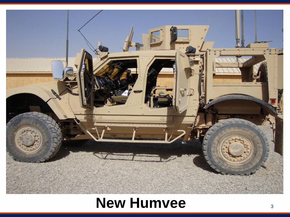
3New Humvee
4
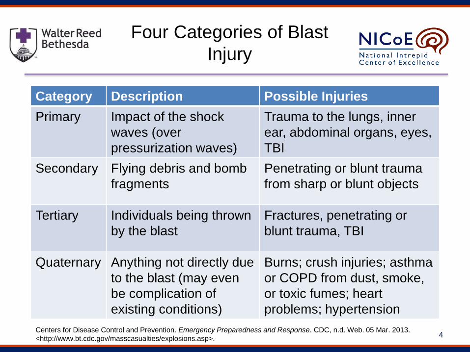
Four Categories of Blast
Injury
Category Description Possible Injuries
Primary Impact of the shock
waves (over
pressurization waves)
Trauma to the lungs, inner
ear, abdominal organs, eyes,
TBI
Secondary Flying debris and bomb
fragments
Penetrating or blunt trauma
from sharp or blunt objects
Tertiary Individuals being thrown
by the blast
Fractures, penetrating or
blunt trauma, TBI
Quaternary Anything not directly due
to the blast (may even
be complication of
existing conditions)
Burns; crush injuries; asthma
or COPD from dust, smoke,
or toxic fumes; heart
problems; hypertension
Centers for Disease Control and Prevention. Emergency Preparedness and Response. CDC, n.d. Web. 05 Mar. 2013.
<http://www.bt.cdc.gov/masscasualties/explosions.asp>.

5
TBI Numbers By Severity
4,213 2,709
21,779
219.921
18,188
Penetrating - 2%
Severe - 1%
Moderate - 8%
Mild - 82%
Not Classifiable - 7%
DoD Numbers for Traumatic Brain Injury
’00 – ’12 Totals
Source: Armed Forces Health Surveillance Center
Total 266,810


UNKNOWN CLINICAL APPLICATION
Injury
Biomechanics
How much is enough to cause concussion?
Diagnosis &
Assessment
What are the most effective methods to
diagnose and evaluate athletes with SRC?
Recovery Time How long does it typically take to recover after
SRC?
Injury
Management
When is it safe for an athlete to return to play?
Long-term Effects Are there potential long-term risks associated
with repetitive concussion?
Sport Concussion: Clinician Challenges
What Does the Science Tell Us?

What is a Concussion?After blow to head, disruption of normal brain cellular activity (“Neurometabolic Cascade”) commonly causes rapid onset of neurologic dysfunction:
• Clinical Symptoms: headache, dizziness, dazed/confused, poor concentration, feeling in a fog, nausea, etc.
• Physical Signs/Acute Injury Characteristics: LOC (<10%), PTA (<25%)
• Neurobehavioral Changes: irritability, mood changes, etc.
• Cognitive Impairment: memory, attention, reaction time, processing speed
• Sleep Disturbance: drowsiness, insomnia, hypersomnia
It Starts at the Beginning:
Concussion is about what
happened to the athlete at
the time of the injury
event
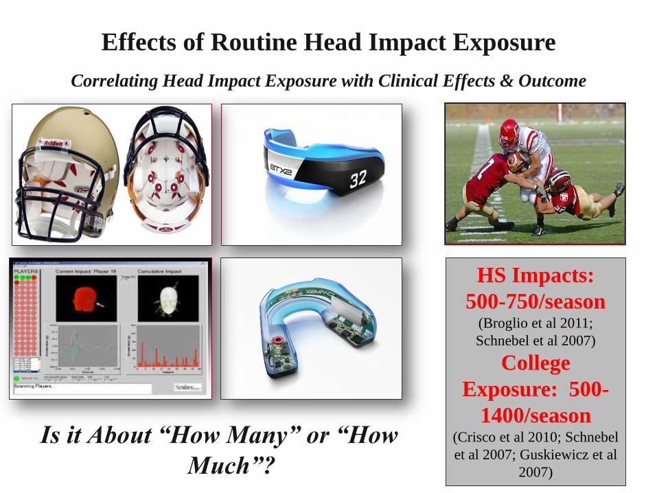
Correlating Head Impact Exposure with Clinical Effects & Outcome
Effects of Routine Head Impact Exposure
Is it About “How Many” or “How
Much”?
HS Impacts:
500-750/season (Broglio et al 2011;
Schnebel et al 2007)
College
Exposure: 500-
1400/season (Crisco et al 2010; Schnebel
et al 2007; Guskiewicz et al
2007)

Symptom Recovery
0
5
10
15
20
25
30
BL CC PG D1 D2 D3 D5 D7 D90
Assessment Point
GS
C T
otal S
co
re
NCAA Control
NCAA Concussion
Higher score indicates more severe symptoms; error bars represent 95% CIMcCrea et al., JAMA 2003
Symptom Recovery
0
5
10
15
20
25
30
BL CC PG D1 D2 D3 D5 D6 D45Assessment Point
GS
C T
otal S
co
re
HS Control
HS Concussion

Cognitive Recovery
23
24
25
26
27
28
29
30
BL CC PG D1 D2 D3 D5 D7 D90
Assessment Point
SA
C T
otal S
co
re
NCAA Control
NCAA Concussion
Lower score indicates more severe cognitive impairment; error bars = 95% CIMcCrea et al., JAMA 2003
Cognitive Recovery
23
24
25
26
27
28
29
30
BL CC PG D1 D2 D3 D5 D6 D45
Assessment Point
SA
C T
otal S
co
re
HS Control
HS Concussion
p < .001 p < .001

McCrea et al., JAMA 2003; 290:2556-2563
> 80% Achieve Complete Recovery in 7-10
Days
75% of repeat concussions within first 7 days
92% of repeat concussions within first 10 days
Association Between Recovery & Risk

• 10% take > 7 days to recover Other reports of higher
percentages
• < 5% take > 45 days
• Acute severity predicts
recovery

14
Amateur boxing
Hässleholm bildbyrå, Sweden

15
Hypothesis
• Diffuse Axonal Injury (DAI)
– Shearing of neurons
• Leakage of proteins in the
cerebrospinal fluid (CSF)
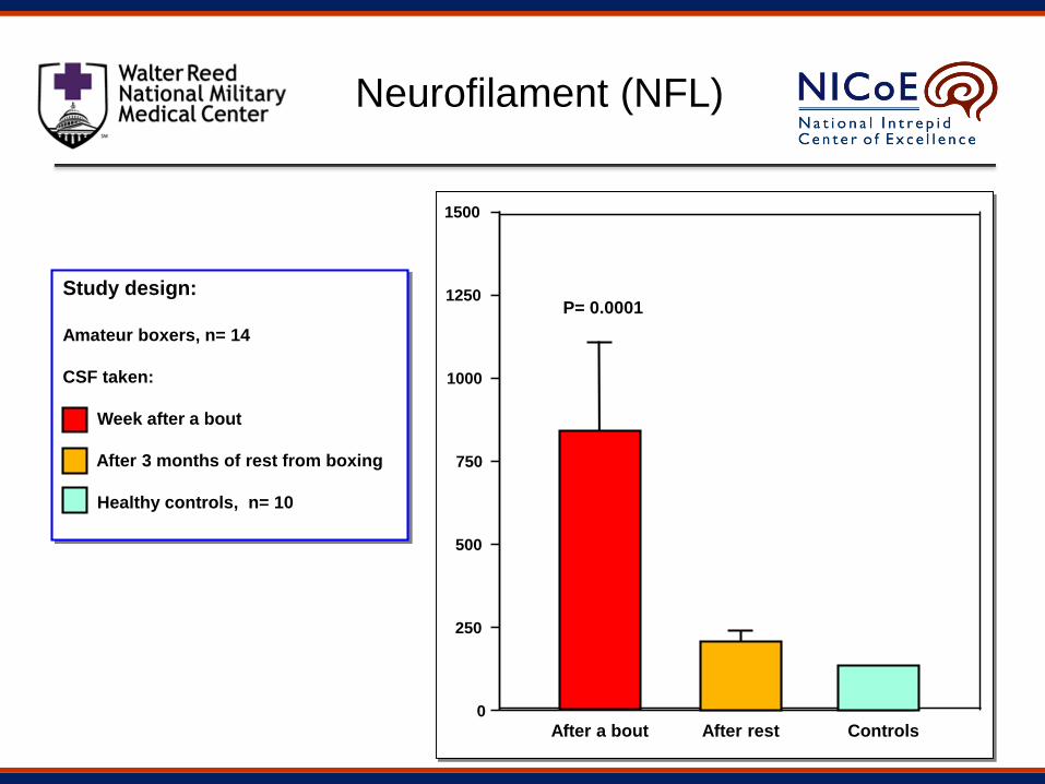
– NFL (neurons)
– GFAP (astrocytes)
– Tau (neurons)
• Lumbar puncture to access
CSF
GFAP
NFL
Tau

16
Boxing Study
• 14 boxers (3 female), age 22±3.8 yrs
• 10 Controls (0 female), age 30±6.3 yrs
• LP 7-10 days after bout
• New LP 3 months after rest
• Interviews at day 7-10
– Amount of hits to the head
– Severity, ie grogginess

17
Total Tau
0
100
200
300
400
500
600
700
P= 0.006
After a bout After rest Controls
Study design:
Amateur boxers, n= 14
CSF taken:
Week after a bout
After 3 months of rest from boxing
Healthy controls, n= 10
18
Neurofilament (NFL)
Study design:
Amateur boxers, n= 14
CSF taken:
Week after a bout
After 3 months of rest from boxing
Healthy controls, n= 10
After a bout After rest Controls 0
250
500
750
1000
1250
1500
P= 0.0001

19
CSF-biomarkers
Zetterberg, Hietala. et al. Arch Neurol 2006;63:1277-1280.
Copyright restrictions may apply.
TAUNFL GFAP
Many (>15) or high impact hits
Few hits

Pathophysiology
• Biomechanical forces cause tissue
deformation, shearing and fluid wave
propagation through the hemisphere
• Irritation leads to rapid, chaotic electrical
depolarization across the cortex

Pathophysiology - 2
• Neurotransmitters / neurochemicals are
released in excessive (excitotoxic)
amounts, driving up cellular metabolism
(hyperglycolysis) and lactic acid levels

Pathophysiology - 3
• Na – K pump failure and axonal stretch
injury lead to Calcium influx and axonal
swelling or disintegration

Pathology
• Rotational injuries lead to diffuse shearing
of small vessels
• Diffuse axonal injury is underlying lesion

24
MRI Findings
CT Routine MRI- GRE New TBI Study- SWI
Read as Normal Possible Lesion
Corpus Callosum
Multiple Lesions Detected
?
25
Brain Trauma-Related Neurodegeneration:Strategies to Define, Detect and Predict
July 22-23, 2013Bethesda, Maryland
Sponsors
• National Institutes of Health (NIH)
• Foundation for the NIH (FNIH)
• National Football League (NFL)

26
Chronic Traumatic Encephalopathy
• Punch-Drunkeness (Martland, 1928)
• Dementia Pugilistica (Millspough, 1937)
• Chronic Progressive Traumatic Encephalopathy of the
Boxer (Critchley, 1957)
• Psychopathic Deterioration of Pugilists (Courville, 1962)

Mechanics of Boxing
• Unterharnscheidt, Friedrich. About boxing: review of historical and medical aspects. Texas Report of Biology and Medicine 1970;28:421-495.

Dementia Pugilistica
• Neuronal loss, gliosis, senile plaques, hydrocephalus, attenuation of the corpus callosum, diffuse axonal injury, neurofibrillary tangles, cerebellar atrophy
• Jack Dempsey, Joe Louis, Floyd Paterson, Jerry Quarry, Mike Quarry, Emile Griffith, Sugar Ray Robinson and Muhammed Ali (Parkinsonism variant)

CT of 338 Active Pro Boxers
• Abnormal in 25 (7%)
• Atrophy most common finding (the number of
bout was correlated with degree of atrophy)
• Focal low density (traumatic encephalopathy) in
only 3 boxers
(Jordan, BD et al 1992)
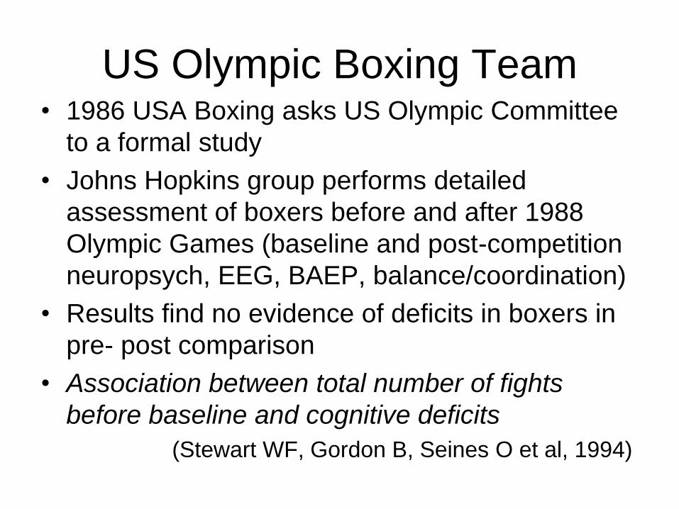
US Olympic Boxing Team• 1986 USA Boxing asks US Olympic Committee
to a formal study
• Johns Hopkins group performs detailed
assessment of boxers before and after 1988
Olympic Games (baseline and post-competition
neuropsych, EEG, BAEP, balance/coordination)
• Results find no evidence of deficits in boxers in
pre- post comparison
• Association between total number of fights
before baseline and cognitive deficits
(Stewart WF, Gordon B, Seines O et al, 1994)

Boxers at USMA – West Point
• 14 boxers who had sustained concussions
(n=6 AAN Grades I, n=8 Grade 2) with full
symptom recovery by day 4 post-injury
• All had pre-injury baseline computerized
neuropsychological testing
• No cadet had returned to baseline on
Simple Reaction Time on day 4 post-injury
(Warden DL et al, 2001)

Boxers at Notre Dame
• 82 collegiate amateur boxers participating in a 7-day single elimination tournament
• 30 non-boxing controls
• Used computerized test battery before and within 2 hours after each bout
• Except for boxers whose bouts were stopped by the referee (n=7), there was no evidence in post-bout cognitive dysfunction
(Moriarty et al, 2004)

Diffusion Tensor MRI
• Regions of increased and decreased
apparent diffusion coefficient (ADC), and
decreased fractional anisotropy (FA) in
boxers compared to controls
• Abnormalities are assumed to reflect
chronic, cumulative brain injury resulting
from repeated mild TBI in boxers
(Chappell MH et al, 2006)

What about football?
Football (UK) = Soccer (US)

Football study
• 23 football players
– 10 headed the ball >10x
– 13 headed the ball >20x
• 10 controls
• 30m “corner kick”
• LP 7-10 days after

Results - Football

Conclusion - Football
No neurochemical evidence for neuronal
injury caused by heading in soccer
Zetterberg et al. Accepted for publication BJSM, 03/28/2007

38
Junior Seau #55

39
2 May 2012
40
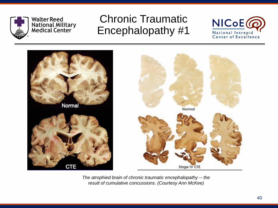
The atrophied brain of chronic traumatic encephalopathy -- the
result of cumulative concussions. (Courtesy Ann McKee)
Chronic Traumatic Encephalopathy #1

41
Top row: Brain sections showing dense tau protein deposition in multiple areas of frontal
cortex (boxes). Bottom row: Microscopic images showing large numbers of tau-
containing neurofibrillary tangles (dark brown spots) in the areas of damage.
Chronic Traumatic Encephalopathy #2
42
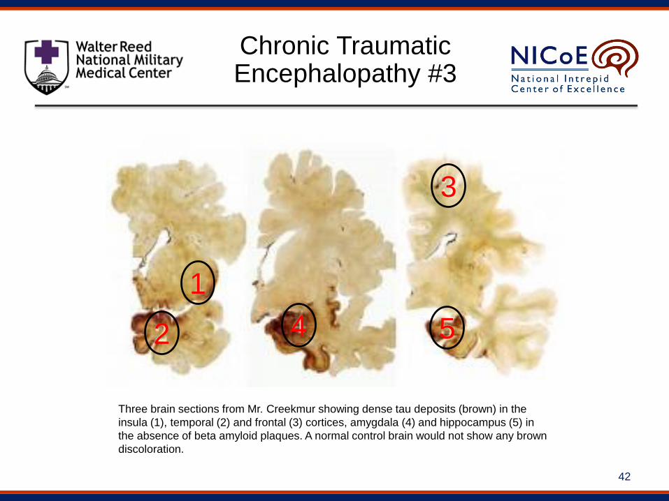
Three brain sections from Mr. Creekmur showing dense tau deposits (brown) in the
insula (1), temporal (2) and frontal (3) cortices, amygdala (4) and hippocampus (5) in
the absence of beta amyloid plaques. A normal control brain would not show any brown
discoloration.
1
2
3
4 5
Chronic Traumatic Encephalopathy #3

43
Photographs by Ann C. McKee, Boston University/Bedford Veterans Hospital
Chronic Traumatic Encephalopathy #4

44
Cerebral Cortex / Bielschowski silver stain
Parkinson-Dementia Complex of Guam (Daniel Perl, MD)

45
Magnified View of Neurofibrillary Tangles in Hippocampus
Parkinson-Dementia Complex of Guam (Daniel Perl, MD)

46
PET Scanning of Brain Tau in Retired National Football League Players: Preliminary Findings
• 5 retired NFL players ages 45-73
• FDDNP-PET signals (tau & amyloid) were higher in players in all
subcortical regions and the amygdala
Small, GW et al. Geriatric Psychiatry 2013;21:138-144

47
Traumatic Brain Injury and Chronic Traumatic
Encephalopathy: A Forensic Neuropsychiatric
Perspective
• There are no uniform diagnostic criteria for
mTBI and CTE
• The evidence for a relationship between TBI
and CTE is mixed and post-mortem only
Wortzel HS, Brenner LA, Arciniegas, DB .
Behavioral Sciences and the Law. 2013

THE NATIONAL INTREPID CENTER OF
EXCELLENCEan instrument of hope, healing, discovery and learning

49
NICoE Satellites(Artist’s Rendering)

SWI T2-FLAIR T2-FLAIR
Extensive microhemorrhage- DAI
76 scattered T2 hyper-intensities
Small focus of encephalomalacia and
gliosis inferior Left frontal Lobe

Radiology Findings: Structural
0.00
10.00
20.00
30.00
40.00
50.00
60.00
1.85 2.775.35
8.49 8.67
13.2814.58 15.31
16.79 17.53
39.67
43.17
51.77 52.03
Pe
rce
nta
ge
Structural Imaging FindingsN = 542
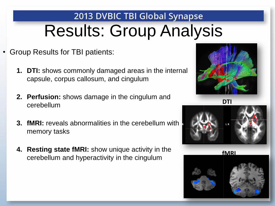
Results: Group Analysis
DTI
• Group Results for TBI patients:
1. DTI: shows commonly damaged areas in the internal
capsule, corpus callosum, and cingulum
2. Perfusion: shows damage in the cingulum and
cerebellum
3. fMRI: reveals abnormalities in the cerebellum with
memory tasks
4. Resting state fMRI: show unique activity in the
cerebellum and hyperactivity in the cingulumfMRI
53
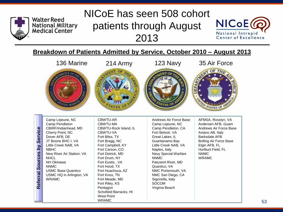
NICoE has seen 508 cohort
patients through August
2013
Breakdown of Patients Admitted by Service, October 2010 – August 2013
CBWTU-AR
CBWTU-MA
CBWTU-Rock Island, IL
CBWTU-VA
Fort Bliss, TX
Fort Bragg, NC
Fort Campbell, KY
Fort Carson, CO
Fort Detrick, MD
Fort Drum, NY
Fort Eustis , VA
Fort Hood, TX
Fort Huachuca, AZ
Fort Knox, TN
Fort Meade, MD
Fort Riley, KS
Pentagon
Schofield Barracks, HI
West Point
WRAMC
Camp Lejeune, NC
Camp Pendleton
CBIRF/Indianhead, MD
Cherry Point, NC
Dover AFB, DE
JT Boone BHC I, VA
Little Creek NAB, VA
NBHC
New River Air Station, VA
NHCL
NH Okinawa
NNMC
USMC Base Quantico
USMC HQ in Arlington, VA
WRAMC
Andrews Air Force Base
Camp Lejeune, NC
Camp Pendleton, CA
Fort Belvoir, VA
Great Lakes, IL
Guantanamo Bay
Little Creek NAB, VA
Naples, Italy
Navy Special Warfare
NNMC
Patuxent River, MD
Quantico, VA
NMC Portsmouth, VA
NMC San Diego, CA
Sigonella, Italy
SOCOM
Virginia Beach
AFMSA, Rosslyn, VA
Andersen AFB, Guam
Andrews Air Force Base
Aviano AB, Italy
Barksdale AFB
Bolling Air Force Base
Elgin AFB, FL
Hurlburt Field, FL
NNMC
WRAMC
123 Navy136 Marine 35 Air Force214 Army
Refe
rra
l S
ou
rce
s b
y S
erv
ice

Corpus Callosum

Results: Single Subject Analysis
• No two TBI injuries are the same- hetrogenous in location and severity
• Individual and personalize analysis are needed• Sample DTI findings in pt with a normal structural MRI scan:
• Right Sided Blast TBI subject presents 70+% less tracks in the injured area
LR
56
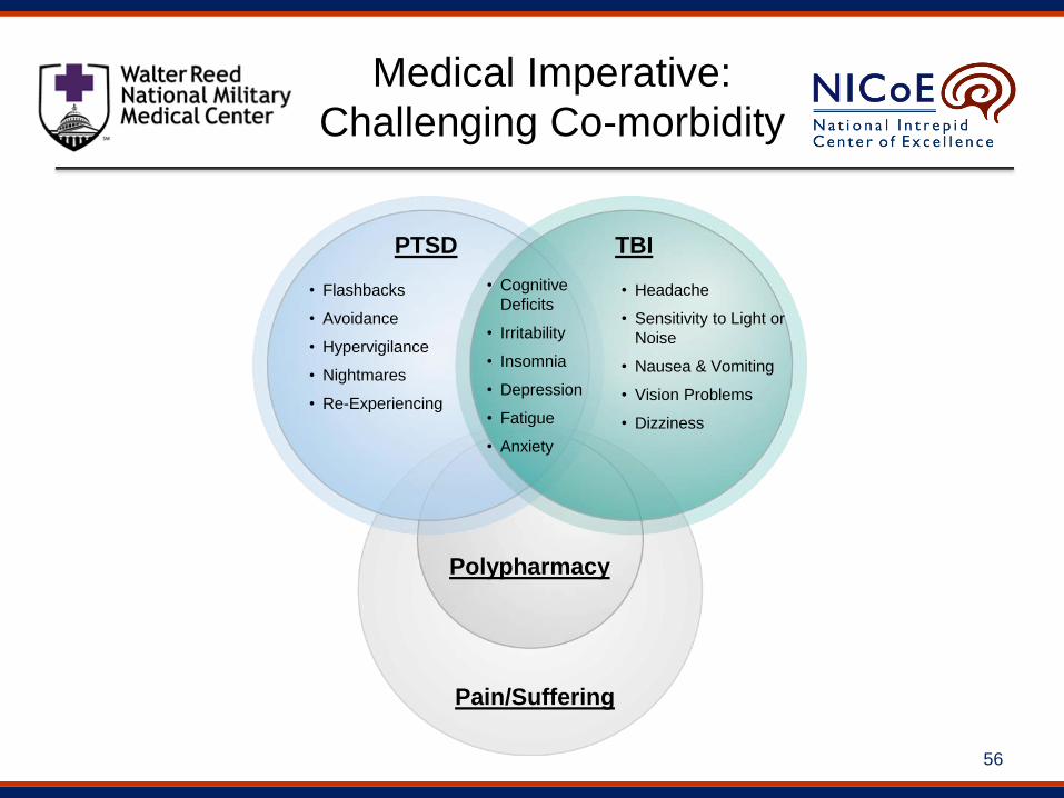
Medical Imperative:
Challenging Co-morbidity
TBIPTSD
• Headache
• Sensitivity to Light or
Noise
• Nausea & Vomiting
• Vision Problems
• Dizziness
• Cognitive
Deficits
• Irritability
• Insomnia
• Depression
• Fatigue
• Anxiety
• Flashbacks
• Avoidance
• Hypervigilance
• Nightmares
• Re-Experiencing
Pain/Suffering
Polypharmacy

57
Interdisciplinary Patient-Centered
Evaluation and Treatment

58
Major Diagnostic and Rehabilitation Equipment
MRI (3-T) / Functional MRI Positron Emission Tomography with
Computed Tomography (PET/CT)
Magneto Encephalography (MEG)
Scanner
Trans-Cranial Doppler
Ultrasound
CAREN (Computer Assisted Rehabilitation
Environment) system
Diffusion Tensor Imaging (DTI)

T38

60C130 60