Concepts in Bone Reconstruction for Implant … · members of the transforming growth factor- β...
24
Chapter 23 Concepts in Bone Reconstruction for Implant Rehabilitation Hany A. Emam and Mark R. Stevens Additional information is available at the end of the chapter http://dx.doi.org/10.5772/53401 1. Introduction The standard of care regarding tooth loss replacement is evolving towards the use of dental implants. The practice of fixed bridges and partial prosthesis can be and are iatrogenic to the existing teeth and bone. Prosthetics in the restoration of partial and complete edentulous conditions with implants has become the most important determinant. Because of this principle the emphasis has focused on optimization of the alveolus to receive a root form implant. Dental implants are a viable treatment option when there is sufficient quantity and quality of bone to achieve the desired functional and esthetic results. The reduction in bone volume has many etiologies. The most common are a result of: Periodontal disease, pneuma‐ tization of the maxillary sinus, long term ill-fitting dentures, and the general progression of osteoporosis with aging. Initially, malposition or short implants were used in areas of deficient bone volume. This often resulted in compromised prosthetic design and poor long term treatment outcomes. Today’s treatment plans first consider the prosthesis options. This necessitates reconstruction and modifications of the pre-existing anatomy provide the ideal environment needed for optimal implant placement. The deformity is often a composite loss of both bone and soft tissue. The alveolar bone loss frequently occurs in a three dimension‐ al pattern. Multiple options and techniques have been advocated for correction and recon‐ struction of the atrophied alveolar bones. They include the following: Guided bone regeneration (GBR), onlay bone grafting (OBG), interpositional bone grafting (IBG), distrac‐ tion osteogenesis (DO), ridge- split (RS), and sinus augmentation techniques (SA). [1-3] The complexity of the defect dictates the selection of the appropriate technique. The reconstruc‐ tion must also take into account the three dimensional spatial relation of one arch to the opposing arch. © 2013 Emam and Stevens; licensee InTech. This is an open access article distributed under the terms of the Creative Commons Attribution License (http://creativecommons.org/licenses/by/3.0), which permits unrestricted use, distribution, and reproduction in any medium, provided the original work is properly cited.
Transcript of Concepts in Bone Reconstruction for Implant … · members of the transforming growth factor- β...

Chapter 23
Concepts in Bone Reconstruction for ImplantRehabilitation
Hany A. Emam and Mark R. Stevens
Additional information is available at the end of the chapter
http://dx.doi.org/10.5772/53401
1. Introduction
The standard of care regarding tooth loss replacement is evolving towards the use of dentalimplants. The practice of fixed bridges and partial prosthesis can be and are iatrogenic to theexisting teeth and bone. Prosthetics in the restoration of partial and complete edentulousconditions with implants has become the most important determinant. Because of thisprinciple the emphasis has focused on optimization of the alveolus to receive a root formimplant. Dental implants are a viable treatment option when there is sufficient quantity andquality of bone to achieve the desired functional and esthetic results. The reduction in bonevolume has many etiologies. The most common are a result of: Periodontal disease, pneuma‐tization of the maxillary sinus, long term ill-fitting dentures, and the general progression ofosteoporosis with aging. Initially, malposition or short implants were used in areas of deficientbone volume. This often resulted in compromised prosthetic design and poor long termtreatment outcomes. Today’s treatment plans first consider the prosthesis options. Thisnecessitates reconstruction and modifications of the pre-existing anatomy provide the idealenvironment needed for optimal implant placement. The deformity is often a composite lossof both bone and soft tissue. The alveolar bone loss frequently occurs in a three dimension‐al pattern. Multiple options and techniques have been advocated for correction and recon‐struction of the atrophied alveolar bones. They include the following: Guided boneregeneration (GBR), onlay bone grafting (OBG), interpositional bone grafting (IBG), distrac‐tion osteogenesis (DO), ridge- split (RS), and sinus augmentation techniques (SA). [1-3] Thecomplexity of the defect dictates the selection of the appropriate technique. The reconstruc‐tion must also take into account the three dimensional spatial relation of one arch to theopposing arch.
© 2013 Emam and Stevens; licensee InTech. This is an open access article distributed under the terms of theCreative Commons Attribution License (http://creativecommons.org/licenses/by/3.0), which permitsunrestricted use, distribution, and reproduction in any medium, provided the original work is properly cited.

2. Considerations for reconstruction
2.1. Bone density
The quality of bone in the jaws is dependent on location and position within the dental archesand alveolus respectively. The most dense bone is observed in the anterior mandible, followedby the anterior maxilla and posterior mandible. The least compact bone is typically found inthe posterior maxilla. Misch classified these bone densities into a spectrum of four categories,ranging from D1 through D4. D1 bone primarily consists of a dense cortical structure. D4 onthe other hand, is the softest, consisting primarily of cancellous bone with a fine trabecularpattern with minimal crestal cortical anatomy. The density of bone is an important quality inthe initial stabilization of the implant and in the loading profile of the prosthesis. Literaturereview of clinical studies from 1981 to 2001 reveals that poor bone density may decreaseimplant loading survival rates. The decrease survival ranged from 16% to 40 %. The primarycause of these failures was directly attributed to the bone density, strength and a lowerpercentage of bone to implant contact. Bone in the posterior maxilla was found to be five toten times weaker in comparison to bone in the anterior when compared to other bone densities.Lesser bone densities also influence stress pattern distribution. Stresses in “soft bone” dem‐onstrate patterns which migrate further towards the apex. Bone loss is more pronounced andoccurs along the implant body rather than crestally, as in denser bone. D4 bone exhibits thegreatest difference in biomechanical modulus of elasticity when compared with titanium.Therefore, afterload results in higher strain conditions at the bone-implant interface acceler‐ating bone resorption and implant failure (Fig. 1).
Figure 1. Types of bone densities
2.2. Bone graft materials and mechanism of bone regeneration
Various bone augmentation materials are used for alveolar reconstruction, they include:Autografts, allografts, alloplasts, and xenografts. Autogenous bone grafts can regenerate bonethrough all three mechanisms: osteogenesis, osteoinduction, and osteoconduction; This is the goldstandard. Other bone substitute materials form bone from osteoinduction and or osteocon‐duction in varying degrees.
A Textbook of Advanced Oral and Maxillofacial Surgery618
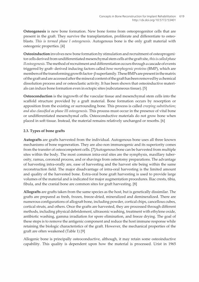
Osteogenesis is new bone formation. New bone forms from osteoprogenitor cells that arepresent in the graft. They survive the transplantation, proliferate and differentiate to osteo‐blasts. This is termed phase I osteogenesis. Autogenous bone is the only graft material withosteogenic properties. [4]
Osteoinduction involves new bone formation by stimulation and recruitment of osteoprogeni‐tor cells derived from undifferentiated mesenchymal stem cells at the graft site, this is called phaseII osteogenesis. The method of recruitment and differentiation occurs through a cascade of eventstriggered by graft- derived inducing factors called bone morphogenic proteins (BMP), which aremembers of the transforming growth factor- β superfamily. These BMPs are present in the matrixof the graft and are accessed after the mineral content of the graft has been removed by a chemicaldissolution process and or osteoclastic activity. It has been shown that osteoinductive materi‐als can induce bone formation even in ectopic sites (subcutaneous tissue). [5]
Osteoconduction is the ingrowth of the vascular tissue and mesenchymal stem cells into thescaffold structure provided by a graft material. Bone formation occurs by resorption orapposition from the existing or surrounding bone. This process is called creeping substitution;and also classified as phase III osteogenesis. This process must occur in the presence of vital boneor undifferentiated mesenchymal cells. Osteoconductive materials do not grow bone whenplaced in soft tissue. Instead, the material remains relatively unchanged or resorbs. [6]
2.3. Types of bone grafts
Autografts are grafts harvested from the individual. Autogenous bone uses all three knownmechanisms of bone regeneration. They are also non immunogenic and its superiority comesfrom the transfer of osteocompetent cells. [7]Autogenous bone can be harvested from multiplesites within the body. The most common intra-oral sites are the symphysis, maxillary tuber‐osity, ramus, coronoid process, and or shavings from osteotomy preparations. The advantageof harvesting intra-orally are, ease of harvesting and the harvest site being within the samereconstruction field. The major disadvantage of intra-oral harvesting is the limited amountand quality of the harvested bone. Extra-oral bone graft harvesting is used to provide largevolumes of the material and is indicated for major augmentation procedures. Iliac crests, tibia,fibula, and the cranial bone are common sites for graft harvesting. [8]
Allografts are grafts taken from the same species as the host, but is genetically dissimilar. Thegrafts are prepared as fresh, frozen, freeze-dried, mineralized and demineralized. There arenumerous configurations of allograft bone, including powder, cortical chips, cancellous cubes,cortical struts, and others. Once the grafts are harvested, they are processed through differentmethods, including physical debridement, ultrasonic washing, treatment with ethylene oxide,antibiotic washing, gamma irradiation for spore elimination, and freeze drying. The goal ofthese steps is to remove the antigenic component and reduce the host immune response whileretaining the biologic characteristics of the graft. However, the mechanical properties of thegraft are often weakened (Table 1) [9]
Allogenic bone is principally osteoconductive, although, it may retain some osteoinductivecapability. This quality is dependent upon how the material is processed. Urist in 1965
Concepts in Bone Reconstruction for Implant Rehabilitationhttp://dx.doi.org/10.5772/53401
619

described the process of acid demineralization of bone before implantation by using hydro‐chloric acid. The organic bone matrix contains bone morphogenic proteins (BMPs). Theseproteins are responsible for the de novo bone formation. BMP is not acid soluble, however thecalcium and phosphate salts of the HA can be removed from the bone in the acid- reducingprocess. This results in demineralization of the freeze-dried bone (FDB) and an increasedexposure of the BMPs with its osteopromotive effect. FDB is primary osteoconductive whiledemineralized freeze dried bone (DFDB) is believed to be osteoinductive. [10] Results ofstudies performed using DFDB are conflicting. Controversy still exists about the osteopromo‐tive effects of DFDB. Some reports raise the question of the concentration variability of BMPsin commercially available grafts. Osteoinductive properties of DFDB vary from one cadaverto another. The product fabrication may also have an effect on the osteoinductivity of theallograft where the demineralization process is very technique sensitive. For example, it hasbeen shown that the osteoinductive properties of the grafts are removed, if the calcium contentis less than 2% by weight. In addition, controversy persists about the use of ethylene oxide forsterilization of the graft materials and its possible destructive affects on the BMPs. [11]Dem‐ineralized cortical bone was found to have higher concentrations of BMPs than trabecular bone.Membranous cortical bone exhibits greater concentration of BMPs than endochondral corticalbone, consequently; the skull and facial bone represent a better source of inductive proteinsthan the remaining appendicular skeleton.
Routine studies are performed to evaluate the safety of allografts. According to the Americanassociation of tissue banks the probability of DFDB to contain HIV virus is 1 in 2.8 billion.When compared with the risk of 1 in 450,000 for blood transfusions, the risk of infection fromallografts seems infinitesimal. Rigorous background checks are performed on the donor andhis/her family before the donor is accepted into the program. Occasionally biopsy specimensof sites containing allograft from human patients sometimes show chronic inflammatory cells.These histologic appearances of a non-specific inflammatory condition cannot be attributed toan immune reaction with certainty.6
Xenografts are derived from the inorganic portion of bone of a genetically different speciesthan the host. One of the most popular used xenografts is the bovine bone. It is a good bonebank material. The process requires complete de-proteinization at high temperature, (1100 °c).This results in total removal of the residual organics that might provoke an immune response(Table 2). [12]
A concern over the risk of disease transmission from cattle to humans through the bone graftmaterial derived from bovine bone used for dental implants has been suggested. The recentincidents of bovine spongiform encephalopathy (BSE) in human have underscored this likelihood.Results from analysis conducted by the German Federal Ministry of Health and by thePharmaceutical Research and Manufacturers Association of America showed that the risk ofdisease transmission was negligible and could be attributed to the stringent protocols followedin sourcing and processing of the raw bovine bone used in the commercial products. [13] Oneof the best known xenografts is Bio-Oss (Osteohealth, Shirley, NY). It has been treated by havingall its organic material removed. This leaves a crystal structure that practically matches humancancellous bone in structure. In 1992, Klinge and colleagues, noted total resorption of Bio-Oss
A Textbook of Advanced Oral and Maxillofacial Surgery620

granules at 14 weeks after placement in rabbit skulls. [14] However, Skoglund and colleaguesreported that granules were present even after 44 months [15].
Another popular alternative xenograft is coralline hydroxyapatite, which is made from oceancorals. This material was created with the intension of producing a graft material with a moreconsistent pore size. Coral, which is composed mainly of calcium carbonate, is processed toremove most of the organic content. Then it is subjected to high pressure and heat in thepresence of an aqueous phosphate solution. When this process is completed, the calciumcarbonate skeleton is totally replaced with a calcium phosphate skeleton (hydrothermalexchange). The material is concurrently sterilized in this process. [16] The generation ofbiomimetic microenvironments, using scaffolds containing cell recognition sequences incombination with bone cells, offers tremendous potential for skeletal tissue regeneration.PepGen P15 (DENTSPLY Friadent CeraMed, Lakewood, CO) is the first man engineeredcollagen I binding domain for potential osteoblasts and is able to multiply the completeregeneration cascade (Figs. 2,3). It is a combination bone replacement graft material composedof natural anorganic bovine-derived hydroxyapatite matrix (ABM) coupled with a syntheticcell-binding peptide (P-15). [17]
Figure 2. Microphotograph (16 weeks 5x 1.25 OP H&E) showing newly formed bone (NB) in an interconnecting tra‐becular pattern (bone bridging) surrounding the remaining graft particles G. (PepGen P-15).
Alloplasts are synthetic bone substitutes that posses osteoconductive potential. The idealsynthetic graft material should be biocompatible and elicit minimal fibrotic changes. The graftshould support new bone growth and undergo remodeling. Other preferred attributes wouldinclude similar toughness, modulus of elasticity, and compressive strength compared to thatof the host cortical or cancellous bone. Many synthetic materials are available including:Bioactive glasses, glass ionomers, aluminum oxide, calcium sulphate, calcium phosphates asα and β tricalcium phosphate (TCP), synthetic hydroxyapatite (HA), and synthetic absorbablepolymers. [16] Synthetic bone substitutes offer many advantages; however, the greatest is theunlimited supply and avoidance of a secondary surgical procedure. The main disadvantageis the material’s lack of the osteoinductive capabilities, experienced in autogenous grafts.Clinicians may prefer performing grafting procedures using combination grafts. This willcombine the osteogenic potential of autogenous bone with the unlimited supply offered by
Concepts in Bone Reconstruction for Implant Rehabilitationhttp://dx.doi.org/10.5772/53401
621

bone substitutes which act as expanders or fillers. Combination grafts also minimize donor sitemorbidity that occurs more frequently when harvesting larger volumes of autogenous bone(Table 3).
Allografts
Material Commercial
source
composition Bone Growth Method Resorption time
DFDB (Demineralized) Pacific Tissue
Bank
Grafton
MTF
DynaGraft
Collagen + Growth
factors
Mainly Osteoinduction varies based
upon processing method
+/- 6 months
FDB (Mineralized) MinerOss
Puross
Minerals + Collagen Mainly Osteoconduction 1 Yr +
Table 1.
Xenografts
Material Brand name Structure
Deprotenized bovine bone mineral Bio-Oss Cancellous or cortical
Anorganic bovine HA+ cell binding peptide PepGen P-15 Peptide + microporous HA
Osteograft N Micro + Macroporous
Coral ( Ca carbonate) Biocoral
Interpore 200
(Coralline)
Natural coral
Table 2.
G NB
Implant
Figure 3. Microphotograph (8 weeks 5x 1.6 OP Paragon) showing the newly formed bone (NB) in an interconnectingtrabecular pattern (bone bridging-arrows) surrounding the remaining graft particles G (PepGen P-15) supporting adental implant.
A Textbook of Advanced Oral and Maxillofacial Surgery622

Alloplasts
Ceramics Polymers
β-tricalcium phosphate (β-TCP) Methylmethacrylate (HTR synthetic bone)
Hydroxyapatite (HA), (Bone source, Norian) Poly- α- hydroxy acids (PLA,PLGA)
Ca2So4 (Plaster of paris)
Calcium phosphate cements (Ceredex, α-BSM)
Bioactive glass ( PerioGlass, BioGran)
Table 3.
2.4. Properties of graft materials
It is important to consider the physical and chemical properties of the graft materials used inthe augmentation procedures. Physical properties include the surface area or form of the product(block, particle), porosity (dense, macroporous, microporous), and crystallinity (crystalline,amorphous). Chemical properties are related to calcium –to- phosphorous ratio, elementimpurities (such as carbonate), and the pH of the surrounding region. These properties playa role in the rate of resorption and clinical applications of the material.7 The larger the particlesize, the longer the material will remain at the augmentation site. It was also reported that thegreater the porosity, the more rapid the resorption of the graft material as this will give thechance for committed cells and blood vessels (bone modeling unit) to invade the spacesbetween the graft particles replacing the graft with the newly formed bone. However, denseHA may lack any micro or macro porosity within the particles with long resorption rate sincethe osteoclasts only attack the surface and cannot penetrate the dense material. With respectto crystallinity, the higher the crystalline structure the harder for the body to break down andabsorb it.7 The resorption of bone substitutes may be cell or solution- mediated. Cell mediatedresorption requires living cells of the body to resorb the material mainly osteoclasts. A solution–mediated resorption is a chemical process; impurities like calcium carbonate permit solution– mediated resorption, which then increases the porosity of the graft. The pH in the region alsoaffects the rate of graft resorption. As the pH decreases (due to infection) the HA componentsresorb by a solution – mediated resorption. Bone, dense HA, macroporous HA, microporousHA, crystalline HA, or amorphous HA may all resorb within a two-week period (Fig. 4).7
Figure 4. Showing the cell - mediated resorption of multinucleated cells (arrow) on the surface of the graft particle (G).
Concepts in Bone Reconstruction for Implant Rehabilitationhttp://dx.doi.org/10.5772/53401
623

Close matching of the resorption rate to the bone deposition rate is important. Selection of graftmaterial should be based on location of graft site, soft tissue environment, and its possible rolein promoting and supporting future implant osseous integration. A rapidly resorbing scaffoldmight reestablish a void filled with connective tissue, whereas one that resorb too slowly, ornot at all, would impede bone deposition and limit creeping substitution. There are, howeverclinical indications in which resorption is not desired, but rather, a permanent implant ispreferred, such as craniofacial onlays for cosmetic augmentation.
2.5. Bone growth factors
The term growth factors comprises a group of polypeptides of approximately 6-45 KD (kiloDalton) which are involved in cellular proliferation, differentiation and morphogenesis oftissues and organs during embryogenesis, postnatal growth, and adulthood. [18] Factors thatare involved in the regeneration and induction of bone tissue have attracted attention as theypossibly can facilitate skeletal reconstruction. These factors include platelet derived growthfactor (PDGF), vascular endothelial growth factor (VEGF), insulin like growth factors (IGF),transforming growth factor β (TGF β), bone morphogenic proteins (BMPs), and platelet richplasma (PRP).
Bone morphogenic proteins (BMPs), particularly BMP2, BMP4 and BMP7, appear to be themost reliable factors of all growth factors currently discussed with regard to enhancement ofbone regeneration in reconstruction of the facial skeleton (Table 4). BMPs stimulate angiogen‐esis, migration, proliferation, and differentiation of mesenchymal stem cells into bone formingcells in the area of bone injury. Although a high washout effect of BMP during the first fewhours in most of the carriers used has to be taken into account, this short-term signal appearsto be sufficient for the initial induction of the cascade of endochondral bone formation toprovide bone regeneration in the defects of the various models. Recombinant techniques arenow used to provide large amounts of BMPs which are normally present in very smallquantities within the organic matrix of bone (accounting for only approximately 0.1% of themass of the organic matrix). [19] Bioactive Proteins, GEM 21S® is a combination of a bioactiveproteins (highly purified recombinant human platelet derived growth factor, rhPDGF-BB) anda biocompatible osteoconductive matrix (beta-tricalcium phosphate, β-TCP). It is presentlybeing used for periodontal regeneration procedures and offers a greater amount of growthfactors as normally found in Platelet Rich Plasma (PRP).
The apparent strong desire of clinicians for the use of growth factors to facilitate reconstructivesurgical procedures by obviating the need for procurement of autogenous grafts is contrastedby their limited availability for clinical application. This has prompted the application ofautogenous growth factors by using platelet rich plasma (PRP) derived from the patient’s ownblood. This preparation has come widely into use recently, despite the fact that currently thereis controversial scientific evidence about the benefit of using this preparation, especially, inreconstructive and preprosthetic bone grafting. According to the characteristics of the growthfactors that are present in PRP and assigned for its biological activity, the use of PRP issupposed to increase proliferation of undifferentiated mesenchymal cells and to enhanceangiogenesis, which then can support bone graft incorporation by enhancing of osteoproge‐
A Textbook of Advanced Oral and Maxillofacial Surgery624

nitor cells in the graft. It may as well improve soft tissue healing by increasing proliferationand matrix synthesis. [20] Recently, inorder to improve the handling characteristics of the graftmaterials to facilitate its use in several clinical situations, several commercial suppliers havebegun to provide several matrices and delivery systems as carriers. The addition of the carrierchanged the consistency of the material from a particulate consistency to a more coherenthydrogel form (flow) or clay like (putty) form with ease in handling during surgical applica‐tion. The carrier must be nontoxic and biocompatible and should not impede any of the stepsof the bone-forming cascade. Also, when used with growth factors they must first bind to them,permit their timed release, facilitate invasion of blood vessels and enable cellular attachment,finally promoting the deposition of new bone. Sodium hyaluronate, carboxymethylcellulose,poly-α- hydroxy acids, absorbable collagen sponges (ACS) and Lecithin are among the carriermaterials used. In addition to the handling characteristics, it is assumed that the carrier materialwhen added to a particulate graft will provide spaces between these particles (lower packingdensity), facilitating the capillary in-growth and the creeping substitution process leading toproper healing with optimum new bone formation in a shorter period of time.
BMPs approved for clinical use and indications
rhBMP-2 (Wyeth/Medtronic)
InductOs (CHMP approved)
Open tibia fracture, 2002
Interbony spinal fusion, 2005
INFUSE Bone Graft (FDA approved)
Interbony spinal fusion, 2002
Open tibia fracture, 2004
Oral/Maxillofacial, 2007
rhOP-1 (Stryker)
OP-1 Implant (FDA HDE & CHMP approved)
Recalcitrant long bone nonunions, 2001/2004
OP-1 Putty (FDA HDE approved)
Osteolateral (intertransverse) lumbar spinal fusion revision, 2004.
Bioactive proteins Gem 21S (Osteohealth), Periodontal defects
Table 4.
3. Treatment plan for bone augmentation
The treatment planning sequence for implant dentistry begins with the design of the finalprosthesis. After the determination of the type of restoration, number and position of teeth tobe restored and the patients force factors are then evaluated. The bone density in the region ofthe implant placement is then considered. The key implant positions and the number and idealimplant sizes are then selected. Finally the available bone volume is evaluated for implantplacement according to the proposed treatment plan. Previous studies have shown that the
Concepts in Bone Reconstruction for Implant Rehabilitationhttp://dx.doi.org/10.5772/53401
625
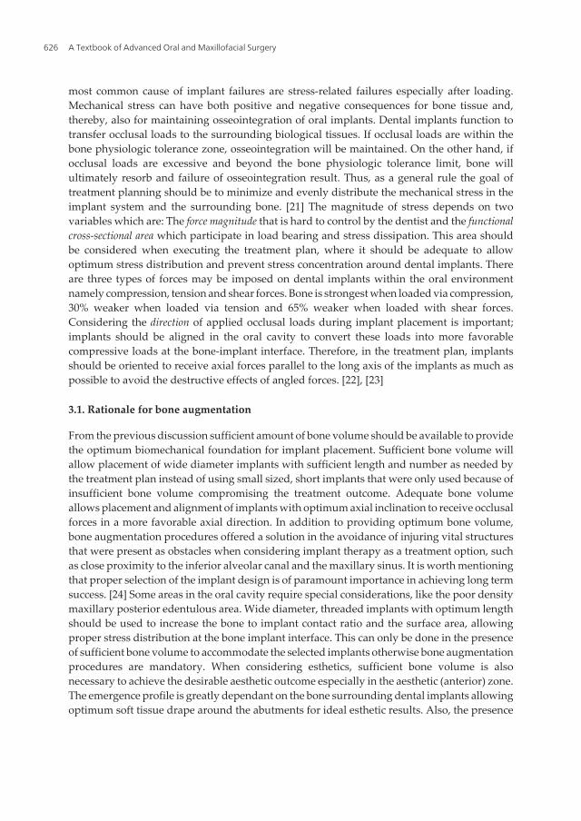
most common cause of implant failures are stress-related failures especially after loading.Mechanical stress can have both positive and negative consequences for bone tissue and,thereby, also for maintaining osseointegration of oral implants. Dental implants function totransfer occlusal loads to the surrounding biological tissues. If occlusal loads are within thebone physiologic tolerance zone, osseointegration will be maintained. On the other hand, ifocclusal loads are excessive and beyond the bone physiologic tolerance limit, bone willultimately resorb and failure of osseointegration result. Thus, as a general rule the goal oftreatment planning should be to minimize and evenly distribute the mechanical stress in theimplant system and the surrounding bone. [21] The magnitude of stress depends on twovariables which are: The force magnitude that is hard to control by the dentist and the functionalcross-sectional area which participate in load bearing and stress dissipation. This area shouldbe considered when executing the treatment plan, where it should be adequate to allowoptimum stress distribution and prevent stress concentration around dental implants. Thereare three types of forces may be imposed on dental implants within the oral environmentnamely compression, tension and shear forces. Bone is strongest when loaded via compression,30% weaker when loaded via tension and 65% weaker when loaded with shear forces.Considering the direction of applied occlusal loads during implant placement is important;implants should be aligned in the oral cavity to convert these loads into more favorablecompressive loads at the bone-implant interface. Therefore, in the treatment plan, implantsshould be oriented to receive axial forces parallel to the long axis of the implants as much aspossible to avoid the destructive effects of angled forces. [22], [23]
3.1. Rationale for bone augmentation
From the previous discussion sufficient amount of bone volume should be available to providethe optimum biomechanical foundation for implant placement. Sufficient bone volume willallow placement of wide diameter implants with sufficient length and number as needed bythe treatment plan instead of using small sized, short implants that were only used because ofinsufficient bone volume compromising the treatment outcome. Adequate bone volumeallows placement and alignment of implants with optimum axial inclination to receive occlusalforces in a more favorable axial direction. In addition to providing optimum bone volume,bone augmentation procedures offered a solution in the avoidance of injuring vital structuresthat were present as obstacles when considering implant therapy as a treatment option, suchas close proximity to the inferior alveolar canal and the maxillary sinus. It is worth mentioningthat proper selection of the implant design is of paramount importance in achieving long termsuccess. [24] Some areas in the oral cavity require special considerations, like the poor densitymaxillary posterior edentulous area. Wide diameter, threaded implants with optimum lengthshould be used to increase the bone to implant contact ratio and the surface area, allowingproper stress distribution at the bone implant interface. This can only be done in the presenceof sufficient bone volume to accommodate the selected implants otherwise bone augmentationprocedures are mandatory. When considering esthetics, sufficient bone volume is alsonecessary to achieve the desirable aesthetic outcome especially in the aesthetic (anterior) zone.The emergence profile is greatly dependant on the bone surrounding dental implants allowingoptimum soft tissue drape around the abutments for ideal esthetic results. Also, the presence
A Textbook of Advanced Oral and Maxillofacial Surgery626
of sufficient bone volume allows flexibility in choosing the properly sized implant for betterabutment emergence profile. [25]
4. Bone augmentation techniques
4.1. Socket preservation/ Guided bone regeneration
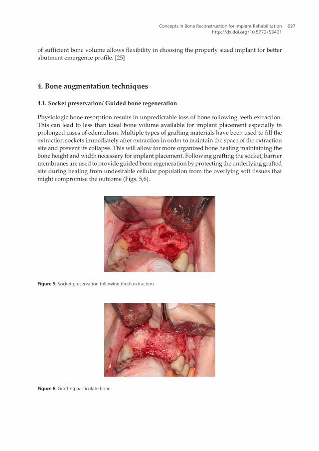
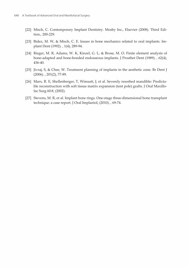
Physiologic bone resorption results in unpredictable loss of bone following teeth extraction.This can lead to less than ideal bone volume available for implant placement especially inprolonged cases of edentulism. Multiple types of grafting materials have been used to fill theextraction sockets immediately after extraction in order to maintain the space of the extractionsite and prevent its collapse. This will allow for more organized bone healing maintaining thebone height and width necessary for implant placement. Following grafting the socket, barriermembranes are used to provide guided bone regeneration by protecting the underlying graftedsite during healing from undesirable cellular population from the overlying soft tissues thatmight compromise the outcome (Figs. 5,6).
Figure 5. Socket preservation following teeth extraction.
Figure 6. Grafting particulate bone
Concepts in Bone Reconstruction for Implant Rehabilitationhttp://dx.doi.org/10.5772/53401
627

4.2. Block bone grafting technique
Block grafting approaches can be used to reconstruct significant deficiency in the vertical andhorizontal dimensions of the alveolar ridge. Autogenous block grafting procedures remain thegold standard for ridge augmentation. However, donor site morbidity associated with graftharvest has turned the attention to using allogenic grafting materials. The locations forharvesting intraoral block grafts include the external oblique ridge of the posterior mandible(ramus), symphysis. With bone defects >2 cm, an extraoral donor site is warranted forharvesting larger bone volumes. The iliac crest (anterior and posterior), cranium, or tibia isoften used as extraoral harvest sites. The detailed description of the harvesting techniques isbeyond the scope of this chapter. Case reports have demonstrated success with FDBA andDFDBA block graft material. However, further research is warranted to evaluate the healingof these blocks histologically (Figs. 7-12).
Figure 7. Ramus bone harvest
Figure 8. Symphysis bone harvest
A Textbook of Advanced Oral and Maxillofacial Surgery628

Figure 9. Calvarial bone harvest
Figure 10. Anterior iliac crest bone harvest
Figure 11. Mandibular bone augmentation using block. grafts. Two screws are used to prevent rotation.
Figure 12. Maxillary ridge augmentation.
Concepts in Bone Reconstruction for Implant Rehabilitationhttp://dx.doi.org/10.5772/53401
629

4.3. Ridge expansion (split) technique
With a narrow ridge, splitting the alveolar bone longitudinally, using chisels, osteotomes, orpeizosurgical devices, can be performed to increase the horizontal ridge with, provided the facialand lingual plates are not fused and some intervening bone is present. With adequate stabilityof the mobile segment, sufficient interpositional grafting and soft tissue protection, compara‐ble results to alternate techniques can be obtained. The decision to place the implants simultane‐ously with the split procedure or delayed placement following bone healing depend on thedegree of stability of the expanded segment and the volume of remaining bone (Figs. 13-17).
Figure 13. Narrow maxillary ridge.
Figure 14. Osteotomy of the ridge
Figure 15. Ridge splitting.
A Textbook of Advanced Oral and Maxillofacial Surgery630

Figure 16. Interpositioning graft between the buccal and the palatal plates of bone. Collagen membrane is used tocover the expanded site
Figure 17. The augmented maxillary ridge 5 weeks postoperatively
4.4. Sinus augmentation
The most commonly used technique use to access the maxillary sinus is the lateral windowtechnique modifying the Caldwell-Luc operation, also called the hinge osteotomy technique,originally described by Tatum then first published by Boyne and James.
A window is then created using a round bur on the lateral wall of the sinus till the bluish hueof the sinus membrane reveals itself. Using specially designed sinus elevation curettes the sinusmembrane is elevated from the bony floor and is freed anteriorly, posteriorly and medially tocreate a tension free elevation to minimize the possibility of perforation. The trap door(window) is intruded medially forming the new sinus floor and the space created below it isthen grafted to provide the platform for implant placement. The flap is then repositioned andclosed. Implants are placed either simultaneously with the graft (one- stage) or after a delayedperiod of up to 8 months to allow for graft maturation (two- stage). The decision about the twooptions mainly depends on the preexisting residual amount of bone required for initialprimary stability of an implant. It was found that if the bone thickness is 4 mm or less, initialimplant stability would be jeopardized. In 1994, Summers published a new less invasiveconservative technique for sinus floor elevation using osteotomes in an attempt to overcomethe drawbacks of the lateral window approach. The technique begins with a crestal incision toexpose the alveolar ridge. An osteotome of the smallest size is then tapped into place by amallet into the bone just shy from the sinus membrane fracturing and moving the sinus floorsuperiorly. Osteotomes of increasing sizes are introduced sequentially to expand the alveolus
Concepts in Bone Reconstruction for Implant Rehabilitationhttp://dx.doi.org/10.5772/53401
631

and with each insertion of a larger osteotome, bone is compressed, pushed laterally andapically. Summers stated that the very nature of this technique improved the bone density ofthe posterior maxilla. Bone graft material is then introduced via the osteotomy followed byimplant fixture insertion. The implant fixture should be slightly larger in diameter than theosteotomy site “tenting” the elevated maxillary sinus membrane. A minimally invasive antralmembrane balloon elevation (MIAMBE) which is a modification of the osteotome techniquehas also been introduced with satisfactory results. It comprises the introduction of a ballooninto the osteotomy site which is then slowly inflated to elevate the sinus membrane. Thisprocedure showed predictable results and required a short learning curve. Recently, someauthors have reported the use of a piezoelectric device in maxillary sinus surgery. Ultrasoundhas been increasingly used in many fields of medicine such as tumor enucleation, fragmenta‐tion of renal calculi and lithotripsy of gall bladder stones. Ultrasonic dissection has beenclassified as tissue-selective technique that might improve the efficiency of dissections and atthe same time reduces the morbidity rate resulting from iatrogenic injuries. In addition,ultrasound energy can induce a cavitational effect in water containing tissues, which can inturn facilitate the tissue separation (Figs. 18,19).
Figure 18. Showing the lateral window approach
Figure 19. Sinus augmentation with immediate implant placement
4.5. Distraction osteogenesis
Distraction Osteogenesis (DO) uses the phenomenon that new bone fills in the gap defectcreated when two pieces of bone are slowly separated under tension. Distraction of thesegment can be achieved in a vertical and /or horizontal direction on the basic principlesinvolved in distraction which include a latency period of 7 days for initial soft callus formation,
A Textbook of Advanced Oral and Maxillofacial Surgery632

a distraction phase during which the 2 segments of bone undergo incremental gradualseparation at a rate ~ 1 mm per day to stretch the formed soft callus, and a consolidation phasethat allows healing of the regenerated bone between the 2 segments. The prerequisites foroptimal bone augmentation of defects using DO are minimum of 6-7 mm of bone height abovevital structures, such as neurovascular bundles or air passages/sinus cavities, a vertical ridgedefect of > 3 -4 mm, and an edentulous span of three or more missing teeth (Figs. 20,21).
Figure 20. Alveolar distraction of the anterior maxillary region
Figure 21. Note: the vertical osteotomy cuts should be divergent to avoid obstructing the path of distracting thetransport segment.
4.6. Tent- Pole technique
Marx et al in 2002 advanced the approach of soft tissue matrix expansion using corticocancel‐lous bone grafting with dental implants to treat severely resorbed mandibles that were shorterthan 6 mm. Using this transcutaneous submental approach, 4 to 6 dental implants were placedto act as “tent poles” to maintain the height of the overlying mucosal soft tissue and preventit from sagging around the iliac crest graft (Figs. 22, 23). [2]
Figure 22. Implant placement in the severely atrophic mandible through a submental approach
Concepts in Bone Reconstruction for Implant Rehabilitationhttp://dx.doi.org/10.5772/53401
633

Figure 23. Corticocancellous bone graft around the implants tenting the soft tissue
4.7. Bone ring technique
Three dimensional bone augmentations with immediate dental implant placement can be doneusing this technique. Using trephine burs corresponding to the extraction socket diameters,bone rings can be harvested from the chin or iliac crest regions. The harvested rings can thenbe secured to the extraction socket using the dental implants restoring the deficient bone atthe crestal portion in a 3D fashion (Figs. 24,25). [27]
Figure 24. Three dimensional crestal bone augmentation using bone rings.
Figure 25. Immediate implant placement in the anterior maxilla
4.8. Reconstruction of segmental bony defects
Ablative loss of both bone and associated soft tissue from treatment of neoplastic or otherpathologic processes represent a far different task from loss of bone from physiologic resorp‐tion, trauma or infection. The goals of reconstruction are to restore jaw continuity, provide
A Textbook of Advanced Oral and Maxillofacial Surgery634

morphology and position of the bone in relation to its opposing jaw, provide adequate heightand width of bone, and provide facial contour and support for soft tissue structures.
Graft malpositioning result in occlusal problems and presents a formidable task to therestorative dentist. The site of the graft harvest depends mainly on the size of the residualdefect (Figs. 26-28).
Figure 26. Reconstruction plate in place.
Figure 27. Free fibula graft.
Figure 28. Reconstruction of mandibular segmental bone defect using free fibula.
Concepts in Bone Reconstruction for Implant Rehabilitationhttp://dx.doi.org/10.5772/53401
635

4.9. Combination grafts
In large defects, the use of grafting materials from different sources can be beneficial. Sometechniques aim to combine the osteogenic potential of autogenous bone with the osteocon‐ductive and space maintaining properties provided by the allogenic or alloplastic sources.Allogenic materials can provide constructs that are close in morphology as the resected partproviding superior esthetic outcome following the grafting procedure (Fig. 29,30).
Figure 29. Hemimandibular reconstruction using a cadaveric mandible in combination with cancellous bone graftharvested from the iliac crest.
Figure 30. Graft in position.
4.10. Future augmentation approaches
Future bone augmentation approaches likely will use molecular, cellular, and genetic tissueengineering technologies. Gene therapy is a relatively new therapeutic modality based on thepotential for delivery of altered genetic material to the cell. Localized gene therapy can be usedto increase the concentration of desired growth or differentiation factors to enhance theregenerative response. Cellular tissue engineering strategies that include the in vitro amplifi‐cation of osteoprogenitor cells grown within three dimensional constructs is currently ofparticular interest. The use of mesenchymal stem cell for construct seeding showed promisefor bone regeneration. These approaches may lead to further refinement and improvement inalveolar bone augmentation techniques.
A Textbook of Advanced Oral and Maxillofacial Surgery636

5. Surgical caveats for bone grafting
There are several factors that may improve the success and predictability of bone graftprocedures, they include the following:
5.1. Surgical asepsis and absence of infection
Contamination of bone grafts due to endogenous bacteria, lack of aseptic surgical technique,inadequate soft tissue closure and salivary exposure may lead to infection with subsequentlowering of the pH. Solution –mediated resorption will follow with resultant graft loss. Someclinicians prefer including antibiotics locally within the graft materials to guard againstbacterial contamination as no blood supply is present early in the graft. Primary soft tissueclosure is also mandatory for the success of the grafting procedure. It allows healing by primaryintension protecting the graft from any surrounding contamination until healing. Dehiscencewith graft loss is one of the most common complications in bone grafting procedures. There‐fore, careful surgical flap planning which ensures adequate blood supply to the site withminimal trauma and primary soft tissue closure without tension are required.
5.2. Space maintenance
Creation of a desired contoured space for bone formation is very important in the graftingprocedure. If the graft material resorbs too rapidly compared with the time required for boneformation, the site may fill with connective tissue rather than bone. Therefore, the space mustbe maintained long enough without collapse for bone to fill the desired area. Titanium-reinforced barrier membranes, tent screws elevated above the bone at the desired heightcovered by a membrane, block grafts (covered by membrane or not) are all used to create andmaintain space for bone growth.
5.3. Graft stability
For predictable bone augmentation, graft stability is a paramount. Bone remodeling and grafthealing requires rigid interface for blood clot adhesion with its associated growth factors. If agraft become mobile its vascularity will be compromised followed by fibrous encapsulationand often sequestrate. If block grafts are used fixation can be achieved using titanium screwsor the graft can be fixed using the inserted implants itself. If particulate graft is used, it can becovered with a barrier membrane fixed with membrane tacks to avoid dislodgement of thegraft particles.
5.4. Regional acceleratory phenomenon (RAP)
The host site during bone augmentation procedures should be decorticated to establishbleeding points in the cortical bone prior to graft placement. This procedure will provideaccess for trabecular bone vessels, encourage revascularization, bring growth factors to thegraft site and increase the availability of osteogenic cells promoting graft union and shortenthe healing time.
Concepts in Bone Reconstruction for Implant Rehabilitationhttp://dx.doi.org/10.5772/53401
637

Acknowledgements
The authors would like to extend their gratitude and acknowledgement to Drs. Solon KaoDDS and Henry “Butch” Ferguson DMD, for allowing the use of several clinical surgicalcases. Their photographic documentation of specific bone grafts and regenerative techniqueswas instrumental in providing comprehensive examples of implant site reconstructions.
Author details
Hany A. Emam1,2 and Mark R. Stevens1
*Address all correspondence to: [email protected]; [email protected]
1 Oral and Maxillofacial Surgery Department, Georgia Health Sciences University, Augusta,Georgia, USA
2 Oral and Maxillofacial Surgery, Cairo University, Egypt
References
[1] Aghaloo, T. L, & Moy, P. K. Which hard tissue augmentation techniques are the mostsuccessful in furnishing bony support for implant placement? Int J Oral MaxillofacImplants (2007). Suppl:, 49-70.
[2] Jensen, J, Sindet-pedersen, S, & Oliver, A. J. Varying treatment strategies for recon‐struction of maxillary atrophy with implants: results in 98 patients. J Oral MaxillofacSurg (1994). discussion 16-8., 52(3), 210-6.
[3] Isaksson, S, & Alberius, P. Maxillary alveolar ridge augmentation with onlay bone-grafts and immediate endosseous implants. J Craniomaxillofac Surg (1992). , 20(1),2-7.
[4] Giannoudis, P. V, Dinopoulos, H, & Tsiridis, E. Bone substitutes: an update. Injury(2005). Suppl 3:S, 20-7.
[5] Browaeys, H, Bouvry, P, & De Bruyn, H. A literature review on biomaterials in sinusaugmentation procedures. Clin Implant Dent Relat Res (2007). , 9(3), 166-77.
[6] Khan, S. N, & Cammisa, F. P. Jr., Sandhu HS, Diwan AD, Girardi FP, Lane JM. Thebiology of bone grafting. J Am Acad Orthop Surg (2005). , 13(1), 77-86.
[7] Misch, C. E, & Dietsh, F. Bone-grafting materials in implant dentistry. Implant Dent(1993). , 2(3), 158-67.
A Textbook of Advanced Oral and Maxillofacial Surgery638

[8] Clavero, J, & Lundgren, S. Ramus or chin grafts for maxillary sinus inlay and localonlay augmentation: comparison of donor site morbidity and complications. Clin Im‐plant Dent Relat Res (2003). , 5(3), 154-60.
[9] Marx, R. E. Bone and bone graft healing. Oral Maxillofac Surg Clin North Am (2007).v., 19(4), 455-66.
[10] Wikesjo, U. M, Sorensen, R. G, Kinoshita, A, & Wozney, J. M. RhBMP-2/alphaBSMinduces significant vertical alveolar ridge augmentation and dental implant osseoin‐tegration. Clin Implant Dent Relat Res (2002). , 4(4), 174-82.
[11] Zhang, M, & Powers, R. M. Jr., Wolfinbarger L, Jr. Effect(s) of the demineralizationprocess on the osteoinductivity of demineralized bone matrix. J Periodontol (1997). ,68(11), 1085-92.
[12] Kao, S. T, & Scott, D. D. A review of bone substitutes. Oral Maxillofac Surg ClinNorth Am (2007). vi., 19(4), 513-21.
[13] Sogal, A, & Tofe, A. J. Risk assessment of bovine spongiform encephalopathy trans‐mission through bone graft material derived from bovine bone used for dental appli‐cations. J Periodontol (1999). , 70(9), 1053-63.
[14] Klinge, B, Alberius, P, Isaksson, S, & Jonsson, J. Osseous response to implanted natu‐ral bone mineral and synthetic hydroxylapatite ceramic in the repair of experimentalskull bone defects. J Oral Maxillofac Surg (1992). , 50(3), 241-9.
[15] Skoglund, A, Hising, P, & Young, C. A clinical and histologic examination in humansof the osseous response to implanted natural bone mineral. Int J Oral Maxillofac Im‐plants (1997). , 12(2), 194-9.
[16] Moore, W. R, Graves, S. E, & Bain, G. I. Synthetic bone graft substitutes. ANZ J Surg(2001). , 71(6), 354-61.
[17] Nguyen, H, Qian, J. J, Bhatnagar, R. S, & Li, S. Enhanced cell attachment and osteo‐blastic activity by peptide-coated matrix in hydrogels. Biochem Biophys Res Com‐mun (2003). , 15.
[18] Schilephake, H. Bone growth factors in maxillofacial skeletal reconstruction. Int J Or‐al Maxillofac Surg (2002). , 31(5), 469-84.
[19] Wikesjo, U. M, Huang, Y. H, Polimeni, G, & Qahash, M. Bone morphogenetic pro‐teins: a realistic alternative to bone grafting for alveolar reconstruction. Oral Maxillo‐fac Surg Clin North Am (2007). vi-vii., 19(4), 535-51.
[20] Marx, R. E, Carlson, E. R, Eichstaedt, R. M, Schimmele, S. R, Strauss, J. E, & Georgeff,K. R. Platelet-rich plasma: Growth factor enhancement for bone grafts. Oral Surg Or‐al Med Oral Pathol Oral Radiol Endod (1998). , 85(6), 638-46.
[21] Isidor, F. Influence of forces on peri-implant bone. Clin Oral Implants Res (2006).Suppl , 2, 8-18.
Concepts in Bone Reconstruction for Implant Rehabilitationhttp://dx.doi.org/10.5772/53401
639
[22] Misch, C. Contemporary Implant Dentistry. Mosby Inc., Elsevier (2008). Third Edi‐tion., 200-229.
[23] Bidez, M. W, & Misch, C. E. Issues in bone mechanics related to oral implants. Im‐plant Dent (1992). , 1(4), 289-94.
[24] Rieger, M. R, Adams, W. K, Kinzel, G. L, & Brose, M. O. Finite element analysis ofbone-adapted and bone-bonded endosseous implants. J Prosthet Dent (1989). , 62(4),436-40.
[25] Jivraj, S, & Chee, W. Treatment planning of implants in the aesthetic zone. Br Dent J(2006). , 201(2), 77-89.
[26] Marx, R. E, Shellenberger, T, Wimsatt, J, et al. Severely resorbed mandible: Predicta‐ble reconstruction with soft tissue matrix expansion (tent pole) grafts. J Oral Maxillo‐fac Surg 60:8, (2002).
[27] Stevens, M. R, et al. Implant bone rings. One-stage three-dimensional bone transplanttechnique: a case report. J Oral Implantol, (2010). , 69-74.
A Textbook of Advanced Oral and Maxillofacial Surgery640