Computed tomography findings of atypical intralobar ...
6
J Res Clin Med, 2021, 9: 10 doi: 10.34172/jrcm.2021.010 https://jrcm.tbzmed.ac.ir Computed tomography findings of atypical intralobar pulmonary sequestration: A case series Sercan Özkaçmaz 1* ID , Mesut Özgökçe 1 ID , Muhammet Bilal Akıncı 1 , Fatma Durmaz 1 ID , İlyas Dündar 1 ID , Cemil Göya 1 , Fuat Sayır 2 1 Department of Radiology, Faculty of Medicine, Yüzüncü Yıl University, Van, Turkey 2 Department of Chest Surgery, Faculty of Medicine, Yüzüncü Yıl University, Van, Turkey Introduction Pulmonary sequestration is a congenital dysplastic lung tissue, which is not associated with the tracheobronchial tree and pulmonary arteries. Arterial supply usually occurs through the thoracic aorta. Pulmonary sequestration has two different types. Intralobar pulmonary sequestration (75%-90%) is located in the lung tissue and does not have its own visceral pleura. It is usually seen as a heterogeneous solid or cystic mass. 1-3 Left lower lobe of the lung is the most frequent location of intralobar pulmonary sequestration and it is supplied from thoracic aorta. 4 Extralobar pulmonary sequestration (10%-25%) completely separates from normal lung tissue and has its own pleura. 4,5 The typical features of intralobar pulmonary sequestration include arterial supply from the thoracic aorta by solitary branch, venous drainage via pulmonary veins, left lower lobe involvement, and heterogenous solid mass appearance. The lesion may be seen as a cystic mass including air when it is infected. 2,6 In this study, we aimed to present atypical computed tomography (CT) findings of intralobar sequestration by reviewing each feature of all our patients. Methods In this retrospective research study, patients with a histopathological proven diagnosis of intralobar pulmonary sequestration who had also a presurgery contrast-enhanced CT scan in our clinic between 2015 and 2019 were evaluated based on the hospital database and radiology department records. Demographic and medical data of each patient were recorded. CT images were obtained by a multislice CT device with 16 detectors (Somatom Emotion 16-slice; CT2012E-Siemens AG Berlin and Munich, Germany). Patients were injected with 1-1.5 cc/kg of non-ionized intravenous contrast agent iohexol (Amersham Health, Ireland) or iopromide (Schering, Germany) at a rate of 3 mL/s via an automated syringe (CT 9000 ADV Liebel-Flarsheim). In thorax CT examination, images were obtained from the distal neck to the upper abdomen at the 25th second after contrast agent administration. The examination was performed by holding the patient’s breath. Axial and multiplanar reformatted images with 3 mm slice thickness were evaluated by two radiologists with 10 to 12 years of experience on thorax radiology. All consensus was reached again by these two radiologists. Images were interpreted for atypical features of sequestration tissue including location, Case Series Abstract Introduction: Bronchopulmonary sequestration is a rare congenital abnormality of the lung that has two different types as intralobar and extralobar. In this study, we aimed to present six cases of intralobar sequestration with atypical findings in terms of feeding, drainage, and localization. Methods: Patients diagnosed with intralobar pulmonary sequestration in our clinic between 2015 and 2019 were evaluated retrospectively. Demographical features and atypical computed tomography (CT) findings of the patients were presented by literature. Results: Among 45 patients with intralobar sequestration, 6 (13.3%) (5 males and 1 female) with a mean age of 43.5±25.4(0-78) years old had atypical pulmonary findings on CT images. Atypical features regarding arterial supply was detected in 8.9%, venous drainage in 2.2%, location in 4.4%, radiological appearance in 4.4% and co-existing lesion in 2.2% of the patient with intralobar sequestration. Conclusion: Typical and atypical features of pulmonary sequestration must be well-known for differential diagnosis of solid or cystic pulmonary lesions. Keywords: Pulmonary sequestration, Intralobar, Computed tomography, Atypical findings Article History: Received: 19 Dec. 2020 Accepted: 3 Jan. 2021 e-Published: 2 Mar. 2021 Keywords: • Pulmonary sequestration • Intralobar • Computed tomography • Atypical findings Article info *Corresponding Author: Sercan Özkaçmaz, Email: [email protected] © 2021 The Author(s). This is an open access article distributed under the terms of the Creative Commons Attribution License (http:// creativecommons.org/licenses/by/4.0/), which permits unrestricted use, distribution, and reproduction in any medium, provided the original work is properly cited. TUOMS PRESS
Transcript of Computed tomography findings of atypical intralobar ...
J Res Clin Med 2021 9 10doi 1034172jrcm2021010
httpsjrcmtbzmedacir
Computed tomography findings of atypical intralobar pulmonary sequestration A case seriesSercan Oumlzkaccedilmaz1 ID Mesut Oumlzgoumlkccedile1 ID Muhammet Bilal Akıncı1 Fatma Durmaz1 ID İlyas Duumlndar1 ID Cemil Goumlya1 Fuat Sayır2
1Department of Radiology Faculty of Medicine Yuumlzuumlncuuml Yıl University Van Turkey 2Department of Chest Surgery Faculty of Medicine Yuumlzuumlncuuml Yıl University Van Turkey
IntroductionPulmonary sequestration is a congenital dysplastic lung tissue which is not associated with the tracheobronchial tree and pulmonary arteries Arterial supply usually occurs through the thoracic aorta Pulmonary sequestration has two different types Intralobar pulmonary sequestration (75-90) is located in the lung tissue and does not have its own visceral pleura It is usually seen as a heterogeneous solid or cystic mass1-3 Left lower lobe of the lung is the most frequent location of intralobar pulmonary sequestration and it is supplied from thoracic aorta4 Extralobar pulmonary sequestration (10-25) completely separates from normal lung tissue and has its own pleura45
The typical features of intralobar pulmonary sequestration include arterial supply from the thoracic aorta by solitary branch venous drainage via pulmonary veins left lower lobe involvement and heterogenous solid mass appearance The lesion may be seen as a cystic mass including air when it is infected26
In this study we aimed to present atypical computed tomography (CT) findings of intralobar sequestration by reviewing each feature of all our patients
MethodsIn this retrospective research study patients with a histopathological proven diagnosis of intralobar pulmonary sequestration who had also a presurgery contrast-enhanced CT scan in our clinic between 2015 and 2019 were evaluated based on the hospital database and radiology department records Demographic and medical data of each patient were recorded CT images were obtained by a multislice CT device with 16 detectors (Somatom Emotion 16-slice CT2012E-Siemens AG Berlin and Munich Germany) Patients were injected with 1-15 cckg of non-ionized intravenous contrast agent iohexol (Amersham Health Ireland) or iopromide (Schering Germany) at a rate of 3 mLs via an automated syringe (CT 9000 ADV Liebel-Flarsheim) In thorax CT examination images were obtained from the distal neck to the upper abdomen at the 25th second after contrast agent administration The examination was performed by holding the patientrsquos breath Axial and multiplanar reformatted images with 3 mm slice thickness were evaluated by two radiologists with 10 to 12 years of experience on thorax radiology All consensus was reached again by these two radiologists Images were interpreted for atypical features of sequestration tissue including location
Case Series
AbstractIntroduction Bronchopulmonary sequestration is a rare congenital abnormality of the lung that has two different types as intralobar and extralobar In this study we aimed to present six cases of intralobar sequestration with atypical findings in terms of feeding drainage and localizationMethods Patients diagnosed with intralobar pulmonary sequestration in our clinic between 2015 and 2019 were evaluated retrospectively Demographical features and atypical computed tomography (CT) findings of the patients were presented by literatureResults Among 45 patients with intralobar sequestration 6 (133) (5 males and 1 female) with a mean age of 435plusmn254(0-78) years old had atypical pulmonary findings on CT images Atypical features regarding arterial supply was detected in 89 venous drainage in 22 location in 44 radiological appearance in 44 and co-existing lesion in 22 of the patient with intralobar sequestrationConclusion Typical and atypical features of pulmonary sequestration must be well-known for differential diagnosis of solid or cystic pulmonary lesionsKeywords Pulmonary sequestration Intralobar Computed tomography Atypical findings
Article HistoryReceived 19 Dec 2020Accepted 3 Jan 2021e-Published 2 Mar 2021
Keywordsbull Pulmonary sequestrationbull Intralobarbull Computed tomographybull Atypical findings
Article info
Corresponding Author Sercan Oumlzkaccedilmaz Email sercanozkacmazhotmailcom
copy 2021 The Author(s) This is an open access article distributed under the terms of the Creative Commons Attribution License (http creativecommonsorglicensesby40) which permits unrestricted use distribution and reproduction in any medium provided the original work is properly cited
TUOMSPRE S S
Oumlzkaccedilmaz et al
J Res Clin Med 2021 9 102
arterial feeding venous drainage radiological appearance and accompanying lesions The variables were expressed as number and mean plusmn standard deviation
ResultsAn intralobar pulmonary sequestration was detected in overall 45 patients All these patients had the diagnosis of intrapulmonary sequestration by histopathological examination after thoracotomy with resection of the lesions Among these 45 patients 6 had atypical findings regarding location arterial supply venous drainage radiological appearance and co-existing lesion The mean age of the 6 patients (133) with atypical pulmonary sequestration was 435plusmn254 (0-78) and the malefemale ratio was 5 (51)
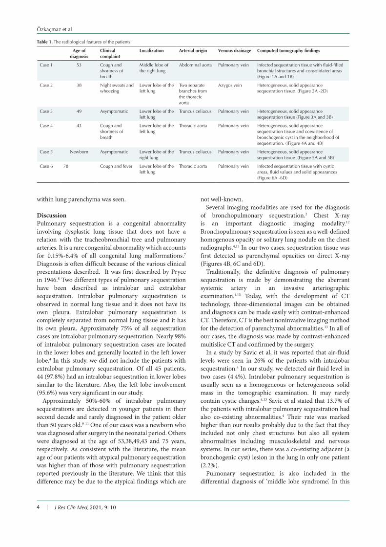
Case 1The first case was a 53-year-old male who presented with cough and shortness of breath The lesion was located in the middle lobe of the right lung and arterial feeding was provided from the abdominal aorta (Figure 1) The venous drainage was via the pulmonary vein The lesion had an infected cystic appearance with fluid-filled bronchial structures and consolidated areas The atypical findings of this case were the location arterial supply and also infected cystic appearance
Case 2The second case was a 38-year-old male who had night sweat and wheezing The lesion was fed by two arterial branches originated from the thoracic aorta and drained to the azygos vein (Figure 2) The lesion had a heterogeneous solid mass appearance The atypical findings of this case were venous drainage and two arterial branches arising from the thoracic aorta
Case 3This case was a 49-year-old asymptomatic female The arterial feeding was provided by truncus celiacus and venous drainage was via the pulmonary vein (Figure 3) The lesion had a heterogeneous solid mass appearance The atypical finding in this case was arterial supply
Case 4The fourth case was a 43-year-old male who had complaints of cough and shortness of breath The lesion with a heterogeneous solid mass appearance was located in the lower lobe of the left lung fed from the thoracic aorta and drained to the pulmonary vein It was associated with a neighboring bronchogenic cyst (Figure 4) The atypical finding in this case was an adjacent bronchogenic cyst
Case 5The fifth case was an asymptomatic newborn female in whom the sequestration was located in the lower lobe of
the right lung and the arterial feeding was from the truncus celiacus (Figure 5) The lesion had a typical heterogeneous mass appearance with venous drainage via the pulmonary vein The atypical finding in this case was arterial supply origin
Case 6The last case was a 75-year-old male who presented
Figure 1 A thin artery arising from abdominal aorta medial to truncus ceilacus (A) feeding a heterogenous sequestration tissue with air-fluid levels (B) in middle lobe of right lung
Figure 2 A sequestration in lower lobe of left lung (A) which has two arteries from abdominal aorta (B C) (red arrows) and draining to azygos vein (D) (black arrow)
Figure 3 A sequestration in lower lobe of left lung (A) that was fed by a branch originated from truncus celiacus (B) (black arrow)
Intalobar pulmonary sequestration
J Res Clin Med 2021 9 10 3
with fever and cough The sequestration tissue with a heterogeneous infected cystic appearance was located in the lower lobe of the left lung which was fed from the thoracic aorta and drained to the pulmonary vein The atypical findings in this case was an infected cystic appearance which could be confused with the pneumonic infiltration (Figure 6)Features of the patients are summarized on Table 1
Complaints of the patientsThree of our 6 patients with atypical intralobar pulmonary sequestration had cough and dyspnea and one of them had night sweating Cough and dyspnea was seen in half of the patients (50) while sweeting in 167 Two of our patients (333) were asymptomatic
Radiological algorithmIn two of our cases the CT scans were performed after detecting lesions on PA chest radiography (Figures 4B 6C and 6B) In the newborn patient CT was performed because of the suspicion of sequestration detected on antenatal ultrasound The remaining three cases were found incidentally on initial CT scans
The diagnosis of sequestrationIntralobar pulmonary sequestration was diagnosed clinically and radiologically in all of the cases and all of them were confirmed by surgery and histopathological examinations
Location of lesionsSequestration tissue was observed in the lower lobe of the left lung in 43 (956) of overall 45 patients in the middle lobe of the right lung in one case (case 1) (22) and in the lower lobe of right lung in the remained one (case 5) (22) Lower lobes involvement was detected in 978 (4445) and left lung involvement in 956 (4345) No bilateral involvement was seen
Arterial supplyA total of 4 (89) atypical arterial supply was observed in the patients In two (44) patients (cases 3 and 5) the artery of sequestration tissue arose from celiac truncus and in one (22) patient (case 1) from the abdominal aorta In one patient (22) (case 2) two nourishing arteries originating from thoracic aorta were observed One artery arising from thoracic aorta was seen in remained 41 patients (911)
Venous drainageAmong 45 patients in only one (22) (case 2) the sequestration tissue drained to azygos vein while in remained 44 ones (978) drained to pulmonary veins
Radiological appearance In two patients (cases 1 and 6) (44) the lesions had a cystic dominant appearance which was due to a probable infectious process while in remained 43 ones (956) typical heterogeneous solid mass appearance was observed Co-existing lesionsIn one patient (22) (Case 4) a bronchogenic cyst adjacent to sequestration tissue was noted since in the other 44 (978) patients no co-existing congenital lesion
Figure 4 A sequestration in lower lobe of left lung (A) and adjacent a bronchogenic cyst (Arrow) located posterolateral to the sequestration tissue (A B)
Figure 5 A sequestration tissue in lower lobe of right lung (A) that has arterial supply from truncus celiacus (B) (black arrow)
Figure 6 The sequestration tissue in lower lobe of left lung (A) that had arterial supply from thoracic aorta (B) with air-fluid levels The lesion is located in posterolateral of left lung (C-D)
Oumlzkaccedilmaz et al
J Res Clin Med 2021 9 104
within lung parenchyma was seen
DiscussionPulmonary sequestration is a congenital abnormality involving dysplastic lung tissue that does not have a relation with the tracheobronchial tree and pulmonary arteries It is a rare congenital abnormality which accounts for 015-64 of all congenital lung malformations7
Diagnosis is often difficult because of the various clinical presentations described It was first described by Pryce in 19468 Two different types of pulmonary sequestration have been described as intralobar and extralobar sequestration Intralobar pulmonary sequestration is observed in normal lung tissue and it does not have its own pleura Extralobar pulmonary sequestration is completely separated from normal lung tissue and it has its own pleura Approximately 75 of all sequestration cases are intralobar pulmonary sequestration Nearly 98 of intralobar pulmonary sequestration cases are located in the lower lobes and generally located in the left lower lobe4 In this study we did not include the patients with extralobar pulmonary sequestration Of all 45 patients 44 (978) had an intralobar sequestration in lower lobes similar to the literature Also the left lobe involvement (956) was very significant in our study
Approximately 50-60 of intralobar pulmonary sequestrations are detected in younger patients in their second decade and rarely diagnosed in the patient older than 50 years old9-11 One of our cases was a newborn who was diagnosed after surgery in the neonatal period Others were diagnosed at the age of 53384943 and 75 years respectively As consistent with the literature the mean age of our patients with atypical pulmonary sequestration was higher than of those with pulmonary sequestration reported previously in the literature We think that this difference may be due to the atypical findings which are
not well-knownSeveral imaging modalities are used for the diagnosis
of bronchopulmonary sequestration2 Chest X-ray is an important diagnostic imaging modality12
Bronchopulmonary sequestration is seen as a well-defined homogenous opacity or solitary lung nodule on the chest radiographs413 In our two cases sequestration tissue was first detected as parenchymal opacities on direct X-ray (Figures 4B 6C and 6D)
Traditionally the definitive diagnosis of pulmonary sequestration is made by demonstrating the aberrant systemic artery in an invasive arteriographic examination413 Today with the development of CT technology three-dimensional images can be obtained and diagnosis can be made easily with contrast-enhanced CT Therefore CT is the best noninvasive imaging method for the detection of parenchymal abnormalities13 In all of our cases the diagnosis was made by contrast-enhanced multislice CT and confirmed by the surgery
In a study by Savic et al it was reported that air-fluid levels were seen in 26 of the patients with intralobar sequestration4 In our study we detected air fluid level in two cases (44) Intralobar pulmonary sequestration is usually seen as a homogeneous or heterogeneous solid mass in the tomographic examination It may rarely contain cystic changes413 Savic et al stated that 137 of the patients with intralobar pulmonary sequestration had also co-existing abnormalities4 Their rate was marked higher than our results probably due to the fact that they included not only chest structures but also all system abnormalities including musculoskeletal and nervous systems In our series there was a co-existing adjacent (a bronchogenic cyst) lesion in the lung in only one patient (22)
Pulmonary sequestration is also included in the differential diagnosis of lsquomiddle lobe syndromersquo In this
Table 1 The radiological features of the patients
Age of diagnosis
Clinical complaint
Localization Arterial origin Venous drainage Computed tomography findings
Case 1 53 Cough and shortness of breath
Middle lobe of the right lung
Abdominal aorta Pulmonary vein Infected sequestration tissue with fluid-filled bronchial structures and consolidated areas (Figure 1A and 1B)
Case 2 38 Night sweats and wheezing
Lower lobe of the left lung
Two separate branches from the thoracic aorta
Azygos vein Heterogeneous solid appearance sequestration tissue (Figure 2A -2D)
Case 3 49 Asymptomatic Lower lobe of the left lung
Truncus celiacus Pulmonary vein Heterogeneous solid appearance sequestration tissue (Figure 3A and 3B)
Case 4 43 Cough and shortness of breath
Lower lobe of the left lung
Thoracic aorta Pulmonary vein Heterogeneous solid appearance sequestration tissue and coexistence of bronchogenic cyst in the neighborhood of sequestration ((Figure 4A and 4B)
Case 5 Newborn Asymptomatic Lower lobe of the right lung
Truncus celiacus Pulmonary vein Heterogeneous solid appearance sequestration tissue (Figure 5A and 5B)
Case 6 78 Cough and fever Lower lobe of the left lung
Thoracic aorta Pulmonary vein Infected sequestration tissue with cystic areas fluid values and solid appearances (Figure 6A -6D)
Intalobar pulmonary sequestration
J Res Clin Med 2021 9 10 5
respect it is important to look at the arterial origin of heterogeneous lesions seen in the middle lobe In our study it was possible to confuse sequestration tissue with middle lobe syndrome in case 1 but the preliminary diagnosis was made by demonstrating the arterial supply from the abdominal aorta For this reason the detailed imaging of all solid or cystic intrapulmonary lesions is essential
According to the study performed by Savic et al the artery of intralobar sequestrations originates from thoracic aorta in 739 and from abdominal aorta in 187 of the patients Truncus celiacus and its branches (splenic and hepatic arteries) may rarely be reported as the origin of arterial supply of intrapulmonary sequestration4 Multiple arterial feeding was reported in 174 of the patients with intralobar sequestration4 In our study arterial feeding occurred from the abdominal aorta in one case (22) and from the celiac artery in two cases (44) In one case there were two arteries arising from thoracic aorta and entering the sequestration tissue In our study multiple feeding artery ratio was 22 (n=1) For the other 41 patients (911) the solitary artery of the sequestration tissue arose from the thoracic aorta
Pulmonary drainage of intralobar sequestration is mostly through the pulmonary veins In less than 5 of the cases venous drainage is via systemic circulation through the vena azygos hemiazygos intercostal veins and inferior-superior vena cava14 In our study similar to the literature venous drainage via azygos vein was detected in only one case (22) while the sequestration tissue drained to pulmonary veins in 978 (n=44) of the patients
The primary treatment modality for bronchopulmonary sequestration is surgery but it carries the risk of infection1516 Preoperative imaging is important in terms of providing information about location arterial nutrition and venous drainage for surgical planning Transcatheter endovascular embolization has been successfully applied since 1998 as another treatment modality17 Transcatheter endovascular embolization treatment is an alternative to surgery in neonates and children as it is a minimally invasive method18 Accurate preoperative diagnosis is important as it will change the surgical approach All of our cases were successfully treated with surgery without any complication or residue
Although we have evaluated the patients with lobar sequestration over a wide time period the small number of cases can be considered as a limitation of our study
ConclusionContrast enhanced CT is a very effective method for the diagnosis of pulmonary sequestration which provides important vascular and structural features of the lesions For an accurate evaluation and differential diagnosis typical and atypical CT findings of pulmonary sequestration must be well-known Since pulmonary
sequestration can mimic various lesions in all age groups as in our patients the arterial supply of all lesions in especially lower lobes must be carefully evaluated
Conflict of InterestAuthors declare no conflict of interest in this study
Ethical ApprovalAuthors declared that procedures followed were in accordance with the ethical standards of the responsible committee on human experimentation (institutional and national) and with the Helsinki Declaration of 1975 as revised in 2008 The study was approved by a university ethics committee at the date of 22052020 with a number of 202003-47
Authorrsquos ContributionSO Editing revision manuscript writing process interpretation of the data MO Review article manuscript writing process and interpretation of data MBA FD IDCG Review article and interpretation of data FS Interpretation of data
AcknowledgementsWe would like to thank the radiology department workers for their help
FundingThere is no funding in this study
References1 Zhang N Zeng Q Chen C Yu J Zhang X Distribution
diagnosis and treatment of pulmonary sequestration report of 208 cases J Pediatr Surg 201954(7)1286-92 doi 101016jjpedsurg201808054
2 Lee EY Boiselle PM Cleveland RH Multidetector CT evaluation of congenital lung anomalies Radiology 2008247(3)632-48 doi 101148radiol2473062124
3 Prasad R Garg R Verma SK Intralobar sequestration of lung Lung India 200926(4)159-61 doi 1041030970-211356357
4 Savic B Birtel FJ Tholen W Funke HD Knoche R Lung sequestration report of seven cases and review of 540 published cases Thorax 197934(1)96-101 doi 101136thx34196
5 Sotto Mayor J Rocha D Esperanccedila S Oliveira e Silva A Intralobar pulmonary sequestration diagnostic expertise BMJ Case Rep 20152015bcr2015212384 doi 101136bcr-2015-212384
6 Frazier AA Rosado de Christenson ML Stocker JT Templeton PA Intralobar sequestration radiologic-pathologic correlation Radiographics 199717(3)725-45 doi 101148radiographics1739153708
7 Montjoy C Hadique S Graeber G Ghamande S Intralobar bronchopulmonary sequestra in adults over age 50 case series and review W V Med J 2012108(5)8-13
8 Pryce DM Lower accessory pulmonary artery with intralobar sequestration of lung a report of seven cases J Pathol Bacteriol 194658(3)457-67
9 Petersen G Martin U Singhal A Criner GJ Intralobar sequestration in the middle-aged and elderly adult recognition and radiographic evaluation J Thorac Cardiovasc Surg 2003126(6)2086-90 doi 101016s0022-
Oumlzkaccedilmaz et al
J Res Clin Med 2021 9 106
5223(03)01297-210 Gustafson RA Murray GF Warden HE Hill RC Rozar GE
Intralobar sequestration A missed diagnosis Ann Thorac Surg 198947(6)841-7 doi 1010160003-4975(89)90016-7
11 Wei Y Li F Pulmonary sequestration a retrospective analysis of 2625 cases in China Eur J Cardiothorac Surg 201140(1)e39-42 doi 101016jejcts201101080
12 Au VW Chan JK Chan FL Pulmonary sequestration diagnosed by contrast enhanced three-dimensional MR angiography Br J Radiol 199972(859)709-11 doi 101259bjr7285910624331
13 Hang JD Guo QY Chen CX Chen LY Imaging approach to the diagnosis of pulmonary sequestration Acta Radiol 199637(6)883-8 doi 10117702841851960373p288
14 Stocker JT Sequestrations of the lung Semin Diagn Pathol 19863(2)106-21
15 Yucel O Gurkok S Gozubuyuk A Caylak H Sapmaz E Kavakli K et al Diagnosis and surgical treatment of pulmonary sequestration Thorac Cardiovasc Surg 200856(3)154-7 doi 101055s-2007-965572
16 Gezer S Taştepe I Sirmali M Findik G Tuumlruumlt H Kaya S et al Pulmonary sequestration a single-institutional series composed of 27 cases J Thorac Cardiovasc Surg 2007133(4)955-9 doi 101016jjtcvs200611003
17 Park ST Yoon CH Sung KB Yoon HK Goo DE Kim KS et al Pulmonary sequestration in a newborn infant treatment with arterial embolization J Vasc Interv Radiol 19989(4)648-50 doi 101016s1051-0443(98)70337-9
18 Chien KJ Huang TC Lin CC Lee CL Hsieh KS Weng KP Early and late outcomes of coil embolization of pulmonary sequestration in children Circ J 200973(5)938-42 doi 101253circjcj-08-0914

Oumlzkaccedilmaz et al
J Res Clin Med 2021 9 102
arterial feeding venous drainage radiological appearance and accompanying lesions The variables were expressed as number and mean plusmn standard deviation
ResultsAn intralobar pulmonary sequestration was detected in overall 45 patients All these patients had the diagnosis of intrapulmonary sequestration by histopathological examination after thoracotomy with resection of the lesions Among these 45 patients 6 had atypical findings regarding location arterial supply venous drainage radiological appearance and co-existing lesion The mean age of the 6 patients (133) with atypical pulmonary sequestration was 435plusmn254 (0-78) and the malefemale ratio was 5 (51)
Case 1The first case was a 53-year-old male who presented with cough and shortness of breath The lesion was located in the middle lobe of the right lung and arterial feeding was provided from the abdominal aorta (Figure 1) The venous drainage was via the pulmonary vein The lesion had an infected cystic appearance with fluid-filled bronchial structures and consolidated areas The atypical findings of this case were the location arterial supply and also infected cystic appearance
Case 2The second case was a 38-year-old male who had night sweat and wheezing The lesion was fed by two arterial branches originated from the thoracic aorta and drained to the azygos vein (Figure 2) The lesion had a heterogeneous solid mass appearance The atypical findings of this case were venous drainage and two arterial branches arising from the thoracic aorta
Case 3This case was a 49-year-old asymptomatic female The arterial feeding was provided by truncus celiacus and venous drainage was via the pulmonary vein (Figure 3) The lesion had a heterogeneous solid mass appearance The atypical finding in this case was arterial supply
Case 4The fourth case was a 43-year-old male who had complaints of cough and shortness of breath The lesion with a heterogeneous solid mass appearance was located in the lower lobe of the left lung fed from the thoracic aorta and drained to the pulmonary vein It was associated with a neighboring bronchogenic cyst (Figure 4) The atypical finding in this case was an adjacent bronchogenic cyst
Case 5The fifth case was an asymptomatic newborn female in whom the sequestration was located in the lower lobe of
the right lung and the arterial feeding was from the truncus celiacus (Figure 5) The lesion had a typical heterogeneous mass appearance with venous drainage via the pulmonary vein The atypical finding in this case was arterial supply origin
Case 6The last case was a 75-year-old male who presented
Figure 1 A thin artery arising from abdominal aorta medial to truncus ceilacus (A) feeding a heterogenous sequestration tissue with air-fluid levels (B) in middle lobe of right lung
Figure 2 A sequestration in lower lobe of left lung (A) which has two arteries from abdominal aorta (B C) (red arrows) and draining to azygos vein (D) (black arrow)
Figure 3 A sequestration in lower lobe of left lung (A) that was fed by a branch originated from truncus celiacus (B) (black arrow)
Intalobar pulmonary sequestration
J Res Clin Med 2021 9 10 3
with fever and cough The sequestration tissue with a heterogeneous infected cystic appearance was located in the lower lobe of the left lung which was fed from the thoracic aorta and drained to the pulmonary vein The atypical findings in this case was an infected cystic appearance which could be confused with the pneumonic infiltration (Figure 6)Features of the patients are summarized on Table 1
Complaints of the patientsThree of our 6 patients with atypical intralobar pulmonary sequestration had cough and dyspnea and one of them had night sweating Cough and dyspnea was seen in half of the patients (50) while sweeting in 167 Two of our patients (333) were asymptomatic
Radiological algorithmIn two of our cases the CT scans were performed after detecting lesions on PA chest radiography (Figures 4B 6C and 6B) In the newborn patient CT was performed because of the suspicion of sequestration detected on antenatal ultrasound The remaining three cases were found incidentally on initial CT scans
The diagnosis of sequestrationIntralobar pulmonary sequestration was diagnosed clinically and radiologically in all of the cases and all of them were confirmed by surgery and histopathological examinations
Location of lesionsSequestration tissue was observed in the lower lobe of the left lung in 43 (956) of overall 45 patients in the middle lobe of the right lung in one case (case 1) (22) and in the lower lobe of right lung in the remained one (case 5) (22) Lower lobes involvement was detected in 978 (4445) and left lung involvement in 956 (4345) No bilateral involvement was seen
Arterial supplyA total of 4 (89) atypical arterial supply was observed in the patients In two (44) patients (cases 3 and 5) the artery of sequestration tissue arose from celiac truncus and in one (22) patient (case 1) from the abdominal aorta In one patient (22) (case 2) two nourishing arteries originating from thoracic aorta were observed One artery arising from thoracic aorta was seen in remained 41 patients (911)
Venous drainageAmong 45 patients in only one (22) (case 2) the sequestration tissue drained to azygos vein while in remained 44 ones (978) drained to pulmonary veins
Radiological appearance In two patients (cases 1 and 6) (44) the lesions had a cystic dominant appearance which was due to a probable infectious process while in remained 43 ones (956) typical heterogeneous solid mass appearance was observed Co-existing lesionsIn one patient (22) (Case 4) a bronchogenic cyst adjacent to sequestration tissue was noted since in the other 44 (978) patients no co-existing congenital lesion
Figure 4 A sequestration in lower lobe of left lung (A) and adjacent a bronchogenic cyst (Arrow) located posterolateral to the sequestration tissue (A B)
Figure 5 A sequestration tissue in lower lobe of right lung (A) that has arterial supply from truncus celiacus (B) (black arrow)
Figure 6 The sequestration tissue in lower lobe of left lung (A) that had arterial supply from thoracic aorta (B) with air-fluid levels The lesion is located in posterolateral of left lung (C-D)
Oumlzkaccedilmaz et al
J Res Clin Med 2021 9 104
within lung parenchyma was seen
DiscussionPulmonary sequestration is a congenital abnormality involving dysplastic lung tissue that does not have a relation with the tracheobronchial tree and pulmonary arteries It is a rare congenital abnormality which accounts for 015-64 of all congenital lung malformations7
Diagnosis is often difficult because of the various clinical presentations described It was first described by Pryce in 19468 Two different types of pulmonary sequestration have been described as intralobar and extralobar sequestration Intralobar pulmonary sequestration is observed in normal lung tissue and it does not have its own pleura Extralobar pulmonary sequestration is completely separated from normal lung tissue and it has its own pleura Approximately 75 of all sequestration cases are intralobar pulmonary sequestration Nearly 98 of intralobar pulmonary sequestration cases are located in the lower lobes and generally located in the left lower lobe4 In this study we did not include the patients with extralobar pulmonary sequestration Of all 45 patients 44 (978) had an intralobar sequestration in lower lobes similar to the literature Also the left lobe involvement (956) was very significant in our study
Approximately 50-60 of intralobar pulmonary sequestrations are detected in younger patients in their second decade and rarely diagnosed in the patient older than 50 years old9-11 One of our cases was a newborn who was diagnosed after surgery in the neonatal period Others were diagnosed at the age of 53384943 and 75 years respectively As consistent with the literature the mean age of our patients with atypical pulmonary sequestration was higher than of those with pulmonary sequestration reported previously in the literature We think that this difference may be due to the atypical findings which are
not well-knownSeveral imaging modalities are used for the diagnosis
of bronchopulmonary sequestration2 Chest X-ray is an important diagnostic imaging modality12
Bronchopulmonary sequestration is seen as a well-defined homogenous opacity or solitary lung nodule on the chest radiographs413 In our two cases sequestration tissue was first detected as parenchymal opacities on direct X-ray (Figures 4B 6C and 6D)
Traditionally the definitive diagnosis of pulmonary sequestration is made by demonstrating the aberrant systemic artery in an invasive arteriographic examination413 Today with the development of CT technology three-dimensional images can be obtained and diagnosis can be made easily with contrast-enhanced CT Therefore CT is the best noninvasive imaging method for the detection of parenchymal abnormalities13 In all of our cases the diagnosis was made by contrast-enhanced multislice CT and confirmed by the surgery
In a study by Savic et al it was reported that air-fluid levels were seen in 26 of the patients with intralobar sequestration4 In our study we detected air fluid level in two cases (44) Intralobar pulmonary sequestration is usually seen as a homogeneous or heterogeneous solid mass in the tomographic examination It may rarely contain cystic changes413 Savic et al stated that 137 of the patients with intralobar pulmonary sequestration had also co-existing abnormalities4 Their rate was marked higher than our results probably due to the fact that they included not only chest structures but also all system abnormalities including musculoskeletal and nervous systems In our series there was a co-existing adjacent (a bronchogenic cyst) lesion in the lung in only one patient (22)
Pulmonary sequestration is also included in the differential diagnosis of lsquomiddle lobe syndromersquo In this
Table 1 The radiological features of the patients
Age of diagnosis
Clinical complaint
Localization Arterial origin Venous drainage Computed tomography findings
Case 1 53 Cough and shortness of breath
Middle lobe of the right lung
Abdominal aorta Pulmonary vein Infected sequestration tissue with fluid-filled bronchial structures and consolidated areas (Figure 1A and 1B)
Case 2 38 Night sweats and wheezing
Lower lobe of the left lung
Two separate branches from the thoracic aorta
Azygos vein Heterogeneous solid appearance sequestration tissue (Figure 2A -2D)
Case 3 49 Asymptomatic Lower lobe of the left lung
Truncus celiacus Pulmonary vein Heterogeneous solid appearance sequestration tissue (Figure 3A and 3B)
Case 4 43 Cough and shortness of breath
Lower lobe of the left lung
Thoracic aorta Pulmonary vein Heterogeneous solid appearance sequestration tissue and coexistence of bronchogenic cyst in the neighborhood of sequestration ((Figure 4A and 4B)
Case 5 Newborn Asymptomatic Lower lobe of the right lung
Truncus celiacus Pulmonary vein Heterogeneous solid appearance sequestration tissue (Figure 5A and 5B)
Case 6 78 Cough and fever Lower lobe of the left lung
Thoracic aorta Pulmonary vein Infected sequestration tissue with cystic areas fluid values and solid appearances (Figure 6A -6D)
Intalobar pulmonary sequestration
J Res Clin Med 2021 9 10 5
respect it is important to look at the arterial origin of heterogeneous lesions seen in the middle lobe In our study it was possible to confuse sequestration tissue with middle lobe syndrome in case 1 but the preliminary diagnosis was made by demonstrating the arterial supply from the abdominal aorta For this reason the detailed imaging of all solid or cystic intrapulmonary lesions is essential
According to the study performed by Savic et al the artery of intralobar sequestrations originates from thoracic aorta in 739 and from abdominal aorta in 187 of the patients Truncus celiacus and its branches (splenic and hepatic arteries) may rarely be reported as the origin of arterial supply of intrapulmonary sequestration4 Multiple arterial feeding was reported in 174 of the patients with intralobar sequestration4 In our study arterial feeding occurred from the abdominal aorta in one case (22) and from the celiac artery in two cases (44) In one case there were two arteries arising from thoracic aorta and entering the sequestration tissue In our study multiple feeding artery ratio was 22 (n=1) For the other 41 patients (911) the solitary artery of the sequestration tissue arose from the thoracic aorta
Pulmonary drainage of intralobar sequestration is mostly through the pulmonary veins In less than 5 of the cases venous drainage is via systemic circulation through the vena azygos hemiazygos intercostal veins and inferior-superior vena cava14 In our study similar to the literature venous drainage via azygos vein was detected in only one case (22) while the sequestration tissue drained to pulmonary veins in 978 (n=44) of the patients
The primary treatment modality for bronchopulmonary sequestration is surgery but it carries the risk of infection1516 Preoperative imaging is important in terms of providing information about location arterial nutrition and venous drainage for surgical planning Transcatheter endovascular embolization has been successfully applied since 1998 as another treatment modality17 Transcatheter endovascular embolization treatment is an alternative to surgery in neonates and children as it is a minimally invasive method18 Accurate preoperative diagnosis is important as it will change the surgical approach All of our cases were successfully treated with surgery without any complication or residue
Although we have evaluated the patients with lobar sequestration over a wide time period the small number of cases can be considered as a limitation of our study
ConclusionContrast enhanced CT is a very effective method for the diagnosis of pulmonary sequestration which provides important vascular and structural features of the lesions For an accurate evaluation and differential diagnosis typical and atypical CT findings of pulmonary sequestration must be well-known Since pulmonary
sequestration can mimic various lesions in all age groups as in our patients the arterial supply of all lesions in especially lower lobes must be carefully evaluated
Conflict of InterestAuthors declare no conflict of interest in this study
Ethical ApprovalAuthors declared that procedures followed were in accordance with the ethical standards of the responsible committee on human experimentation (institutional and national) and with the Helsinki Declaration of 1975 as revised in 2008 The study was approved by a university ethics committee at the date of 22052020 with a number of 202003-47
Authorrsquos ContributionSO Editing revision manuscript writing process interpretation of the data MO Review article manuscript writing process and interpretation of data MBA FD IDCG Review article and interpretation of data FS Interpretation of data
AcknowledgementsWe would like to thank the radiology department workers for their help
FundingThere is no funding in this study
References1 Zhang N Zeng Q Chen C Yu J Zhang X Distribution
diagnosis and treatment of pulmonary sequestration report of 208 cases J Pediatr Surg 201954(7)1286-92 doi 101016jjpedsurg201808054
2 Lee EY Boiselle PM Cleveland RH Multidetector CT evaluation of congenital lung anomalies Radiology 2008247(3)632-48 doi 101148radiol2473062124
3 Prasad R Garg R Verma SK Intralobar sequestration of lung Lung India 200926(4)159-61 doi 1041030970-211356357
4 Savic B Birtel FJ Tholen W Funke HD Knoche R Lung sequestration report of seven cases and review of 540 published cases Thorax 197934(1)96-101 doi 101136thx34196
5 Sotto Mayor J Rocha D Esperanccedila S Oliveira e Silva A Intralobar pulmonary sequestration diagnostic expertise BMJ Case Rep 20152015bcr2015212384 doi 101136bcr-2015-212384
6 Frazier AA Rosado de Christenson ML Stocker JT Templeton PA Intralobar sequestration radiologic-pathologic correlation Radiographics 199717(3)725-45 doi 101148radiographics1739153708
7 Montjoy C Hadique S Graeber G Ghamande S Intralobar bronchopulmonary sequestra in adults over age 50 case series and review W V Med J 2012108(5)8-13
8 Pryce DM Lower accessory pulmonary artery with intralobar sequestration of lung a report of seven cases J Pathol Bacteriol 194658(3)457-67
9 Petersen G Martin U Singhal A Criner GJ Intralobar sequestration in the middle-aged and elderly adult recognition and radiographic evaluation J Thorac Cardiovasc Surg 2003126(6)2086-90 doi 101016s0022-
Oumlzkaccedilmaz et al
J Res Clin Med 2021 9 106
5223(03)01297-210 Gustafson RA Murray GF Warden HE Hill RC Rozar GE
Intralobar sequestration A missed diagnosis Ann Thorac Surg 198947(6)841-7 doi 1010160003-4975(89)90016-7
11 Wei Y Li F Pulmonary sequestration a retrospective analysis of 2625 cases in China Eur J Cardiothorac Surg 201140(1)e39-42 doi 101016jejcts201101080
12 Au VW Chan JK Chan FL Pulmonary sequestration diagnosed by contrast enhanced three-dimensional MR angiography Br J Radiol 199972(859)709-11 doi 101259bjr7285910624331
13 Hang JD Guo QY Chen CX Chen LY Imaging approach to the diagnosis of pulmonary sequestration Acta Radiol 199637(6)883-8 doi 10117702841851960373p288
14 Stocker JT Sequestrations of the lung Semin Diagn Pathol 19863(2)106-21
15 Yucel O Gurkok S Gozubuyuk A Caylak H Sapmaz E Kavakli K et al Diagnosis and surgical treatment of pulmonary sequestration Thorac Cardiovasc Surg 200856(3)154-7 doi 101055s-2007-965572
16 Gezer S Taştepe I Sirmali M Findik G Tuumlruumlt H Kaya S et al Pulmonary sequestration a single-institutional series composed of 27 cases J Thorac Cardiovasc Surg 2007133(4)955-9 doi 101016jjtcvs200611003
17 Park ST Yoon CH Sung KB Yoon HK Goo DE Kim KS et al Pulmonary sequestration in a newborn infant treatment with arterial embolization J Vasc Interv Radiol 19989(4)648-50 doi 101016s1051-0443(98)70337-9
18 Chien KJ Huang TC Lin CC Lee CL Hsieh KS Weng KP Early and late outcomes of coil embolization of pulmonary sequestration in children Circ J 200973(5)938-42 doi 101253circjcj-08-0914

Intalobar pulmonary sequestration
J Res Clin Med 2021 9 10 3
with fever and cough The sequestration tissue with a heterogeneous infected cystic appearance was located in the lower lobe of the left lung which was fed from the thoracic aorta and drained to the pulmonary vein The atypical findings in this case was an infected cystic appearance which could be confused with the pneumonic infiltration (Figure 6)Features of the patients are summarized on Table 1
Complaints of the patientsThree of our 6 patients with atypical intralobar pulmonary sequestration had cough and dyspnea and one of them had night sweating Cough and dyspnea was seen in half of the patients (50) while sweeting in 167 Two of our patients (333) were asymptomatic
Radiological algorithmIn two of our cases the CT scans were performed after detecting lesions on PA chest radiography (Figures 4B 6C and 6B) In the newborn patient CT was performed because of the suspicion of sequestration detected on antenatal ultrasound The remaining three cases were found incidentally on initial CT scans
The diagnosis of sequestrationIntralobar pulmonary sequestration was diagnosed clinically and radiologically in all of the cases and all of them were confirmed by surgery and histopathological examinations
Location of lesionsSequestration tissue was observed in the lower lobe of the left lung in 43 (956) of overall 45 patients in the middle lobe of the right lung in one case (case 1) (22) and in the lower lobe of right lung in the remained one (case 5) (22) Lower lobes involvement was detected in 978 (4445) and left lung involvement in 956 (4345) No bilateral involvement was seen
Arterial supplyA total of 4 (89) atypical arterial supply was observed in the patients In two (44) patients (cases 3 and 5) the artery of sequestration tissue arose from celiac truncus and in one (22) patient (case 1) from the abdominal aorta In one patient (22) (case 2) two nourishing arteries originating from thoracic aorta were observed One artery arising from thoracic aorta was seen in remained 41 patients (911)
Venous drainageAmong 45 patients in only one (22) (case 2) the sequestration tissue drained to azygos vein while in remained 44 ones (978) drained to pulmonary veins
Radiological appearance In two patients (cases 1 and 6) (44) the lesions had a cystic dominant appearance which was due to a probable infectious process while in remained 43 ones (956) typical heterogeneous solid mass appearance was observed Co-existing lesionsIn one patient (22) (Case 4) a bronchogenic cyst adjacent to sequestration tissue was noted since in the other 44 (978) patients no co-existing congenital lesion
Figure 4 A sequestration in lower lobe of left lung (A) and adjacent a bronchogenic cyst (Arrow) located posterolateral to the sequestration tissue (A B)
Figure 5 A sequestration tissue in lower lobe of right lung (A) that has arterial supply from truncus celiacus (B) (black arrow)
Figure 6 The sequestration tissue in lower lobe of left lung (A) that had arterial supply from thoracic aorta (B) with air-fluid levels The lesion is located in posterolateral of left lung (C-D)
Oumlzkaccedilmaz et al
J Res Clin Med 2021 9 104
within lung parenchyma was seen
DiscussionPulmonary sequestration is a congenital abnormality involving dysplastic lung tissue that does not have a relation with the tracheobronchial tree and pulmonary arteries It is a rare congenital abnormality which accounts for 015-64 of all congenital lung malformations7
Diagnosis is often difficult because of the various clinical presentations described It was first described by Pryce in 19468 Two different types of pulmonary sequestration have been described as intralobar and extralobar sequestration Intralobar pulmonary sequestration is observed in normal lung tissue and it does not have its own pleura Extralobar pulmonary sequestration is completely separated from normal lung tissue and it has its own pleura Approximately 75 of all sequestration cases are intralobar pulmonary sequestration Nearly 98 of intralobar pulmonary sequestration cases are located in the lower lobes and generally located in the left lower lobe4 In this study we did not include the patients with extralobar pulmonary sequestration Of all 45 patients 44 (978) had an intralobar sequestration in lower lobes similar to the literature Also the left lobe involvement (956) was very significant in our study
Approximately 50-60 of intralobar pulmonary sequestrations are detected in younger patients in their second decade and rarely diagnosed in the patient older than 50 years old9-11 One of our cases was a newborn who was diagnosed after surgery in the neonatal period Others were diagnosed at the age of 53384943 and 75 years respectively As consistent with the literature the mean age of our patients with atypical pulmonary sequestration was higher than of those with pulmonary sequestration reported previously in the literature We think that this difference may be due to the atypical findings which are
not well-knownSeveral imaging modalities are used for the diagnosis
of bronchopulmonary sequestration2 Chest X-ray is an important diagnostic imaging modality12
Bronchopulmonary sequestration is seen as a well-defined homogenous opacity or solitary lung nodule on the chest radiographs413 In our two cases sequestration tissue was first detected as parenchymal opacities on direct X-ray (Figures 4B 6C and 6D)
Traditionally the definitive diagnosis of pulmonary sequestration is made by demonstrating the aberrant systemic artery in an invasive arteriographic examination413 Today with the development of CT technology three-dimensional images can be obtained and diagnosis can be made easily with contrast-enhanced CT Therefore CT is the best noninvasive imaging method for the detection of parenchymal abnormalities13 In all of our cases the diagnosis was made by contrast-enhanced multislice CT and confirmed by the surgery
In a study by Savic et al it was reported that air-fluid levels were seen in 26 of the patients with intralobar sequestration4 In our study we detected air fluid level in two cases (44) Intralobar pulmonary sequestration is usually seen as a homogeneous or heterogeneous solid mass in the tomographic examination It may rarely contain cystic changes413 Savic et al stated that 137 of the patients with intralobar pulmonary sequestration had also co-existing abnormalities4 Their rate was marked higher than our results probably due to the fact that they included not only chest structures but also all system abnormalities including musculoskeletal and nervous systems In our series there was a co-existing adjacent (a bronchogenic cyst) lesion in the lung in only one patient (22)
Pulmonary sequestration is also included in the differential diagnosis of lsquomiddle lobe syndromersquo In this
Table 1 The radiological features of the patients
Age of diagnosis
Clinical complaint
Localization Arterial origin Venous drainage Computed tomography findings
Case 1 53 Cough and shortness of breath
Middle lobe of the right lung
Abdominal aorta Pulmonary vein Infected sequestration tissue with fluid-filled bronchial structures and consolidated areas (Figure 1A and 1B)
Case 2 38 Night sweats and wheezing
Lower lobe of the left lung
Two separate branches from the thoracic aorta
Azygos vein Heterogeneous solid appearance sequestration tissue (Figure 2A -2D)
Case 3 49 Asymptomatic Lower lobe of the left lung
Truncus celiacus Pulmonary vein Heterogeneous solid appearance sequestration tissue (Figure 3A and 3B)
Case 4 43 Cough and shortness of breath
Lower lobe of the left lung
Thoracic aorta Pulmonary vein Heterogeneous solid appearance sequestration tissue and coexistence of bronchogenic cyst in the neighborhood of sequestration ((Figure 4A and 4B)
Case 5 Newborn Asymptomatic Lower lobe of the right lung
Truncus celiacus Pulmonary vein Heterogeneous solid appearance sequestration tissue (Figure 5A and 5B)
Case 6 78 Cough and fever Lower lobe of the left lung
Thoracic aorta Pulmonary vein Infected sequestration tissue with cystic areas fluid values and solid appearances (Figure 6A -6D)
Intalobar pulmonary sequestration
J Res Clin Med 2021 9 10 5
respect it is important to look at the arterial origin of heterogeneous lesions seen in the middle lobe In our study it was possible to confuse sequestration tissue with middle lobe syndrome in case 1 but the preliminary diagnosis was made by demonstrating the arterial supply from the abdominal aorta For this reason the detailed imaging of all solid or cystic intrapulmonary lesions is essential
According to the study performed by Savic et al the artery of intralobar sequestrations originates from thoracic aorta in 739 and from abdominal aorta in 187 of the patients Truncus celiacus and its branches (splenic and hepatic arteries) may rarely be reported as the origin of arterial supply of intrapulmonary sequestration4 Multiple arterial feeding was reported in 174 of the patients with intralobar sequestration4 In our study arterial feeding occurred from the abdominal aorta in one case (22) and from the celiac artery in two cases (44) In one case there were two arteries arising from thoracic aorta and entering the sequestration tissue In our study multiple feeding artery ratio was 22 (n=1) For the other 41 patients (911) the solitary artery of the sequestration tissue arose from the thoracic aorta
Pulmonary drainage of intralobar sequestration is mostly through the pulmonary veins In less than 5 of the cases venous drainage is via systemic circulation through the vena azygos hemiazygos intercostal veins and inferior-superior vena cava14 In our study similar to the literature venous drainage via azygos vein was detected in only one case (22) while the sequestration tissue drained to pulmonary veins in 978 (n=44) of the patients
The primary treatment modality for bronchopulmonary sequestration is surgery but it carries the risk of infection1516 Preoperative imaging is important in terms of providing information about location arterial nutrition and venous drainage for surgical planning Transcatheter endovascular embolization has been successfully applied since 1998 as another treatment modality17 Transcatheter endovascular embolization treatment is an alternative to surgery in neonates and children as it is a minimally invasive method18 Accurate preoperative diagnosis is important as it will change the surgical approach All of our cases were successfully treated with surgery without any complication or residue
Although we have evaluated the patients with lobar sequestration over a wide time period the small number of cases can be considered as a limitation of our study
ConclusionContrast enhanced CT is a very effective method for the diagnosis of pulmonary sequestration which provides important vascular and structural features of the lesions For an accurate evaluation and differential diagnosis typical and atypical CT findings of pulmonary sequestration must be well-known Since pulmonary
sequestration can mimic various lesions in all age groups as in our patients the arterial supply of all lesions in especially lower lobes must be carefully evaluated
Conflict of InterestAuthors declare no conflict of interest in this study
Ethical ApprovalAuthors declared that procedures followed were in accordance with the ethical standards of the responsible committee on human experimentation (institutional and national) and with the Helsinki Declaration of 1975 as revised in 2008 The study was approved by a university ethics committee at the date of 22052020 with a number of 202003-47
Authorrsquos ContributionSO Editing revision manuscript writing process interpretation of the data MO Review article manuscript writing process and interpretation of data MBA FD IDCG Review article and interpretation of data FS Interpretation of data
AcknowledgementsWe would like to thank the radiology department workers for their help
FundingThere is no funding in this study
References1 Zhang N Zeng Q Chen C Yu J Zhang X Distribution
diagnosis and treatment of pulmonary sequestration report of 208 cases J Pediatr Surg 201954(7)1286-92 doi 101016jjpedsurg201808054
2 Lee EY Boiselle PM Cleveland RH Multidetector CT evaluation of congenital lung anomalies Radiology 2008247(3)632-48 doi 101148radiol2473062124
3 Prasad R Garg R Verma SK Intralobar sequestration of lung Lung India 200926(4)159-61 doi 1041030970-211356357
4 Savic B Birtel FJ Tholen W Funke HD Knoche R Lung sequestration report of seven cases and review of 540 published cases Thorax 197934(1)96-101 doi 101136thx34196
5 Sotto Mayor J Rocha D Esperanccedila S Oliveira e Silva A Intralobar pulmonary sequestration diagnostic expertise BMJ Case Rep 20152015bcr2015212384 doi 101136bcr-2015-212384
6 Frazier AA Rosado de Christenson ML Stocker JT Templeton PA Intralobar sequestration radiologic-pathologic correlation Radiographics 199717(3)725-45 doi 101148radiographics1739153708
7 Montjoy C Hadique S Graeber G Ghamande S Intralobar bronchopulmonary sequestra in adults over age 50 case series and review W V Med J 2012108(5)8-13
8 Pryce DM Lower accessory pulmonary artery with intralobar sequestration of lung a report of seven cases J Pathol Bacteriol 194658(3)457-67
9 Petersen G Martin U Singhal A Criner GJ Intralobar sequestration in the middle-aged and elderly adult recognition and radiographic evaluation J Thorac Cardiovasc Surg 2003126(6)2086-90 doi 101016s0022-
Oumlzkaccedilmaz et al
J Res Clin Med 2021 9 106
5223(03)01297-210 Gustafson RA Murray GF Warden HE Hill RC Rozar GE
Intralobar sequestration A missed diagnosis Ann Thorac Surg 198947(6)841-7 doi 1010160003-4975(89)90016-7
11 Wei Y Li F Pulmonary sequestration a retrospective analysis of 2625 cases in China Eur J Cardiothorac Surg 201140(1)e39-42 doi 101016jejcts201101080
12 Au VW Chan JK Chan FL Pulmonary sequestration diagnosed by contrast enhanced three-dimensional MR angiography Br J Radiol 199972(859)709-11 doi 101259bjr7285910624331
13 Hang JD Guo QY Chen CX Chen LY Imaging approach to the diagnosis of pulmonary sequestration Acta Radiol 199637(6)883-8 doi 10117702841851960373p288
14 Stocker JT Sequestrations of the lung Semin Diagn Pathol 19863(2)106-21
15 Yucel O Gurkok S Gozubuyuk A Caylak H Sapmaz E Kavakli K et al Diagnosis and surgical treatment of pulmonary sequestration Thorac Cardiovasc Surg 200856(3)154-7 doi 101055s-2007-965572
16 Gezer S Taştepe I Sirmali M Findik G Tuumlruumlt H Kaya S et al Pulmonary sequestration a single-institutional series composed of 27 cases J Thorac Cardiovasc Surg 2007133(4)955-9 doi 101016jjtcvs200611003
17 Park ST Yoon CH Sung KB Yoon HK Goo DE Kim KS et al Pulmonary sequestration in a newborn infant treatment with arterial embolization J Vasc Interv Radiol 19989(4)648-50 doi 101016s1051-0443(98)70337-9
18 Chien KJ Huang TC Lin CC Lee CL Hsieh KS Weng KP Early and late outcomes of coil embolization of pulmonary sequestration in children Circ J 200973(5)938-42 doi 101253circjcj-08-0914
Oumlzkaccedilmaz et al
J Res Clin Med 2021 9 104
within lung parenchyma was seen
DiscussionPulmonary sequestration is a congenital abnormality involving dysplastic lung tissue that does not have a relation with the tracheobronchial tree and pulmonary arteries It is a rare congenital abnormality which accounts for 015-64 of all congenital lung malformations7
Diagnosis is often difficult because of the various clinical presentations described It was first described by Pryce in 19468 Two different types of pulmonary sequestration have been described as intralobar and extralobar sequestration Intralobar pulmonary sequestration is observed in normal lung tissue and it does not have its own pleura Extralobar pulmonary sequestration is completely separated from normal lung tissue and it has its own pleura Approximately 75 of all sequestration cases are intralobar pulmonary sequestration Nearly 98 of intralobar pulmonary sequestration cases are located in the lower lobes and generally located in the left lower lobe4 In this study we did not include the patients with extralobar pulmonary sequestration Of all 45 patients 44 (978) had an intralobar sequestration in lower lobes similar to the literature Also the left lobe involvement (956) was very significant in our study
Approximately 50-60 of intralobar pulmonary sequestrations are detected in younger patients in their second decade and rarely diagnosed in the patient older than 50 years old9-11 One of our cases was a newborn who was diagnosed after surgery in the neonatal period Others were diagnosed at the age of 53384943 and 75 years respectively As consistent with the literature the mean age of our patients with atypical pulmonary sequestration was higher than of those with pulmonary sequestration reported previously in the literature We think that this difference may be due to the atypical findings which are
not well-knownSeveral imaging modalities are used for the diagnosis
of bronchopulmonary sequestration2 Chest X-ray is an important diagnostic imaging modality12
Bronchopulmonary sequestration is seen as a well-defined homogenous opacity or solitary lung nodule on the chest radiographs413 In our two cases sequestration tissue was first detected as parenchymal opacities on direct X-ray (Figures 4B 6C and 6D)
Traditionally the definitive diagnosis of pulmonary sequestration is made by demonstrating the aberrant systemic artery in an invasive arteriographic examination413 Today with the development of CT technology three-dimensional images can be obtained and diagnosis can be made easily with contrast-enhanced CT Therefore CT is the best noninvasive imaging method for the detection of parenchymal abnormalities13 In all of our cases the diagnosis was made by contrast-enhanced multislice CT and confirmed by the surgery
In a study by Savic et al it was reported that air-fluid levels were seen in 26 of the patients with intralobar sequestration4 In our study we detected air fluid level in two cases (44) Intralobar pulmonary sequestration is usually seen as a homogeneous or heterogeneous solid mass in the tomographic examination It may rarely contain cystic changes413 Savic et al stated that 137 of the patients with intralobar pulmonary sequestration had also co-existing abnormalities4 Their rate was marked higher than our results probably due to the fact that they included not only chest structures but also all system abnormalities including musculoskeletal and nervous systems In our series there was a co-existing adjacent (a bronchogenic cyst) lesion in the lung in only one patient (22)
Pulmonary sequestration is also included in the differential diagnosis of lsquomiddle lobe syndromersquo In this
Table 1 The radiological features of the patients
Age of diagnosis
Clinical complaint
Localization Arterial origin Venous drainage Computed tomography findings
Case 1 53 Cough and shortness of breath
Middle lobe of the right lung
Abdominal aorta Pulmonary vein Infected sequestration tissue with fluid-filled bronchial structures and consolidated areas (Figure 1A and 1B)
Case 2 38 Night sweats and wheezing
Lower lobe of the left lung
Two separate branches from the thoracic aorta
Azygos vein Heterogeneous solid appearance sequestration tissue (Figure 2A -2D)
Case 3 49 Asymptomatic Lower lobe of the left lung
Truncus celiacus Pulmonary vein Heterogeneous solid appearance sequestration tissue (Figure 3A and 3B)
Case 4 43 Cough and shortness of breath
Lower lobe of the left lung
Thoracic aorta Pulmonary vein Heterogeneous solid appearance sequestration tissue and coexistence of bronchogenic cyst in the neighborhood of sequestration ((Figure 4A and 4B)
Case 5 Newborn Asymptomatic Lower lobe of the right lung
Truncus celiacus Pulmonary vein Heterogeneous solid appearance sequestration tissue (Figure 5A and 5B)
Case 6 78 Cough and fever Lower lobe of the left lung
Thoracic aorta Pulmonary vein Infected sequestration tissue with cystic areas fluid values and solid appearances (Figure 6A -6D)
Intalobar pulmonary sequestration
J Res Clin Med 2021 9 10 5
respect it is important to look at the arterial origin of heterogeneous lesions seen in the middle lobe In our study it was possible to confuse sequestration tissue with middle lobe syndrome in case 1 but the preliminary diagnosis was made by demonstrating the arterial supply from the abdominal aorta For this reason the detailed imaging of all solid or cystic intrapulmonary lesions is essential
According to the study performed by Savic et al the artery of intralobar sequestrations originates from thoracic aorta in 739 and from abdominal aorta in 187 of the patients Truncus celiacus and its branches (splenic and hepatic arteries) may rarely be reported as the origin of arterial supply of intrapulmonary sequestration4 Multiple arterial feeding was reported in 174 of the patients with intralobar sequestration4 In our study arterial feeding occurred from the abdominal aorta in one case (22) and from the celiac artery in two cases (44) In one case there were two arteries arising from thoracic aorta and entering the sequestration tissue In our study multiple feeding artery ratio was 22 (n=1) For the other 41 patients (911) the solitary artery of the sequestration tissue arose from the thoracic aorta
Pulmonary drainage of intralobar sequestration is mostly through the pulmonary veins In less than 5 of the cases venous drainage is via systemic circulation through the vena azygos hemiazygos intercostal veins and inferior-superior vena cava14 In our study similar to the literature venous drainage via azygos vein was detected in only one case (22) while the sequestration tissue drained to pulmonary veins in 978 (n=44) of the patients
The primary treatment modality for bronchopulmonary sequestration is surgery but it carries the risk of infection1516 Preoperative imaging is important in terms of providing information about location arterial nutrition and venous drainage for surgical planning Transcatheter endovascular embolization has been successfully applied since 1998 as another treatment modality17 Transcatheter endovascular embolization treatment is an alternative to surgery in neonates and children as it is a minimally invasive method18 Accurate preoperative diagnosis is important as it will change the surgical approach All of our cases were successfully treated with surgery without any complication or residue
Although we have evaluated the patients with lobar sequestration over a wide time period the small number of cases can be considered as a limitation of our study
ConclusionContrast enhanced CT is a very effective method for the diagnosis of pulmonary sequestration which provides important vascular and structural features of the lesions For an accurate evaluation and differential diagnosis typical and atypical CT findings of pulmonary sequestration must be well-known Since pulmonary
sequestration can mimic various lesions in all age groups as in our patients the arterial supply of all lesions in especially lower lobes must be carefully evaluated
Conflict of InterestAuthors declare no conflict of interest in this study
Ethical ApprovalAuthors declared that procedures followed were in accordance with the ethical standards of the responsible committee on human experimentation (institutional and national) and with the Helsinki Declaration of 1975 as revised in 2008 The study was approved by a university ethics committee at the date of 22052020 with a number of 202003-47
Authorrsquos ContributionSO Editing revision manuscript writing process interpretation of the data MO Review article manuscript writing process and interpretation of data MBA FD IDCG Review article and interpretation of data FS Interpretation of data
AcknowledgementsWe would like to thank the radiology department workers for their help
FundingThere is no funding in this study
References1 Zhang N Zeng Q Chen C Yu J Zhang X Distribution
diagnosis and treatment of pulmonary sequestration report of 208 cases J Pediatr Surg 201954(7)1286-92 doi 101016jjpedsurg201808054
2 Lee EY Boiselle PM Cleveland RH Multidetector CT evaluation of congenital lung anomalies Radiology 2008247(3)632-48 doi 101148radiol2473062124
3 Prasad R Garg R Verma SK Intralobar sequestration of lung Lung India 200926(4)159-61 doi 1041030970-211356357
4 Savic B Birtel FJ Tholen W Funke HD Knoche R Lung sequestration report of seven cases and review of 540 published cases Thorax 197934(1)96-101 doi 101136thx34196
5 Sotto Mayor J Rocha D Esperanccedila S Oliveira e Silva A Intralobar pulmonary sequestration diagnostic expertise BMJ Case Rep 20152015bcr2015212384 doi 101136bcr-2015-212384
6 Frazier AA Rosado de Christenson ML Stocker JT Templeton PA Intralobar sequestration radiologic-pathologic correlation Radiographics 199717(3)725-45 doi 101148radiographics1739153708
7 Montjoy C Hadique S Graeber G Ghamande S Intralobar bronchopulmonary sequestra in adults over age 50 case series and review W V Med J 2012108(5)8-13
8 Pryce DM Lower accessory pulmonary artery with intralobar sequestration of lung a report of seven cases J Pathol Bacteriol 194658(3)457-67
9 Petersen G Martin U Singhal A Criner GJ Intralobar sequestration in the middle-aged and elderly adult recognition and radiographic evaluation J Thorac Cardiovasc Surg 2003126(6)2086-90 doi 101016s0022-
Oumlzkaccedilmaz et al
J Res Clin Med 2021 9 106
5223(03)01297-210 Gustafson RA Murray GF Warden HE Hill RC Rozar GE
Intralobar sequestration A missed diagnosis Ann Thorac Surg 198947(6)841-7 doi 1010160003-4975(89)90016-7
11 Wei Y Li F Pulmonary sequestration a retrospective analysis of 2625 cases in China Eur J Cardiothorac Surg 201140(1)e39-42 doi 101016jejcts201101080
12 Au VW Chan JK Chan FL Pulmonary sequestration diagnosed by contrast enhanced three-dimensional MR angiography Br J Radiol 199972(859)709-11 doi 101259bjr7285910624331
13 Hang JD Guo QY Chen CX Chen LY Imaging approach to the diagnosis of pulmonary sequestration Acta Radiol 199637(6)883-8 doi 10117702841851960373p288
14 Stocker JT Sequestrations of the lung Semin Diagn Pathol 19863(2)106-21
15 Yucel O Gurkok S Gozubuyuk A Caylak H Sapmaz E Kavakli K et al Diagnosis and surgical treatment of pulmonary sequestration Thorac Cardiovasc Surg 200856(3)154-7 doi 101055s-2007-965572
16 Gezer S Taştepe I Sirmali M Findik G Tuumlruumlt H Kaya S et al Pulmonary sequestration a single-institutional series composed of 27 cases J Thorac Cardiovasc Surg 2007133(4)955-9 doi 101016jjtcvs200611003
17 Park ST Yoon CH Sung KB Yoon HK Goo DE Kim KS et al Pulmonary sequestration in a newborn infant treatment with arterial embolization J Vasc Interv Radiol 19989(4)648-50 doi 101016s1051-0443(98)70337-9
18 Chien KJ Huang TC Lin CC Lee CL Hsieh KS Weng KP Early and late outcomes of coil embolization of pulmonary sequestration in children Circ J 200973(5)938-42 doi 101253circjcj-08-0914