Computed tomographic imaging of the brain of normal … · Computed tomographic imaging of the...
6
209 Arch Med Vet 47, 209-214 (2015) ORIGINAL ARTICLE Accepted: 30.10.2014. * [email protected] Computed tomographic imaging of the brain of normal neonatal foals Tomografía computarizada del cerebro en potrillos neonatos L Cabrera a , A Arencibia a , C Rizkallal a , D Blanco a , D Farray a , ML Díaz-Bertrana b , C Carrascosa b , JR Jaber a* a Departamento de Morfología, Facultad de Veterinaria, Universidad de Las Palmas de Gran Canaria, Las Palmas, España. b Departamento de Patología Animal, Producción Animal, Bromatología y Tecnología de los Alimentos, Facultad de Veterinaria, Universidad de Las Palmas de Gran Canaria, Las Palmas, España. RESUMEN El objetivo de este estudio fue proporcionar una descripción detallada de la anatomía normal del cerebro de potrillos neonatos y de las estructuras asociadas mediante tomografía computarizada (TC) y cortes anatómicos. Se utilizó un escáner de cuarta generación y se adquirieron imágenes transversales con un grosor de dos milímetros. Tras el estudio por TC, los animales fueron sacrificados por razones no relacionadas con patologías de la cabeza. Para ayudar en la identificación precisa del cerebro y las estructuras asociadas, las imágenes de TC fueron comparadas con las correspondientes secciones de la cabeza. Las imágenes de TC se correlacionaron de buena manera con sus correspondientes secciones anatómicas, mostrando una buena diferenciación entre el tejido óseo y el tejido blando de la cabeza. Las imágenes obtenidas mediante TC pretenden ser una referencia anatómica inicial para la interpretación de los estudios clínicos del cerebro y las estructuras asociadas en potros vivos recién nacidos. Palabras clave: tomografía computarizada, anatomía, cerebro, potro neonato. SUMMARY The aim of this study was to provide a more complete description of normal cross-sectional anatomy of the neonatal brain of the foal and associated structures by computed tomography (CT) and gross anatomical sections. Using a fourth-generation CT scanner, 2-mm contiguous transverse images were acquired from two neonatal 5-days-old Quarter horse foals. After the study the animals were euthanised for reasons unrelated to head pathology. To assist in the accurate identification of brain and associated structures, transverse CT images were obtained and compared with the corresponding frozen cross-sections of the head. CT images matched well with their corresponding transverse gross sections and provided good differentiation between the bones and the soft tissues of the head. These CT images are intended to be a useful initial anatomic reference in the interpretation for clinical CT imaging studies of the brain and associated structures in live neonatal foals. Key words: computed tomography, anatomy, brain, neonatal foal. INTRODUCTION In the last years, the use of modern images techniques such as computed tomography (CT) or magnetic resonance imaging (MRI) has greatly improved the way of making a diagnosis in veterinary medicine. CT is a non-invasive imaging modality that provides excellent spatial resolu- tion and good discrimination between bone and soft tissue (Barbee et al 1987, Peterson and Bowman 1988). These characteristics are very useful to obtain anatomical infor- mation about different regions of the animal body. Most of the published reports deal with the normal description of cranioencephalic structures in small and large animals (Fike et al 1981, Kaufman et al 1981, Feeney et al 1991, George and Smallwood 1992, Solano and Brawer 2004). In equine medicine, the contribution of CT or MRI to anatomical and clinical knowledge is very limited due to the high cost and the lack of suitable design of this equipment when treating older foals and adult horses. The CT scanners used in veterinary medicine are designed to use in human patients; therefore, this type of machines are only suitable for neonatal foals due to size constraints of the CT gantry and scanning table. Due to all these limitations, reports of CT in the horse have been focused on technical procedures (Barbee et al 1987), studies of the head (Vink-Nooteboom et al 1998, Morrow et al 2000, De Zani et al 2010), distal extremities (Peterson and Bowman 1988) and cervical spine (Zafra et al 2012). However, most of these studies were performed on cadaver specimens, but to the author’s knowledge similar CT studies in live neonatal foals have not been reported. The aim of this study was to provide a more detailed description of the normal cross-sectional anatomy of the equine neonatal brain and associated structures using CT images and transverse gross anatomic sections. These CT images are intended to be a useful and complementary an- atomic reference for clinical studies of the head of neonatal foals such as fractures, malformations, hydrocephalus and other neurological disorders.
Transcript of Computed tomographic imaging of the brain of normal … · Computed tomographic imaging of the...
209
Arch Med Vet 47, 209-214 (2015)
ORIGINAL ARTICLE
Accepted: 30.10.2014.
Computed tomographic imaging of the brain of normal neonatal foals
Tomografía computarizada del cerebro en potrillos neonatos
L Cabreraa, A Arencibiaa, C Rizkallala, D Blancoa, D Farraya, ML Díaz-Bertranab, C Carrascosab, JR Jabera*
aDepartamento de Morfología, Facultad de Veterinaria, Universidad de Las Palmas de Gran Canaria, Las Palmas, España.bDepartamento de Patología Animal, Producción Animal, Bromatología y Tecnología de los Alimentos, Facultad de Veterinaria,
Universidad de Las Palmas de Gran Canaria, Las Palmas, España.
RESUMEN
El objetivo de este estudio fue proporcionar una descripción detallada de la anatomía normal del cerebro de potrillos neonatos y de las estructuras asociadas mediante tomografía computarizada (TC) y cortes anatómicos. Se utilizó un escáner de cuarta generación y se adquirieron imágenes transversales con un grosor de dos milímetros. Tras el estudio por TC, los animales fueron sacrificados por razones no relacionadas con patologías de la cabeza. Para ayudar en la identificación precisa del cerebro y las estructuras asociadas, las imágenes de TC fueron comparadas con las correspondientes secciones de la cabeza. Las imágenes de TC se correlacionaron de buena manera con sus correspondientes secciones anatómicas, mostrando una buena diferenciación entre el tejido óseo y el tejido blando de la cabeza. Las imágenes obtenidas mediante TC pretenden ser una referencia anatómica inicial para la interpretación de los estudios clínicos del cerebro y las estructuras asociadas en potros vivos recién nacidos.
Palabras clave: tomografía computarizada, anatomía, cerebro, potro neonato.
SUMMARY
The aim of this study was to provide a more complete description of normal cross-sectional anatomy of the neonatal brain of the foal and associated structures by computed tomography (CT) and gross anatomical sections. Using a fourth-generation CT scanner, 2-mm contiguous transverse images were acquired from two neonatal 5-days-old Quarter horse foals. After the study the animals were euthanised for reasons unrelated to head pathology. To assist in the accurate identification of brain and associated structures, transverse CT images were obtained and compared with the corresponding frozen cross-sections of the head. CT images matched well with their corresponding transverse gross sections and provided good differentiation between the bones and the soft tissues of the head. These CT images are intended to be a useful initial anatomic reference in the interpretation for clinical CT imaging studies of the brain and associated structures in live neonatal foals.
Key words: computed tomography, anatomy, brain, neonatal foal.
INTRODUCTION
In the last years, the use of modern images techniques such as computed tomography (CT) or magnetic resonance imaging (MRI) has greatly improved the way of making a diagnosis in veterinary medicine. CT is a non-invasive imaging modality that provides excellent spatial resolu-tion and good discrimination between bone and soft tissue (Barbee et al 1987, Peterson and Bowman 1988). These characteristics are very useful to obtain anatomical infor-mation about different regions of the animal body. Most of the published reports deal with the normal description of cranioencephalic structures in small and large animals (Fike et al 1981, Kaufman et al 1981, Feeney et al 1991, George and Smallwood 1992, Solano and Brawer 2004). In equine medicine, the contribution of CT or MRI to anatomical and clinical knowledge is very limited due to the high cost and the
lack of suitable design of this equipment when treating older foals and adult horses. The CT scanners used in veterinary medicine are designed to use in human patients; therefore, this type of machines are only suitable for neonatal foals due to size constraints of the CT gantry and scanning table. Due to all these limitations, reports of CT in the horse have been focused on technical procedures (Barbee et al 1987), studies of the head (Vink-Nooteboom et al 1998, Morrow et al 2000, De Zani et al 2010), distal extremities (Peterson and Bowman 1988) and cervical spine (Zafra et al 2012). However, most of these studies were performed on cadaver specimens, but to the author’s knowledge similar CT studies in live neonatal foals have not been reported.
The aim of this study was to provide a more detailed description of the normal cross-sectional anatomy of the equine neonatal brain and associated structures using CT images and transverse gross anatomic sections. These CT images are intended to be a useful and complementary an-atomic reference for clinical studies of the head of neonatal foals such as fractures, malformations, hydrocephalus and other neurological disorders.
210
CABRERA ET AL
MATERIAL AND METHODS
CT images were obtained from two neonatal 5-days-old Quarter horse foals. Before the evaluation of the head by CT, the animals were pre-anesthetised with xylazine at 0.5 mg/kg, IV (Rompun®, Bayer HealthCare AG, Germany). The anesthesia was induced with a bolus of propofol at 2.0-2.5 mg/kg IV (Diprivan®, AstraZeneca, UK) administered through a jugular catheter. An infusion of propofol (0.2-0.4 mg/kg IV) was initiated after intubation. The infusion rate was regulated to maintain immobiliza-tion without over anaesthetising the foal. The foals were intubated with a cuffed endotracheal tube to maintain an airway. Anesthetised foals were placed in sternal recum-bence on the scanning table. Foals were monitored by counting respiratory and pulse rates. A serie of 2-mm thick transverse CT images from the occipital condyles to the olfactory bulb was obtained with a fourth generation CT equipment (General Electric Medical System, Milwaukee, WI). The parameters used for CT imaging were 120 kVp, 560 mAs. A soft tissue window setting (WW = 190; WL = 35) was applied. After CT study, the foals were euthanised for reasons unrelated to head diseases. This protocol was done following the recommendations approved by the Ethical Committee of the Veterinary Faculty of Las Palmas de Gran Canaria University (Protocol 52/2009). To better evaluate CT images, the heads were frozen and sectioned (10 mm thick), using an electric saw with the slabs cut transversely to correspond with the CT images. All sections were cleaned, photographed and replaced in the freezer for future studies. CT images that most closely matched each gross section were compared to the corresponding gross anatomic sections and textbooks of anatomy (Feeney et al 1991, Vazquez Auton et al 1992, Schaller 2007) to identify the normal anatomy of the central nervous system and associate structures of the head.
RESULTS
Six CT images with their corresponding transverse cryosections were presented from caudal to rostral pro-gression, from the level of the myelencephalon (figure 1) to the level of the olfactory bulbs (figure 6). The CT images were displayed to the viewer’s left, whereas the gross anatomic sections were displayed to the viewer’s right. No internal head lesions were seen on CT images. Clinically relevant anatomic structures of the brain and associated structures were evaluated and labelled according to location and the characteristics of degree of attenuation of the different tissues. No anatomic or degree of attenuation variations were detected in the head of the two neonatal foals used in the study because of the similar size of the animals. The transverse CT images provided excellent depictions of anatomical structures of the brain. Detailed anatomy of the central nervous system and associated formations was acquired. Thus, bones of
the head were easily identifiable by their high degree of attenuation compared to adjacent or surrounding struc-tures, such as the paranasal sinuses, guttural pouch and encephalic ventricular system. Several structures of the central nervous system such as myelencephalon (figure 2), cerebellar vermis and hemispheres (figures 2-3), cerebral hemispheres (figures 3-7), hippocampus (figure 4), roof of mid-brain (figure 4), thalamus, hypothalamus and hyphophysis (figure 5), optic nerves (figure 6), olfac-tory bulb (figure 7), as well as the muscles of the head and glands showed intermediate degree of attenuation. Cerebrospinal fluid appeared black on the CT images and could be visualized in the ventricular system and the subarachnoid spaces. In addition to these structures, there are the eyeball and retrobulbar area, as well as different component of the ear were clearly seen.
DISCUSSION
In the present study, the CT images of the brain and associated structures of normal neonatal foals provid-ed excellent detail of clinically relevant anatomy and correlated well with corresponding gross specimens. Some reports that described the normal anatomy of the equine head showed similar results (Arencibia et al 2000, Smallwood et al 2002, De Zani et al 2010). However, in most of these studies the normal anatomy of the brain was scarcely described.
Most of the anatomic atlases for tomographic imag-ing modalities are made by direct comparison between images obtained in vivo and the corresponding anatomic slices obtained ex vivo. Therefore, the use of live neonatal foals instead of cadaver specimens or isolated heads was an important aspect to consider. Several reports of CT imaging of the head used live animals (Hathcock et al 1995, Solano and Brawer 2004, Probst et al 2005), but they were not focused in the study of the brain anatomy. Other reports used isolated heads for the study of the equine adult head (Morrow et al 2000), the foal head (Smallwood et al 2002), and the paranasal sinuses of adult horses (De Zani et al 2010). In these studies there were soft tissue findings associated with post-mortem changes such as the brain retraction or enlargement of cerebral ventricles due to the effect of air-filled cavity. In contrast, in the present study, the normal appearance of the different structures of the central nervous system such as the hippocampus, the pineal gland or specific ear anatomic structures were better visualised. In addition, the use of contrast enhanced CT images provided more anatomic detailed information regarding to different encephalic structures such as the choroid plexus of the lateral ventricles or the falx cerebri that were not well identified by other CT studies (Arencibia et al 2000, Morrow et al 2000, Smallwood et al 2002).
Despite the higher usage in the last years of CT imaging in veterinary medicine, there are limiting factors such
211
COMPUTED TOMOGRAPHY, ANATOMY, BRAIN, NEONATAL FOAL
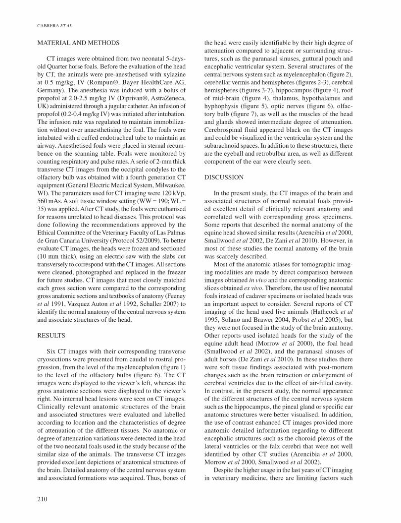
Figure 1. Representative image of equine brain. Each line and number (I-VI) represents the approximate level of the following trans-verse CT images (Figures 2-7). Imagen representativa de un encéfalo equino. Cada línea y número (I-VI) representa el nivel aproximado de las TC transversales (Figuras 2-7).
Figure 2. (A) Computed tomography image and (B) anatomic section at the level of myelencephalon. 1. Squamous part of occipital bone; 2. Basilar part of occipital bone; 3. Lateral part of occipital bone; 4. Myelencephalon; 5. Fourth ventricle; 6. Cerebellar vermis; 7. Cerebellar hemisphere; 8. Auricle; 9. Auricular cartilage; 10. Auricular fat; 11. Guttural pouch (caudal part). (A) Imagen de tomografía computarizada y (B) sección anatómica a nivel del mielencéfalo. 1. Porción escamosa del hueso occipital; 2. Porción basilar del hueso occipital; 3. Porción lateral del hueso occipital; 4. Mielencéfalo; 5. Cuarto ventrículo; 6. Vermis del cerebelo; 7. Hemisferio del cerebelo; 8. Pabellón auricular; 9. Cartílago auricular; 10. Cuerpo adiposo auricular; 11. Bolsa gutural (parte caudal).
212
CABRERA ET AL
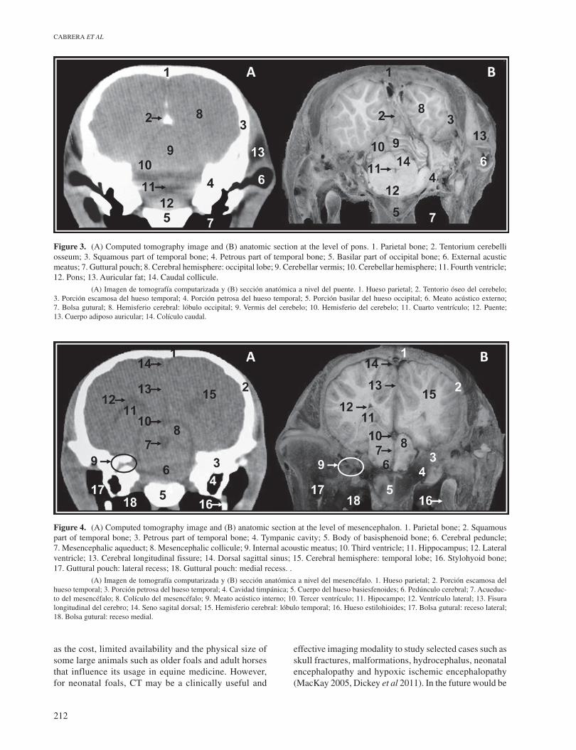
Figure 3. (A) Computed tomography image and (B) anatomic section at the level of pons. 1. Parietal bone; 2. Tentorium cerebelli osseum; 3. Squamous part of temporal bone; 4. Petrous part of temporal bone; 5. Basilar part of occipital bone; 6. External acustic meatus; 7. Guttural pouch; 8. Cerebral hemisphere: occipital lobe; 9. Cerebellar vermis; 10. Cerebellar hemisphere; 11. Fourth ventricle; 12. Pons; 13. Auricular fat; 14. Caudal collicule. (A) Imagen de tomografía computarizada y (B) sección anatómica a nivel del puente. 1. Hueso parietal; 2. Tentorio óseo del cerebelo; 3. Porción escamosa del hueso temporal; 4. Porción petrosa del hueso temporal; 5. Porción basilar del hueso occipital; 6. Meato acústico externo; 7. Bolsa gutural; 8. Hemisferio cerebral: lóbulo occipital; 9. Vermis del cerebelo; 10. Hemisferio del cerebelo; 11. Cuarto ventrículo; 12. Puente; 13. Cuerpo adiposo auricular; 14. Colículo caudal.
Figure 4. (A) Computed tomography image and (B) anatomic section at the level of mesencephalon. 1. Parietal bone; 2. Squamous part of temporal bone; 3. Petrous part of temporal bone; 4. Tympanic cavity; 5. Body of basisphenoid bone; 6. Cerebral peduncle; 7. Mesencephalic aqueduct; 8. Mesencephalic collicule; 9. Internal acoustic meatus; 10. Third ventricle; 11. Hippocampus; 12. Lateral ventricle; 13. Cerebral longitudinal fissure; 14. Dorsal sagittal sinus; 15. Cerebral hemisphere: temporal lobe; 16. Stylohyoid bone; 17. Guttural pouch: lateral recess; 18. Guttural pouch: medial recess. . (A) Imagen de tomografía computarizada y (B) sección anatómica a nivel del mesencéfalo. 1. Hueso parietal; 2. Porción escamosa del hueso temporal; 3. Porción petrosa del hueso temporal; 4. Cavidad timpánica; 5. Cuerpo del hueso basiesfenoides; 6. Pedúnculo cerebral; 7. Acueduc-to del mesencéfalo; 8. Colículo del mesencéfalo; 9. Meato acústico interno; 10. Tercer ventrículo; 11. Hipocampo; 12. Ventrículo lateral; 13. Fisura longitudinal del cerebro; 14. Seno sagital dorsal; 15. Hemisferio cerebral: lóbulo temporal; 16. Hueso estilohioides; 17. Bolsa gutural: receso lateral; 18. Bolsa gutural: receso medial.
as the cost, limited availability and the physical size of some large animals such as older foals and adult horses that influence its usage in equine medicine. However, for neonatal foals, CT may be a clinically useful and
effective imaging modality to study selected cases such as skull fractures, malformations, hydrocephalus, neonatal encephalopathy and hypoxic ischemic encephalopathy (MacKay 2005, Dickey et al 2011). In the future would be
213
COMPUTED TOMOGRAPHY, ANATOMY, BRAIN, NEONATAL FOAL
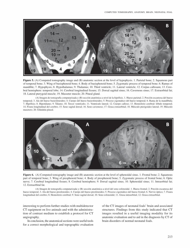
Figure 5. (A) Computed tomography image and (B) anatomic section at the level of hypophysis. 1. Parietal bone; 2. Squamous part of temporal bone; 3. Wing of basisphenoid bone; 4. Body of basisphenoid bone; 5. Zygomatic process of temporal bone; 6. Ramus of mandible; 7. Hypophysis; 8. Hypothalamus; 9. Thalamus; 10. Third ventricle; 11. Lateral ventricle; 12. Corpus callosum; 13. Cere-bral hemisphere: temporal lobe; 14. Cerebral longitudinal fissure; 15. Dorsal sagittal sinus; 16. Cavernous sinus; 17. Extraorbital fat; 18. Lateral pterygoid muscle; 19. Masseter muscle; 20. Pineal gland. (A) Imagen de tomografía computarizada y (B) sección anatómica a nivel de la hipófisis. 1. Hueso parietal; 2. Porción escamosa del hueso temporal; 3. Ala del hueso basiesfenoides; 4. Cuerpo del hueso basiesfenoides; 5. Proceso cigomático del hueso temporal; 6. Rama de la mandíbula; 7. Hipófisis; 8. Hipotálamo; 9. Tálamo; 10. Tercer ventrículo; 11. Ventrículo lateral; 12. Cuerpo calloso; 13. Hemisferio cerebral: lóbulo temporal; 14. Fisura longitudinal del cerebro; 15. Seno sagital dorsal; 16. Seno cavernoso; 17. Grasa extraorbital; 18. Músculo pterigoideo lateral; 19. Músculo masetero; 20. Glándula pineal.
Figure 6. (A) Computed tomography image and (B) anatomic section at the level of sphenoidal sinus. 1. Frontal bone; 2. Squamous part of temporal bone; 3. Wing of presphenoid bone; 4. Body of presphenoid bone; 5. Zygomatic process of frontal bone; 6. Optic nerve; 7. Cerebral longitudinal fissure; 8. Cerebral hemisphere; 9. Dorsal sagittal sinus; 10. Sphenoidal sinus; 11. Intraorbital fat; 12. Extraorbital fat. (A) Imagen de tomografía computarizada y (B) sección anatómica a nivel del seno esfenoidal. 1. Hueso frontal; 2. Porción escamosa del hueso temporal; 3. Ala del hueso presfenoides; 4. Cuerpo del hueso presfenoides; 5. Proceso cigomático del hueso frontal; 6. Nervio óptico; 7. Fisura longitudinal del cerebro; 8. Hemisferio cerebral; 9. Seno sagital dorsal; 10. Seno esfenoidal; 11. Grasa intraorbital; 12. Grasa extraorbital.
interesting to perform further studies with multidetector CT equipment on live animals and with the administra-tion of contrast medium to establish a protocol for CT angiography.
In conclusion, the anatomical sections were useful tools for a correct morphological and topographic evaluation
of the CT images of neonatal foals’ brain and associated structures. Findings from this study indicated that CT images resulted in a useful imaging modality for its anatomic evaluation and to aid in the diagnosis by CT of brain disorders of normal neonatal foals.
214
CABRERA ET AL
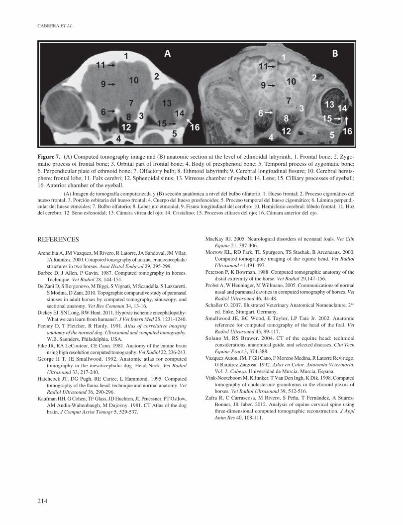
Figure 7. (A) Computed tomography image and (B) anatomic section at the level of ethmoidal labyrinth. 1. Frontal bone; 2. Zygo-matic process of frontal bone; 3. Orbital part of frontal bone; 4. Body of presphenoid bone; 5. Temporal process of zygomatic bone; 6. Perpendicular plate of ethmoid bone; 7. Olfactory bulb; 8. Ethmoid labyrinth; 9. Cerebral longitudinal fissure; 10. Cerebral hemis-phere: frontal lobe; 11. Falx cerebri; 12. Sphenoidal sinus; 13. Vitreous chamber of eyeball; 14. Lens; 15. Cilliary processes of eyeball; 16. Anterior chamber of the eyeball. (A) Imagen de tomografía computarizada y (B) sección anatómica a nivel del bulbo olfatorio. 1. Hueso frontal; 2. Proceso cigomático del hueso frontal; 3. Porción orbitaria del hueso frontal; 4. Cuerpo del hueso presfenoides; 5. Proceso temporal del hueso cigomático; 6. Lámina perpendi-cular del hueso etmoides; 7. Bulbo olfatorio; 8. Laberinto etmoidal; 9. Fisura longitudinal del cerebro; 10. Hemisferio cerebral: lóbulo frontal; 11. Hoz del cerebro; 12. Seno esfenoidal; 13. Cámara vítrea del ojo; 14. Cristalino; 15. Procesos ciliares del ojo; 16. Cámara anterior del ojo.
REFERENCES
Arencibia A, JM Vazquez, M Rivero, R Latorre, JA Sandoval, JM Vilar, JA Ramírez. 2000. Computed tomography of normal cranioencephalic structures in two horses. Anat Histol Embryol 29, 295-299.
Barbee D, J Allen, P Gavin. 1987. Computed tomography in horses. Technique. Vet Radiol 28, 144-151.
De Zani D, S Borgonovo, M Biggi, S Vignati, M Scandella, S Lazzaretti, S Modina, D Zani. 2010. Topographic comparative study of paranasal sinuses in adult horses by computed tomography, sinuscopy, and sectional anatomy. Vet Res Commun 34, 13-16.
Dickey EJ, SN Long, RW Hunt. 2011. Hypoxic ischemic encephalopathy- What we can learn from humans?. J Vet Intern Med 25, 1231-1240.
Feeney D, T Fletcher, R Hardy. 1991. Atlas of correlative imaging anatomy of the normal dog. Ultrasound and computed tomography. W.B. Saunders, Philadelphia, USA.
Fike JR, RA LeCouteur, CE Cann. 1981. Anatomy of the canine brain using high resolution computed tomography. Vet Radiol 22, 236-243.
George II T, JE Smallwood. 1992. Anatomic atlas for computed tomography in the mesaticephalic dog. Head Neck. Vet Radiol Ultrasound 33, 217-240.
Hatchcock JT, DG Pugh, RE Cartee, L Hammond. 1995. Computed tomography of the llama head: technique and normal anatomy. Vet Radiol Ultrasound 36, 290-296.
Kaufman HH, G Cohen, TF Glass, JD Huchton, JL Pruessner, PT Ostlow, AM Andia-Waltenbaugh, M Dujovny. 1981. CT Atlas of the dog brain. J Comput Assist Tomogr 5, 529-537.
MacKay RJ. 2005. Neurological disorders of neonatal foals. Vet Clin Equine 21, 387-406.
Morrow KL, RD Park, TL Spurgeon, TS Stashak, B Arceneaux. 2000. Computed tomographic imaging of the equine head. Vet Radiol Ultrasound 41,491-497.
Peterson P, K Bowman. 1988. Computed tomographic anatomy of the distal extremity of the horse. Vet Radiol 29,147-156.
Probst A, W Henninger, M Willmann. 2005. Communications of normal nasal and paranasal cavities in computed tomography of horses. Vet Radiol Ultrasound 46, 44-48.
Schaller O. 2007. Illustrated Veterinary Anatomical Nomenclature. 2nd ed. Enke, Sttutgart, Germany.
Smallwood JE, BC Wood, E Taylor, LP Tate Jr. 2002. Anatomic reference for computed tomography of the head of the foal. Vet Radiol Ultrasound 43, 99-117.
Solano M, RS Brawer. 2004. CT of the equine head: technical considerations, anatomical guide, and selected diseases. Clin Tech Equine Pract 3, 374-388.
Vazquez Auton, JM, F Gil Cano, F Moreno Medina, R Latorre Reviriego, G Ramírez Zarzosa. 1992. Atlas en Color. Anatomía Veterinaria. Vol. 1. Cabeza. Universidad de Murcia, Murcia, España.
Vink-Nooteboom M, K Junker, T Van Den Ingh, K Dik. 1998. Computed tomography of cholesterinic granulomas in the choroid plexus of horses. Vet Radiol Ultrasound 39, 512-516.
Zafra R, C Carrascosa, M Rivero, S Peña, T Fernández, A Suárez-Bonnet, JR Jaber. 2012. Analysis of equine cervical spine using three-dimensional computed tomographic reconstruction. J Appl Anim Res 40, 108-111.