Comprehensive Biochemistry for Dentistry
45
Anil Gupta Comprehensive Biochemistry for Dentistry Textbook for Dental Students
Transcript of Comprehensive Biochemistry for Dentistry

Anil Gupta
Comprehensive Biochemistry for DentistryTextbook for Dental Students

Comprehensive Biochemistry for Dentistry

Anil Gupta
Comprehensive Biochemistry for Dentistry
Textbook for Dental Students

Anil GuptaDepartment of Physiology and BiochemistryEklavya Dental College and HospitalKotputli Rajasthan India
ISBN 978-981-13-1034-8 ISBN 978-981-13-1035-5 (eBook)https://doi.org/10.1007/978-981-13-1035-5
Library of Congress Control Number: 2018950138
© Springer Nature Singapore Pte Ltd. 2019This work is subject to copyright. All rights are reserved by the Publisher, whether the whole or part of the material is concerned, specifically the rights of translation, reprinting, reuse of illustrations, recitation, broadcasting, reproduction on microfilms or in any other physical way, and transmission or information storage and retrieval, electronic adaptation, computer software, or by similar or dissimilar methodology now known or hereafter developed.The use of general descriptive names, registered names, trademarks, service marks, etc. in this publication does not imply, even in the absence of a specific statement, that such names are exempt from the relevant protective laws and regulations and therefore free for general use.The publisher, the authors, and the editors are safe to assume that the advice and information in this book are believed to be true and accurate at the date of publication. Neither the publisher nor the authors or the editors give a warranty, express or implied, with respect to the material contained herein or for any errors or omissions that may have been made. The publisher remains neutral with regard to jurisdictional claims in published maps and institutional affiliations.
This Springer imprint is published by the registered company Springer Nature Singapore Pte Ltd.The registered company address is: 152 Beach Road, #21-01/04 Gateway East, Singapore 189721, Singapore

I dedicate my book to Almighty Shirdi Sai Baba.

vii
Foreword
It gives me a great satisfaction to write a brief note on Comprehensive Biochemistry for Dentistry by Dr. Anil Gupta. In this book, the latest information has been incor-porated to create a comprehensive text that is suitable for beginners.
Every effort has been made to update the text with the possible latest informa-tion. The information is clearly presented, logically arranged, and well-illustrated. References have been included to guide readers to the classical and current litera-ture. I am confident that this textbook will prove to be a useful compendium for all students at the graduate and postgraduate levels, igniting their interest and enabling a greater involvement in the growing dental field.
O. P. JangirDirector Academics
FELM, IASE (Deemed to be) University Gandhi Vidya Mandir
SardarshahrRajasthan, India

Foreword
It is my privilege to review and comment on the book Comprehensive Biochemistry for Dentistry. The 24 chapters of this book begin with cells and organelles—the fundamental units of life. The next two sections contain information on structural and molecular biology components and intermediary metabolism. The fourth sec-tion is dedicated to medical biochemistry, explaining the importance of acid–base balance, nutrition, and diagnostic enzymes. The final section focuses on dental bio-chemistry, which is of more clinical interest. This unique section examines the role of various molecules and macromolecules in the normal functioning of oral tissues and in the instigation of dental diseases. The index is quite good and easily steers the reader toward references of interest. Most chapters present information on applied biochemistry, including the biochemistry and treatment of specific diseases, making the book useful for both physicians and students. It also serves as a refresher on the fundamentals of biochemistry for students and physicians. The author pres-ents up-to-date information on biochemistry and emphasizes relevant physiologic and pathophysiologic biochemical concepts. The book uses simple and clear lan-guage that can be easily understood by students, physicians, and researchers. I am sure this book will be welcomed by many undergraduate and postgraduate students, physicians, and researchers.
Rajesh DaburProfessor and Head
Department of BiochemistryM. D. University
Rohtak, India

ix
Foreword
I am glad to know about the publication of Comprehensive Biochemistry for Dentistry. This book covers the complete syllabus for undergraduate courses, as well as being a source of knowledge for professionals. I wish the author great suc-cess with this book.
S. P. S. Sodhi Principal
Dashmesh Institute of Research and Dental SciencesFaridkot, India
Executive Member Dental Council of India
New Delhi, India
Member–Academic Council Member–Board of Studies Member–Planning Board
Baba Farid University of Health SciencesFaridkot, India

Foreword
It is a pleasure to write a foreword for Comprehensive Biochemistry for Dentistry, which was written by my younger brother, Dr. Anil Gupta. What impressed me most was his deep insight on the subject and his method of presenting even the most complex concepts in a simple, easily understandable, and convincing manner. With each chapter, he provides the details of biochemistry as applied to the understanding of dental disorders. The book is organized in a manner to help students. It is a book to be opened with expectation and closed with profit.
I have reviewed this book and appreciate the simple language, straightforward style, and clear presentation. This book will definitely prove to be very useful for all dental students and practitioners in India.
S. K. Singhal MD, LLB
Medico Legal Consultant and Author of Seven Books Medical Director
Professor, and Head of the Department of Forensic Medicine and ToxicologyA.C.P.M. Medical College
Dhule, India

xi
Foreword
Biochemistry is a vast and challenging field for dental students. Comprehensive Biochemistry for Dentistry is a complete and student-friendly book. It offers the users an excellent opportunity to understand the basic fundamentals of biochemistry and its applications to the medical field. A dedicated effort has been made by Prof. (Dr.) Anil Gupta to systematically compile the book into six units covering the entire syllabus. I am sure that the textbook will be an informative read for under-graduate students, as well as clinicians. The author deserves appreciation and recog-nition for his efforts to carry out this commendable and arduous task.
Sanjay Bansal, MDSPrincipal
EDCH Kotputli, India
Foreword
I wish great success and my heartiest congratulations to my senior colleague, Dr. Anil Gupta, for publishing Comprehensive Biochemistry for Dentistry. This book fills a great need of students, especially those studying dentistry. I have known Dr. Anil for years; he is deeply loved and respected by our students for his skills in the subject, his teaching methodology, and his constant pursuit for excellence. He is a great academician, a great person, and above all, a kind human being. Dr. Anil has been continuously working on this title for years, trying to create the best possible textbook on biochemistry with up-to-date content and simple language, allowing its readers to understand the complexities of the biochemicals in the human body. It is a great fortune of mine to be associated with Dr. Anil Gupta. I believe this title will be a superb choice for students of biochemistry thanks to its comprehensive content, simplicity, and ease of understanding.
Nitul Jain, MDS Professor and Head Department of Oral Pathology
Eklavya Dental College and Hospital Kotputli, Rajasthan, India

xiii
Foreword
It gives me great pleasure to unveil yet another noteworthy work penned by the author.
Dr. Anil Gupta is a prolific writer and no stranger to the world of books. His exceptional style of writing, creativity, and groundbreaking ideas have only improved with time.
Biochemistry is one of the most important basic sciences in the preclinical years of medical and dental schools. For me personally, it was a daunting subject.
This book complements the classical textbooks of biochemistry. It blends the fundamental and dynamic concepts of biochemistry and provides exhaustive knowl-edge for dental students in a single, clearly presented volume. The chapters are skillfully written and easy to understand.
I recommend this book with enthusiasm and give my best wishes to Dr. Anil Gupta for undertaking this endeavor.
Prithvi Raj Singh MD Pathology
Reader, Department of PathologyEDCH, Kotputli, India

Foreword
Dr. Anil Gupta, the author of this book, deserves to be congratulated on presenting the speciality of biochemistry to medical and dental students at different levels of education, as well as to professionals of different specialists. This book provides a comprehensive and clear account of the current principles of biochemistry knowl-edge. The author has done this by drawing on his rich experience as a teacher of biochemistry for more than 10 years. This book defines all topics in a narrative format so that the information is palatable to students and professionals. In this book, Dr. Gupta has adopted a conventional approach to characterize food constitu-ents, describing their formulae, properties, and place in human biological and chemical reactions. The language is simple and easily understandable. The chapters on the metabolism of carbohydrates, lipids, proteins, and nucleic acids are exhaus-tive and naturally presented. Students will benefit greatly from the descriptive account of vitamins, hormones, and enzymes, with special emphasis on dental top-ics. This book will be welcomed by teachers of biochemistry, dental students, medi-cal students, and professionals for revising and refreshing one’s knowledge in biochemistry. I am confident that this book will go through many editions in the coming years and the author will add more details on the topics herein.
P. K. Garg, MBBS, MD (Pathology)Professor, Pathology
Venkateshwara Institute of Medical SciecnesGajraula, UP, India

xv
Foreword
This book, Comprehensive Biochemistry for Dentistry, is a good idea and great effort by the author. The book is very educative and informative for graduates. I wish Dr. Gupta good luck for this great endeavour.
Naveen Bansal Head, Department of Orthodontics
Genesis Institute of Dental Sciences and ResearchFerozepur, India

Foreword
I sincerely thank the author for allowing me to be part of his precious book. The chapters within are unique and contain a plethora of information, not only for under-graduate studies but also for postgraduate entrance examinations. The genetics sec-tion may be especially helpful for this purpose. As a faculty member at a reputed coaching centre for postgraduate entrance examinations, I find that most under-graduate students lack a clear understanding of the concepts of biochemistry. Therefore, I think this book is very helpful for clarifying the concepts. It includes a section on dental biochemistry that is an additional benefit for students. To con-clude, I want to quote Francis Bacon: “Some books are to be tasted, others swal-lowed, and some few to be chewed and digested.” The content of this book should be chewed and absorbed properly for undergraduate and postgraduate entrance examinations.
Puneet Garg, MBBS, MD (Pathology) PCMS-I, Govt. Medical College and Rajindra Hospital
Patiala, India

xvii
Foreword
I congratulate Dr. Anil Gupta for his extensive teaching and research experience, which were utilized to write this book, Comprehensive Biochemistry for Dentistry. I am absolutely sure that students of dentistry will find this book highly useful.
Hasan Kamal, PhD (Biochemistry) Associate Professor
Department of BiochemistryIDST, ModinagarGhaziabad, India

xix
Preface
Biochemistry is an essential subject in the medical, dental, nursing, paramedical, physiotherapy and homeopathy streams in various universities across the world. Knowledge of the fundamentals of biochemistry is necessary for the study of normal structure and functioning of organs and organ systems, as well as the pathogenesis of diseases.
A number of biochemistry books are available in the market. This book is unique in its approach to the vital concepts of biochemistry. It blends the funda-mentals and dynamic concepts of biochemistry with the theory of dental sciences to provide valuable, coherent, and exhaustive knowledge for dental students and professionals.
Comprehensive Biochemistry for Dentistry is composed of 24 chapters on the structure, function, and metabolism of biomolecules. The information is presented in a simplified approach and enriched with diagrams, flow charts, tables,, and graphs. Important concepts, definitions, and clinical significance are provided in a summary format for easy understanding and recapitulation. Topics such as cell and organelles, vitamins, hormones, nucleic acids, biological oxidation, metabolism, nutrition, and serum enzymes have been prepared in such a way to provide exhaus-tive knowledge to students using clear and straightforward language. In particular, the information on dental biochemistry is unique to this book. Proteins, lipids, car-bohydrates, vitamins, and hormones are discussed in relation to the normal structure and functioning of oral tissues, as well as their roles in dental diseases.
This book is a unique attempt to integrate the fundamentals of biochemistry with the science of dentistry for the purpose of imparting knowledge to dental students and professionals. I hope it will disseminate the knowledge of biochemistry to undergraduate students and serve as a reference book for professionals and acade-micians. I have made sincere and honest efforts to prepare and present a factually accurate book. However, I welcome criticisms, comments, and suggestions for improvements to forthcoming editions of this book.
Kotputli, India Anil Gupta 27 March 2018

xxi
Acknowledgements
The Almighty Shri Shirdi Sai Baba bestowed upon me the knowledge and persever-ance for writing the book. My father, Shri Ved Parkash Gupta, always inspires me to undertake great endeavours. I am highly indebted to my father for instilling the habit of learning in me since my school days.My wife persistently motivates me to achieve my goals and she stands by me in odd hours.
I owe my gratitude to Dr. Bhavik Sawhney, Associate Editor–Biomedicine, Springer (India) Pvt. Ltd, for his support, cooperation, and understanding, which he offered to me to complete the writing of this book. I am also thankful to Ms. Saanthi Shankhararaman, Project Coordinator (Books) and her production team for creating an attractive and appealing book design.

xxiii
Contents
1 Introduction to Biochemistry . . . . . . . . . . . . . . . . . . . . . . . . . . . . . . . . . . 1 1.1 Definition . . . . . . . . . . . . . . . . . . . . . . . . . . . . . . . . . . . . . . . . . . . . . . 1 1.2 Historical Development of Biochemistry . . . . . . . . . . . . . . . . . . . . . 1
Part I Cellular Biochemistry
2 Cell and Organelles . . . . . . . . . . . . . . . . . . . . . . . . . . . . . . . . . . . . . . . . . . 5 2.1 Cell . . . . . . . . . . . . . . . . . . . . . . . . . . . . . . . . . . . . . . . . . . . . . . . . . . . 5
2.1.1 Definition . . . . . . . . . . . . . . . . . . . . . . . . . . . . . . . . . . . . . . . . 5 2.2 Landmark Discoveries . . . . . . . . . . . . . . . . . . . . . . . . . . . . . . . . . . . . 5 2.3 Cell Theory Postulates . . . . . . . . . . . . . . . . . . . . . . . . . . . . . . . . . . . . 5 2.4 Modern Concept of Cell . . . . . . . . . . . . . . . . . . . . . . . . . . . . . . . . . . . 6 2.5 Prokaryotic Cell . . . . . . . . . . . . . . . . . . . . . . . . . . . . . . . . . . . . . . . . . 6 2.6 Eukaryotic Cell . . . . . . . . . . . . . . . . . . . . . . . . . . . . . . . . . . . . . . . . . 6 2.7 Plasma Membrane . . . . . . . . . . . . . . . . . . . . . . . . . . . . . . . . . . . . . . . 8
2.7.1 Definition . . . . . . . . . . . . . . . . . . . . . . . . . . . . . . . . . . . . . . . . 8 2.7.2 History . . . . . . . . . . . . . . . . . . . . . . . . . . . . . . . . . . . . . . . . . . 8 2.7.3 Chemical Composition . . . . . . . . . . . . . . . . . . . . . . . . . . . . . 8
2.8 Models of Plasma Membrane Structure . . . . . . . . . . . . . . . . . . . . . . . 10 2.8.1 Lipid Bilayer Model . . . . . . . . . . . . . . . . . . . . . . . . . . . . . . . . 10 2.8.2 Sandwich Model . . . . . . . . . . . . . . . . . . . . . . . . . . . . . . . . . . 11 2.8.3 Unit Membrane Model . . . . . . . . . . . . . . . . . . . . . . . . . . . . . 11 2.8.4 Fluid Mosaic Model . . . . . . . . . . . . . . . . . . . . . . . . . . . . . . . . 12 2.8.5 Functions of Plasma Membrane. . . . . . . . . . . . . . . . . . . . . . . 13
2.9 Cytoplasmic Organelles . . . . . . . . . . . . . . . . . . . . . . . . . . . . . . . . . . . 14 2.9.1 Mitochondria . . . . . . . . . . . . . . . . . . . . . . . . . . . . . . . . . . . . . 14 2.9.2 Ribosomes . . . . . . . . . . . . . . . . . . . . . . . . . . . . . . . . . . . . . . . 18
2.10 Golgi Body . . . . . . . . . . . . . . . . . . . . . . . . . . . . . . . . . . . . . . . . . . . . . 21 2.10.1 Functions of Golgi Body . . . . . . . . . . . . . . . . . . . . . . . . . . . . 24
2.11 Endoplasmic Reticulum . . . . . . . . . . . . . . . . . . . . . . . . . . . . . . . . . . . 25 2.12 Nucleus . . . . . . . . . . . . . . . . . . . . . . . . . . . . . . . . . . . . . . . . . . . . . . . 28 2.13 Nucleolus . . . . . . . . . . . . . . . . . . . . . . . . . . . . . . . . . . . . . . . . . . . . . . 32Suggested Readings . . . . . . . . . . . . . . . . . . . . . . . . . . . . . . . . . . . . . . . . . . 32

xxiv
Part II Structural Biochemistry
3 Protein and Amino Acids . . . . . . . . . . . . . . . . . . . . . . . . . . . . . . . . . . . . . 35 3.1 Historical Facts . . . . . . . . . . . . . . . . . . . . . . . . . . . . . . . . . . . . . . . . . 35 3.2 Definition . . . . . . . . . . . . . . . . . . . . . . . . . . . . . . . . . . . . . . . . . . . . . . 35 3.3 Classification of Proteins . . . . . . . . . . . . . . . . . . . . . . . . . . . . . . . . . . 35
3.3.1 Classification Based upon Size and Shape. . . . . . . . . . . . . . . 36 3.3.2 Classification Based upon Biological Functions . . . . . . . . . . 36 3.3.3 Classification Based upon Chemical Composition
and Physical Properties . . . . . . . . . . . . . . . . . . . . . . . . . . . . . 37 3.3.4 Classification Based upon Quality
and Nutritional Value . . . . . . . . . . . . . . . . . . . . . . . . . . . . . . . 41 3.4 Structural Organization of Proteins . . . . . . . . . . . . . . . . . . . . . . . . . . 42
3.4.1 Primary Structure . . . . . . . . . . . . . . . . . . . . . . . . . . . . . . . . . . 43 3.4.2 Insulin Structure . . . . . . . . . . . . . . . . . . . . . . . . . . . . . . . . . . . 46 3.4.3 Secondary Structure . . . . . . . . . . . . . . . . . . . . . . . . . . . . . . . . 47 3.4.4 Tertiary Structure . . . . . . . . . . . . . . . . . . . . . . . . . . . . . . . . . . 51 3.4.5 Quaternary Structure of Protein . . . . . . . . . . . . . . . . . . . . . . . 52
3.5 Amino Acids Definition . . . . . . . . . . . . . . . . . . . . . . . . . . . . . . . . . . . 54 3.6 Number of Amino Acids . . . . . . . . . . . . . . . . . . . . . . . . . . . . . . . . . . 55 3.7 Structure of Amino Acid . . . . . . . . . . . . . . . . . . . . . . . . . . . . . . . . . . 55 3.8 Important Characteristics of Amino Acids . . . . . . . . . . . . . . . . . . . . . 55 3.9 Classification of Amino Acids . . . . . . . . . . . . . . . . . . . . . . . . . . . . . . 56
3.9.1 Classification Depending on Position of Amino Group . . . . . 56 3.9.2 Classification Depending on Proteinogenic Property . . . . . . 57 3.9.3 Classification Depending on Polarity of Side Chain . . . . . . . 58 3.9.4 Classification Depending on Nutritional Value . . . . . . . . . . . 58 3.9.5 Classification Depending on Chemical Structure
of Side Chain . . . . . . . . . . . . . . . . . . . . . . . . . . . . . . . . . . . . . 59 3.9.6 Classification Depending on Chemical Property . . . . . . . . . . 63
3.10 Applied Biochemistry . . . . . . . . . . . . . . . . . . . . . . . . . . . . . . . . . . . . 64 3.10.1 Arginine . . . . . . . . . . . . . . . . . . . . . . . . . . . . . . . . . . . . . . . . . 64 3.10.2 Casein Phosphopeptides (CPP) . . . . . . . . . . . . . . . . . . . . . . . 64
Suggested Readings . . . . . . . . . . . . . . . . . . . . . . . . . . . . . . . . . . . . . . . . . . 65
4 Plasma Proteins . . . . . . . . . . . . . . . . . . . . . . . . . . . . . . . . . . . . . . . . . . . . . 67 4.1 Classification of Plasma Proteins . . . . . . . . . . . . . . . . . . . . . . . . . . . . 67 4.2 Plasma Proteins . . . . . . . . . . . . . . . . . . . . . . . . . . . . . . . . . . . . . . . . . 67 4.3 Albumin . . . . . . . . . . . . . . . . . . . . . . . . . . . . . . . . . . . . . . . . . . . . . . . 67 4.4 Globulins . . . . . . . . . . . . . . . . . . . . . . . . . . . . . . . . . . . . . . . . . . . . . . 69 4.5 α1-Globulins . . . . . . . . . . . . . . . . . . . . . . . . . . . . . . . . . . . . . . . . . . . . 69
4.5.1 Alpha-1: Acid Glycoprotein . . . . . . . . . . . . . . . . . . . . . . . . . . 69 4.5.2 Alpha-1 Fetoglobulin . . . . . . . . . . . . . . . . . . . . . . . . . . . . . . . 69 4.5.3 Alpha-1 Antitrypsin . . . . . . . . . . . . . . . . . . . . . . . . . . . . . . . . 70
4.6 α2-Globulins . . . . . . . . . . . . . . . . . . . . . . . . . . . . . . . . . . . . . . . . . . . . 71 4.6.1 Ceruloplasmin . . . . . . . . . . . . . . . . . . . . . . . . . . . . . . . . . . . . 71 4.6.2 Haptoglobin . . . . . . . . . . . . . . . . . . . . . . . . . . . . . . . . . . . . . . 71
Contents

xxv
4.7 β-Globulins. . . . . . . . . . . . . . . . . . . . . . . . . . . . . . . . . . . . . . . . . . . . . 72 4.7.1 Transferrin . . . . . . . . . . . . . . . . . . . . . . . . . . . . . . . . . . . . . . . 72 4.7.2 C-Reactive Protein . . . . . . . . . . . . . . . . . . . . . . . . . . . . . . . . . 72 4.7.3 Hemopexin . . . . . . . . . . . . . . . . . . . . . . . . . . . . . . . . . . . . . . . 73
4.8 Other Important Plasma Proteins . . . . . . . . . . . . . . . . . . . . . . . . . . . . 73 4.8.1 Bence-Jones Protein . . . . . . . . . . . . . . . . . . . . . . . . . . . . . . . . 73 4.8.2 Fibrinogen . . . . . . . . . . . . . . . . . . . . . . . . . . . . . . . . . . . . . . . 74
4.9 Biological Functions of Plasma Proteins . . . . . . . . . . . . . . . . . . . . . . 74Suggested Readings . . . . . . . . . . . . . . . . . . . . . . . . . . . . . . . . . . . . . . . . . . 75
5 Hemoglobin . . . . . . . . . . . . . . . . . . . . . . . . . . . . . . . . . . . . . . . . . . . . . . . . 77 5.1 Definition . . . . . . . . . . . . . . . . . . . . . . . . . . . . . . . . . . . . . . . . . . . . . . 77 5.2 Characteristics of Hemoglobin . . . . . . . . . . . . . . . . . . . . . . . . . . . . . 77 5.3 Structure of Hemoglobin . . . . . . . . . . . . . . . . . . . . . . . . . . . . . . . . . . 78
5.3.1 Heme . . . . . . . . . . . . . . . . . . . . . . . . . . . . . . . . . . . . . . . . . . . 78 5.4 Two-State Model of Hemoglobin . . . . . . . . . . . . . . . . . . . . . . . . . . . 80 5.5 Functions of Hemoglobin . . . . . . . . . . . . . . . . . . . . . . . . . . . . . . . . . 82 5.6 Variants of Hemoglobin . . . . . . . . . . . . . . . . . . . . . . . . . . . . . . . . . . . 82
5.6.1 Adult Hemoglobin (HbA) . . . . . . . . . . . . . . . . . . . . . . . . . . . 83 5.6.2 Fetal Hemoglobin (HbF) . . . . . . . . . . . . . . . . . . . . . . . . . . . . 83 5.6.3 Glycosylated Hemoglobin (HbA1C) . . . . . . . . . . . . . . . . . . . 83
5.7 Hemoglobinopathies . . . . . . . . . . . . . . . . . . . . . . . . . . . . . . . . . . . . . 84 5.7.1 Sickle-Cell Hemoglobin (HbS) . . . . . . . . . . . . . . . . . . . . . . . 84 5.7.2 Sickle-Cell Trait . . . . . . . . . . . . . . . . . . . . . . . . . . . . . . . . . . . 86 5.7.3 Hemoglobin C (HbC) . . . . . . . . . . . . . . . . . . . . . . . . . . . . . . . 87 5.7.4 Hemoglobin D (HbD) . . . . . . . . . . . . . . . . . . . . . . . . . . . . . . 87 5.7.5 Hemoglobin E (HbE) . . . . . . . . . . . . . . . . . . . . . . . . . . . . . . . 87 5.7.6 Thalassemia . . . . . . . . . . . . . . . . . . . . . . . . . . . . . . . . . . . . . . 87
5.8 Derivatives of Hemoglobin . . . . . . . . . . . . . . . . . . . . . . . . . . . . . . . . 89 5.8.1 Oxyhemoglobin . . . . . . . . . . . . . . . . . . . . . . . . . . . . . . . . . . . 89 5.8.2 Carboxyhemoglobin . . . . . . . . . . . . . . . . . . . . . . . . . . . . . . . . 89 5.8.3 Carbamino-Hemoglobin . . . . . . . . . . . . . . . . . . . . . . . . . . . . 90 5.8.4 Sulfhemoglobin . . . . . . . . . . . . . . . . . . . . . . . . . . . . . . . . . . . 90 5.8.5 Methemoglobin . . . . . . . . . . . . . . . . . . . . . . . . . . . . . . . . . . . 90
5.9 Biodegradation of Hemoglobin . . . . . . . . . . . . . . . . . . . . . . . . . . . . . 91 5.10 Biosynthesis of Hemoglobin . . . . . . . . . . . . . . . . . . . . . . . . . . . . . . . 94
5.10.1 Biosynthesis of Heme . . . . . . . . . . . . . . . . . . . . . . . . . . . . . . 94 5.10.2 Biosynthesis of Globin . . . . . . . . . . . . . . . . . . . . . . . . . . . . . . 97 5.10.3 Regulation of Heme Biosynthesis . . . . . . . . . . . . . . . . . . . . . 99
Suggested Readings . . . . . . . . . . . . . . . . . . . . . . . . . . . . . . . . . . . . . . . . . . 100
6 Carbohydrates . . . . . . . . . . . . . . . . . . . . . . . . . . . . . . . . . . . . . . . . . . . . . . 101 6.1 Definition . . . . . . . . . . . . . . . . . . . . . . . . . . . . . . . . . . . . . . . . . . . . . . 101 6.2 Classification . . . . . . . . . . . . . . . . . . . . . . . . . . . . . . . . . . . . . . . . . . . 101
6.2.1 Monosaccharides . . . . . . . . . . . . . . . . . . . . . . . . . . . . . . . . . . 101 6.2.2 Disaccharides . . . . . . . . . . . . . . . . . . . . . . . . . . . . . . . . . . . . . 102 6.2.3 Oligosaccharides . . . . . . . . . . . . . . . . . . . . . . . . . . . . . . . . . . 102 6.2.4 Polysaccharides . . . . . . . . . . . . . . . . . . . . . . . . . . . . . . . . . . . 103
Contents

xxvi
6.3 Characteristics of Monosaccharides . . . . . . . . . . . . . . . . . . . . . . . . . 103 6.3.1 Asymmetric Carbon Atom . . . . . . . . . . . . . . . . . . . . . . . . . . . 103 6.3.2 Vant Hoff’s Rule of “n” for Number of Isomers . . . . . . . . . . 103 6.3.3 Isomerism in Monosaccharides . . . . . . . . . . . . . . . . . . . . . . . 104 6.3.4 Optical Isomerism . . . . . . . . . . . . . . . . . . . . . . . . . . . . . . . . . 106 6.3.5 Racemic Mixture . . . . . . . . . . . . . . . . . . . . . . . . . . . . . . . . . . 107
6.4 Biologically Important Carbohydrates . . . . . . . . . . . . . . . . . . . . . . . . 107 6.4.1 Monosaccharides . . . . . . . . . . . . . . . . . . . . . . . . . . . . . . . . . . 107 6.4.2 Disaccharides . . . . . . . . . . . . . . . . . . . . . . . . . . . . . . . . . . . . . 108 6.4.3 Polysaccharides (Glycans) . . . . . . . . . . . . . . . . . . . . . . . . . . . 111
6.5 Classification of Mucopolysaccharides . . . . . . . . . . . . . . . . . . . . . . . 116 6.5.1 Acidic and Non-sulfated Mucopolysaccharides . . . . . . . . . . 116 6.5.2 Acidic and Sulfated Mucopolysaccharides . . . . . . . . . . . . . . 117 6.5.3 Neutral Mucopolysaccharides . . . . . . . . . . . . . . . . . . . . . . . . 119
Suggested Readings . . . . . . . . . . . . . . . . . . . . . . . . . . . . . . . . . . . . . . . . . . 121
7 Lipids . . . . . . . . . . . . . . . . . . . . . . . . . . . . . . . . . . . . . . . . . . . . . . . . . . . . . 123 7.1 Definition . . . . . . . . . . . . . . . . . . . . . . . . . . . . . . . . . . . . . . . . . . . . . . 123 7.2 Classification of Lipids . . . . . . . . . . . . . . . . . . . . . . . . . . . . . . . . . . . 123
7.2.1 Simple Lipids . . . . . . . . . . . . . . . . . . . . . . . . . . . . . . . . . . . . . 123 7.2.2 Compound Lipids . . . . . . . . . . . . . . . . . . . . . . . . . . . . . . . . . 124 7.2.3 Derived Lipids . . . . . . . . . . . . . . . . . . . . . . . . . . . . . . . . . . . . 125
7.3 Derived Lipids . . . . . . . . . . . . . . . . . . . . . . . . . . . . . . . . . . . . . . . . . . 125 7.3.1 Fatty Acids . . . . . . . . . . . . . . . . . . . . . . . . . . . . . . . . . . . . . . . 125 7.3.2 Types of Fatty Acids . . . . . . . . . . . . . . . . . . . . . . . . . . . . . . . 126
7.4 Essential Fatty Acids (EFA) . . . . . . . . . . . . . . . . . . . . . . . . . . . . . . . . 128 7.4.1 History . . . . . . . . . . . . . . . . . . . . . . . . . . . . . . . . . . . . . . . . . . 128 7.4.2 Types of Essential Fatty Acids . . . . . . . . . . . . . . . . . . . . . . . . 128 7.4.3 Functions of EFA . . . . . . . . . . . . . . . . . . . . . . . . . . . . . . . . . . 129 7.4.4 Clinical Significance of EFA . . . . . . . . . . . . . . . . . . . . . . . . . 130
7.5 Cholesterol . . . . . . . . . . . . . . . . . . . . . . . . . . . . . . . . . . . . . . . . . . . . . 130 7.5.1 Occurrence . . . . . . . . . . . . . . . . . . . . . . . . . . . . . . . . . . . . . . . 130 7.5.2 Dietary Sources . . . . . . . . . . . . . . . . . . . . . . . . . . . . . . . . . . . 131 7.5.3 Properties . . . . . . . . . . . . . . . . . . . . . . . . . . . . . . . . . . . . . . . . 131 7.5.4 Chemical Structure. . . . . . . . . . . . . . . . . . . . . . . . . . . . . . . . . 131 7.5.5 Functions . . . . . . . . . . . . . . . . . . . . . . . . . . . . . . . . . . . . . . . . 132
7.6 Compound Lipids. . . . . . . . . . . . . . . . . . . . . . . . . . . . . . . . . . . . . . . . 132 7.6.1 Phospholipids . . . . . . . . . . . . . . . . . . . . . . . . . . . . . . . . . . . . . 132 7.6.2 Glycolipids . . . . . . . . . . . . . . . . . . . . . . . . . . . . . . . . . . . . . . . 139 7.6.3 Sulfolipids . . . . . . . . . . . . . . . . . . . . . . . . . . . . . . . . . . . . . . . 142
7.7 Simple Lipids . . . . . . . . . . . . . . . . . . . . . . . . . . . . . . . . . . . . . . . . . . . 142 7.8 Applied Biochemistry . . . . . . . . . . . . . . . . . . . . . . . . . . . . . . . . . . . . 144
7.8.1 Dietary Omega: Three Fatty Acids and Dental Diseases . . . . 144Suggested Readings . . . . . . . . . . . . . . . . . . . . . . . . . . . . . . . . . . . . . . . . . . 144
Contents

xxvii
8 Nucleic Acids . . . . . . . . . . . . . . . . . . . . . . . . . . . . . . . . . . . . . . . . . . . . . . . 145 8.1 Historical Background . . . . . . . . . . . . . . . . . . . . . . . . . . . . . . . . . . . . 145 8.2 Occurrence . . . . . . . . . . . . . . . . . . . . . . . . . . . . . . . . . . . . . . . . . . . . . 146 8.3 Chemical Composition of DNA . . . . . . . . . . . . . . . . . . . . . . . . . . . . . 146 8.4 Types of Deoxyribonucleotides in DNA . . . . . . . . . . . . . . . . . . . . . . 149 8.5 Structure of DNA Strand . . . . . . . . . . . . . . . . . . . . . . . . . . . . . . . . . . 149 8.6 Watson and Crick DNA Model . . . . . . . . . . . . . . . . . . . . . . . . . . . . . 152 8.7 Functions of DNA . . . . . . . . . . . . . . . . . . . . . . . . . . . . . . . . . . . . . . . 156 8.8 Types of DNA . . . . . . . . . . . . . . . . . . . . . . . . . . . . . . . . . . . . . . . . . . 156 8.9 Replication of DNA . . . . . . . . . . . . . . . . . . . . . . . . . . . . . . . . . . . . . . 157
8.9.1 Definition . . . . . . . . . . . . . . . . . . . . . . . . . . . . . . . . . . . . . . . . 157 8.9.2 Site of Occurrence . . . . . . . . . . . . . . . . . . . . . . . . . . . . . . . . . 158 8.9.3 Models of Replication of DNA . . . . . . . . . . . . . . . . . . . . . . . 158 8.9.4 Semiconservative Model of DNA Replication . . . . . . . . . . . . 158 8.9.5 Mechanism of Semiconservative Model . . . . . . . . . . . . . . . . 159
8.10 Transcription . . . . . . . . . . . . . . . . . . . . . . . . . . . . . . . . . . . . . . . . . . . 165 8.10.1 Site of Occurrence . . . . . . . . . . . . . . . . . . . . . . . . . . . . . . . . . 165 8.10.2 Prerequisites of Transcription . . . . . . . . . . . . . . . . . . . . . . . . 165 8.10.3 Mechanism of Transcription . . . . . . . . . . . . . . . . . . . . . . . . . 168
8.11 Ribonucleic Acid (RNA) . . . . . . . . . . . . . . . . . . . . . . . . . . . . . . . . . . 172 8.11.1 Structure . . . . . . . . . . . . . . . . . . . . . . . . . . . . . . . . . . . . . . . . . 172 8.11.2 Types of RNA . . . . . . . . . . . . . . . . . . . . . . . . . . . . . . . . . . . . 172
8.12 Translation (Protein Synthesis) . . . . . . . . . . . . . . . . . . . . . . . . . . . . . 177 8.12.1 Site of Occurrence . . . . . . . . . . . . . . . . . . . . . . . . . . . . . . . . . 177 8.12.2 Prerequisite of Translation . . . . . . . . . . . . . . . . . . . . . . . . . . . 177 8.12.3 Mechanism of Translation . . . . . . . . . . . . . . . . . . . . . . . . . . . 178 8.12.4 Post-translational Modification of Polypeptide Chain . . . . . . 184
Suggested Readings . . . . . . . . . . . . . . . . . . . . . . . . . . . . . . . . . . . . . . . . . . 184
9 Enzymes . . . . . . . . . . . . . . . . . . . . . . . . . . . . . . . . . . . . . . . . . . . . . . . . . . . 185 9.1 Definition . . . . . . . . . . . . . . . . . . . . . . . . . . . . . . . . . . . . . . . . . . . . . . 185 9.2 Characteristics of Enzymes . . . . . . . . . . . . . . . . . . . . . . . . . . . . . . . . 185 9.3 Nomenclature and Classification of Enzymes . . . . . . . . . . . . . . . . . . 187 9.4 Enzyme Specificity . . . . . . . . . . . . . . . . . . . . . . . . . . . . . . . . . . . . . . 188 9.5 Mechanism of Enzyme Action . . . . . . . . . . . . . . . . . . . . . . . . . . . . . . 189
9.5.1 Theory of Activation Energy . . . . . . . . . . . . . . . . . . . . . . . . . 189 9.5.2 Enzyme-Substrate Complex Theory . . . . . . . . . . . . . . . . . . . 191 9.5.3 Lock and Key Model of Enzyme Action . . . . . . . . . . . . . . . . 191 9.5.4 Induced-Fit Model . . . . . . . . . . . . . . . . . . . . . . . . . . . . . . . . . 192
9.6 Factors Regulating Enzyme Action . . . . . . . . . . . . . . . . . . . . . . . . . . 194 9.6.1 Effect of Enzyme Concentration . . . . . . . . . . . . . . . . . . . . . . 194 9.6.2 Effect of Substrate Concentration . . . . . . . . . . . . . . . . . . . . . 194 9.6.3 Effect of Product Concentration . . . . . . . . . . . . . . . . . . . . . . 195 9.6.4 Effect of Temperature . . . . . . . . . . . . . . . . . . . . . . . . . . . . . . 195
Contents

xxviii
9.6.5 Effect of pH . . . . . . . . . . . . . . . . . . . . . . . . . . . . . . . . . . . . . . 195 9.6.6 Effect of Coenzymes and Cofactors . . . . . . . . . . . . . . . . . . . . 196 9.6.7 Effect of Inhibitors . . . . . . . . . . . . . . . . . . . . . . . . . . . . . . . . . 196
9.7 Enzyme Inhibition . . . . . . . . . . . . . . . . . . . . . . . . . . . . . . . . . . . . . . . 197 9.7.1 Definition . . . . . . . . . . . . . . . . . . . . . . . . . . . . . . . . . . . . . . . . 197 9.7.2 Competitive Enzyme Inhibition Features . . . . . . . . . . . . . . . 197 9.7.3 Noncompetitive Enzyme Inhibition Features . . . . . . . . . . . . 198 9.7.4 Uncompetitive Enzyme Inhibition Features . . . . . . . . . . . . . 198 9.7.5 Suicide Inhibition Features . . . . . . . . . . . . . . . . . . . . . . . . . . 199
9.8 Isoenzymes or Isozymes . . . . . . . . . . . . . . . . . . . . . . . . . . . . . . . . . . 201 9.8.1 Definition . . . . . . . . . . . . . . . . . . . . . . . . . . . . . . . . . . . . . . . . 201 9.8.2 Occurrence . . . . . . . . . . . . . . . . . . . . . . . . . . . . . . . . . . . . . . . 201 9.8.3 Structure of LDH Isoenzymes . . . . . . . . . . . . . . . . . . . . . . . . 201 9.8.4 Function of LDH . . . . . . . . . . . . . . . . . . . . . . . . . . . . . . . . . . 202 9.8.5 Clinical Importance . . . . . . . . . . . . . . . . . . . . . . . . . . . . . . . . 203
9.9 Ribozymes . . . . . . . . . . . . . . . . . . . . . . . . . . . . . . . . . . . . . . . . . . . . . 203 9.10 Lysozyme . . . . . . . . . . . . . . . . . . . . . . . . . . . . . . . . . . . . . . . . . . . . . . 204Suggested Readings . . . . . . . . . . . . . . . . . . . . . . . . . . . . . . . . . . . . . . . . . . 204
10 Hormones . . . . . . . . . . . . . . . . . . . . . . . . . . . . . . . . . . . . . . . . . . . . . . . . . . 207 10.1 Hormones . . . . . . . . . . . . . . . . . . . . . . . . . . . . . . . . . . . . . . . . . . . . . 207
10.1.1 Definition . . . . . . . . . . . . . . . . . . . . . . . . . . . . . . . . . . . . . . 207 10.2 Comparison and Contrast . . . . . . . . . . . . . . . . . . . . . . . . . . . . . . . . . 207 10.3 Classification of Hormones . . . . . . . . . . . . . . . . . . . . . . . . . . . . . . . 208
10.3.1 Depending Upon Chemical Structure . . . . . . . . . . . . . . . . 208 10.3.2 Depending Upon Nature of Site of Action . . . . . . . . . . . . . 210
10.4 Mechanism of Action of Hormones . . . . . . . . . . . . . . . . . . . . . . . . . 211 10.4.1 Intracellular Receptor-Based Mechanism
of Hormonal Action . . . . . . . . . . . . . . . . . . . . . . . . . . . . . . 211 10.4.2 Cell-Surface Receptor-Based Mechanism
of Hormonal Action . . . . . . . . . . . . . . . . . . . . . . . . . . . . . . 215 10.5 Hormones from Hypothalamus . . . . . . . . . . . . . . . . . . . . . . . . . . . . 223 10.6 Hormones from Pituitary Gland . . . . . . . . . . . . . . . . . . . . . . . . . . . . 224
10.6.1 Prolactin . . . . . . . . . . . . . . . . . . . . . . . . . . . . . . . . . . . . . . . 224 10.6.2 Thyroid-Stimulating Hormone . . . . . . . . . . . . . . . . . . . . . . 225 10.6.3 Adrenocorticotropic Hormone . . . . . . . . . . . . . . . . . . . . . . 226 10.6.4 Follicle-Stimulating Hormone (FSH) . . . . . . . . . . . . . . . . 227 10.6.5 Luteinizing Hormone (LH) . . . . . . . . . . . . . . . . . . . . . . . . 228 10.6.6 Growth Hormone . . . . . . . . . . . . . . . . . . . . . . . . . . . . . . . . 228
10.7 Hormone from Posterior Pituitary Gland . . . . . . . . . . . . . . . . . . . . . 230 10.7.1 Antidiuretic Hormone (ADH) . . . . . . . . . . . . . . . . . . . . . . 230 10.7.2 Oxytocin . . . . . . . . . . . . . . . . . . . . . . . . . . . . . . . . . . . . . . . 233
10.8 Parathormone (Parathyroid Hormone) . . . . . . . . . . . . . . . . . . . . . . . 235 10.9 Insulin . . . . . . . . . . . . . . . . . . . . . . . . . . . . . . . . . . . . . . . . . . . . . . . 238 10.10 Glucagon . . . . . . . . . . . . . . . . . . . . . . . . . . . . . . . . . . . . . . . . . . . . . 241
Contents

xxix
10.11 Thyroid Hormones . . . . . . . . . . . . . . . . . . . . . . . . . . . . . . . . . . . . . . 243 10.11.1 Biosynthesis . . . . . . . . . . . . . . . . . . . . . . . . . . . . . . . . . . . . 244
10.12 Calcitonin . . . . . . . . . . . . . . . . . . . . . . . . . . . . . . . . . . . . . . . . . . . . . 251 10.13 Somatostatin . . . . . . . . . . . . . . . . . . . . . . . . . . . . . . . . . . . . . . . . . . . 252 10.14 Adrenal Cortical Hormones (Corticosteroids) . . . . . . . . . . . . . . . . . 253
10.14.1 Glucocorticoids . . . . . . . . . . . . . . . . . . . . . . . . . . . . . . . . . 256 10.14.2 Mineralocorticoids . . . . . . . . . . . . . . . . . . . . . . . . . . . . . . . 261 10.14.3 Adrenal Medulla Hormones . . . . . . . . . . . . . . . . . . . . . . . . 264
10.15 Hormones of Gonads . . . . . . . . . . . . . . . . . . . . . . . . . . . . . . . . . . . . 266 10.15.1 Androgens . . . . . . . . . . . . . . . . . . . . . . . . . . . . . . . . . . . . . 267 10.15.2 Female Sex Hormones . . . . . . . . . . . . . . . . . . . . . . . . . . . . 270
10.16 Applied Biochemistry . . . . . . . . . . . . . . . . . . . . . . . . . . . . . . . . . . . 277Suggested Readings . . . . . . . . . . . . . . . . . . . . . . . . . . . . . . . . . . . . . . . . . . 278
11 Vitamins . . . . . . . . . . . . . . . . . . . . . . . . . . . . . . . . . . . . . . . . . . . . . . . . . . . 279 11.1 Definition . . . . . . . . . . . . . . . . . . . . . . . . . . . . . . . . . . . . . . . . . . . . . 279 11.2 Important Facts About Vitamins . . . . . . . . . . . . . . . . . . . . . . . . . . . 279 11.3 Classification of Vitamins . . . . . . . . . . . . . . . . . . . . . . . . . . . . . . . . 279 11.4 Fat Soluble Vitamins . . . . . . . . . . . . . . . . . . . . . . . . . . . . . . . . . . . . 280
11.4.1 Vitamin A . . . . . . . . . . . . . . . . . . . . . . . . . . . . . . . . . . . . . . 280 11.4.2 Vitamin D . . . . . . . . . . . . . . . . . . . . . . . . . . . . . . . . . . . . . . 292 11.4.3 Vitamin E . . . . . . . . . . . . . . . . . . . . . . . . . . . . . . . . . . . . . . 301 11.4.4 Vitamin K . . . . . . . . . . . . . . . . . . . . . . . . . . . . . . . . . . . . . . 307
11.5 Water Soluble Vitamins . . . . . . . . . . . . . . . . . . . . . . . . . . . . . . . . . . 312 11.5.1 Vitamin C . . . . . . . . . . . . . . . . . . . . . . . . . . . . . . . . . . . . . . 312 11.5.2 Vitamin B Complex . . . . . . . . . . . . . . . . . . . . . . . . . . . . . . 319 11.5.3 Vitamin B1 (Thiamine) . . . . . . . . . . . . . . . . . . . . . . . . . . . 319 11.5.4 Vitamin B2 . . . . . . . . . . . . . . . . . . . . . . . . . . . . . . . . . . . . . 326 11.5.5 Nicotinic Acid . . . . . . . . . . . . . . . . . . . . . . . . . . . . . . . . . . 331 11.5.6 Pantothenic Acid . . . . . . . . . . . . . . . . . . . . . . . . . . . . . . . . 335 11.5.7 Vitamin B6 . . . . . . . . . . . . . . . . . . . . . . . . . . . . . . . . . . . . . 339 11.5.8 Biotin . . . . . . . . . . . . . . . . . . . . . . . . . . . . . . . . . . . . . . . . . 344 11.5.9 Folic Acid . . . . . . . . . . . . . . . . . . . . . . . . . . . . . . . . . . . . . . 346 11.5.10 Vitamin B12 . . . . . . . . . . . . . . . . . . . . . . . . . . . . . . . . . . . . . 355
Suggested Readings . . . . . . . . . . . . . . . . . . . . . . . . . . . . . . . . . . . . . . . . . . 364
Part III Metabolism
12 Digestion and Absorption of Proteins . . . . . . . . . . . . . . . . . . . . . . . . . . . 367 12.1 Dietary Sources . . . . . . . . . . . . . . . . . . . . . . . . . . . . . . . . . . . . . . . . 367 12.2 Digestion of Proteins . . . . . . . . . . . . . . . . . . . . . . . . . . . . . . . . . . . . 368
12.2.1 Digestion in Oral Cavity . . . . . . . . . . . . . . . . . . . . . . . . . . 368 12.2.2 Digestion in Stomach . . . . . . . . . . . . . . . . . . . . . . . . . . . . . 368 12.2.3 Digestion in the Small Intestine . . . . . . . . . . . . . . . . . . . . . 369
12.3 Absorption of Amino Acids . . . . . . . . . . . . . . . . . . . . . . . . . . . . . . . 372
Contents

xxx
12.4 Mechanism of Absorption . . . . . . . . . . . . . . . . . . . . . . . . . . . . . . . . 372 12.4.1 Passive Diffusion of Amino Acids . . . . . . . . . . . . . . . . . . . 372 12.4.2 Active Transport of Amino Acids . . . . . . . . . . . . . . . . . . . . 372 12.4.3 Influence of Meister Cycle (Gamma-Glutamyl Cycle)
in Amino Acid Absorption . . . . . . . . . . . . . . . . . . . . . . . . . 373 12.5 Clinical Significance . . . . . . . . . . . . . . . . . . . . . . . . . . . . . . . . . . . . 373Suggested Readings . . . . . . . . . . . . . . . . . . . . . . . . . . . . . . . . . . . . . . . . . . 374
13 Metabolism of Proteins and Amino Acids . . . . . . . . . . . . . . . . . . . . . . . . 377 13.1 Amino Acid Pool . . . . . . . . . . . . . . . . . . . . . . . . . . . . . . . . . . . . . . . 377
13.1.1 Sources Providing Free Amino Acids into Amino Acid Pool . . . . . . . . . . . . . . . . . . . . . . . . . . . . . 378
13.1.2 Sources Utilizing Free Amino Acids from Amino Acid Pool . . . . . . . . . . . . . . . . . . . . . . . . . . . . 379
13.2 Transamination . . . . . . . . . . . . . . . . . . . . . . . . . . . . . . . . . . . . . . . . . 380 13.3 Deamination . . . . . . . . . . . . . . . . . . . . . . . . . . . . . . . . . . . . . . . . . . . 383
13.3.1 Types of Deamination . . . . . . . . . . . . . . . . . . . . . . . . . . . . 383 13.4 Transdeamination . . . . . . . . . . . . . . . . . . . . . . . . . . . . . . . . . . . . . . . 387 13.5 Urea Cycle . . . . . . . . . . . . . . . . . . . . . . . . . . . . . . . . . . . . . . . . . . . . 388
13.5.1 Steps in Urea Cycle . . . . . . . . . . . . . . . . . . . . . . . . . . . . . . 389 13.5.2 Biological Significance . . . . . . . . . . . . . . . . . . . . . . . . . . . 392 13.5.3 Regulation of Urea Cycle . . . . . . . . . . . . . . . . . . . . . . . . . . 392 13.5.4 Clinical Significance . . . . . . . . . . . . . . . . . . . . . . . . . . . . . 392
Suggested Readings . . . . . . . . . . . . . . . . . . . . . . . . . . . . . . . . . . . . . . . . . . 393
14 Digestion and Absorption of Carbohydrates . . . . . . . . . . . . . . . . . . . . . 395 14.1 Definition of Digestion . . . . . . . . . . . . . . . . . . . . . . . . . . . . . . . . . . 395 14.2 Dietary Sources . . . . . . . . . . . . . . . . . . . . . . . . . . . . . . . . . . . . . . . . 395 14.3 Digestion of Carbohydrates . . . . . . . . . . . . . . . . . . . . . . . . . . . . . . . 396
14.3.1 Digestion in Oral Cavity . . . . . . . . . . . . . . . . . . . . . . . . . . 396 14.3.2 Digestion in the Stomach . . . . . . . . . . . . . . . . . . . . . . . . . . 396 14.3.3 Digestion in the Duodenum . . . . . . . . . . . . . . . . . . . . . . . . 396 14.3.4 Digestion in the Small Intestine . . . . . . . . . . . . . . . . . . . . . 397
14.4 Absorption of Carbohydrates . . . . . . . . . . . . . . . . . . . . . . . . . . . . . . 397 14.4.1 Definition . . . . . . . . . . . . . . . . . . . . . . . . . . . . . . . . . . . . . . 397 14.4.2 Rate of Absorption of Monosaccharides . . . . . . . . . . . . . . 397 14.4.3 Mechanism of Monosaccharide Absorption. . . . . . . . . . . . 398
14.5 Clinical Significance . . . . . . . . . . . . . . . . . . . . . . . . . . . . . . . . . . . . 401Suggested Readings . . . . . . . . . . . . . . . . . . . . . . . . . . . . . . . . . . . . . . . . . . 401
15 Metabolism of Carbohydrates . . . . . . . . . . . . . . . . . . . . . . . . . . . . . . . . . 403 15.1 Introduction . . . . . . . . . . . . . . . . . . . . . . . . . . . . . . . . . . . . . . . . . . . 403 15.2 Glycolysis . . . . . . . . . . . . . . . . . . . . . . . . . . . . . . . . . . . . . . . . . . . . 403 15.3 Pyruvate Metabolism . . . . . . . . . . . . . . . . . . . . . . . . . . . . . . . . . . . . 412 15.4 Citric Acid Cycle . . . . . . . . . . . . . . . . . . . . . . . . . . . . . . . . . . . . . . . 414 15.5 Shuttle System . . . . . . . . . . . . . . . . . . . . . . . . . . . . . . . . . . . . . . . . . 418
Contents

xxxi
15.6 Hexose Monophosphate Shunt (HMP) . . . . . . . . . . . . . . . . . . . . . . 420 15.7 Gluconeogenesis . . . . . . . . . . . . . . . . . . . . . . . . . . . . . . . . . . . . . . . 423 15.8 Gluconeogenesis from Lactic Acid (Cori’s Cycle) . . . . . . . . . . . . . 424 15.9 Glycogenolysis . . . . . . . . . . . . . . . . . . . . . . . . . . . . . . . . . . . . . . . . . 426 15.10 Glycogenesis . . . . . . . . . . . . . . . . . . . . . . . . . . . . . . . . . . . . . . . . . . 428 15.11 Inherited Disorders of Glycogen Metabolism . . . . . . . . . . . . . . . . . 433 15.12 Disorders of Carbohydrate Metabolism . . . . . . . . . . . . . . . . . . . . . . 435
15.12.1 Diabetes Mellitus . . . . . . . . . . . . . . . . . . . . . . . . . . . . . . . . 435 15.12.2 Types of Diabetes Mellitus . . . . . . . . . . . . . . . . . . . . . . . . . 435 15.12.3 Laboratory Investigation of Diabetes Mellitus . . . . . . . . . . 437
Suggested Readings . . . . . . . . . . . . . . . . . . . . . . . . . . . . . . . . . . . . . . . . . . 440
16 Digestion and Absorption of Lipids . . . . . . . . . . . . . . . . . . . . . . . . . . . . . 441 16.1 Dietary Sources . . . . . . . . . . . . . . . . . . . . . . . . . . . . . . . . . . . . . . . . 441
16.1.1 Digestion of Triglycerides . . . . . . . . . . . . . . . . . . . . . . . . . 441 16.1.2 Digestion of Cholesteryl Ester . . . . . . . . . . . . . . . . . . . . . . 444 16.1.3 Digestion of Phospholipids . . . . . . . . . . . . . . . . . . . . . . . . 444
16.2 Absorption of Lipids . . . . . . . . . . . . . . . . . . . . . . . . . . . . . . . . . . . . 444 16.2.1 Lipolytic Theory . . . . . . . . . . . . . . . . . . . . . . . . . . . . . . . . . 445 16.2.2 Frazer’s Partition Theory . . . . . . . . . . . . . . . . . . . . . . . . . . 445 16.2.3 Bergstrom Theory . . . . . . . . . . . . . . . . . . . . . . . . . . . . . . . 445
16.3 Clinical Significance . . . . . . . . . . . . . . . . . . . . . . . . . . . . . . . . . . . . 450Suggested Readings . . . . . . . . . . . . . . . . . . . . . . . . . . . . . . . . . . . . . . . . . . 450
17 Metabolism of Lipids . . . . . . . . . . . . . . . . . . . . . . . . . . . . . . . . . . . . . . . . 451 17.1 Introduction . . . . . . . . . . . . . . . . . . . . . . . . . . . . . . . . . . . . . . . . . . . 451 17.2 Oxidation of Fatty Acids . . . . . . . . . . . . . . . . . . . . . . . . . . . . . . . . . 452
17.2.1 Types of Fatty Acids Oxidation . . . . . . . . . . . . . . . . . . . . . 452 17.3 Biosynthesis of Cholesterol . . . . . . . . . . . . . . . . . . . . . . . . . . . . . . . 459
17.3.1 Steps in Biosynthesis . . . . . . . . . . . . . . . . . . . . . . . . . . . . . 460 17.3.2 Regulation of Cholesterol Biosynthesis . . . . . . . . . . . . . . . 463
17.4 Biodegradation of Cholesterol . . . . . . . . . . . . . . . . . . . . . . . . . . . . . 464 17.5 Ketogenesis . . . . . . . . . . . . . . . . . . . . . . . . . . . . . . . . . . . . . . . . . . . 465
17.5.1 Steps in Ketogenesis. . . . . . . . . . . . . . . . . . . . . . . . . . . . . . 466 17.5.2 Biological Significance of Ketone Bodies . . . . . . . . . . . . . 467
17.6 Ketosis . . . . . . . . . . . . . . . . . . . . . . . . . . . . . . . . . . . . . . . . . . . . . . . 468 17.7 Lipoproteins . . . . . . . . . . . . . . . . . . . . . . . . . . . . . . . . . . . . . . . . . . . 469
17.7.1 Classification of Lipoproteins . . . . . . . . . . . . . . . . . . . . . . 469Suggested Readings . . . . . . . . . . . . . . . . . . . . . . . . . . . . . . . . . . . . . . . . . . 472
18 Metabolism of Minerals . . . . . . . . . . . . . . . . . . . . . . . . . . . . . . . . . . . . . . 473 18.1 Calcium . . . . . . . . . . . . . . . . . . . . . . . . . . . . . . . . . . . . . . . . . . . . . . 473
18.1.1 Functions of Calcium . . . . . . . . . . . . . . . . . . . . . . . . . . . . . 476 18.1.2 Regulation of Plasma Calcium Level . . . . . . . . . . . . . . . . . 477 18.1.3 Disorders of Calcium Metabolism . . . . . . . . . . . . . . . . . . . 479
18.2 Phosphorus . . . . . . . . . . . . . . . . . . . . . . . . . . . . . . . . . . . . . . . . . . . . 481
Contents

xxxii
18.3 Selenium . . . . . . . . . . . . . . . . . . . . . . . . . . . . . . . . . . . . . . . . . . . . . 482 18.4 Copper . . . . . . . . . . . . . . . . . . . . . . . . . . . . . . . . . . . . . . . . . . . . . . . 483 18.5 Zinc . . . . . . . . . . . . . . . . . . . . . . . . . . . . . . . . . . . . . . . . . . . . . . . . . 484 18.6 Fluorine . . . . . . . . . . . . . . . . . . . . . . . . . . . . . . . . . . . . . . . . . . . . . . 485
18.6.1 Fluoride Toxicity . . . . . . . . . . . . . . . . . . . . . . . . . . . . . . . . 486 18.7 Iron Metabolism . . . . . . . . . . . . . . . . . . . . . . . . . . . . . . . . . . . . . . . . 488
18.7.1 Clinical Significance . . . . . . . . . . . . . . . . . . . . . . . . . . . . . 491Suggested Readings . . . . . . . . . . . . . . . . . . . . . . . . . . . . . . . . . . . . . . . . . . 493
19 Biological Oxidation . . . . . . . . . . . . . . . . . . . . . . . . . . . . . . . . . . . . . . . . . 495 19.1 Definition . . . . . . . . . . . . . . . . . . . . . . . . . . . . . . . . . . . . . . . . . . . . . 495 19.2 Methods of Biological Oxidation . . . . . . . . . . . . . . . . . . . . . . . . . . . 495 19.3 Standard Reduction Potential (Redox Potential) . . . . . . . . . . . . . . . 497 19.4 Oxidoreductase Enzymes . . . . . . . . . . . . . . . . . . . . . . . . . . . . . . . . . 498 19.5 Electron Transport System (ETS) . . . . . . . . . . . . . . . . . . . . . . . . . . 500
19.5.1 Structural Components of Electron Transport System (ETS) . . . . . . . . . . . . . . . . . . . . . . . . . . . . . . . . . . . 500
19.5.2 Electron Carriers (Acceptors) . . . . . . . . . . . . . . . . . . . . . . 500 19.5.3 Enzyme Complexes in Electron Transport System . . . . . . 504 19.5.4 Physiology of Electron Transport System . . . . . . . . . . . . . 504
19.6 Oxidative Phosphorylation . . . . . . . . . . . . . . . . . . . . . . . . . . . . . . . . 508 19.6.1 Theories of Oxidative Phosphorylation . . . . . . . . . . . . . . . 509 19.6.2 Inhibitors of Electron Transport System and Oxidative
Phosphorylation . . . . . . . . . . . . . . . . . . . . . . . . . . . . . . . . . 511Suggested Readings . . . . . . . . . . . . . . . . . . . . . . . . . . . . . . . . . . . . . . . . . . 512
Part IV Medical Biochemistry
20 Acid-Base Balance . . . . . . . . . . . . . . . . . . . . . . . . . . . . . . . . . . . . . . . . . . . 517 20.1 Definition . . . . . . . . . . . . . . . . . . . . . . . . . . . . . . . . . . . . . . . . . . . . . 517 20.2 Sources of Acids in Body . . . . . . . . . . . . . . . . . . . . . . . . . . . . . . . . . 517 20.3 Sources of Bases in Body . . . . . . . . . . . . . . . . . . . . . . . . . . . . . . . . 519 20.4 Acid-Base Homeostasis . . . . . . . . . . . . . . . . . . . . . . . . . . . . . . . . . . 520
20.4.1 Buffer Systems . . . . . . . . . . . . . . . . . . . . . . . . . . . . . . . . . . 520 20.4.2 Role of Buffer System in Acid-Base Homeostasis. . . . . . . 520 20.4.3 Types of Buffer Systems . . . . . . . . . . . . . . . . . . . . . . . . . . 520 20.4.4 Role of Respiratory System in Acid-Base Homeostasis . . 525 20.4.5 Role of Renal System in Acid-Base Homeostasis . . . . . . . 526
20.5 Disorders of Acid-Base Balance . . . . . . . . . . . . . . . . . . . . . . . . . . . 529 20.5.1 Acidosis . . . . . . . . . . . . . . . . . . . . . . . . . . . . . . . . . . . . . . . 529 20.5.2 Alkalosis . . . . . . . . . . . . . . . . . . . . . . . . . . . . . . . . . . . . . . . 530
Suggested Readings . . . . . . . . . . . . . . . . . . . . . . . . . . . . . . . . . . . . . . . . . . 531
21 Nutrition . . . . . . . . . . . . . . . . . . . . . . . . . . . . . . . . . . . . . . . . . . . . . . . . . . . 533 21.1 Definition . . . . . . . . . . . . . . . . . . . . . . . . . . . . . . . . . . . . . . . . . . . . . 533 21.2 Food . . . . . . . . . . . . . . . . . . . . . . . . . . . . . . . . . . . . . . . . . . . . . . . . . 533 21.3 Definition of Calorific Value of Food . . . . . . . . . . . . . . . . . . . . . . . . 533
Contents

xxxiii
21.4 Measurement of Calorific Value. . . . . . . . . . . . . . . . . . . . . . . . . . . . 534 21.5 Respiratory Quotient of Foods (RQ) . . . . . . . . . . . . . . . . . . . . . . . . 535 21.6 Basal Metabolic Rate (BMR) . . . . . . . . . . . . . . . . . . . . . . . . . . . . . . 536
21.6.1 Estimation of Basal Metabolic Rate . . . . . . . . . . . . . . . . . . 536 21.6.2 Factors Regulating BMR . . . . . . . . . . . . . . . . . . . . . . . . . . 538 21.6.3 Clinical Significance of BMR . . . . . . . . . . . . . . . . . . . . . . 540
21.7 Specific Dynamic Action (SDA) . . . . . . . . . . . . . . . . . . . . . . . . . . . 540 21.8 Balance Diet . . . . . . . . . . . . . . . . . . . . . . . . . . . . . . . . . . . . . . . . . . . 541
21.8.1 Basic Food Groups . . . . . . . . . . . . . . . . . . . . . . . . . . . . . . . 542 21.8.2 Types of Basic Food Groups . . . . . . . . . . . . . . . . . . . . . . . 542 21.8.3 Types of Balanced Diet . . . . . . . . . . . . . . . . . . . . . . . . . . . 544
21.9 Nutritional Value of Dietary Carbohydrates . . . . . . . . . . . . . . . . . . 547 21.10 Nutritional Value of Dietary Proteins . . . . . . . . . . . . . . . . . . . . . . . . 548
21.10.1 Quality of Dietary Proteins . . . . . . . . . . . . . . . . . . . . . . . . 548 21.10.2 Quantity of Dietary Proteins . . . . . . . . . . . . . . . . . . . . . . . 552
21.11 Nutritional Value of Dietary Lipids . . . . . . . . . . . . . . . . . . . . . . . . . 552 21.12 Protein Energy Malnutrition . . . . . . . . . . . . . . . . . . . . . . . . . . . . . . 553
21.12.1 Kwashiorkor . . . . . . . . . . . . . . . . . . . . . . . . . . . . . . . . . . . . 554 21.12.2 Marasmus . . . . . . . . . . . . . . . . . . . . . . . . . . . . . . . . . . . . . . 555
Suggested Readings . . . . . . . . . . . . . . . . . . . . . . . . . . . . . . . . . . . . . . . . . . 556
22 Serum Enzymes and Organ Function Tests . . . . . . . . . . . . . . . . . . . . . . 557 22.1 Definition . . . . . . . . . . . . . . . . . . . . . . . . . . . . . . . . . . . . . . . . . . . . . 557 22.2 Types of Plasma Enzymes . . . . . . . . . . . . . . . . . . . . . . . . . . . . . . . . 557 22.3 Significance of Clinical Enzymology . . . . . . . . . . . . . . . . . . . . . . . 558 22.4 Serum Enzymes in Heart Diseases . . . . . . . . . . . . . . . . . . . . . . . . . 559
22.4.1 Creatine Kinase (CK or CPK) . . . . . . . . . . . . . . . . . . . . . . 559 22.4.2 Serum Glutamate Oxaloacetate Transaminase (SGOT) . . . 560 22.4.3 Lactate Dehydrogenase (LDH) . . . . . . . . . . . . . . . . . . . . . 561
22.5 Serum Enzymes in Liver Diseases . . . . . . . . . . . . . . . . . . . . . . . . . . 561 22.5.1 Serum Transaminases . . . . . . . . . . . . . . . . . . . . . . . . . . . . . 562
22.6 Serum Enzymes in Gastrointestinal Tract Diseases . . . . . . . . . . . . . 564 22.6.1 Serum Amylase . . . . . . . . . . . . . . . . . . . . . . . . . . . . . . . . . 564 22.6.2 Serum Lipase . . . . . . . . . . . . . . . . . . . . . . . . . . . . . . . . . . . 564
22.7 Serum Enzymes in Bone Diseases . . . . . . . . . . . . . . . . . . . . . . . . . . 565 22.8 Liver Function Tests . . . . . . . . . . . . . . . . . . . . . . . . . . . . . . . . . . . . . 565
22.8.1 Classification of Liver Function Tests . . . . . . . . . . . . . . . . 566 22.8.2 Group I (Tests for Excretory Function of the Liver) . . . . . 567 22.8.3 Group II (Tests for Enzymes of the Liver) . . . . . . . . . . . . . 570 22.8.4 Group III (Tests for Synthesizing Function of the Liver) . 572 22.8.5 Group IV (Test for Carbohydrate Metabolism) . . . . . . . . . 573 22.8.6 Types of Glucose Tolerance Test . . . . . . . . . . . . . . . . . . . . 573 22.8.7 Group V (Test for Lipid Metabolism) . . . . . . . . . . . . . . . . 574 22.8.8 Group VI (Test for Amino Acid Metabolism) . . . . . . . . . . 575 22.8.9 Group VII (Test for Detoxification Function) . . . . . . . . . . 575
22.9 Renal Function Tests (RFT) . . . . . . . . . . . . . . . . . . . . . . . . . . . . . . . 576Suggested Readings . . . . . . . . . . . . . . . . . . . . . . . . . . . . . . . . . . . . . . . . . . 581
Contents

xxxiv
Part V Immunochemistry
23 Immunoglobulins . . . . . . . . . . . . . . . . . . . . . . . . . . . . . . . . . . . . . . . . . . . 585 23.1 Definition . . . . . . . . . . . . . . . . . . . . . . . . . . . . . . . . . . . . . . . . . . . . . 585 23.2 Structure of Immunoglobulin . . . . . . . . . . . . . . . . . . . . . . . . . . . . . . 586
23.2.1 Edelman-Gally Model of Immunoglobulin . . . . . . . . . . . . 586 23.3 Characteristics of Individual Immunoglobulins . . . . . . . . . . . . . . . . 588
23.3.1 Immunoglobulin A (IgA) . . . . . . . . . . . . . . . . . . . . . . . . . . 588 23.3.2 Immunoglobulin G (IgG) . . . . . . . . . . . . . . . . . . . . . . . . . . 588 23.3.3 Immunoglobulin M (IgM) . . . . . . . . . . . . . . . . . . . . . . . . . 589 23.3.4 Immunoglobulin D (IgD) . . . . . . . . . . . . . . . . . . . . . . . . . . 589 23.3.5 Immunoglobulin E (IgE) . . . . . . . . . . . . . . . . . . . . . . . . . . 590
Suggested Readings . . . . . . . . . . . . . . . . . . . . . . . . . . . . . . . . . . . . . . . . . . 591
Part VI Dental Biochemistry
24 Dental Biochemistry . . . . . . . . . . . . . . . . . . . . . . . . . . . . . . . . . . . . . . . . . 595 24.1 Introduction . . . . . . . . . . . . . . . . . . . . . . . . . . . . . . . . . . . . . . . . . . . 595 24.2 Enamel . . . . . . . . . . . . . . . . . . . . . . . . . . . . . . . . . . . . . . . . . . . . . . . 595
24.2.1 Chemical Composition of Enamel . . . . . . . . . . . . . . . . . . . 596 24.3 Dentine . . . . . . . . . . . . . . . . . . . . . . . . . . . . . . . . . . . . . . . . . . . . . . . 598
24.3.1 Chemical Composition of Dentine . . . . . . . . . . . . . . . . . . . 598 24.4 Cementum . . . . . . . . . . . . . . . . . . . . . . . . . . . . . . . . . . . . . . . . . . . . 599
24.4.1 Chemical Composition of Cementum . . . . . . . . . . . . . . . . 599 24.5 Biochemical Basis of Dental Caries . . . . . . . . . . . . . . . . . . . . . . . . 600
24.5.1 Dental Caries . . . . . . . . . . . . . . . . . . . . . . . . . . . . . . . . . . . 600 24.5.2 Role of Biochemical Compounds Involved
in Dental Caries . . . . . . . . . . . . . . . . . . . . . . . . . . . . . . . . . 600 24.5.3 Fluoride as Anticaries Agent . . . . . . . . . . . . . . . . . . . . . . . 601 24.5.4 Salivary Proteins in Dental Caries . . . . . . . . . . . . . . . . . . . 601
24.6 Chemical Composition of Saliva . . . . . . . . . . . . . . . . . . . . . . . . . . . 602 24.7 Periodontal Diseases and Immunity . . . . . . . . . . . . . . . . . . . . . . . . . 604Suggested Readings . . . . . . . . . . . . . . . . . . . . . . . . . . . . . . . . . . . . . . . . . . 604
Contents

xxxv
About the Author
Anil Gupta is currently working as Professor and Head of the Department of Physiology and Biochemistry at Eklavya Dental College and Hospital, Kotputli (affiliated to the Rajasthan University of Health Sciences, Jaipur, India). He gradu-ated in biosciences from Punjab University in 1989. He obtained his Bachelors in Dental Surgery from the University of Poona in 1984. Later, he completed his Masters in Biochemistry in 2009 and PhD in Biochemistry in 2012 from SJJT University, Rajasthan. Over the past 6 years, he has been independently, pursuing postdoctoral research on the nutritional status of children aged 2–5 years. He has presented research papers at reputed universities, including Thaper University, Patiala; Birla Institute of Technology and Sciences, Pilani; Punjabi University, Patiala; M.D. University, Rohtak; and Arya P.G. College of Krukshetra University. Dr. Gupta has had more than 30 research papers accepted and published in high-impact, peer-reviewed, and indexed journals. During his academic period, he was accorded with merit certificates, merit scholarships, and medals.Dr. Gupta has 9 years of teaching experience, 6 years of postdoctoral experience, and 4 years of PhD supervisor experience, with more than 21 years of clinical experience. He is a guide to PhD scholars at universities and supervises research scholars in distinctive fields such as heavy metal contamination of water, quality analysis of drinking water, and the predisposition of blood types to diabetes mellitus and dyslipidemia). One research scholar has been awarded PhD degree under his supervision. He serves as adjunct faculty, teaching research methodology to research scholars at universities. Additionally, he serves as a reviewer and editorial board member of national and international journals. His research interests are focused on human physiology, nutrition, and associated pathophysiology.
1© Springer Nature Singapore Pte Ltd. 2019A. Gupta, Comprehensive Biochemistry for Dentistry, https://doi.org/10.1007/978-981-13-1035-5_1
1Introduction to Biochemistry
Biochemistry is the study of chemical compounds that are structural and functional components of living organisms. These chemical compounds exist in organic and inorganic forms in the body of living organisms. Life is the highly organized form of biochemicals that exhibit proliferation, growth, development, irritation, and reproduction.
Biochemistry deals with study of basic structure of these biochemicals and pro-portions in which biochemicals aggregate to form a living cell. Study of biochemis-try also emphasizes upon the transformation of biochemicals within the body in health and diseases. The subject of biochemistry also deals with molecular changes that are inherently associated with pathogenesis of diseases. Field of biochemistry is highly extensive. It also describes the role of biomolecules as biomarkers in the diagnosis and prognosis of diseases.
1.1 Definition
Biochemistry is a branch of life science (medical science) which deals with study of the structure, functions, and transformation of biomolecules within living organisms together with their role in diagnosis and pathogenesis of diseases.
The word biochemistry is derived from “bio” which means “living” and “chemistry” which means the study of chemicals. Biochemistry deals with the study of chemicals that are essential part of living matter.
1.2 Historical Development of Biochemistry
• Roots of modern biochemistry are traceable to the science of Indian medicine called as Ayurveda. Its origin coincides with the development of mankind. Ayurvedic science focuses on seven tissues of the body called as DHATUS. They

2
are rasa, rakta, mamsa (muscles), meda (fats), asthi (skeletal system), majja (bone marrow), and shukra (semen). Ayurvedic principles strongly emphasize upon the positive interrelationship among AHARA (quality of diet), VIHARA (lifestyle), and DHATUS.
• Charaka was an Ayurvedic physician which dates back to 300 BC. He compiled a compendium of Ayurveda called as Charak Samhita. It describes types of foods, quality of food, and role of nutrition in health and diseases.
• Modern medical science relies on biochemical assaying of blood, urine, and cerebrospinal fluid to diagnose diseases.
• Paracelsus (1493–1541) introduced the chemicals in the field of medicine.• Carl Wilhelm Scheele (1742–1786) studied organic compounds like tartaric
acid, citric acid, and lactic acid.• Antoine Lavoisier (1743–1794) was recognized as father of modern chemistry.
He was pioneer in the study of oxidation of foods inside body cells.• J. von Liebig (1803–1973) contributed to biological chemistry. In 1842, he
wrote a book of organic chemistry with applications to physiology and pathology.
• JJ Berzelius (1779–1848) is considered as one of the founders of modern bio-chemistry. He coined the term protein. He wrote a book named as animal chemistry.
• Friedrich Miescher discovered nucleic acid in 1869.• Kuhne used the term enzyme in 1877.• Emil Fischer proposed lock and key theory to explain mechanism of enzyme
action in 1998.• Carl Neuberg was a German scientist. He is credited with title of Father of
Modern Biochemistry. The term biochemistry was proposed by Carl Neuberg in 1903.
• Embden-Meyerhof-Parnas provided understanding about oxidation of glucose.
• A. Szent-Gyorgyi contributed to discovery of fumaric acid in 1930.• Kendell was credited with isolation of thyroxine in 1914.• Hans A Krebs described TCA cycle in 1937.• Watson and Crick lead to pioneering research in molecular biology. They dis-
covered double helical model of DNA in 1953.• Har Gobind Khorana (1922–2011) led to remarkable research. He demon-
strated significance of ribonucleotides (genetic codes) (UCUCUCU) in coding serine and leucine. He synthesized polyribonucleotides.
• Herbert Boyer in 1937 synthesized recombinant DNA by transducing DNA fragment into plasmid. Human insulin has been synthesized by DNA recombi-nant technology.
• Today, modern medicine is heavily relied on biochemistry in the diagnosis of diseases and in designing drugs.
• In the future, mapping of human genome will lead to gene therapy.
1 Introduction to Biochemistry

Part I
Cellular Biochemistry
5© Springer Nature Singapore Pte Ltd. 2019A. Gupta, Comprehensive Biochemistry for Dentistry, https://doi.org/10.1007/978-981-13-1035-5_2
2Cell and Organelles
2.1 Cell
2.1.1 Definition
Cell can be defined as fundamental structural and functional unit of life bound by plasma membrane that can reproduce independently.
All living organisms are composed of basic structural and functional fundamen-tal units, which are called as Cells. The body of living organisms like bacteria, protozoans and blue-green algae is unicellular. Single cell in these organisms per-forms all the functions. Plants, fungi, animals are multicellular organisms and their bodies are made up of millions of cells. The body of man is composed of nearly one trillion cells. The cell are highly specialized in their structure and functions in mul-ticellular organisms.
2.2 Landmark Discoveries
In 1665, Robert Hooke investigated a piece of cork under microscope. He found that cork was made up of small compartments; he called them as cells.
In 1672, Leeuwenhoek was the first who observed sperms, bacteria, and red blood cells under microscope.
In 1831, Robert Brown postulated that all cells had a nucleus in the center.In 1839, Schleiden and Schwann postulated cell theory.
2.3 Cell Theory Postulates
• Schleiden (1838) and Schwann (1839) together proposed Cell theory. All living organisms are made up of one or more than one cells.
• Cell is the fundamental unit of life.

6
• Later on, Rudolph Virchow in 1885 described that “Omnis Cellula e Cellula”. The modified cell theory is as follows:
1. The body of all living organisms is made up of cells. 2. The cell is the basic structural and functional unit of life. 3. All cells arise from preexisting cells (Omnis Cellula e Cellula).Exception to Cell theory.
i. Viruses ii. Viriods iii. Prions
2.4 Modern Concept of Cell
• All living organisms are made up of one or more cells.• All living cells arise from preexisting cells by division.• The cell is the basic unit of structure and function among all living organisms.• Biochemical reactions involving catabolism and anabolism in organism occur
inside cells.• Cells contain genetic material which is transferred from one cell to other cell.
2.5 Prokaryotic Cell
Pro- means “primitive,” and karyon means “nucleus.”
Features
• Prokaryotic cell has primitive nucleus. It is not enclosed by nuclear membrane.• Cell has a single double-stranded circular DNA molecule in cytoplasm. It is
called as nucleoid as in Fig. 2.1.• Cell membrane is covered by cell wall.• Cytoplasm lacks membrane-bound cytoplasmic organelles.• Cell contains 70S ribosomes freely scattered in cytoplasm.• Cell lacks internal cytoskeleton.• Cell lacks mitochondria. Enzymes for respiration in prokaryotes are found within
the infoldings of cell membrane and these infoldings are called as mesosomes.• Cell division occurs by fission.
2.6 Eukaryotic Cell
The word Eu means “true” and karyon means “nucleus.”
Features
• Eukaryotic cells have properly defined nucleus. It is surrounded by nuclear membrane.
2 Cell and Organelles

7
• Cell contains single double-stranded helically coiled DNA which associates with histone proteins to form chromosomes as in Fig. 2.2.
• Cell membrane has phospholipid bilayer structure.• Cytoplasm has membrane-bound organelles.• Cell contains internal cytoskeleton.
CircularDNA [nucleoid]
Ribosome
Cytoplasm
Plasmamembrane
Cell wall
Capsule
Pilli
Fig. 2.1 Diagram of prokaryotic cell
Fig. 2.2 Diagram of eukaryotic cell
Cell Membrane
Roughendoplasmicreticulum
Nuclear pore
Chromatin network
Nucleolus
Nucleus
Mitochondria
Ribosome
Cytosol
Smoothendoplasmic
reticulum
Golgi body
Ribosomes
Lysosomes
2.6 Eukaryotic Cell

8
• Cell contains 80S ribosomes attached to endoplasmic reticulum.• Cell contains mitochondria containing enzymes for oxidative phosphorylation
and energy production.• Cells divide by mitosis.
2.7 Plasma Membrane
2.7.1 Definition
Plasma membrane is a lipoprotein aceous semipermeable structure that sur-rounds and supports protoplasm.
2.7.2 History
• In 1855, Nageli and Cramer proposed the term “cell membrane” for membrane surrounding protoplasm.
• In 1895, Charles Ernest Overton described lipid nature of plasma membrane.• In 1917, Irving Langmuir proposed an orientation of hydrophilic heads and
hydrophobic tails of phospholipid molecules in plasma membrane.• In 1925, Gorter and Grendel described lipid bilayer model of plasma mem-
brane without protein.• In 1935, Danielli and Davson proposed sandwich model (protein-lipid-protein).• In 1950, Robertson proposed a unit membrane model of plasma membrane.• In 1972, Singer and Nicholson proposed fluid mosaic model of plasma membrane.
2.7.3 Chemical Composition
Plasma membrane is essentially composed of carbohydrates, lipids, and proteins in varying proportions. The relative proportion of macromolecules present in plasma membrane is dependent on type of cells.
In general, plasma membrane contains the following components:
• Example: Lipid contents vary between 20% and 75%• [24% lipids in inner membrane of mitochondria and 74% in myelin sheath].• Protein contents vary between 20% and 76%• Example: [20% lipids in myelin sheath and 77% lipids in inner membrane of
mitochondria].• Carbohydrate contents vary between 1% and 10%• Example: [1% carbohydrates in endoplasmic reticulum and 7% in plasma
membrane].
Lipids in Plasma MembraneLipids constitute the essential component of plasma membrane. Each lipid mole-cule is amphipathic in nature. It is composed of a hydrophilic (polar) head and
2 Cell and Organelles

9
hydrophobic (nonpolar) tail. Hydrophilic head is oriented outward towards the aquous medium and hydrophobic tail is directed inwardly.
Types of Lipids
• Fatty acidsFatty acids are the building blocks of phospholipids and glycolipids in plasma membrane. Nearly 50% of the total fatty acids in membranes are the saturated fatty acids like palmitic acid and steric acid. Remaining 50% of fatty acids are unsaturated fatty acids like are oleic acids, arachidonic acid, linoleic acid and linolenic acids. The number of fatty acids in lipids is variable.
• PhospholipidsPhospholipids are the principal structural components of membrane lipids. Glycerophospholipids are important part of lipids in biomembranes. Lecithin and cephalin are common glycerophospholipids in biomembranes of animals.
• SphingolipidsSphingolipids are important constituents of biomembranes of neurons in animals. Cerebrosides, gangliosides, and sphingomyelin are common sphingolipids.
• CholesterolCholesterol is an additional component of biological membranes in animals. Cholesterol is a sterol that is exclusively found in the biological membranes of animals. It is not found in plant tissues. Cholesterol forms about 20% of the total lipids of cell membrane.
Proteins in Plasma MembraneProteins constitute another essential structural part of biological membranes.
Types of Proteins
• Integral membrane proteinThey are deeply implanted in biological membrane. Integral membrane proteins are permanently attached to hydrophobic region of lipid layer by van der Waals forces. They are also called as intrinsic proteins.
• Peripheral membrane proteinsThey are weakly attached to hydrophilic regions of phospholipid layer by ionic bonds. These proteins are present on surface of membrane. They are also called as extrinsic proteins. They can be easily separated from membrane by denatur-ation and detergents.
• Transmembrane proteinsTransmembrane proteins span across the membrane extending from its outer sur-face to inner surface. They are intrinsic proteins. They are linked by hydrophobic amino acid residues to nonpolar region of phospholipids. Transmembrane pro-teins serve as receptors for vast variety of drugs. These proteins also serve as ion channels for the transport of ions, solutes across the plasma membrane.
2.7 Plasma Membrane

10
Carbohydrates in Plasma MembraneCarbohydrates constitute a small fraction of biological membranes. Carbohydrates are covalently linked to membrane proteins. This process is called as glycosylation of proteins. Common monosaccharides are D-galactose, D-mannose, N-acetylglucosamine, and N-acetylneuraminic acid.
Glycosylation can be achieved through two types of glycosidic bonding as:
• N-glycosidic linkageIt is a bonding between glycans and nitrogen of either asparagine or arginine residue. It occurs in lumen of endoplasmic reticulum in eukaryotic cells.
• O-glycosidic linkageIt is a linkage between glycans to oxygen of hydroxyl group on serine, Hydroxyproline, hydroxylysine, tyrosine and threonine residues. It occurs in lumen of Golgi bodies in eukaryotic cells.
2.8 Models of Plasma Membrane Structure
2.8.1 Lipid Bilayer Model
Characteristics
• Lipid bilayer model of plasma membrane was proposed by Gorter and Grendel in 1925.
• Two workers studied the plasma membrane of RBCs.• Plasma membrane is composed of two layers of lipid molecules.• Each lipid molecule has hydrophilic head and hydrophobic tail. Heads of all lipid
molecules are oriented toward aqueous medium, while tails of molecules are oriented inwardly as in Fig. 2.3.
Phospholipid molecule
Outerphospholipid
layer
Innerphospholipid
layer
Polar head
Non-polar tail
Fig. 2.3 Diagram Showing Lipid Bilayer Model
2 Cell and Organelles

11
2.8.2 Sandwich Model
Characteristics
• Sandwich model was proposed by Danielli and Davson in 1935.• Plasma membrane is made up of a phospholipid bilayer and two layers of pro-
teins to form a protein-lipid-protein structure of plasma membrane.• Lipid bilayer in Danielli and Davson model has the same structure as was pro-
posed in lipid bilayer model.• Lipid bilayer is surrounded (sandwiched) on either side by sheets of beta proteins
as in Fig. 2.4.
2.8.3 Unit Membrane Model
Characteristics
• Unit membrane model was proposed by Robertson in 1950.• All biological membranes have unit membrane structure.• Unit membrane is made up of a central phospholipid bilayer, and it is sandwiched
between two sheets of proteins. This arrangement of lipoprotein layers results in formation of a trilaminar structure of plasma membrane as in Fig. 2.5. The trila-mella together constitute as a unit to surround and support cell and organelles.
• Thickness of unit membrane is 75 Å. – Thickness of lipid bilayer is 35 Å. – Thickness of each layer of protein is 20 Å.
Outer protein layer
Phospholipid molecule
Inner protein layer
Polar head
Non-polar tail
Fig. 2.4 Diagram of Sandwich Model
2.8 Models of Plasma Membrane Structure

12
2.8.4 Fluid Mosaic Model
Features
• It is a highly accepted model of plasma membrane.• Fluid mosaic model of plasma membrane structure was proposed by Singer and
Nicolson in 1972. It is also called as Singer and Nicolson model.
Characteristics of Fluid Mosaic Model
• Plasma membrane has a thickness of 75 Å.• Plasma membrane is a dynamic structure. It has a feature of expansion, con-
traction, invagination, evagination, and repair.• Plasma membrane has a quasi-fluid nature. It is partly solid as it surrounds and
protects cell and organelles. It is partly liquid as it allows passage of substances through it.
• Quasi-fluid nature of membrane allows lateral movement of protein mole-cules through lipid bilayer as in Fig. 2.6.
• Plasma membrane structure represents a mosaic of lipids and proteins. The protein molecules like icebergs float in a sea of lipid molecules.
• On outer surface of plasma membrane, oligosaccharides are linked to extrinsic proteins and phospholipid molecules to form glycoproteins and glycolipids.
Nature of Lipids in Fluid Mosaic Model
• Lipid molecules have amphipathic nature. Lipid molecules are arranged into two layers forming a lipid bilayer.
• Each lipid molecule has a polar head (hydrophilic) and a nonpolar tail (hydro-phobic). Polar heads of all lipid molecules are directed outward toward aqueous
Outer protein layer [20A°]
Phospholipid bilayer [35A°]Unitmembrane[75A°]
Inner protein layer [20A°]
Fig. 2.5 Diagram of Unit Membrane Model
2 Cell and Organelles
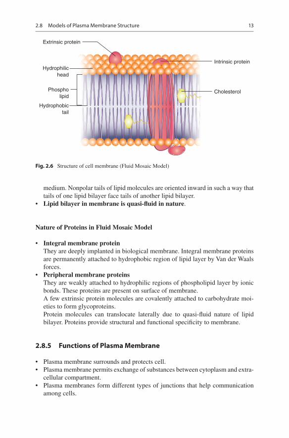
13
medium. Nonpolar tails of lipid molecules are oriented inward in such a way that tails of one lipid bilayer face tails of another lipid bilayer.
• Lipid bilayer in membrane is quasi-fluid in nature.
Nature of Proteins in Fluid Mosaic Model
• Integral membrane proteinThey are deeply implanted in biological membrane. Integral membrane proteins are permanently attached to hydrophobic region of lipid layer by Van der Waals forces.
• Peripheral membrane proteinsThey are weakly attached to hydrophilic regions of phospholipid layer by ionic bonds. These proteins are present on surface of membrane.A few extrinsic protein molecules are covalently attached to carbohydrate moi-eties to form glycoproteins.Protein molecules can translocate laterally due to quasi-fluid nature of lipid bilayer. Proteins provide structural and functional specificity to membrane.
2.8.5 Functions of Plasma Membrane
• Plasma membrane surrounds and protects cell.• Plasma membrane permits exchange of substances between cytoplasm and extra-
cellular compartment.• Plasma membranes form different types of junctions that help communication
among cells.
Fig. 2.6 Structure of cell membrane (Fluid Mosaic Model)
Intrinsic protein
CholesterolPhospholipid
Hydrophilichead
Extrinsic protein
Hydrophobictail
2.8 Models of Plasma Membrane Structure

14
• Plasma membrane allows movement of selected molecules to pass through it (selective permeable), while other molecules cannot pass through membrane.
• Plasma membrane contains receptors for drugs and hormones.• Plasma membrane contains carrier protein molecules for active transport of
molecules.• Plasma membrane of mitochondria carries ATPase for synthesis of ATP.• Plasma membrane of enterocytes has fingerlike outgrowths and microvilli that
increase absorptive area.• Plasma membrane of neurons helps in nerve impulse generation and conduction.
Plasma membrane of bacteria contains respiratory enzymes.
2.9 Cytoplasmic Organelles
2.9.1 Mitochondria
DefinitionMitochondria are filamentous, self-replicating, double-walled organelles located in cytoplasm of eukaryotic cells.
Mitochondria are termed as powerhouse of cell.The term mitochondrion is derived from Greek words mitos which means
thread and chondros which means granule (owing to appearance of mitochon-dria in spermatogenesis).
History
• In 1880, Kolliker discovered mitochondria in insect flight muscles.• In 1890, Altman described mitochondria in cells and called them as bioblasts
(living organelles inside cell).• In 1898, Benda coined the term mitochondria.• In 1941, Claude separated mitochondria from other fractions of cell.
OccurrenceMitochondria are located in cytoplasm of eukaryotic cells.
ColorMitochondria have brownish red color.
SizeSize of mitochondria is variable. Mitochondria may have 0.5–3 μm length and 0.1–0.5 μm diameter.
ShapeMitochondria have variable shapes. Under simple microscope, mitochondria appear as minute threadlike structures. They may have sausage, spherical, or filament- like shape.
2 Cell and Organelles

15
NumberThe number of mitochondria in a cell is dependent on metabolic activity.
• Minimum number is one mitochondrion per cell. Example: green algae belonging to genera Micromonas, Trypanosoma, and Chlorella, yeast
• The highest number is 50,000 mitochondria/cell. Example: insect flight muscles
• Absence of mitochondria. Example: human red blood cells
Generally, the number of mitochondria varies between 100 and 2000 mitochon-dria per cell.
StructureMitochondria structure can be described in three separate headings as:
Mitochondrial MembranesMitochondria are bounded by two successive membranes. Each one has unique bio-logical functions.
Outer Mitochondrial Membrane
• Outer membrane has a thickness of about 60 Å.• Its chemical composition is similar to that of plasma membrane of eukaryotic
cell. It is composed of phospholipids and proteins which are present in a 1:1 ratio.
• Outer membrane proteinsOuter membrane contains integral membrane proteins (transmembrane pro-tein). These proteins act as channels and hence proteins are called as porins. – Porin proteins allow transport of molecules (7000 dalton mw) across the outer
mitochondrial membrane.
Inner Mitochondrial Membrane
• Inner mitochondrial membrane has high protein contents than phospholipids. The ratio of proteins to phospholipids is 3:1.
• Inner membrane phospholipidInner membrane contains a prominent phospholipid called as cardiolipin (exclu-sively found in inner membrane in eukaryotes, in bacterial cell wall).
• Inner membrane proteinsInner membrane contains more than 100 polypeptides. They act as ATP synthase enzyme, carrier proteins, and proteins for mitochondrial fission and fusion.
• Inner membrane is impermeable to ionic and polar compounds. These molecules require carrier proteins to pass across inner membrane as in Fig. 2.7.
2.9 Cytoplasmic Organelles