
Discuss the Indications and Complications of Blood Transfusion

Handout
Complications of Collagen Cross-Linking: Indications, Applications, results, complications and evolving technology
Senior Instructor: A. John Kanellopoulos Instructrors: D. Stulting Gr. Pamel
J.Vryghem P. Binder
Synopsis: Didactic approach to the management of progressive cornea ectasia associated with keratoconus and refractive surgery. Several surgical treatment modalities utilized internationally will be presented, including: collagen cross-linking with ultraviolet radiation A in order to halt ectasia, combined in some cases with a customised excimer laser ablation to facilitate visual rehabilitation (as presented in previous ESCRS meetings by the author), these alternatives to Intracornea ring segment implantation, lamelllar grafts as well as penetrating graft techniques will be analyzed. Surgical and medical treatment technique, indications, potential complications and their management as well as clinical experience pearls will be presented Objective: The participants will share our vast experience in managing progressive keratoconus and post-LASIK ectasia in order to visually rehabilitate these patients. Pearls on indications, patient selection, surgical technique and complication management for safe and effective results will be presented and discussed with the participants.
A. John Kanellopoulos, MD - IC 72 ESCRS 2011 Vienna

Post-LASIK Ectasia
Dear Editor:I report a patient who had post-LASIK ectasia and wasmanaged in a novel fashion, without keratoplasty.
A 29-year-old male underwent uniocular LASIK 38 monthsago. Little detail was available from the patient and the sur-geon. His original uncorrected visual acuity (UCVA) beforeLASIK was 20/80, and his spectacle-corrected visual acuity(BSCVA) was 20/20 with refraction of sphere being�2.00�175�85. Initially after the LASIK procedure, thepatient reported that vision was good. During the followingmonths, vision in that eye deteriorated. The original LASIKsurgeon diagnosed ectasia and recommended the placementof Intacs (Addition Technology, Des Plaines, IL). AfterIntacs placement, his vision did not improve, and the patientdeveloped severe night vision halos.
The treating LASIK surgeon then recommended pene-trating keratoplasty (PK) as the next step, and the patientcame for a second opinion for PK, 11 months after theoriginal LASIK procedure and 3 months after Intacs im-plantation. Corneal topography is shown in Figure 1 (avail-able at http://aaojournal.org); the central corneal thicknesswas 410 �m, and the endothelial cell count was 2750cells/mm2 (Conan, Boston, MA). I discussed with the pa-tient the following:
1. The poor long-term experience with Intacs in post-LASIK ectasia that I have reported.1
2. The benefits and risks of PK.3. Combined ultraviolet radiation and riboflavin treat-
ment to achieve collagen cross-linking and biome-chanical stabilization of the ectasia.
After informed consent was given, I removed the Intacs.Two weeks later, I treated the ecstatic cornea with a singleapplication of combined ultraviolet radiation and riboflavintreatment to achieve collagen cross-linking at 3 mW/cm2 for30 minutes (KeraCure, Priavision, Menlo Park, CA) com-bined with the use of 0.1% riboflavin ophthalmic solution in20% dextran T-500.
The treatment was performed after 20% alcohol-assistedepithelial removal. The riboflavin solution was then applied forapproximately 2 minutes to soak the stromal bed and protectthe iris, crystalline lens, and retina from the ultraviolet Airradiation, and then 1 drop every 2 minutes for a total of 30minutes. A bandage contact lens was placed onto the corneafor 5 days and the patient treated with topical ofloxacin 1%(Ocuflox, Allergan, Irvine, CA) and prednisolone acetate 1%(Predforte, Allergan) 4 times a day for 10 days.
At 3 months, his UCVA improved from 20/400 to 20/70and his BSCVA from 20/200 to 20/40. Refraction changedfrom �4.50�4.50�120 to �4.00�3.50�115, and cornealtopography changed as seen in Figure 1. The stability ofthese parameters and the corneal topography between
months 1 and 3 of this treatment encouraged me to proceedwith topography-guided photorefractive keratectomy (PRK)to reduce the irregular astigmatism and try to provide thepatient with a visual acuity not requiring the use of specta-cles or a soft contact lens.
The corneal thickness at that point of 420 �m enabled aPRK of his full spectacle correction with a topography-guided customized ablation on top of the LASIK flap(T-CAT software, Wavelight excimer laser, Wavelight, Er-langen, Germany). At the first post-PRK month, UCVA was20/20� and BSCVA 20/20, with a refraction of�0.50�0.50�160. There was no corneal endotheliumcount change. It is now 24 months after the operation andthe patient enjoys UCVA of 20/20, although there aresome mild night vision problems. Postoperative cornealtopography is shown on Figure 1.
The most frequent management for post-LASIK ectasiahas been PK.2 Previous reports of the use of combinedultraviolet radiation and riboflavin treatment to achievecollagen cross-linking mention a slowing down of kerato-conus.3 We have reported the management of extreme cor-nea irregularity with topography-guided ablations.4 This isthe first report of management of post-LASIK ectasia withcombined ultraviolet radiation and riboflavin treatment toachieve collagen cross-linking followed by customizedPRK for visual rehabilitation. The apparent corneal stabili-zation, along with the successful visual rehabilitation, sug-gests that this approach may have a wider application as analternative to therapeutic PK.5
Larger comparative studies and longer follow-up areobviously necessary to validate the long-term efficacy ofthis combined ultraviolet radiation and riboflavin treatmentfollowed by a surface excimer laser treatment. Nevertheless,the refractive and topographic stability for 2 years appearsto validate this minimally invasive treatment of iatrogenickeratectasia and leads me to believe that it may have aneven wider application in the near future.
A. JOHN KANELLOPOULOS, MDAthens, Greece
References
1. Kanellopoulos AJ, Pe LH, Perry HD, Donnenfeld ED. Mod-ified intracorneal ring segment implantations (INTACS) forthe management of moderate to advanced keratoconus: effi-cacy and complications. Cornea 2006;25:29–33.
2. Binder PS. Ectasia after laser in situ keratomileusis. J CataractRefract Surg 2003;29:2419–29.
3. Wollensak G, Spoerl E, Seiler T. Riboflavin/ultraviolet-A-induced collagen crosslinking for the treatment of keratoco-nus. Am J Ophthalmol 2003;135:620–7.
4. Kanellopoulos AJ. Topography-guided custom retreatments in27 symptomatic eyes. J Refract Surg 2005;21:S513–8.
5. Donnenfeld ED, Kanellopoulos AJ. Therapeutic keratoplasty.In: Krachmer JH, Mannis MJ, Holland EJ, eds. Cornea. Vol. 3.St. Louis: Mosby; 1997:1843–54.
1230
A. John Kanellopoulos, MD - IC 72 ESCRS 2011 Vienna

Figure 1. Display of topographies. 1, Corneal topography of this case when first seen by the authors, with central cornea ectasia and midperipheryflattening as an effect of the Intacs that were present. At this point, best spectacle-corrected visual acuity (BSCVA) is 20/200. 2, Corneal topography 2months after the removal of Intacs and 1 month after combined ultraviolet radiation and riboflavin treatment to achieve collagen cross-linking. Thecentral steepening is still present, and the effect of the Intacs removal relative to the previous image is appreciated mostly at the midperiphery, whichappears steeper now. At this point, BSCVA is 20/200. Bottom center, An estimated corneal topographic ablation pattern as a laser treatment plan of thetopography-guided procedure. It is notable that this ablation pattern is highly irregular, with a deeper ablation plan just inferior to and right of the center,which matches, however, the central cornea irregularity in the previous topographies. 4, Corneal topography 6 months after topography-guidedphotorefractive keratectomy. The central cornea appears more regular and much flatter. At this point, BSCVA and UCVA are 20/20�. Bottom left,Comparison map depicting the result of subtraction of corneal topography 4 (final result) from corneal topography 1 (state of the complication when weencountered it). Impressively, the difference resembles the topography-guided ablation pattern (bottom center), demonstrating effectively the specificityof this treatment in reducing the pathogenic cornea irregularity, which, we theorize, contributed to the drastic improvement in BSCVA.
1230.e1
A. John Kanellopoulos, MD - IC 72 ESCRS 2011 Vienna

CASE REPORT
Collagen Cross-Linking (CCL) With SequentialTopography-Guided PRK
A Temporizing Alternative for Keratoconusto Penetrating Keratoplasty
A. John Kanellopoulos, MD*†‡ and Perry S. Binder, MS, MD§
Purpose: To assess the effectiveness of ultraviolet A (UVA)
irradiation–induced collagen cross-linking (CCL) on keratoconus
(KC) progression.
Methods: A patient with bilateral, progressive KC underwent
UVA irradiation (3 mW/cm2 for 30 minutes) after topical 0.1%
riboflavin drops over a deepithelialized cornea. Twelve months later, a
topography-guided penetrating keratoplasty (PRK; wavelight 400 Hz
Eye-Q excimer) was performed in 1 eye for a refractive error of
23.50 24.00 3 155 by using an attempted treatment of 22.50
23.00 3 155. At all postoperative follow-up visits to 18 months,
uncorrected visual acuity (UCVA), best spectacle-corrected visual
acuity (BSCVA), pachymetry, and topography were performed.
Results: In the treated left eye, the UCVA after the UVA CCL
improved from 20/100 to 20/80, and the BSCVA improved from
20/50 to 20/40. Eighteen months after the topography-guided PRK,
the UCVA was 20/20, and the BSCVA was 20/15, with a refractive
error of Plano 20.50 3 150. The cornea was clear, and the endo-
thelial cell count remained unchanged. The untreated right mate eye
continued to progress during the same period.
Conclusions: The significant clinical improvement and the
apparent stability of more than a year after UVA CCL, and subse-
quent PRK compared with the untreated mate eye, seems to validate
this treatment approach for KC. An adjusted nomogram may be
considered in the ablation of cross-linked cornea tissue to avoid
overcorrections.
Key Words: keratoconus, cornea ectasia, surgical management,
collagen cross-linking, ultraviolet A, riboflavin, customized topography-
guided cornea ablation, visual rehabilitation
(Cornea 2007;26:891–895)
Keratoconus is a bilateral, nonsymmetric, and noninflam-matory progressive corneal degeneration. Its incidence has
been thought to be 1 in 2000 in the general population,1 but theincreased number of eyes undergoing screening for laserrefractive surgery suggests the prevalence may be higher. Itcan be diagnosed at puberty, with up to 20% of the eyes pro-gressing to the extent that penetrating keratoplasty isindicated.2 Although spectacles and contact lenses can provideuseful vision in many cases, there are several surgical optionsfor those cases that can no longer benefit from them: implan-tation of intracorneal ring segments (Intacs or Ferrera rings),3
lamellar keratoplasty,4 or penetrating keratoplasty.2 Otherectatic corneal disorders such as Pellucid marginal degener-ation5 and post-LASIK ectasia6 require similar treatmentapproaches. Although penetrating keratoplasty for ectatic cor-neal disorders is highly successful, many eyes require contactlenses to correct the unpredictable topographic changes thatare associated with sutures and postsuture abnormal cornealshapes, and sometimes the contact lens is not successful.7
In recent years, basic laboratory studies and subsequentclinical studies have suggested that by increasing the collagencross-linking (CCL) of the corneal stromal collagen, one isable to increase the stiffness (biomechanics?) of the corneawith attendant stabilization of the normally progressivecorneal disorder.8–16 We present a case of bilateral progressivekeratoconus that underwent unilateral CCL followed by PRKwith an excellent outcome.
CASE REPORTA 26-year-old male patient had been treated with gas-
permeable contact lenses for 8 years before his presentation. Becauseof debilitating giant papillary conjunctivitis he was no longer able towear the contact lens; spectacles were unable to provide functionalvision because of poor vision and asthenopia. At the time of hisexamination, his uncorrected visual acuity (UCVA) was 20/40 in theright eye and 20/100 in the left eye, and his best spectacle-correctedvisual acuity (BSCVA) was 20/15 OD (manifest refraction 20.7520.75 3 165) and 20/50 OS (manifest refraction 23.75 24.50 3155). The keratometry readings were as follows: OD, 43.25 310/44.253 100; OS, 45.503 05/48.503 95 (Topolyzer; Wavelight,Erlagen, Germany).
Slit-lamp examination of the right eye failed to show clinicalfindings associated with keratoconus such as a Fleischer ring, Vogtstriae, or a noticeable excessive thinning of the central or paracentralcornea. The central pachymetry was 520 mm (Orbscan II; Bausch and
Received for publication June 19, 2006; revision received April 6, 2007;accepted April 15, 2007.
From the *Laservision.gr Institute, Athens, Greece; the †New York UniversityMedical College, New York, NY; the ‡Manhattan Eye, Ear and ThroatHospital, New York, NY; and the §Gordon Binder & Weiss VisionInstitute, San Diego, CA.
Reprints: A. John Kanellopoulos, Laservision.gr Institute, 2 MesogeionAvenue, Athens 11527, Greece (e-mail: [email protected]).
Copyright � 2007 by Lippincott Williams & Wilkins
Cornea � Volume 26, Number 7, August 2007 891
Copyright © Lippincott Williams & Wilkins. Unauthorized reproduction of this article is prohibited.
A. John Kanellopoulos, MD - IC 72 ESCRS 2011 Vienna

Lomb, Rochester, NY; ultrasound; Echoscan US-1800; NIDEK,Gamamory, Aichi, Japan). The left cornea had significant centralcorneal thinning of 440 mm and a Fleisher ring and Vogt lines withoutapical scarring. The endothelial cell density was 2800 cells/mm2 ODand 2750 cells/mm2 OS (Konan Medical, Boston, MA). Cornealtopography (Topolyzer; Wavelight) in the OS (Fig. 1A) clearlyshowed a steep ‘‘island’’ in the infero-temporal cornea consistent withthe cone apex. Figures 1A–F show the storyline of our treatment inthe OS eye. Figures 1G and H show the fellow untreated OD at thebeginning and at the end of our treatment to the OS. The OD (Fig.1G) revealed with-the-rule cylinder that seems irregular. The lowercomponent of the cornea cylinder is steeper than the upper and doesnot continue in a straight diametric line in respect to the center of thecornea. It is obvious from the topographies of the 2 eyes that, at thebeginning of our treatment, the left eye was significantly moreaffected by keratoconus. This made the patient and us decide to treatthe OS first and observe the less affected OD.
Intracorneal ring segments as a means of visual rehabilitationfor the OS were discussed, but because of our experiences with thismodality,17 we presented the risks, benefits, and alternatives ofpenetrating keratoplasty. The patient asked if there were any otheralternatives to penetrating keratoplasty. Because of our preliminarysuccess with CCL in a case of post-LASIK ectasia,18 we counseledhim about CCL, which he elected to undergo knowing thata subsequent penetrating keratoplasty might be needed.
CCL ProcedureTwo weeks after the initial examination, a Keracure prototype
device was used (Priavision, Menlo Park, CA). The epithelium wasremoved in a 9-mm diameter by using 20% alcohol applied to thesurface for 20 seconds. For the next 30 minutes, 0.1% riboflavinophthalmic solution was applied topically every 2 minutes. Riboflavinwas used to facilitate CLL while protecting the iris, crystalline lens,and retina.12 After the riboflavin drops, 4 light-emitting diodes,ultraviolet light of 370-nm wavelength and 3-mW/cm2 radiance, wasprojected onto the surface for 30 minutes, after which a bandagecontact lens was inserted. The device has a built-in beeper that resetsat the beginning of the treatment and alerts clinicians every 2 minutesduring the 30 minutes of treatment to instill the riboflavin solution.
After CCL, topical Ofloxacin (Allergan, Irvine, CA) andprednisolone acetate 1% (Pred Forte; Allergan) were used 4 timesa day for 10 days. The contact lens was removed at day 4 afterreepithelialization.
Clinical CourseThree months after the CCL procedure, the UCVA had
improved to 20/80 and the BSCVA improved to 20/40, with therefraction improving to 23.50 24.00 3 165. These parameters(UCVA, BSCVA, and refraction) remained stable for the next 12months. At this 12-month period, the thinnest part of the corneameasured 450 mm by Orbscan and ultrasonic pachymetry. Thetopography at the 12-month follow-up for the CCL procedure in thetreated OS is depicted in Figure 1B. There is some reduction in thecone steepness in the treated left eye, better shown in the differencemap (Fig. 1C). The difference map clearly shows that the CCLtreatment in the left eye resulted in cone flattening and improvementof the keratoconus. This was evident clinically as well, by theimprovement in UCVA, BSCVA, and refraction as noted above.
The patient remained unable to wear contact lenses because ofthe giant papillary conjunctivitis and it was difficult to wear spectaclesbecause of the anisometropia. In our attempt to visually rehabilitatethe eye and taking into account the 12-month stability after the CCL,we proceeded with a limited topography-guided PRK in an attempt toreduce the irregular astigmatism, and we hope that it will facilitatevisual rehabilitation. We decreased the effective optical zone diameter
to 5.5 mm from our standard routine of 6.5 mm and partially treatedthe sphere to not exceed 50 mm in planned stromal removal. Theattempted myopic sphere was 22.50 D and the planned astigmatismtreatment was 23.00 D by using the Allegretto–Wave topography-guided customized program (T-CAT).19 This proprietary softwareuses topographic data from the linked topography device (Topolyzer;Wavelight). By default, it permits the consideration of 8 topographies(of predetermined threshold accuracy), averages the data, and enablesthe surgeon to adjust the desired postoperative cornea asphericity; theinclusion, or not, of tilt correction; and the adjustment of sphere,cylinder, axis, and treatment zone. The image of the planned surgerygenerated by the laser software is displayed in Figure 1D.
For the PRK procedure, the epithelium was removed by using20% alcohol placed on the surface for 20 seconds, after which the lasertreatment was performed. A cellulose sponge soaked in mitomycin-C0.02% solution was applied over the ablated tissue for 30 seconds,followed by irrigation with 10 mL of chilled balanced salt solution.Finally, a bandage lens was placed onto the cornea. Ofloxacin andprednisolone acetate 1% were used topically 4 times a day for 10 days.Prednisolone acetate 1% was used 4 times a day for 3 more weeks andtwice a day for an additional 4 weeks. Protection from all natural lightwith sunglasses was encouraged, along with oral 1000 mg of vitamin Cdaily for 60 days, which is our standard PRK treatment. The bandagecontact lens was removed at day 5 after complete reepithelialization.
One month after the laser treatment, the UCVA improved to20/20, with subjective good nighttime vision. Thirty months after theUVACCL treatment and 18 months after the laser treatment, the UCVAremained 20/20 and the BSCVAwas 20/15, with a manifest refraction ofPlano 20.50 3 150. The cornea was clear and compact, and bybiomicroscopy, the Vogt striae were removed by the PRK, but theFleischer ring was still present. No hazewas noted over the healed area ofthe PRK treatment. The endothelial cell density remained stable at 2750cells/mm2, which was the same 1 month after the UVA CCL treatment,as well as at months 1 and 12 after the PRK. The post-PRK topographyfor the operated left eye is displayed in Figure 1E. The difference mapfor the left eye, from before the PRK (Fig. 1B) to final measurement18 months after the PRK (Fig. 1E), is depicted in Figure 1F.
Meanwhile the untreated mate eye had worsened because ofthe natural progression of the keratoconus. The patient wasdiscouraged from eye rubbing. During the 30 months after thebeginning of the treatment of the OS, the UCVA in the ODdeteriorated to 20/70 and a BSCVA to 20/25 with a refractive error of21.25 21.75 3 160.
The keratometry readings had increased to 43.75 3 05/44.753 95. The topography 30 months after treatment of the fellow eye isshown for the untreated OD (Fig. 1H).
In Figure 1D, the reader can evaluate the topography-guidedPRK treatment plan. There seems to be a ‘‘deeper’’ ablation over thesteep cone area and evidence of peripheral ‘‘flattening’’ effect of thecornea 180 degrees opposite of the cone center. The flattening of thispart of the peripheral cornea will result in steepening the middlecornea just away from the cone apex. The combined action on coneapex flattening while the rest of the central cornea is steepened is theunique modality that the topography-guided treatment offers. Itresembles a part-myopic and part-hyperopic treatment blendedtogether. Because we combined this flattening on the cone apexwith steepening of the rest of the central cornea, little stroma tissue isremoved from the cone apex area in relation to the normalizationachieved. It is remarkable how similar Figures 1D and F are. Theyrepresent the topography-guided PRK treatment plan and the actualachieved topographic difference just before and 18 months after thistreatment, respectively. The similarity of Figures 1D and F establishesthe accuracy of ablation delivery and achieved effect with this novelPRK treatment.
892 q 2007 Lippincott Williams & Wilkins
Kanellopoulos and Binder Cornea � Volume 26, Number 7, August 2007
Copyright © Lippincott Williams & Wilkins. Unauthorized reproduction of this article is prohibited.
A. John Kanellopoulos, MD - IC 72 ESCRS 2011 Vienna

Cornea Volume 26, Number 7, August 2007 CCL With Sequential Topography-Guided PRK
FIGURE 1. This is a sequence of topographic maps for both the left treated eye and the right untreated eye. A–F, Storyline through our treatment to the OS. G and H, Fellow untreated OD at the beginning and at the end of our treatment to the OS. A, Pretreatment topography of the left, most affected eye. The OS clearly shows a steep ‘‘island’’ in the infero–temporal cornea consistent with the cone apex. At the time of his examination, his UCVA was 20/100 in the left eye, and his BSCVA was 20/50 OS (manifest refraction, 23.75 24.50 3 155). The keratometry readings were 45.50 3 05/48.50 3 95. B, Left, treated eye 12 months after UVA CCL. The central steepening is still present, although reduced. At this point, the UCVA was 20/80, the BSCVA was 20/40, and the manifest refraction was 23.50 24.00 3 115. This image serves also as the pre-PRK topography forthe same eye. C, This is the difference of the initial topography for the OS (A) minus the 12-month post-UVA CCL treatment topography of the same eye (B). It clearly showed that the UVA CCL treatment in the left eye resulted in cone flattening and improvement of the keratoconus also evident clinically by the improvement in UCVA, BSCA, and refraction as noted above. Itseems that the CCL effect resulted in some cone flattening, shown by this comparison topography. D, This is thetopography-guided PRK treatment plan. There seems to be a ‘‘deeper’’ ablation over the steep cone area and evidence ofperipheral ‘‘flattening’’ effect of the cornea 180 degrees opposite of the cone center. The flattening of this part of the peripheral cornea will result in steepening the middle cornea just away from the cone apex. The combined action on coneapex flattening while the rest of the central cornea is steepened is the unique modality that the topography-guided treatment offers. It resembles a part-myopic and part-hyperopic treatment blended together. Because we combined this flattening on the cone apex with steepening of the rest of the central cornea, little stroma tissue is removed from the cone apex area in relationto the normalization achieved. E, Left eye 18 months after topography-guided PRK. The central cornea seems more regularand much flatter than the pre-PRK topography (B) and the initial topography just before initial UVA CCL treatment (A). Thereis no topographic evidence of keratoconus progression 18 months after the topography-guided PRK and a total of 30 months of the stabilizing UVA CCL treatment in the same eye. F, The difference between B and E of the left treated eye. It is the difference of the topography before and 18 months after the topography-guided PRK in the left cornea. The reader canappreciate a ‘‘deeper’’ ablation over the steep cone area and evidence of peripheral ‘‘flattening’’ effect of the cornea 180 degrees opposite to the cone center. The flattening of this part of the peripheral cornea resulted in steepening the middlecornea just away from the cone apex. The combined action on cone apex flattening while the rest of the central cornea is steepened is the unique modality that the topography-guided treatment offers. It resembles a part-myopic and part-hyperopic treatment blended together. Because we combined this flattening on the cone apex with steepening of the rest of the central cornea, little stroma tissue is removed from the cone apex area in relation to the normalization achieved. It is remarkable howsimilar D and F look. G, This is the topography of the right, less affected eye at the beginning of this study. The topographyreveals with-the-rule cylinder that appears irregular. The keratometry readings had increased to 43.75 3 05/44.75 3 95. Comparing this topography with G, which depicts the same untreated right eye at the beginning of this study, is important.There is topographic evidence of deterioration of the cone compared to the previous topography G.
© 2007 Lippincott Williams & Wilkins 893
Copyright © Lippincott Williams & Wilkins. Unauthorized reproduction of this article is prohibited.
A. John Kanellopoulos, MD - IC 72 ESCRS 2011 Vienna

DISCUSSIONThe technique of CCL by the photosensitizer, similar to
photopolymerization in polymers, has been previously de-scribed.10,20 Experimental studies in rabbit and porcine eyeshave shown an approximate increase in corneal rigidity by70% after CCL.11 A clinical study of 22 cases showed thestabilization of keratoconus with 4-year follow-up. In thatstudy, there was a 70% mean keratometry regression of 2 D atthe corneal plane and a regression of 1.14 D of the manifestspherical equivalent refractive error. Corneal and lens trans-parency, as well as endothelial cell density and intraocularpressure, remained unchanged, whereas visual acuity im-proved slightly in 65% of the eyes.8,11–14
Although multiple approaches have been used to treatectatic corneal disorders, in cases of progressive disease, the‘‘gold standard’’ treatment has become penetrating kerato-plasty,2 with its established attendant risks. As an alternative topenetrating keratoplasty, riboflavin/UVA CCL to decrease theprogression of keratoconus8,9,16 and progressive iatrogenicectasia15,18 has been studied.
We present 1 case that had bilateral keratoconus thatunderwent CCL and subsequent PRK on 1 eye with an excellentand stable outcome for 12 months after UVA CCL and 18months after a subsequent PRK, whereas the mate eyeunderwent progression of the keratoconus. The first step ofour treatment was used to stabilize the keratoconus. The PRKwas attempted to improve the post-CCL refractive error. Figures1E and H display the latest topography for both eyes. Figures 1Band E demonstrate the original and final topography of the left(treated) cornea. One can appreciate the difference map (Fig.1C) between pre- (Fig. 1A) and post-UVACCL (Fig. 1B) for theoperative eye and the difference map (Fig. 1F) between pre-(Fig. 1B) and post-topography-guided treatment (Fig. 1E).
In contrast, the topography map (Fig. 1G) of theuntreated mate eye before and at the conclusion of thetreatment in the left (Fig. 1H) documents progression ofthe keratoconus. In the PRK treatment plan (Fig. 1D), as wellas the difference map before PRK to 1 year after PRK (Fig.1F), there seems to be a deeper ablation over the steep conearea and evidence of a midperipheral ‘‘steepening’’ effect ofthe cornea 180 degrees opposite of the cone center. This wasachieved by flattening just peripheral to the cornea the area thatis planned to be ‘‘steepened.’’ It almost looks like a 90-degreepart in circumference of a hyperopic treatment.
The CCL procedure used riboflavin solution over thedeepithelialized cornea to protect the crystalline lens,endothelium, and possibly the retina from UVA radianceand to enhance UVA absorption in the anterior stroma andfacilitate the cross-linking process.8,10–14,16
Clinically, it has been reported that one can see the depthof the protective effect of the riboflavin in the cornea,9 but wedid not see this in our case; at the end of the UVA CCLtreatment, the entire cornea seemed to be ‘‘soaked’’ withyellow-tinged riboflavin by slit-lamp evaluation and in theanterior chamber. We did not detect a ‘‘line’’ in the stroma atthe last visit.
We subsequently performed a topographic-guided PRKprocedure19 in hopes of prolonging the need for a corneal
transplant. The clinical results have been rewarding duringa post-PRK follow-up of 18 months. The visual and topographicresults document the improvement.
We concluded that the spherical outcome was achievedby both flattening the cone apex combined with steepening theopposite side of the cone, which results in a ‘‘steepening’’ ofthe central part of the cornea opposite of the cone with respectto the center of the cornea. This treatment offers the clinicallysignificant advantage of reduced need for tissue removal thatwould be required with a wavefront-guided treatment19 fromthe cone apex, which corresponds to the thinnest part of thecornea. If a wavefront-guided approach was attempted ina similar case, and we assume that the wavefront analyzerwould be able to image such an irregular eye, the wavefront-guided software would ‘‘read’’ and attempt to flatten the steepcone irregularity to the reference point of more peripheral andequally flatter cornea. This would require significant ablationthickness specifically in stroma underlying the cone apex. Thiscustomized approach that uses topography addresses andmeasures effectively the extreme cornea irregularity that thesecases may have. It is a significant tool in enhancing visualrehabilitation. In cases where the refractive error will notpermit full correction, we propose the correction of only theirregular astigmatism to improve the BSCVA but not neces-sarily the UCVA, without risking significant cornea thinning.
Corneal stabilization, followed by full visual rehabili-tation, leads us to believe that this combined approach mayhave wider applications and may become a temporizingalternative to cornea transplantation. Special emphasis shouldbe taken preoperatively in cases with a minimal cornealthickness ,400 mm because of potential cytotoxic effects ofUVA on corneal endothelial cells.12 In addition, the lasertreatment must be applied with caution because the more rigidcornea may have an ablation rate different from that ofa normal cornea. For this reason, we elected to attempt anundercorrection of the sphere and cylinder by 25%; weobtained a 100% effect. We therefore recommend attempting75%–80% of the measured sphere and cylinder as a correctionparameter when planning the Excimer ablation with T-CATsoftware or other wavefront-guided software until we can moreaccurately determine the new ablation rate of CCL stroma.Larger, comparative studies establishing the safety andefficacy of this treatment and longer follow-up are necessaryto further validate these results and potentially make thistreatment available for ectatic corneal disorders.
REFERENCES1. Rabinowitz Y. Keratoconus. Surv Ophthalmol. 1998;42:297–319.2. Pramanik S, Musch DC, Sutphin JE, et al. Extended long-term outcomes
of penetrating keratoplasty for keratoconus. Ophthalmology. 2006;113:1633–1638.
3. Rabinowitz YS, Li X, Ignacio TS, et al. Intacs inserts using thefemtosecond laser compared to the mechanical spreader in the treatmentof keratoconus. J Refract Surg. 2006;22:764–771.
4. Amayem AF, Anwar M. Fluid lamellar keratoplasty in keratoconus.Ophthalmology. 2000;107:76–80.
5. Ertan A, Bahadir M. Management of superior pellucid marginaldegeneration with a single intracorneal ring segment using femtosecondlaser J Refract Surg. 2006;23:205–208.
894 q 2007 Lippincott Williams & Wilkins
Kanellopoulos and Binder Cornea � Volume 26, Number 7, August 2007
Copyright © Lippincott Williams & Wilkins. Unauthorized reproduction of this article is prohibited.
A. John Kanellopoulos, MD - IC 72 ESCRS 2011 Vienna

6. Binder PS. Ectasia after laser in situ keratomileusis. J Cataract RefractSurg. 2003;29:2419–2429.
7. Gordon M, Steger-May K, Szczotka-Flynn L, et al. Baseline factorspredictive of incident penetrating keratoplasty in keratoconus. Am JOphthalmol. 2006;142:923–930.
8. Wollensak G, Spoerl E, Seiler T. Riboflavin/ultraviolet -a-inducedcollagen cross linking for the treatment of keratoconus. Am J Ophthalmol.2003;135:620–627.
9. Seiler T, Hafezi F. Corneal cross-linking-induced stromal demarcationline. Cornea. 2006;25:1057–1059.
10. Spoerl E, Huhle M, Seiler T. Induction of cross-links in corneal tissue. ExpEye Res. 1998;66:97–103.
11. Wollensak G, Spoerl E, Seiler T. Stress-strain measurements of human andporcine corneas after riboflavin-ultraviolet-A-induced cross-linking.J Cataract Refract Surg. 2003;29:1780–1785.
12. Wollensak G, Spoerl E. Wilsch M, et al. Endothelial cell damage afterriboflavin-ultraviolet-A treatment in the rabbit. J Cataract Refract Surg.2003;29:1786–1790.
13. Wollensak G, Spoerl E, Reber F, et al. Keratocyte cytotoxicity ofriboflavin/UVA-treatment in vitro. Eye. 2004;18:718–722.
14. Wollensak G, Spoerl E, Wilsch M, et al. Keratocyte apoptosis after cornealcollagen cross-linking using riboflavin/UVA treatment. Cornea. 2004;23:43–49.
15. Hafezi F, Mrochen M, Jankov M, et al. Corneal collagen crosslinkingwith riboflavin/UVA for the treatment of induced kerectasia after laser insitu keratomileusis. 2007; (in press).
16. Spoerl E, Seiler T. Techniques for stiffening the cornea. J Refract Surg.1999;15:711–713.
17. Kanellopoulos A, Pe L, Perry H, et al. Modified intracorneal ring segmentimplantations (Intacs) for the management of moderate to advancedkeratoconus: efficacy and complications. Cornea. 2006;25:29–33.
18. Kanellopoulos A. Management of post-LASIK ectasiawith UVA collagen-cross linking followed by customized topography-guided PRK—anefficient approach to avoid corneal transplantation. J Ophthalmol. 2007;(in press).
19. Kanellopoulos A, Pe L. Wavefront-guided enhancements using thewavelight excimer laser in symptomatic eyes previously treated withLASIK. J Refract Surg. 2006;22:345–349.
20. Hettlick H, Lucke K, Kreiner C. Light induced endocapsular polymerizationof injectable lens refilling materials. Ger J Ophthalmol. 1992;1:346–349.
q 2007 Lippincott Williams & Wilkins 895
Cornea � Volume 26, Number 7, August 2007 CCL With Sequential Topography-Guided PRK
Copyright © Lippincott Williams & Wilkins. Unauthorized reproduction of this article is prohibited.
A. John Kanellopoulos, MD - IC 72 ESCRS 2011 Vienna

journalofrefractivesurgery.comS812
Comparison of Sequential vs Same-day Simultaneous Collagen Cross-linking and Topography-guided PRK for Treatment of KeratoconusAnastasios John Kanellopoulos, MD
From the Laservision.gr Institute, Athens, Greece; New York University Medical College and Manhattan Eye, Ear and Throat Hospital, New York, NY.
Presented in part as a poster at the annual ARVO meetings in 2004-2008; an invited lecture at the Annual ISRS/AAO Refractive Surgery Subspecialty Day, November 9-10, 2007, Las Vegas, Nev; a paper at the annual ESCRS symposium, September 11, 2008, Berlin, Germany; and a paper at the 4th International Congress of Corneal Cross-linking, December 5-6, 2008, Dresden, Germany.
Correspondence: Anastasios John Kanellopoulos, MD, Laservision.gr Institute, 17 Tsocha St, Athens 11521, Greece. Tel: 30 210 7472777; Fax: 30 210 7472789; E-mail: [email protected]
ABSTRACT
PURPOSE: The safety and effi cacy of corneal collagen cross-linking (CXL) and topography-guided photorefractive keratectomy (PRK) using a different sequence and timing were evaluated in consecutive keratoconus cases.
METHODS: This study included a total of 325 eyes with keratoconus. Eyes were divided into two groups. The fi rst group (n=127 eyes) underwent CXL with subse-quent topography-guided PRK performed 6 months later (sequential group) and the second group (n=198 eyes) underwent CXL and PRK in a combined procedure on the same day (simultaneous group). Statistical differ-ences were examined for pre- to postoperative changes in uncorrected (UCVA, logMAR) and best-spectacle-corrected visual acuity (BSCVA, logMAR), manifest re-fraction spherical equivalent (MRSE), keratometry (K), topography, central corneal thickness, endothelial cell count, corneal haze, and ectatic progression. Mean fol-low-up was 36�18 months (range: 24 to 68 months).
RESULTS: At last follow-up in the sequential group, the mean UCVA improved from 0.9�0.3 logMAR to 0.49�0.25 logMAR, and mean BSCVA from 0.41�0.25 logMAR to 0.16�0.22 logMAR. Mean reduction in spherical equivalent refraction was 2.50�1.20 diop-ters (D), mean haze score was 1.2�0.5, and mean reduction in K was 2.75�1.30 D. In the simultaneous group, mean UCVA improved from 0.96�0.2 logMAR to 0.3�0.2 logMAR, and mean BSCVA from 0.39�0.3 logMAR to 0.11�0.16 logMAR. Mean reduction in spherical equivalent refraction was 3.20�1.40 D, mean haze score was 0.5�0.3, and mean reduction in K was 3.50�1.3 D. Endothelial cell count preopera-tively and at last follow-up was unchanged (P�.05) in both groups. Statistically, the simultaneous group did better (P�.05) in all fi elds evaluated, with improve-ment in UCVA and BSCVA, a greater mean reduction in spherical equivalent refraction and keratometry, and less corneal haze.
CONCLUSIONS: Same-day simultaneous topography-guided PRK and CXL appears to be superior to sequential CXL with later PRK in the visual rehabilitation of progress-ing keratoconus. [J Refract Surg. 2009;25:S812-S818.]doi:10.3928/1081597X-20090813-10
K eratoconus is a bilateral, nonsymmetric, noninfl am-matory progressive corneal degeneration that fre-quently manifests in post-pubescent young adults as
progressive steepening attributed to biomechanical stromal collagen weakening. Its incidence has been reported to be 1 in 2000 in the general population.1,2 The increased number among eyes undergoing screening for laser refractive surgery suggests the prevalence may be higher. Current surgical/non-surgical interventions such as spectacles and contact lenses, intracorneal ring segment implantation,3,4 lamellar kerato-plasty,5 and, the gold standard, penetrating keratoplasty,6
although popular, have limitations.7,8
In recent years, basic laboratory studies and subsequent clinical studies have demonstrated stiffening of the cornea with use of ultraviolet A (UVA) light and ribofl avin solution as a photosensitizer-initiated corneal collagen cross-linking (CXL) with no loss in corneal transparency. The CXL proce-dure has demonstrated the revolutionary potential for retard-ing or eliminating the progression of keratoconus and postop-erative LASIK ectasia.9-17
We have performed over 1000 CXL treatments in our fa-cility over the past 7 years both for ectasia after LASIK and keratoconus with satisfactory outcomes. We have also dem-onstrated that topography-guided ablation of the cross-linked corneal stroma can “normalize” the highly irregular corneal surface in these eyes by reducing irregular astigmatism and often reducing the refractive error as well, providing patients with improved visual outcomes.18-21
The clinical results of a novel, same-day, simultaneous approach of topography-guided photorefractive keratectomy (PRK) and CXL for keratoconus22 are presented and retrospec-
A. John Kanellopoulos, MD - IC 72 ESCRS 2011 Vienna

S813Journal of Refractive Surgery Volume 25 September 2009
Sequential vs Simultaneous Topography-guided PRK and CXL/Kanellopoulos
tively compared to the data from our previous experi-ence of performing CXL fi rst and PRK at least 6 months later. The outcomes of these two groups are compared in a retrospective case series with follow-up of 2 to 5 years.
PATIENTS AND METHODS
PATIENT SELECTIONAll patients were enrolled in our Athens clinical facil-
ity. Once a diagnosis of keratoconus was confi rmed (see below), patients were informed of various popular op-tions, including CXL. Being neither FDA-approved nor CE Marked (in cases treated prior to December 2006), informed consent was obtained from all patients and the surgery was performed in the following sequence: CXL fi rst with topography-guided PRK at least 6 months later in eyes treated from August 2003 to May 2005 (se-quential group). After gaining signifi cant experience, we shifted our approach to same-day simultaneous custom-ized topography-guided PRK and CXL in eyes treated from June 2005 to May 2008 (simultaneous group).
DIAGNOSIS OF KERATOCONUSA diagnosis of progressive keratoconus was made in
all patients (all aged �30 years), who developed pro-gressive corneal steepening of at least 1.00 diopter (D) in keratometry, associated with a documented progression of increasing myopia and/or astigmatism over a period of 3 or more months. These fi ndings were in parallel with increasing inferior corneal steepening and thinning to no less than 350 µm after PRK, based on videokeratog-raphy and pachymetry. Preoperative clinical data and topography were used as a baseline. Progression of the myopic refractive error with or without progression of the manifest astigmatism, decreasing uncorrected visu-al acuity (UCVA), loss of best spectacle-corrected visual acuity (BSCVA), progressive inferior corneal steepen-ing on topography, and/or decreasing inferior corneal thickness were fi ndings in all cases.
CLINICAL EXAMINATIONEach patient underwent manifest refraction before
and 30 minutes after administration of one drop of 1% tropicamide solution (Akorn Inc, Lake Forest, Ill), as well as measurement of UCVA and BSCVA that was recorded in a 20-foot lane using high contrast Snellen visual acuity testing and then converted to logMAR scale values. A slit-lamp microscopic examination per-formed by the author confi rmed invariably in all cases the presence of keratoconus either by the presence of a Fleischer ring, central or paracentral corneal thinning with prominent cornea nerves, and/or Vogt’s striae.
Keratometry readings were obtained by videokerato-graphy (Topolyzer; WaveLight Laser Technologie AG, Erlangen, Germany) and by two tomography-based to-pography devices—Orbscan II (Bausch & Lomb, Roch-ester, NY) and Pentacam (Oculus Optikgeräte, Wetz-lar, Germany). Pachymetry was performed using all of the following instruments: Pentacam, Orbscan II, and ultrasonic EchoScan US-1800 (NIDEK Co Ltd, Gama-gori, Japan). The minimal measurement in each case was used as the cornea thickness value, due to the im-portance of referencing the thinnest point. Specular microscopy was performed using the Konan specular microscope (Konan Medical, Boston, Mass).
Our technique of sequential CXL followed by topog-raphy-guided PRK at a later date has been reported pre-viously.20,21 Both procedures have received a CE Mark for clinical use (CXL in 2006, topography-guided PRK in 2003) within the countries of the European Union, including Greece. Topography-guided ablations with the WaveLight platform and CXL have not received FDA approval to date. As this is a novel approach and may be unfamiliar to most readers, the same-day simultaneous approach of topography-guided PRK and then CXL is described.
STEP 1: PARTIAL, SPHERICALLY CORRECTEDTOPOGRAPHY-GUIDED PRK
We have devised this technique based on the proprie-tary WaveLight customized platform (Topolyzer). Use of the topography-guided platform with this device to normalize irregular corneas, including those with ecta-sia, has been reported previously.23-25
This proprietary software utilizes topographic data from the linked topography device (Topolyzer). By default, it permits the consideration of eight topogra-phies (of predetermined threshold accuracy), averages the data, and enables the surgeon to adjust the desired postoperative cornea asphericity (chosen as zero in all cases), the inclusion, or not, of tilt correction (no tilt was chosen in all cases), as well as the adjustment of sphere, cylinder, axis, and treatment zone (an optical zone of 5.5 mm was chosen in all cases). The image of the planned surgery is generated by the laser software.20,21
We used topography-guided PRK to normalize the cornea, by reducing irregular astigmatism and also treating part of the refractive error. To ensure minimal tissue removal, the effective optical zone diameter was decreased to 5.5 mm (compared to our usual treatment diameter in routine PRK and LASIK cases of at least 6.5 mm). The transition zone was 1.5 mm. We also planned ~70% treatment of cylinder and whatever level of sphere (up to 70%), so as not to exceed 50 µm in planned stromal removal. The value of 50 µm was
A. John Kanellopoulos, MD - IC 72 ESCRS 2011 Vienna

journalofrefractivesurgery.comS814
Sequential vs Simultaneous Topography-guided PRK and CXL/Kanellopoulos
chosen arbitrarily by the author, based on experience with this platform in irregular corneas.
Following the placement of an aspirating lid specu-lum (Rumex, St Petersburg, Fla), 20% alcohol solution was placed within a 9-mm titanium LASEK trephine (Rumex) for 20 seconds after which the epithelium was wiped with a dry Weck-cell sponge. The laser treat-ment was then applied. A cellulose sponge soaked in mitomycin C 0.02% solution was applied over the ab-lated tissue for 20 seconds followed by irrigation with 10 mL of chilled balanced salt solution.
STEP 2: COLLAGEN CROSS-LINKINGFor the next 10 minutes, 0.1% ribofl avin sodium
phosphate ophthalmic solution (PriaVision Inc, Men-lo Park, Calif) was applied topically every 2 minutes. The solution appeared to “soak” into the corneal stro-ma rapidly, as it was centrally devoid of Bowman’s layer. Following the initial ribofl avin administration, four diodes, emitting UVA light of approximately 370 nm wavelength (365 to 375 nm) and 3 mW/cm2 ra-diance at 2.5 cm was projected onto the surface of the cornea for 30 minutes. A “Keracure” prototype device was used (Priavision). The Keracure device has a built-in beeper that alerts the clinician every 2 minutes during the 30-minute treatment to install the ribofl avin solution in a timely fashion. A bandage contact lens was placed on the cornea at the comple-tion of the procedure.
After CXL, topical ofl oxacin (Allergan Inc, Irvine, Ca-lif) was used four times a day for the fi rst 10 days and prednisolone acetate 1% (Pred Forte, Allergan) was used four times a day for 60 days. Protection from all natural light with sunglasses was encouraged along with oral Vi-tamin C 1000 mg daily for 60 days postoperatively (our standard postoperative management following PRK). The bandage contact lens was removed at approximately day 5 following complete reepithelialization.
All cases were evaluated before and after both treat-ments for age, sex, UCVA, BSCVA, refraction, keratom-etry (K), topography, central corneal thickness (CCT), endothelial cell count, corneal haze on a scale 0 to 4 (0=clear cornea, 1=mild haze, 2=moderate haze, 3=severe haze, and 4=reticular haze [obstructing iris anatomy]), and ectasia stability. These parameters were retrospec-tively collected and then statistically analyzed using paired t test.
RESULTSApproximately 40% of patients complained of sig-
nifi cant pain during the fi rst postoperative night after both CXL alone or when combined with simultaneous topography-guided PRK the same day, whereas others
reported minimal discomfort. Reepithelialization oc-curred by postoperative day 4 in 90% of patients.
Specifi cally, 127 consecutive eyes (sequential group) had topography-guided PRK at least 6 months follow-ing CXL, whereas 198 eyes (simultaneous group) un-derwent topography-guided PRK followed immediate-ly by CXL as a single procedure. Mean follow-up was 36 months (range: 24 to 68 months) from the time of the last procedure performed.
Mean endothelial cell count and morphology were unchanged in both groups (2650�150 cells/mm2 preop-eratively and 2700�140 cells/mm2 postoperatively).
Mean patient age in the sequential group was 21.5 years (range: 17 and 29 years) and comprised 44 males and 32 females. At last follow-up, preoperative UCVA of 0.9�0.3 logMAR improved to 0.49�0.25 logMAR postoperatively, and BSCVA improved from 0.41�0.25logMAR preoperatively to 0.16�0.22 logMAR postop-eratively. Mean reduction in spherical equivalent re-fraction was 2.50�1.2 D, and mean reduction in K was 2.75�1.3 D. Mean haze score was 1.2�0.5. Mean CCT decreased from 465�45 µm preoperatively to 395�25 µm postoperatively.
Mean patient age in the simultaneous group was 20.5 years (range: 16 to 29 years) and comprised 53 males and 42 females. Mean UCVA improved from 0.96�0.2 logMAR preoperatively to 0.3�0.2 logMAR postoperatively, and BSCVA from 0.39�0.3 logMAR to 0.11�0.16 logMAR. Mean reduction in spherical equivalent refraction was 3.20�1.4 D, and mean re-duction in K was 3.50�1.3 D. Mean haze score was 0.5�0.3, and CCT decreased from 475�55 µm preop-eratively to 405�35 µm postoperatively.
Statistical comparison using paired t test revealed that the simultaneous group performed superiorly with a better BSCVA (P�.001), spherical equivalent reduction (P�.005), mean K reduction (P�.005), and corneal haze score (P�.002) at fi nal follow-up. No eye lost lines of UCVA or BSCVA.
Figure 1 shows representative corneal topogra-phies from sequential Pentacam examinations in a 29-year-old patient from the simultaneous group (same-day simultaneous topography-guided PRK and CXL). The keratometric data changes ~3.50 D from 49.0 and 44.3 @ 60.9° preoperatively to 44.0 and 41.8 @ 46° postoperatively, whereas the visual and refrac-tion data changed from UCVA of 20/100 to 20/25 and �2.75 �3.50 @ 65 (20/30) to �0.50 �1.00 @ 35 (20/20) at 2-year follow-up.
DISCUSSIONThe technique of collagen CXL via a photosensitiz-
ing agent is similar to photopolymerization in polymers
A. John Kanellopoulos, MD - IC 72 ESCRS 2011 Vienna

S815Journal of Refractive Surgery Volume 25 September 2009
Sequential vs Simultaneous Topography-guided PRK and CXL/Kanellopoulos
and has found a broad international application for ker-atoconus in recent years. Although multiple approaches have been used to treat ectatic corneal disorders, in cases of progressive disease, the gold standard treatment in many countries is penetrating keratoplasty with its es-tablished costs, morbidity, and attendant risks.
The mechanism of topography-guided ablation is the fi tting of an ideal cornea shape (usually a sphere) under the present topography map with the ablation of tissue in between.
We have been able to use topography-guided treat-ments in highly irregular corneas that are beyond the limits of wavefront measuring devices,23-25 making this approach more effi cient in treating highly irregular astigmatism, such as in keratoconus. It may also be ap-plied in cases with some media opacity, such as in ker-atoconus eyes with corneal scars, as its measurements are based solely on the cornea surface refl ection.
Topography-guided PRK fl attens not only some of the cone “peak” but also an arcuate, broader area of the cornea away from the cone, usually in the supe-rior nasal periphery; this ablation pattern will resem-ble part of a hyperopic treatment and thus will cause some amount of steepening, or elevation adjacent to the cone, effectively normalizing the cornea.26 It is this core concept in the topography-guided PRK treatment that makes it, in our opinion, more therapeutic than refractive. We theorize additionally that the new, “fl at-ter” and less irregular cornea shape may perform better biomechanically in keratoconus. Specifi cally, as the cornea cone gives way to a fl atter and “broader” cone, this may redistribute the biomechanical strain from the eye’s intraocular pressure and other factors (such
as eye rubbing). This effect may be further enhanced with the CXL adjunct.
We have converted to same-day simultaneous topography-guided PRK and CXL in our clinical prac-tice, represented herein by the cases in the simultane-ous group, for three reasons: 1) the combination reduces the patient’s time away from work, 2) performing both procedures at the same time with topography-guided PRK fi rst appeared to minimize the potential superfi -cial stromal scarring resulting from PRK, and 3) when topography-guided PRK is performed after the CXL procedure, some of the cross-linked anterior cornea is removed, minimizing the potential benefi t of CXL.
Regarding the second reason, our initial experience with the cases in the sequential group suggested that if the practitioner waited between procedures, the dam-aged keratocytes would replenish and may become activated as fi broblasts after PRK, causing scarring even with the use of mitomycin C (MMC).27 In these cases, we performed topography-guided PRK at least 6 months after CXL and encountered signifi cant haze in 17 eyes, despite using MMC. When we employed CXL immediately after topography-guided PRK, we encoun-tered minimal haze formation (2 cases with signifi cant haze). The CXL procedure has been shown to “kill” keratocytes as deep as 300 µm,28 which may explain why this late haze formation was prevented when CXL was performed the same day after topography-guided PRK in the eyes in the simultaneous group.
Regarding the fi nal reason, we believe it is counter-intuitive to remove the cross-linked tissue with topog-raphy-guided PRK at a later time, as we are potentially removing a benefi cial layer of the stiffer, cross-linked
Figure 1. Representative corneal topogra-phies from sequential Pentacam examinations are shown in a 29-year-old patient from the simultaneous group (same-day, simultaneous topography-guided photorefractive keratectomy [PRK] and corneal collagen cross-linking [CXL]). The difference map (right topography) is shown for the changes from preoperative (left topog-raphy) to 2 years postoperative (middle topog-raphy). The Pentacam data demonstrate the change of keratometric data from 49 and 44.3 @ 60.9° to 44 and 41.8 @ 46°. Changes in visual acuity and refraction data from pre- to postoperative were: uncorrected visual acuity 20/100 to 20/25 and best spectacle-correct-ed visual acuity (BSCVA) 20/30 with �2.75 �3.50 @ 65 to BSCVA 20/20 with �0.50 �1.00 @ 35 at 2 years. The difference map illustrates the surface change achieved by the simultaneous topography-guided PRK and CXL technique, which resembles a combined myo-pic PRK over the cone apex and an asymmetric hyperopic PRK in the periphery.
A. John Kanellopoulos, MD - IC 72 ESCRS 2011 Vienna

journalofrefractivesurgery.comS816
Sequential vs Simultaneous Topography-guided PRK and CXL/Kanellopoulos
cornea, which helps maintain the normalized corneal shape.
Although a patient with keratoconus can have an improved visual result with the addition of the topog-raphy-guided PRK procedure, completely removing a high level of refraction was not our goal. We have placed an arbitrary “ceiling” of 50 µm to the amount of tissue that we safely remove centrally, anticipating that further thinning might destabilize the cornea’s biomechanical integrity, even with the effect of CXL.
Special attention should be given to eyes with a pre-operative minimal corneal thickness �350 µm because of potential cytotoxic effects of UVA light on corneal endothelial cells. It should be noted that the propri-etary ribofl avin solution used was a slightly hypotonic (340 mOsm) formulation, resulting in slight “swelling” of the cornea intraoperatively (during CXL). This re-stored the corneal thickness to ~400 µm to protect the corneal endothelium, and may be the reason we did not encounter any corneal endothelial decompensa-tion in any of the eyes studied.
In addition, the laser treatment was applied with caution, as the refractive effect of the CXL (cornea fl attening), had to be anticipated. For this reason, we elected to always attempt a signifi cant undercorrection of both the sphere and cylinder by at least 30%. As de-scribed previously, we suggest attempting at most 70% of the measured sphere and cylinder when planning the excimer ablation with T-CAT software (WaveLight Laser Technologie AG) or other wavefront-guided soft-ware after CXL. In the future, we hope to more accu-rately determine the new ablation rate of CXL stroma.
Simultaneous topography-guided PRK and CXL ap-peared in this study to be superior to sequential treat-ment in the rehabilitation of keratoconus. A possible reason for the difference may be an “enhanced” CXL in the former group, either due to better penetration of the ribofl avin solution through the ablated stroma or to the absence of Bowman’s layer. In addition, cross-link-ing the more “normal” corneal shape in the laser pre-
treated keratoconus eyes (simultaneous group) makes them more “resistant” to factors affecting ectasia pro-gression.
Why bother with topography-guided PRK in the fi rst place? Our long-term follow-up of CXL alone in kerato-conus appears to halt ectasia progression, but it often leaves the patient with diffi culty in visual rehabilita-tion, as most cases had contact lens intolerance. In the patient population we encountered, the great majority fell into this category even when CXL had successfully halted the progression of ectasia.
The reality of the effi cacy of topography-guided PRK and CXL has been the reduction of penetrating keratoplasty cases performed for the indication of ker-atoconus in our clinical practice over the past 4 years. The same-day, simultaneous topography-guided PRK/CXL procedure was easy to perform, but in some cases the central epithelial surface took up to 1 month to smoothen and become lucent. It took from 1 to 4 weeks for us to detect stable changes in the K and topogra-phy, which seemed to match the visual and refractive changes.
A specifi c demarcation line of separation was noted between the suspected cross-linked collagen and the deeper cornea both clinically29 as well as with corneal optical coherence tomography (Optivue, Freemont, Calif) (Fig 2). The cross-linking effect in the stroma was clinically assessed at the slit lamp following the procedure by the “ground-glass” appearance of the an-terior stroma, and in most cases, by the presence of ul-tra-thin, curved, whitish fi ne lines in the anterior and mid-stroma (Fig 3). These lines do not appear to affect vision and tend to fade away by postoperative month 12. In our clinical assessment, the presence of this fi nding over the anterior half of the stroma confi rms suffi cient CXL treatment has occurred. As we perform more CXL procedures, we hope to learn which candi-dates might best benefi t from the procedure.
Questions such as “How much ectasia?” and “What types of ectasia can we safely and predictably correct?”
Figure 2. Corneal optical coherence tomography demonstrates hyper-reflective intracorneal stromal “lines” at 2/3 depth (arrows) corresponding with the clinical presence of the corneal collagen cross-link-ing (CXL) demarcation line in a patient from the simultaneous group 3 years following the combined topography-guided photore-fractive keratectomy and CXL procedure.
A. John Kanellopoulos, MD - IC 72 ESCRS 2011 Vienna
S817Journal of Refractive Surgery Volume 25 September 2009
Sequential vs Simultaneous Topography-guided PRK and CXL/Kanellopoulos
as well as “Is there a minimum preoperative CXL cor-neal thickness that will not respond to the procedure?” require further investigation. Strategies need to be de-veloped to determine the attempted correction and ab-lation depth for the topography-guided PRK portion of this process. The proper concentration of ribofl avin, its delivery within the corneal stroma, and the proper UVAlight exposure and duration will need to be adjusted as we move from animal model studies into clinical pro-cedures. Perhaps CXL will have a wider application as prophylaxis in laser refractive surgery, as we reported in PRK30and LASIK.31
Our fi ndings suggest better results with same-day, simultaneous topography-guided PRK and collagen CXL, as a therapeutic intervention in highly irregular corneas with progressive keratoconus. Our goal was to stabilize the ectasia (with CXL) and rehabilitate the vision (with topography-guided PRK) in young adults with advancing keratoconus to delay or even avoid corneal transplantation. Larger, prospective and ran-domized, comparative studies, establishing the safety and effi cacy of this treatment, and longer follow-up, are necessary to further validate these results.
REFERENCES 1. Krachmer JH, Feder RS, Belin MW. Keratoconus and related
noninfl ammatory corneal thinning disorders. Surv Ophthal-mol. 1984;28:293-322.
2. Rabinowitz Y. Keratoconus. Surv Ophthalmol. 1998;42:297-319.
3. Kanellopoulos AJ, Pe LH, Perry HD, Donnenfeld ED. Modifi ed intracorneal ring segment implantations (INTACS) for the man-agement of moderate to advanced keratoconus: effi cacy and complications. Cornea. 2006;25:29-33.
4. Ertan A, Colin J. Intracorneal rings for keratoconus and keratec-tasia. J Cataract Refract Surg. 2007;33:1303-1314.
5. Amayem AF, Anwar M. Fluid lamellar keratoplasty in kerato-conus. Ophthalmology. 2000;107:76-79.
6. Koralewska-Makár A, Florén I, Stenevi U. The results of pen-etrating keratoplasty for keratoconus. Acta Ophthalmol Scand.1996;74:187-190.
7. Kanellopoulos AJ, Perry HD, Donnenfeld ED. Visual reha-bilitation following penetrating keratoplasty. Ophthalmology.1996;103:142.
8. Doyle SJ, Harper C, Marcyniuk B, Ridgway AE. Prediction of refractive outcome in penetrating keratoplasty for keratoconus. Cornea. 1996;15:441-445.
9. Wollensak G, Spoerl E, Seiler T. Stress-strain measurements of human and porcine corneas after ribofl avin-ultraviolet-A-induced cross-linking. J Cataract Refract Surg. 2003;29:1780-1785.
10. Wollensak G, Spoerl E, Seiler T. Ribofl avin/ultraviolet-a-in-duced collagen crosslinking for the treatment of keratoconus. Am J Ophthalmol. 2003;135:620-627.
11. Wollensak G. Crosslinking treatment of progressive keratoco-nus: new hope. Curr Opin Ophthalmol. 2006;17:356-360.
12. Mazzotta C, Balestrazzi A, Traversi C, Baiocchi S, Caporossi T, Tommasi C, Caporossi A. Treatment of progressive keratoconus
by ribofl avin-UVA-induced cross-linking of corneal collagen: ultrastructural analysis by Heidelberg Retinal Tomograph II in vivo confocal microscopy in humans. Cornea. 2007;26:390-397.
13. Braun E, Kanellopoulos J, Pe L, Sperber LM. Ribofl avin/ultra-violet A–induced collagen cross-linking in the management of keratoconus. Invest Ophthalmol Vis Sci. 2005;46:E-Abstract 4964.
14. Barbarino SC, Papakostas AD, Sperber L, Jue AT, Park L, Kanel-lopoulos J. Post-LASIK ectasia: stabilization and effective man-agement with ribofl avin/ultraviolet A–Induced collagen cross-linking. Invest Ophthalmol Vis Sci. 2006;47:E-Abstract 536.
15. Spoerl E, Mrochen M, Sliney D, Trokel S, Seiler T. Safety of UVA-ribofl avin cross-linking of the cornea. Cornea. 2007;26:385-389.
16. Spoerl E, Huhle M, Seiler T. Induction of cross-links in corneal tissue. Exp Eye Res. 1998;66:97-103.
17. Hafezi F, Kanellopoulos J, Wiltfang R, Seiler T. Corneal col-lagen crosslinking with ribofl avin and ultraviolet A to treat in-duced keratectasia after laser in situ keratomileusis. J Cataract Refract Surg. 2007;33:2035-2040.
18. Wong JJ, Papakostas AD, Kanellopoulos AJ, Sperber LT. Post-LASIK ectasia: PRK following previous stabilization and effec-tive management with ribofl avin/ultraviolet A–induced collagen cross-linking. Invest Ophthalmol Vis Sci. 2006;47:E-Abstract 557.
19. Lai EC, Kanellopoulos AJ. Keratoconus management: ribofl a-vin/ultraviolet A-induced collagen cross-linking followed by surface excimer ablation. Invest Ophthalmol Vis Sci. 2007;48:E-Abstract 5324.
20. Kanellopoulos AJ. Post-LASIK ectasia. Ophthalmology.2007;114:1230.
21. Kanellopoulos AJ, Binder PS. Collagen cross-linking (CCL) with sequential topography-guided PRK. A temporizing alternative for keratoconus to penetrating keratoplasty. Cornea. 2007;26:891-895.
22. Ewald M, Kanellopoulos J. Limited topography-guided surface ab-lation (TGSA) followed by stabilization with collagen cross-link-ing with UV irradiation and ribofl avin (UVACXL) for keratoconus (KC). Invest Ophthalmol Vis Sci. 2008;49:E-Abstract 4338.
23. Lustig MJ, Kanellopoulos A. Topography-guided retreatment in 11 symptomatic eyes following LASIK. Invest Ophthalmol Vis Sci. 2004;45:E-Abstract 285.
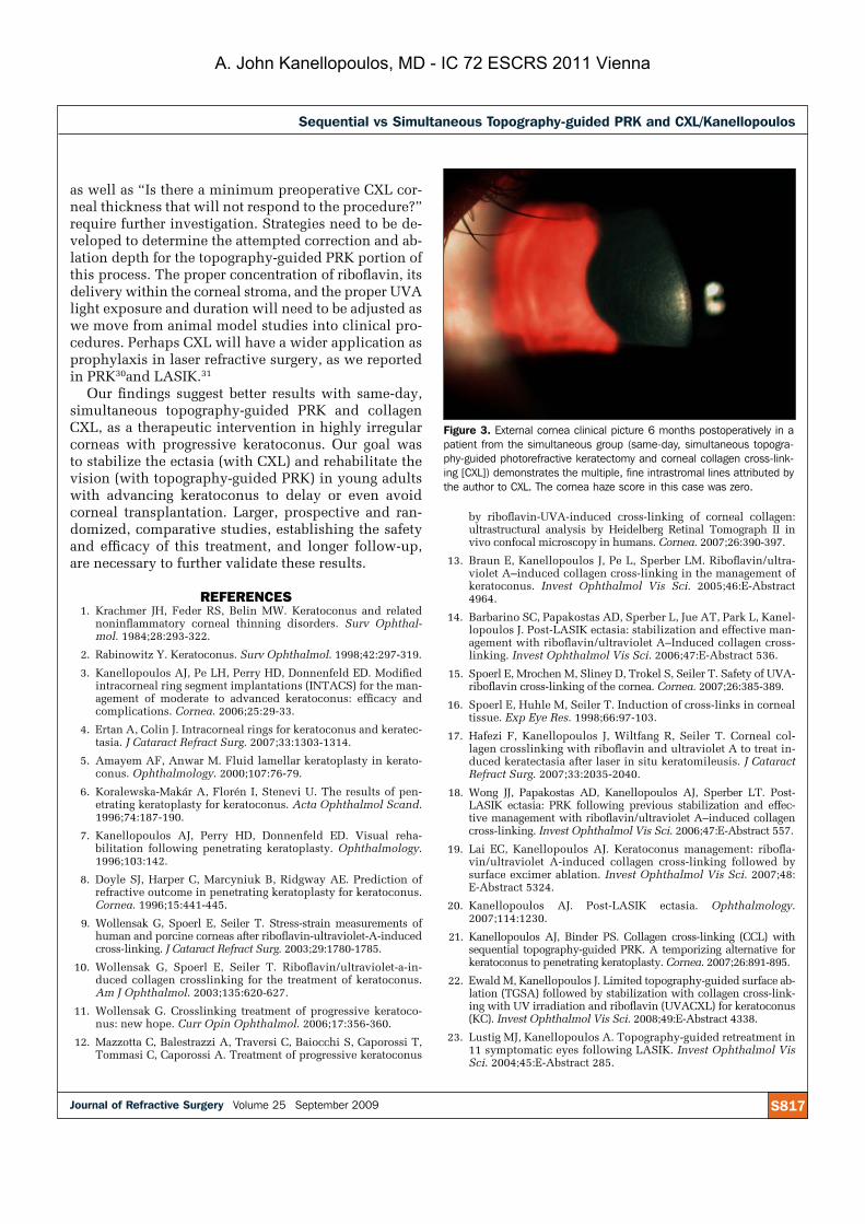
Figure 3. External cornea clinical picture 6 months postoperatively in a patient from the simultaneous group (same-day, simultaneous topogra-phy-guided photorefractive keratectomy and corneal collagen cross-link-ing [CXL]) demonstrates the multiple, fine intrastromal lines attributed by the author to CXL. The cornea haze score in this case was zero.
A. John Kanellopoulos, MD - IC 72 ESCRS 2011 Vienna
journalofrefractivesurgery.comS818
Sequential vs Simultaneous Topography-guided PRK and CXL/Kanellopoulos
24. Ledoux DM, Kanellopoulos A. Topography-guided LASIK, early ex-perience in 7 irregular eyes. Invest Ophthalmol Vis Sci. 2004;45:E-Abstract 2821.
25. Kanellopoulos AJ. Topography-guided custom retreatments in 27 symptomatic eyes. J Refract Surg. 2005;21:S513-S518.
26. Kanellopoulos AJ. Managing highly distorted corneas with to-pography-guided treatment. In: ISRS/AAO 2007 Subspecialty Day/Refractive Surgery Syllabus. Section II: Ablation Strategies.San Francisco, Calif: American Academy of Ophthalmology; 2007:13-15.
27. Xu H, Liu S, Xia X, Huang P, Wang P, Wu X. Mitomycin C re-duces haze formation in rabbits after excimer laser photorefrac-tive keratectomy. J Refract Surg. 2001;17:342-349.
28. Wollensak G, Spoerl E, Reber F, Seiler T. Keratocyte cytotoxic-ity of ribofl avin/UVA treatment in vitro. Eye. 2004;18:718-722.
29. Seiler T, Hafezi F. Corneal cross-linking-induced stromal de-marcation line. Cornea. 2006;25:1057-1059.
30. Kanellopoulos AJ. Safety and effi cacy of prophylactic, ultravio-let A irradiation cross-linking for high-risk myopic PRK cases. Poster presentation at: ISRS/AAO Refractive Surgery Subspe-cialty Day American Academy of Ophthalmology annual meet-ing; November 7, 2008; Atlanta, Ga.
31. Kanellopoulos AJ. Prophylactic, ultraviolet A cross-linking combined at the completion of high risk myopic LASIK cases. Presented at: ISRS/AAO Refractive Surgery Subspecialty Day American Academy of Ophthalmology annual meeting; November 8, 2008; Atlanta, Ga.
A. John Kanellopoulos, MD - IC 72 ESCRS 2011 Vienna
S827Journal of Refractive Surgery • Vol. 26, No. 10, 2010 Commercially Sponsored Section
Stability of Simultaneous Topography-guided Photorefractive Keratectomy and Ribofl avin/UVA Cross-linking for Progressive Keratoconus: Case ReportsRonald R. Krueger, MD, MSE; A. John Kanellopoulos, MD
From Cole Eye Institute, Cleveland Clinic Foundation, Cleveland, Ohio (Krueger); and Laservision.gr Institute, Athens, Greece, and NYU Medical School, New York, New York (Kanellopoulos).
Dr Krueger receives consulting and research funds from Alcon Laboratories Inc, Ft Worth, Texas. Dr Kanellopoulos has no financial interest in the materials presented herein.
Correspondence: Ronald R. Krueger, MD, MSE, Cole Eye Institute, Cleveland Clinic Foundation, 9500 Euclid Ave, i32, Cleveland, OH 44195. Tel: 216.444.8158; Fax: 216.445.8475; E-mail: [email protected]
ABSTRACT
PURPOSE: To follow the stability of a simultaneously delivered therapy that corrects aberrations and stiffens the corneal collagen of eyes with progressive keratoconus.
METHODS: Two patients with progressive keratoconus underwent partial treatment (70% cylinder and sphere up to 50-µm central depth) with topographic custom-ized photorefractive keratectomy (PRK) using the T-CAT module of the ALLEGRETTO WAVE Eye-Q excimer laser (Alcon Laboratories Inc), and then immediate corneal collagen cross-linking (CXL) with ribofl avin 0.1% drops every 2 minutes while exposed to mean 365-nm ultra-violet A (UVA) light at 3.0 mW/cm2 for 30 minutes (the Athens Protocol). Pre- and postoperative evaluations included manifest and cycloplegic refraction, Scheimp-fl ug corneal tomography and pachymetry, and slit-lamp examination of corneal clarity with a minimum follow-up of 30 months.
RESULTS: Both treated eyes experienced rapid healing of the epithelial surface within 5 days and progressive improvement of vision. In the fi rst case, partial treat-ment reduced the astigmatism and aberrations, allow-ing for successful soft contact lens wear at 3 months. Follow-up at 13, 19, 30, and 36 months showed pro-gressive reduction of refractive myopia and keratometric power. In the second case, laser treatment led to a near emmetropic refraction with an uncorrected visual acuity of 20/20 at 3 months, which remained unchanged at 21 and 30 months postoperative.
CONCLUSIONS: Partial topography-guided PRK followed by ribofl avin/UVA CXL is a safe and effective therapy that halts the progression of keratoectasia and reduces the spherocylindrical refraction and aberrations to improve the visual function of patients with progressive keratoconus. Stability and progressive improvement over time is ob-served, although limitations may exist for steeper and thin-ner corneas. [J Refract Surg. 2010;26(10):S827-S832.] doi:10.3928/1081597X-20100921-11
K eratoconus is a disease of corneal collagen that leads to progressive and irregular steepening of the cor-neal shape with a loss of corrected vision. When
advanced, the irregular corneal shape can no longer be fi t with a contact lens, and keratoplasty must be considered to rehabilitate visual function. Ribofl avin/ultraviolet A (UVA) corneal collagen cross-linking (CXL) has been shown to effec-tively halt the progression of keratoconus, and in some cases, gradually reduce the refractive and keratometric irregularity.1
When exposed to UVA light, the ribofl avin absorbed within the cornea is photoactivated in the presence of oxygen to cre-ate a reactive singlet oxygen species, which interacts with collagen to form the cross-links within the exposed tissue.2
Although CXL halts the progression of keratoconus, it does not improve or restore the irregularity in corneal shape suffi ciently to rehabilitate the visual function of the patient. Hence, the need for further refractive correction is required after CXL, which may be problematic if the patient remains contact lens intolerant. As one alternative, intrastromal cor-neal ring segments (Intacs; Addition Technology Inc, Des Plaines, Illinois) have been proposed as a method of regular-izing the corneal shape in association with ribofl avin/UVA CXL.3,4 Although effective in many patients, some eyes still have suffi cient irregularity limiting the full potential return of corrected vision, and must undergo keratoplasty.
Topography-guided photorefractive keratectomy (PRK) has been proposed as a palliative method for correcting irregular astigmatism in keratoconus.5-7 However, in the absence of cross-linking, the weakened corneal structure is still vulner-able to progression, so that the ectasia may worsen. Herein, we evaluate the stability of simultaneous topography-guided PRK with ribofl avin/UVA CXL as a method for both regular-
A. John Kanellopoulos, MD - IC 72 ESCRS 2011 Vienna
Commercially Sponsored Section Copyright © SLACK IncorporatedS828
Sequential Topography-guided PRK and CXL for Keratoconus/Krueger & Kanellopoulos
izing the cornea and strengthening the weakened col-lagen of keratoconus over time.
SURGICAL TECHNIQUE AND EVALUATION
THE ATHENS PROTOCOLThis technique was recently reported for the man-
agement of keratoconus.8-12
Step 1: The (Partial, Spherically Corrected) Topography-guided Transepithelial PRK Technique.We devised this technique based on the proprietary WaveLight ALLEGRETTO WAVE Eye-Q laser (Alcon Laboratories Inc, Ft Worth, Texas) customized plat-form. As noted above, we previously described the use of the topography-guided platform with this device to normalize irregular corneas as well as ectasia.
This customized excimer laser treatment is guided by topographic images and is different from wavefront-guided treatments. It received CE Mark approval for clinical use in the European Union in 2003; however, it has yet to receive US Food and Drug Administration (FDA) approval.
This proprietary software utilizes topographic data from the linked topography device (Topolyzer; Wave-Light GmbH, Erlangen, Germany). By default, it permits the consideration of eight topographies (of pre-deter-mined threshold accuracy), averages the data and en-ables the surgeon to adjust the desired postoperative cornea asphericity (chosen as zero in all cases), pro-vides the option of including tilt correction (no tilt was chosen in all cases), as well as adjustment of sphere, cylinder, axis, and treatment zone (optical zone of 5.5 mm was chosen in all cases). The image of the planned surgery is generated by the laser software.
We used topography-guided PRK to normalize the cornea by reducing irregular astigmatism while treat-ing part of the refractive error. To remove the mini-mum possible tissue, we decreased the effective opti-cal zone diameter to 5.5 mm in all cases (compared to our usual treatment diameter of at least 6.5 mm in routine PRK and LASIK). We also planned ~70% treat-ment of cylinder and sphere (up to 70%), so as not to exceed 50 µm in planned stromal removal. We chose the value of 50 µm as the maximum ablation depth effected, based on our experience of treating irregular corneas with this platform.7-10
Following the placement of an aspirating lid specu-lum (Rumex, St Petersburg, Florida), a 6.5-mm, 50-µm phototherapeutic keratectomy (PTK) was performed to remove the corneal epithelium. Partial topography-guided PRK laser treatment was applied. A cellulose sponge soaked in mitomycin C (MMC) 0.02% solution was applied over the ablated tissue for 20 seconds fol-
lowed by irrigation with 10 mL of chilled balanced salt solution.
Step 2: Collagen CXL Procedure. For the next 10 minutes, the proprietary 0.1% ribofl avin sodium phos-phate ophthalmic solution (Priavision, Menlo Park, California) was applied topically every 2 minutes. The solution appeared to “soak” in the corneal stroma rap-idly, as it was centrally devoid of Bowman layer. Fol-lowing the initial ribofl avin administration, 4 diodes, emitting UVA light of mean 370-nm wavelength (range: 365 to 375 nm) and 3 mW/cm2 radiance at 2.5 cm was projected onto the surface of the cornea for 30 minutes (Keracure prototype device, Priavision). The Keracure device, which has a built-in beeper, alerts clinicians every 2 minutes during the 30-minute treatment to install the ribofl avin solution in a timely fashion. A bandage contact lens was placed on the cornea upon completion of the combined procedures.
Postoperatively, topical ofl oxacin (Ocufl ox 0.3%; Allergan Inc, Irvine, California) was used four times a day for the fi rst 10 days and prednisolone acetate 1% (Pred Forte, Allergan) was used four times a day for 60 days. Protection from all natural light with sunglasses was encouraged, with administration of oral 1000 mg Vitamin C daily for 60 days postoperative. The bandage contact lens was removed at or around day 5 following complete re-epithelialization.
EVALUATIONThe following evaluations were completed before
and after both treatments: age, sex, uncorrected distance visual acuity (UDVA), corrected distance visual acu-ity (CDVA), refraction, keratometry (K), tomography, pachymetry, endothelial cell count, corneal haze on a scale of 0 to 4 (0=clear cornea, 1=mild haze, 2=moderate haze, 3=severe haze, and 4=reticular haze [obstructing iris anatomy]), and ectasia stability as defi ned by stabil-ity in mean keratometry and tomography.
Both cases reported were performed at Laservision.gr Institute, Athens, Greece.
CASE REPORTS
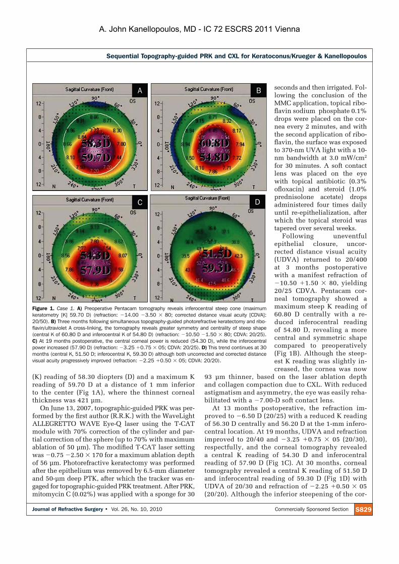
CASE 1A 24-year-old man with advanced keratoconus
and contact lens intolerance was recruited for se-quential therapy using the Athens Protocol instead of undergoing corneal transplantation. Manifest re-fraction was �14.00 �3.50 � 80 in the right eye with a corrected distance visual acuity (CDVA) of 20/50. Scheimpfl ug corneal tomography (Pentacam; Oculus Optikgeräte GmbH, Wetzlar, Germany) revealed a steep inferocentral cone with a central keratometry
A. John Kanellopoulos, MD - IC 72 ESCRS 2011 Vienna
S829Journal of Refractive Surgery • Vol. 26, No. 10, 2010 Commercially Sponsored Section
Sequential Topography-guided PRK and CXL for Keratoconus/Krueger & Kanellopoulos
(K) reading of 58.30 diopters (D) and a maximum K reading of 59.70 D at a distance of 1 mm inferior to the center (Fig 1A), where the thinnest corneal thickness was 421 µm.
On June 13, 2007, topographic-guided PRK was per-formed by the fi rst author (R.R.K.) with the WaveLight ALLEGRETTO WAVE Eye-Q laser using the T-CAT module with 70% correction of the cylinder and par-tial correction of the sphere (up to 70% with maximum ablation of 50 µm). The modifi ed T-CAT laser setting was �0.75 �2.50 � 170 for a maximum ablation depth of 56 µm. Photorefractive keratectomy was performed after the epithelium was removed by 6.5-mm diameter and 50-µm deep PTK, after which the tracker was en-gaged for topographic-guided PRK treatment. After PRK, mitomycin C (0.02%) was applied with a sponge for 30
seconds and then irrigated. Fol-lowing the conclusion of the MMC application, topical ribo-fl avin sodium phosphate 0.1% drops were placed on the cor-nea every 2 minutes, and with the second application of ribo-fl avin, the surface was exposed to 370-nm UVA light with a 10-nm bandwidth at 3.0 mW/cm2
for 30 minutes. A soft contact lens was placed on the eye with topical antibiotic (0.3% ofl oxacin) and steroid (1.0% prednisolone acetate) drops administered four times daily until re-epithelialization, after which the topical steroid was tapered over several weeks.
Following uneventful epithelial closure, uncor-rected distance visual acuity (UDVA) returned to 20/400 at 3 months postoperative with a manifest refraction of �10.50 �1.50 � 80, yielding 20/25 CDVA. Pentacam cor-neal tomography showed a maximum steep K reading of 60.80 D centrally with a re-duced inferocentral reading of 54.80 D, revealing a more central and symmetric shape compared to preoperatively (Fig 1B). Although the steep-est K reading was slightly in-creased, the cornea was now
93 µm thinner, based on the laser ablation depth and collagen compaction due to CXL. With reduced astigmatism and asymmetry, the eye was easily reha-bilitated with a �7.00-D soft contact lens.
At 13 months postoperative, the refraction im-proved to �6.50 D (20/25) with a reduced K reading of 56.30 D centrally and 56.20 D at the 1-mm infero-central location. At 19 months, UDVA and refraction improved to 20/40 and �3.25 �0.75 � 05 (20/30), respectfully, and the corneal tomography revealed a central K reading of 54.30 D and inferocentral reading of 57.90 D (Fig 1C). At 30 months, corneal tomography revealed a central K reading of 51.50 D and inferocentral reading of 59.30 D (Fig 1D) with UDVA of 20/30 and refraction of �2.25 �0.50 � 05(20/20). Although the inferior steepening of the cor-
Figure 1. Case 1. A) Preoperative Pentacam tomography reveals inferocentral steep cone (maximum keratometry [K] 59.70 D) (refraction: �14.00 �3.50 � 80; corrected distance visual acuity [CDVA]: 20/50). B) Three months following simultaneous topography-guided photorefractive keratectomy and ribo-flavin/ultraviolet A cross-linking, the tomography reveals greater symmetry and centrality of steep shape (central K of 60.80 D and inferocentral K of 54.80 D) (refraction: �10.50 �1.50 � 80; CDVA: 20/25). C) At 19 months postoperative, the central corneal power is reduced (54.30 D), while the inferocentral power increased (57.90 D) (refraction: �3.25 �0.75 � 05; CDVA: 20/25). D) This trend continues at 30 months (central K, 51.50 D; inferocentral K, 59.30 D) although both uncorrected and corrected distance visual acuity progressively improved (refraction: �2.25 �0.50 � 05; CDVA: 20/20).
A B
DC
A. John Kanellopoulos, MD - IC 72 ESCRS 2011 Vienna

Commercially Sponsored Section Copyright © SLACK IncorporatedS830
Sequential Topography-guided PRK and CXL for Keratoconus/Krueger & Kanellopoulos
neal shape progressively returned between 3 and 30 months (from 54.80 to 59.30 D), with progressive thickening of the central corneal thickness (~44 µm), a progressive reduction in the magnitude of the central K reading (~8.00 D) was also noted with marked improvement in UDVA and CDVA. At 36-month follow-up, the progressive inferior steepen-ing seemed to be stabilizing, as both the central K reading at 49.10 D and inferocentral reading at 56.40 D improved.
The fellow eye had milder keratoconus with a re-fraction of �4.00 �4.00 � 140 (20/30), steepest K reading of 51.20 D inferiorly and 47.60 D centrally, and thinnest corneal thickness of 461 µm. The eye was treated 3 months after the fi rst eye with only CXL (A.J.K.). Now, 3 years later, manifest refraction is �3.25 �1.50 � 175 (20/20) with a steepest K of
52.10 D inferiorly and 44.80 D centrally, and thinnest corneal thickness of 469 µm.
CASE 2A 21-year-old man with pro-
gressive keratoconus was also recruited for sequential thera-py using the Athens Protocol prior to corneal transplanta-tion. Uncorrected distance vi-sual acuity was 20/200 and, with a manifest refraction of �6.00 �1.50 � 90 in the right eye, CDVA was 20/30. Scheimpfl ug corneal tomog-raphy (Pentacam) revealed a steep inferocentral cone with a maximum K reading of 53.10 D (Fig 2A) and thinnest corneal thickness of 496 µm. On June 13, 2007, topography-guided PRK with the WaveLight AL-LEGRETTO WAVE Eye-Q laser using the T-CAT module with a partial correction of sphere and 75% correction of cylin-der for a maximum depth of 50 µm was performed by the fi rst author (R.R.K.). The modi-fi ed T-CAT laser setting was �2.25 �1.50 � 80 (the topo-graphy calculated a refrac-tion of �1.00 �3.00 � 80) for a maximum depth of 48 µm. Photorefractive keratectomy
was performed as in case 1, and topical ribofl avin sodium phosphate 1% drops and UVA light were applied in the same manner with the same postop-erative antibiotic and steroid drop regimen as the Athens Protocol.
The epithelium closed uneventfully within 5 days, and at 3 months postoperative, CDVA was 20/20 with a manifest refraction of �1.00 �0.75 � 124. Pentacam tomography revealed a maximum steep K reading of 45.70 D (Fig 2B) and thinner apical corneal thickness, being reduced by 113 µm. The patient did not return for follow-up at 6, 12, or 18 months. At 21-month follow-up, examination revealed a stable UDVA of 20/20 with a manifest refraction of �0.25 �0.50 � 125 (20/20+). Pentacam corneal tomography revealed a maximally steep K reading of 45.60 D (Fig 2C), with a nearly identical appearance to the map
Figure 2. Case 2. A) Preoperative Pentacam tomography reveals inferocentral steep cone (maximum kera-tometry [K] 53.10 D) (refraction: �6.00 �1.50 � 90; corrected distance visual acuity [CDVA]: 20/30). B)Three months following simultaneous topography-guided photorefractive keratectomy and riboflavin/ultra-violet A cross-linking, the tomography reveals greater symmetry and reduced steepness of corneal shape (maximum K 45.70 D) (refraction: �1.00 �0.75 � 124; CDVA: 20/20). C) At 21 months postoperative, the keratometric power and shape is stable (maximum K 45.60 D) (refraction: �0.25 �0.50 � 125;CDVA: 20/20). D) Keratometric power and shape remain unchanged at 30 months postoperative (refrac-tion: �0.75 �0.75 � 125; CDVA: 20/15).
A B
DC
A. John Kanellopoulos, MD - IC 72 ESCRS 2011 Vienna

S831Journal of Refractive Surgery • Vol. 26, No. 10, 2010 Commercially Sponsored Section
Sequential Topography-guided PRK and CXL for Keratoconus/Krueger & Kanellopoulos
taken 18 months earlier. This same appearance was once again seen at 30 months with a steep K reading of 45.70 D (Fig 2D) and CDVA of 20/15 and manifest refraction of �0.75 �0.75 � 125. The thinnest Pen-tacam corneal thickness was 387 µm at 21 months and 385 µm at 30 months, which was identical to the 385-µm reading taken at 3 months postoperative.
The fellow eye had a greater degree of myopia, �11.25 �1.25 � 82 (20/30), with a steepest inferior K reading of 57.00 D and a thinnest corneal thickness of 459 µm. This eye was previously treated with simulta-neous topography-guided PRK and CXL in September 2006 (A.J.K.). On last examination at 3 years postoper-ative, refraction and CDVA were �5.50 D (20/25), and steepest K reading was reduced to 52.90 D centrally and thinnest corneal thickness was 363 µm.
DISCUSSIONSimultaneous topography-guided PRK and ribofl a-
vin/UVA CXL—the Athens Protocol—is a new com-bined therapy that takes into account both the irregu-lar corneal shape and structural collagen weakening of progressive keratoconus. At the time of this writing, neither topographic-guided PRK nor ribofl avin/UVA CXL have been approved for clinical use by the FDA, although each of these therapies is commonly used internationally. The success of each of these two mo-dalities in the management of irregular corneal astig-matism and ectasia makes their combined clinical use worthy of consideration for progressive keratoconus. The combined use of both modalities during the same surgical setting has been reported recently.13 To prop-erly evaluate the full merit of simultaneous therapy, one should fi rst consider the pros and cons of one therapy without the other, as well as sequential CXL followed by topography-guided PRK, to access the validity of the simultaneous strategy.
First, the concept of ribofl avin/UVA CXL for progres-sive keratoconus has become an internationally accepted therapy over the past 2 years with more than 1000 procedures being performed worldwide each month (personal communication, Michael Mrochen, PhD; February 16, 2008). Although the effectiveness of this therapy in halting the progression of keratoconus has been important in stabilizing the condition, it does not address the residual refractive error with marked ir-regular astigmatism. Many of these patients who were dependent on contact lenses prior to the therapy still require contact lens wear. Some, however, have become contact lens intolerant based on progressive steepening, and remain so after CXL therapy. Even if the therapy makes it possible to once again wear contact lenses, its investigational nature (especially within the US FDA
clinical trial) makes it necessary for the patient to cease wearing contact lenses during the investigation, which for many patients is functionally unacceptable. The positive side of ribofl avin/UVA CXL is the stability of progressive keratometric steepening, whereas the nega-tive side is the lack of refractive rehabilitation, which fails to address the visual dysfunction of the disease.
Second, the irregular astigmatism of keratoconus, as with other highly aberrated eyes, can be effectively minimized with customized laser ablation procedures. Topography-guided PRK has been proposed as a rea-sonable intervention for symptomatic, highly aber-rated corneas, including those with keratoconus.6,7 Al-though effective in regularizing the aberrated corneal shape, the structural weakening of this ectatic disease leaves the cornea vulnerable to progressive change and inadequately addresses the fundamental problem. Yet myopic astigmatic PRK and topography-guided PRK are still being proposed by some for cases of early ker-atoconus. Although correcting the irregularity of kera-toconus with customized PRK is in itself functionally benefi cial, the long-term progressive instability with-out CXL makes this therapy controversial at best.
Combining these two procedures sequentially offers a solution to both the structural weakening and irregular corneal shape. Ribofl avin/UVA CXL followed at least 6 months later by topography-guided PRK has been proposed previously by one of the authors (A.J.K.),8
and has been successfully implemented in an unpub-lished study of 27 progressive keratoconic eyes.14,15 In this evaluation, 22 of the 27 eyes experienced a mean improvement of 2.00 D in the steepest K and 2.40 D of the spherical equivalent refraction at 6 months afterCXL. One year following topography-guided PRK with MMC as a second step further revealed a reduction of the overall mean spherical equivalent refraction by another 6.40 D and the steepest K from a mean of 54.00 to 47.00 D. These refractive changes were met by a mean reduction in UDVA from 20/400 to 20/60 and CDVA from 20/100 to 20/40. Although impressive, some of these sequentially treated eyes also had a mild to moderate amount of corneal haze, which was not observed in the two patients reported herein, or in the larger series of simultaneously treated eyes reported by Kanellopoulos.13
With simultaneous topography-guided PRK and CXL, the initial step of epithelial removal is utilized by both PRK and CXL, making the simultaneous process more effi cient and less traumatic. Overall, the concept of both strengthening and reshaping the cornea in ker-atoconus is a revolutionary idea that appears to work well in corneas that are not too steep or too thin. In our second case, with a steepest K reading of 53.10 D
A. John Kanellopoulos, MD - IC 72 ESCRS 2011 Vienna

Commercially Sponsored Section Copyright © SLACK IncorporatedS832
Sequential Topography-guided PRK and CXL for Keratoconus/Krueger & Kanellopoulos
and thinnest pachymetric reading of 496 µm, moderate myopia and astigmatism were fully corrected with the modifi ed T-CAT laser settings, in addition to the ir-regular astigmatism. Excellent stability in this example over 3 to 30 months demonstrates keratometry of 53.00 D and thinnest corneal thickness of 496 µm as being ac-ceptable when considering simultaneous treatment. In our fi rst case, with a steepest K reading of 59.70 D and thinnest pachymetric reading of 421 µm, the cor-rection of both regular and irregular astigmatism was possible with the modifi ed T-CAT laser settings, re-sulting in greater corneal symmetry and the fi tting of a soft contact lens. However, the residual myopia at 3 months underwent a progressive reduction in magni-tude over the next 27 months, demonstrating a lack of stability, even though the progression was desirable. Topographically, the improved corneal symmetry at 3 months gradually regressed to a pattern of inferior steepening, with the central keratometric values being reduced by ~8.00 D in magnitude, but the inferocentral keratometry being increased by ~4.50 D. This change together with the progressive increase in central cor-neal thickness of ~44 µm suggests a possible loss of CXL effect, yet with progressive reduction of central K reading and improvement in UDVA and CDVA.
The progressive reduction of keratometric power over time following corneal CXL is a poorly under-stood process, but has been documented in several studies.1,8,16 This likely explains the progressive re-duction of corneal power and myopic refraction in our fi rst case, although the relative inferocentral steepen-ing at 19 and 30 months suggests the degree of CDVA improvement may not have been enough. Despite the keratometric fi ndings, the visual outcome of this pa-tient was more than satisfactory.
Simultaneous topography-guided PRK using the T-CAT module of the ALLEGRETTO WAVE Eye-Q excimer laser with corneal CXL appears to be an ac-ceptable alternative for the management of progressive keratoconus. These results are further validated by the larger comparative series of simultaneous versus se-quential CXL with topography-guided PRK published previously.13 The detailed analysis of these two indi-viduals and the longer follow-up have demonstrated a satisfactory outcome in both cases, making this therapy worthy of consideration in advancing keratoconus.
AUTHOR CONTRIBUTIONSStudy concept and design (R.R.K., A.J.K.); data collection (A.J.K.);
analysis and interpretation of data (R.R.K., A.J.K.); drafting of the man-
uscript (R.R.K.); critical revision of the manuscript (A.J.K.); adminis-
trative, technical, or material support (A.J.K.); supervision (R.R.K.)
REFERENCES 1. Wollensak G, Spoerl E, Seiler T. Ribofl avin/ultraviolet-a-in-
duced collagen crosslinking for the treatment of keratoconus. Am J Ophthalmol. 2003;135(5):620-627.
2. Spoerl E, Mrochen M, Sliney D, Trokel S, Seiler T. Safety of UVA-ribofl avin cross-linking of the cornea. Cornea. 2007;26(4):385-389.
3. Chan CC, Sharma M, Wachler BS. Effect of inferior-segment Intacs with and without C3-R on keratoconus. J Cataract Refract Surg.2007;33(1):75-80.
4. Kamburoglu G, Ertan A. Intacs implantation with sequential collagen cross-linking treatment in postoperative LASIK ectasia. J Refract Surg. 2008;24(7):S726-S729.
5. Tamayo GE, Serrano MG. Treatment of irregular astigmatism and keratoconus with the VISX C-CAP method. Int Ophthalmol Clin. 2003;43(3):103-110.
6. Cennamo G, Intravaja A, Boccuzzi D, Marotta G, Cennamo G. Treatment of keratoconus by topography-guided custom-ized photorefractive keratectomy: two-year follow-up study. J Refract Surg. 2008;24(2):145-149.
7. Lin DT, Holland SR, Rocha KM, Krueger RR. Method for op-timizing topography-guided ablation of highly aberrated eyes with the ALLEGRETTO WAVE excimer laser. J Refract Surg.2008;24(4):S439-S445.
8. Kanellopoulos AJ, Binder PS. Collagen cross-linking (CCL) with sequential topography-guided PRK: a temporizing alter-native for keratoconus to penetrating keratoplasty. Cornea.2007;26(7):891-895.
9. Ewald M, Kanellopoulos J. Limited topography-guided surface ab-lation (TGSA) followed by stabilization with collagen cross-link-ing with UV irradiation and ribofl avin (UVACXL) for keratoconus (KC). Invest Ophthalmol Vis Sci. 2008;49:E-Abstract 4338.
10. Kanellopoulos AJ. Cross-linking plus topography-guided PRK for post-LASIK ectasia management. In: Garg A, ed. Master-ing Advanced Surface Ablation Techniques. New Delhi, India: Jaypee Brothers; 2007:204-214.
11. Kanellopoulos AJ. Cross-linking plus topography-guided PRK for post-LASIK ectasia management. In: Garg A, Rosen E, eds. Instant Clinical Diagnosis in Ophthalmology Refractive Sur-gery. New Delhi, India: Jaypee Brothers; 2008:258-269.
12. Kanellopoulos AJ. Cross-linking plus topography-guided PRK for post-LASIK ectasia management. In: Garg A, Pinelli R, O’Brart D, Lovisolo CF, eds. Mastering Corneal Collagen Cross-linking Techniques. New Delhi, India: Jaypee Brothers; 2008:69-80.
13. Kanellopoulos AJ. Comparison of sequential vs same-day simul-taneous collagen cross-linking and topography-guided PRK for treatment of keratoconus. J Refract Surg. 2009;25(9):S812-S818.
14. Kanellopoulos AJ. Managing highly distorted corneas. Presented at: American Academy of Ophthalmology Refractive Surgery Subspecialty Day; November 9, 2007; New Orleans, LA.
15. Kanellopoulos AJ. Limited topography-guided surface ablation followed by stabilization with collagen cross-linking with UV irradiation and ribofl avin for keratoconus. Presented at: Ameri-can Academy of Ophthalmology annual meeting; November 12, 2007; New Orleans, LA.
16. Wittig-Silva C, Whiting M, Lamoureux E, Lindsay RG, Sulli-van LJ, Snibson GR. A randomized controlled trial of corneal collagen cross-linking in progressive keratoconus: preliminary results. J Refract Surg. 2008;24(7):S720-S725.
A. John Kanellopoulos, MD - IC 72 ESCRS 2011 Vienna

1Journal of Refractive Surgery • Vol. xx, No. x, 2010
O R I G I N A L A R T I C L E
Management of Corneal Ectasia After LASIK With Combined, Same-day, Topography-guided Partial Transepithelial PRK and Collagen Cross-linking: The Athens ProtocolAnastasios John Kanellopoulos, MD; Perry S. Binder, MS, MD
From LaserVision.gr Institute, Athens, Greece (Kanellopoulos); New York University Medical College (Kanellopoulos) and Manhattan Eye, Ear and Throat Hospital (Kanellopoulos), New York, New York; and Gavin Herbert Eye Institute Department of Ophthalmology, University of California, Irvine, California (Binder).
The authors have no financial interest in the materials presented herein.
Presented as a paper at the American Society of Cataract and Refractive Surgery annual meeting; April 9-14, 2010; Boston, Massachusetts.
Correspondence: A. John Kanellopoulos, MD, LaserVision.gr Institute, 17 Tsocha St, Athens, 11521 Greece. Tel: 30 210 7472777; Fax: 30 210 7472789; E-mail: [email protected]
Received: April 20, 2010; Accepted: October 13, 2010
Posted online: November 5, 2010
ABSTRACT
PURPOSE: To evaluate a series of patients with corneal ectasia after LASIK that underwent the Athens Protocol: combined topography-guided photorefractive keratec-tomy (PRK) to reduce or eliminate induced myopia and astigmatism followed by sequential, same-day ultraviolet A (UVA) corneal collagen cross-linking (CXL).
METHODS: Thirty-two consecutive corneal ecta-sia cases underwent transepithelial PRK (WaveLight ALLEGRETTO) immediately followed by CXL (3 mW/cm2)for 30 minutes using 0.1% topical ribofl avin sodium phosphate. Uncorrected distance visual acuity (UDVA), corrected distance visual acuity (CDVA), manifest refrac-tion spherical equivalent, keratometry, central ultrasonic pachymetry, corneal tomography (Oculus Pentacam), and endothelial cell counts were analyzed. Mean follow-up was 27 months (range: 6 to 59 months).
RESULTS: Twenty-seven of 32 eyes had an improvement in UDVA and CDVA of 20/45 or better (2.25 logMAR) at last follow-up. Four eyes showed some topographic improvement but no improvement in CDVA. One of the treated eyes required a subsequent penetrating kerato-plasty. Corneal haze grade 2 was present in 2 eyes.
CONCLUSIONS: Same-day, combined topography-guided PRK and CXL appeared to offer tomographic stability, even after long-term follow-up. Only 2 of 32 eyes had corneal ectasia progression after the intervention. Seven-teen of 32 eyes appeared to have improvement in UDVA and CDVA with follow-up �1.5 years. This technique may offer an alternative in the management of iatrogen-ic corneal ectasia. [J Refract Surg. 2010;xx(x):xxx-xxx.] doi:10.3928/1081597X-20101105-01
P rogressive, asymmetrical corneal steepening associ-ated with an increase in myopic and astigmatic re-fractive errors, combined with midperipheral and/or
peripheral corneal thinning, represents a constellation of fi ndings in ectatic corneal disorders, such as keratoconus and pellucid marginal degeneration. Asymmetry in presen-tation and unpredictability of progression associated with a myriad of abnormal topographic fi ndings describe these enti-ties. Similar fi ndings following LASIK have been described as corneal ectasia.1-3 Analysis of different series of eyes de-veloping corneal ectasia after LASIK has suggested that cer-tain preoperative and/or operative features may be associated with this adverse outcome of LASIK or photorefractive kera-tectomy (PRK).4 The fact that corneal ectasia can occur in the absence of these features, or that it does not occur despite the presence of these features,5 has confounded our understand-ing of this entity. Nevertheless, corneal ectasia after LASIK is a visually disabling complication with an ultimate surgical treatment of penetrating keratoplasty when spectacles or con-tact lenses can no longer provide patients with the quality of vision to permit activities of daily living.
Over the past 10 years, the use of topical ribofl avin com-bined with ultraviolet A (UVA) irradiation to increase col-lagen cross-linking (CXL) has demonstrated the potential for retarding or eliminating the progression of keratoconus and corneal ectasia after LASIK. We have previously reported the application of CXL in corneal ectasia after LASIK.6 Once the
A. John Kanellopoulos, MD - IC 72 ESCRS 2011 Vienna

2 Copyright © SLACK Incorporated
Management of Corneal Ectasia After LASIK/Kanellopoulos & Binder
progression has stabilized, it is possible to treat the surface of the eye with customized PRK to normalize the corneal surface by reducing irregular astigmatism and potentially reducing the refractive error as well as providing improved visual outcomes in addition to stabilizing the disease process.7,8 We have subsequently introduced the combined, same-day use of these two intervention modalities in the management of kerato-conus.9-11
We present a series of patients with corneal ectasia after LASIK who have undergone combined, same-day topography-guided PRK and subsequent UVA colla-gen CXL to achieve stabilization of corneal ectasia and enhance visual rehabilitation.
PATIENTS AND METHODS
PATIENT SELECTIONPatients entered into this study were seen by one
of the authors (A.J.K.) in his private practice, either through individual patient referral, referral from other eye care practitioners, or were his own patients. Once a diagnosis of corneal ectasia after LASIK was confi rmed (see below), patients were presented with the options of contact lens fi tting, intracorneal ring segment im-plantation, or, in advanced cases, penetrating kerato-plasty. If these modalities did not serve the needs of the patient, he/she was then presented with the option of undergoing topography-guided PRK and UVA colla-gen CXL as a possible technique to prolong or prevent the need for penetrating keratoplasty. Patients provided verbal and written consent prior to undergoing the combined topography-guided PRK/CXL procedure.
A diagnosis of corneal ectasia was made when patients developed progressive corneal steepening associated with an increasing myopic and/or astig-matic refractive error 2 or more months after LASIK surgery. These fi ndings were combined with increas-ing inferior corneal steepening and thinning based on videokeratography and ultrasound pachymetry. Preop-erative LASIK clinical data and topography were re-quested from the referring physician or primary LASIK surgeon for analysis. Progression of the myopic refrac-tive error with or without progression of the manifest astigmatism, decreasing uncorrected distance visual acuity (UDVA), loss of corrected distance visual acu-ity (CDVA), progressive inferior corneal steepening on topography, and/or decreasing inferior corneal thick-ness were fi ndings in all cases.
CLINICAL EXAMINATIONEach patient underwent manifest refraction as well
as measurement of UDVA and CDVA, which was re-
corded in a 20-foot lane using high-contrast Snellen visual acuity testing. Cycloplegic refractions were performed using 1% tropicamide solution (Alcon Laboratories Inc, Ft Worth, Texas). Slit-lamp micros-copy confi rmed the presence of a LASIK fl ap. Kera-tometry readings were obtained by videokeratography (Topolyzer; WaveLight AG, Erlangen, Germany) and/or manual keratometry (model 71-21-35; Bausch & Lomb, Rochester, New York). Pachymetry was performed us-ing at least one of the following devices/instruments: Pentacam (Oculus Optikgeräte GmbH, Wetzlar, Germany), Orbscan II (Bausch & Lomb), or EchoScan US-1800 (NIDEK Co Ltd, Gamagori, Japan). Specular microscopy was performed using the Konan specular microscope (Konan Medical, Boston, Massachusetts). Topography was performed using the Orbscan II or Pentacam.
SURGICAL TECHNIQUE—THE ATHENS PROTOCOLWe have reported this technique in the management
of keratoconus.9-11
Step 1. The (Partial, Spherically Corrected) Topography-guided Transepithelial PRK Technique.We devised this technique based on the proprietary WaveLight customized platform. As noted above, we previously described the use of the topography-guided platform with this device to normalize irregular cor-neas as well as corneal ectasia.
This customized excimer laser treatment is guided by topographic images and is different from wavefront-guided treatments. It received CE mark for clinical use in the European Union in 2003; however, it has yet to receive US Food and Drug Administration approval.
This proprietary software utilizes topographic data from the linked topography device (Topolyzer). By default, it permits the consideration of eight topogra-phies (of predetermined threshold accuracy), averages the data and enables the surgeon to adjust the desired postoperative cornea asphericity (chosen as zero in all cases), provides the option of including tilt correction (no tilt was chosen in all cases), as well as the adjust-ment of sphere, cylinder, axis, and treatment zone (opti-cal zone of 5.5 mm was chosen in all cases). The image of the planned surgery is generated by the laser software.
We used topography-guided PRK to normalize the cornea by reducing irregular astigmatism while treat-ing part of the refractive error. To remove the minimum possible tissue, we decreased the effective optical zone diameter to 5.5 mm in all cases (compared to our usual treatment diameter of at least 6.5 mm in routine PRK and LASIK). We also planned ~70% treatment of cyl-inder and sphere (up to 70%) so as not to exceed 50 µmin planned stromal removal. We chose the value of 50 µm as the maximum ablation depth effected, based
A. John Kanellopoulos, MD - IC 72 ESCRS 2011 Vienna
3Journal of Refractive Surgery • Vol. xx, No. x, 2010
Management of Corneal Ectasia After LASIK/Kanellopoulos & Binder
on our experience of treating irregular corneas with this platform.7-10
Following the placement of an aspirating lid specu-lum (Rumex, St Petersburg, Florida), a 6.5-mm, 50-µm phototherapeutic keratectomy (PTK) was performed to remove the corneal epithelium. Partial topography-guided PRK laser treatment was applied. A cellulose sponge soaked in mitomycin C (MMC) 0.02% solution was applied over the ablated tissue for 20 seconds fol-lowed by irrigation with 10 mL of chilled balanced salt solution.
Step 2. Collagen CXL Procedure. For the next 10 minutes, the proprietary 0.1% ribofl avin sodium phos-phate ophthalmic solution (Priavision, Menlo Park, California) was applied topically every 2 minutes. The solution appeared to “soak” into the corneal stroma rapidly, as it was centrally devoid of Bowman layer. Following the initial ribofl avin administration, 4 di-odes emitting UVA light of mean 370-nm wavelength (range: 365 to 375 nm) and 3 mW/cm2 radiance at 2.5 cm were projected onto the surface of the cornea for 30 minutes (Keracure prototype device, Priavision). The Keracure device, which has a built-in beeper, alerts clinicians every 2 minutes during the 30-minute treat-ment to install the ribofl avin solution in a timely fash-ion. A bandage contact lens was placed on the cornea at the completion of the combined procedures.
Postoperatively, topical ofl oxacin (Ocufl ox 0.3%; Allergan Inc, Irvine, California) was used four times a day for the fi rst 10 days and prednisolone acetate 1% (Pred Forte, Allergan Inc) was used four times a day for 60 days. Protection from all natural light with sun-glasses was encouraged, with administration of oral 1000 mg Vitamin C daily for 60 days postoperative. The bandage contact lens was removed at or around day 5 following complete re-epithelialization.
EVALUATIONThe following evaluations were completed before and
after both treatments: age, sex, UDVA, CDVA, refrac-tion, keratometry, tomography, pachymetry, endothe-lial cell count, corneal haze on a scale of 0 to 4: (0=clear cornea, 1=mild haze, 2=moderate haze, 3=severe haze, and 4=reticular haze [obstructing iris anatomy]), and corneal ectasia stability as defi ned by stability in mean keratometry and tomography.
CASE REPORTS
CASE 1A 39-year-old man had undergone LASIK in May
2004 at another institution. At that time, according to patient history, UDVA was counting fi ngers in both
eyes. Manifest refraction was �6.50 �0.50 � 020 (20/20) in the right eye and �6.00 �0.50 � 165 (20/20) in the left eye. Preoperative keratometry and corneal thickness readings were not available. No surgical data were available. The patient achieved UDVA of 20/20 in each eye, and reportedly plano refraction in both. In October 2005, he complained of progressively decreas-ing vision in both eyes. At that time, UDVA was 20/50 in the right eye and 20/40 in the left eye and he was told that “astigmatism was developing.”
The patient presented in March 2006, 26 months after LASIK, with a manifest refraction of �2.25 �1.75� 090 (20/20) in the right eye and �1.25 �0.75 � 010 (20/20) in the left eye. Uncorrected distance visual acu-ity was 20/40 in the right eye and 20/30 in the left eye. Keratometry was 38.75@90/35.62@180 in the right eye and 40.65@05/39.55@95 in the left eye. Central corneal thickness (measured with Pentacam and ultrasound) was 495 µm in the right eye and 505 µm in the left eye, respectively. A diagnosis of bilateral corneal ectasia was made.
Because of the decrease in UDVA and the presence of corneal ectasia, the patient was informed of the risks, benefi ts, and alternatives of the combined topography-guided PRK/CXL technique. This procedure was per-formed on both eyes in January 2007, 32 months after LASIK. Based on the clinical manifest refraction of right (�2.25 �1.75 � 90 [20/20]) and left (�1.25 �0.50� 005 [20/20]) eyes, the attempted correction was re-duced to �1.75 �1.50 � 90 and �0.75 �0.50 � 005 for the right and left eyes, respectively. (The goal in the treatment was modifi ed to anticipate the possible long-term fl attening effect that CXL may have on these corneas.)
In February 2010, 37 months after topography-guided PRK/CXL, UDVA improved to 20/40 in the right eye and 20/20 in the left eye with a manifest refraction of �0.75(20/20) in the right eye and �0.25 �0.25 � 95 (20/20) in the left eye. Keratometry was 37.50@85/36.62@175 in the right eye and 37.75@79/37.87@169 in the left eye. Ultrasound pachymetry was 440 µm and 414 µm in the right and left eyes, respectively. Figure 1 demon-strates the pre- and postoperative topographies of the right eye as well as the difference map after treatment with the Athens Protocol.
CASE 2A 33-year-old woman reportedly had a manifest re-
fraction of �4.00 �2.50 � 90 (20/20) in the right eye and �1.50 �2.00 � 100 (20/20) in the left eye. No other preoperative data were available. The patient had a history of eye rubbing.
Sometime in 2002, the patient underwent bilateral
A. John Kanellopoulos, MD - IC 72 ESCRS 2011 Vienna
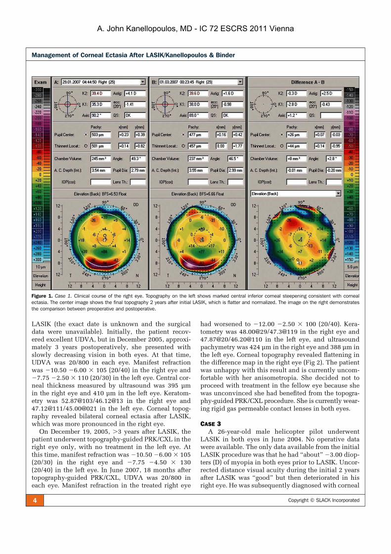
4 Copyright © SLACK Incorporated
Management of Corneal Ectasia After LASIK/Kanellopoulos & Binder
LASIK (the exact date is unknown and the surgical data were unavailable). Initially, the patient recov-ered excellent UDVA, but in December 2005, approxi-mately 3 years postoperatively, she presented with slowly decreasing vision in both eyes. At that time, UDVA was 20/800 in each eye. Manifest refraction was �10.50 �6.00 � 105 (20/40) in the right eye and �7.75 �2.50 � 110 (20/30) in the left eye. Central cor-neal thickness measured by ultrasound was 395 µmin the right eye and 410 µm in the left eye. Keratom-etry was 52.87@103/46.12@13 in the right eye and 47.12@111/45.00@021 in the left eye. Corneal topog-raphy revealed bilateral corneal ectasia after LASIK, which was more pronounced in the right eye.
On December 19, 2005, �3 years after LASIK, the patient underwent topography-guided PRK/CXL in the right eye only, with no treatment in the left eye. Atthis time, manifest refraction was �10.50 �6.00 � 105(20/30) in the right eye and �7.75 �4.50 � 130 (20/40) in the left eye. In June 2007, 18 months after topography-guided PRK/CXL, UDVA was 20/800 in each eye. Manifest refraction in the treated right eye
had worsened to �12.00 �2.50 � 100 (20/40). Kera-tometry was 48.00@29/47.3@119 in the right eye and 47.87@20/46.20@110 in the left eye, and ultrasound pachymetry was 424 µm in the right eye and 388 µm in the left eye. Corneal topography revealed fl attening in the difference map in the right eye (Fig 2). The patient was unhappy with this result and is currently uncom-fortable with her anisometropia. She decided not to proceed with treatment in the fellow eye because she was unconvinced she had benefi ted from the topogra-phy-guided PRK/CXL procedure. She is currently wear-ing rigid gas permeable contact lenses in both eyes.
CASE 3A 26-year-old male helicopter pilot underwent
LASIK in both eyes in June 2004. No operative data were available. The only data available from the initial LASIK procedure was that he had “about” �3.00 diop-ters (D) of myopia in both eyes prior to LASIK. Uncor-rected distance visual acuity during the initial 2 years after LASIK was “good” but then deteriorated in his right eye. He was subsequently diagnosed with corneal
Figure 1. Case 1. Clinical course of the right eye. Topography on the left shows marked central inferior corneal steepening consistent with cornealectasia. The center image shows the final topography 2 years after initial LASIK, which is flatter and normalized. The image on the right demonstrates the comparison between preoperative and postoperative.
A. John Kanellopoulos, MD - IC 72 ESCRS 2011 Vienna

5Journal of Refractive Surgery • Vol. xx, No. x, 2010
Management of Corneal Ectasia After LASIK/Kanellopoulos & Binder
ectasia and was offered Intacs (Addition Technology Inc, Des Plaines, Illinois) or a corneal transplant.
He presented to our institution in September 2007, 3 years after LASIK. Uncorrected distance visual acu-ity was 20/40 the right eye and 20/15 in the left eye. Manifest refraction was �1.50 �2.00 � 65 (20/20) in
the right eye and plano (20/15) in the left eye. Kera-tometry was 41.62@65/43.62@155 in the right eye and 41.75/42.12@10 in the left eye. Central ultrasound pachymetry was 476 µm in the right eye and 490 µm in the left eye.
On September 13, 2007, 39 months after LASIK,
Figure 2. Case 2. Topography on the left shows marked inferior steepening before topography-guided PRK/CXL treatment. The topography on the right shows the same cornea 18 months after topography-guided PRK/CXL with marked flattening of the corneal ectasia and normalization of the cornea.
Figure 3. Case 3. Clinical course of the right eye. A) Topography 3 years after LASIK demonstrates irregular astigmatism and marked inferior corneal steepen-ing. Uncorrected distance visual acu-ity was 20/40 and corrected distance visual acuity was 20/20 with refraction of �1.50 �2.00 � 65. B) Topography 3 months after topography-guided PRK/CXL procedure demonstrates a flatter and normalized cornea. Uncorrected distance visual acuity was 20/15. C) Topographic reproduction of the topography-guided PRK treatment plan with the WaveLight platform. This platform plans to remove tissue in an irregular fashion to normalize the corneal ectasia seen in Figure 3A. D)Comparison map, derived from subtract-ing image B from A, represents the topo-graphic difference in this case 3 months after the combined treatment. The para-central flattening is self-explanatory, as the PRK and CXL have flattened the cone apex. The superior nasal arcuate flatten-ing represents the actual part-hyperopic correction, which the topography-guided treatment has achieved, to accomplish steepening in the area central to this arc. Thus, the topography-guided treatment has normalized the ectatic cornea by flat-tening the cone apex and at the same time by “steepening” the remainder of the central cornea.
A. John Kanellopoulos, MD - IC 72 ESCRS 2011 Vienna
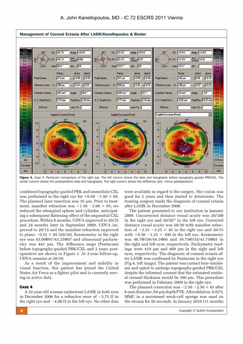
6 Copyright © SLACK Incorporated
Management of Corneal Ectasia After LASIK/Kanellopoulos & Binder
combined topography-guided PRK and immediate CXL was performed in the right eye for �0.50 �1.50 � 60.The planned laser resection was 35 µm. Prior to treat-ment, manifest refraction was �1.50 �2.00 � 65; we reduced the attempted sphere and cylinder, anticipat-ing a subsequent fl attening effect of the sequential CXL procedure. Within 6 months, UDVA improved to 20/25 and 24 months later in September 2009, UDVA im-proved to 20/15 and the manifest refraction improved to plano �0.25 � 05 (20/10). Keratometry in the right eye was 43.00@97/43.25@07 and ultrasound pachym-etry was 441 µm. The difference maps (Pentacam) before topography-guided PRK/CXL and 2 years post-operative are shown in Figure 3. At 3-year follow-up, UDVA remains at 20/10.
As a result of the improvement and stability in visual function, this patient has joined the United States Air Force as a fi ghter pilot and is currently serv-ing in active duty.
CASE 4A 32-year-old woman underwent LASIK in both eyes
in December 2006 for a refractive error of �3.75 D in the right eye and �4.00 D in the left eye. No other data
were available in regard to the surgery. Her vision was good for 2 years and then started to deteriorate. The treating surgeon made the diagnosis of corneal ectasia after LASIK in December 2008.
The patient presented to our institution in January 2009. Uncorrected distance visual acuity was 20/100 in the right eye and 20/20-2 in the left eye. Corrected distance visual acuity was 20/30 with manifest refrac-tion of �3.25 �3.25 � 45 in the right eye and 20/15 with �0.50 �1.25 � 100 in the left eye. Keratometry was 46.7@156/44.1@66 and 39.75@155/41.75@65 in the right and left eyes, respectively. Pachymetry read-ings were 419 µm and 460 µm in the right and left eyes, respectively. The diagnosis of corneal ectasia af-ter LASIK was confi rmed by Pentacam in the right eye (Fig 4, left image). The patient was contact lens–intoler-ant and opted to undergo topography-guided PRK/CXL despite the informed consent that the estimated residu-al corneal thickness would be 360 µm. This procedure was performed in February 2009 in the right eye.
The planned correction was �2.50 �2.50 � 45 after 6-mm diameter, 50-µm depth PTK. After ablation, 0.02% MMC in a moistened weck-cell sponge was used on the stroma for 20 seconds. In January 2010 (11 months
Figure 4. Case 4. Pentacam comparison of the right eye. The left column shows the data and topography before topography-guided PRK/CXL. The center column shows the postoperative data and topography. The right column shows the difference (pre- minus postoperative).
A. John Kanellopoulos, MD - IC 72 ESCRS 2011 Vienna

7Journal of Refractive Surgery • Vol. xx, No. x, 2010
Management of Corneal Ectasia After LASIK/Kanellopoulos & Binder
following treatment), UDVA was 20/30, CDVA was 20/20-1 with manifest refraction of �0.50 �0.75 � 141.Keratometry was 43.3 and 42.7@103. Central corneal thickness was 330 µm. The pre- and postoperative difference map is shown in Figure 4. Endothelial cell count was unchanged at 20 months (2600 from 2650 prior to application of the Athen’s protocol).
Optical coherence tomography (OCT) of the cen-tral cornea in the right eye at 11 months postoperative shows hyper-refl ectivity of the anterior 2/3 of the cor-nea (Fig 5) demonstrating the CXL effect, which we re-ported previously when applying similar treatment in cases of keratoconus.10,11 The hyper-refl ective demar-cation in the middle of the cornea in this case suggests a thick LASIK fl ap calculated to �200 µm by corneal OCT prior to application of the Athen’s protocol.
SUMMARY OF ALL CASESA total of 32 eyes in 22 patients with corneal ectasia
occurring 1 to 4 years after LASIK were treated. Preop-erative LASIK data were not available in most cases. In the 5 patients who had available data, no irregularity on topography or tomography was noted and no oth-er factor of the LASIK procedure was evaluated to be high risk (eg, thick fl ap, residual stromal bed �250 µm). All patients had documented progression of inferior steepening in topography and/or tomography maps. Patient age ranged from 23 to 66 years (mean: 32 years) with gender divided (women:men=11:11). The mean measurements representing values after corneal ecta-sia were confi rmed and preoperative to our technique were as follows. Mean sphere was �7.50 D and mean preoperative astigmatism was �2.40 D in the 32 eyes. Mean preoperative to the original LASIK central corne-al thickness was �525 µm in 25 of 32 eyes. The origi-nal LASIK laser resection data were unavailable in 27 eyes, and fl ap thickness was assumed or calculated us-ing corneal OCT (Optovue, Fremont, California). The mean residual stromal thickness was 285 µm (range: 210 to 355 µm). Of all 32 ectasia cases, 15 were thought to have resulted from thicker than planned fl aps (mean
residual stromal bed 230 µm), 10 showed signs of corne-al irregularity on preoperative LASIK topography, and 7 had no recognizable risk factor for the development of corneal ectasia.
All topography-guided PRK procedures were planned to reduce corneal thickness by a maximum of 50 µm, de-spite the existing refractive error, to avoid exacerbation of the ectasia. Most patients (19 patients, 25 eyes) com-plained of signifi cant pain the fi rst postoperative night whereas others reported minimal discomfort. Mean fol-low-up after the procedure was 27 months.
Uncorrected distance visual acuity improved in 27 eyes, was unchanged in 4 eyes, and worsened in 1 eye; it was 20/30 or better (�0.18 logMAR) in 11 of 32 eyes and 20/60 or worse (�0.47 logMAR) in 2 eyes. Corrected distance visual acuity was 20/40 or better (�0.30 logMAR) in 27 of 32 eyes and 20/25 or better (�0.10 logMAR) in 14 eyes.
Mean refractive error decreased by more than 2.50 D in 27 of 32 eyes, appeared to increase by 0.75 D in 3 eyes, and remained stable in 2 eyes. Mean fi nal spheri-cal equivalent refraction was �1.75 D, indicating the reduction of cornea irregularity was the target and not emmetropia.
DISCUSSIONTopography-guided PRK fl attens some of the cone
apex (in a fashion similar to an eccentric partial myo-pic PRK) but simultaneously fl attens an arcuate, broader area of the cornea away from the cone, usually in the superior nasal periphery; this ablation pattern (see Fig 3C) resembles part of a hyperopic treatment and thus will cause some amount of steepening or elevation ad-jacent to the cone, effectively normalizing the cornea. We have introduced this concept as an effective tissue-sparing ablation pattern in highly irregular corneas such as ectasia in keratoconus.12 It is this core concept in the topography-guided PRK treatment that makes it, in our opinion, more therapeutic than refractive. We have reported7-10 that in theory, the new “fl atter” and less irregular corneal shape may perform better biome-
Figure 5. Case 4. Optical coherence tomog-raphy of the central cornea in the right eye 11 months after topography-guided PRK/CXL. The hyper-reflectivity of the anterior 2/3 of the cornea suggests (as reported pre-viously10) the CXL effect (yellow arrows). The hyper-reflective demarcation in the middle of the cornea (white arrows) suggests a thick LASIK flap calculated to �200 µm.
A. John Kanellopoulos, MD - IC 72 ESCRS 2011 Vienna

8 Copyright © SLACK Incorporated
Management of Corneal Ectasia After LASIK/Kanellopoulos & Binder
chanically in eyes with corneal ectasia. Specifi cally, as the corneal apex becomes a fl atter and “broader” cone (see Figs 3A and 3B), this may redistribute the biome-chanical strain from the eye’s intraocular pressure and other external factors (eg, eye rubbing, blinking, etc). This effect may be further enhanced with additional collagen CXL strengthening.
Same-day simultaneous topography-guided PRK and CXL has several advantages: 1) the combination reduces the patient’s time away from work, 2) perform-ing both procedures at the same time with topography-guided PRK appears to minimize the potential superfi -cial stromal scarring resulting from topography-guided PRK (unpublished observations, December 2005), and 3) when topography-guided PRK is performed fol-lowing the CXL procedure, some of the cross-linked anterior cornea is removed, minimizing the potential benefi t of CXL (unpublished observations, December 2005). We believe it may be counterintuitive to remove the cross-linked tissue with topography-guided PRK at a later time, as we are potentially removing a benefi cial layer of the stiffer, cross-linked cornea, which helps maintain the normalized corneal shape. Lastly, 4) by removing the Bowman layer with topography-guided PRK, this may facilitate ribofl avin solution penetration in the corneal stroma and less “shielding” of UVA light in its passage through the cornea, resulting in more ef-fective CXL.
Although a patient with corneal ectasia can have an improved visual result with the addition of the topogra-phy-guided PRK, completely removing signifi cant refrac-tive errors was not our goal. We have placed an arbitrary “ceiling” of 50 µm to the amount of tissue that we safely removed centrally, anticipating that further thinning might destabilize the cornea’s biomechanical integrity, even following the “stiffening” effect of CXL.
It should be noted that the proprietary ribofl avin so-lution used was a slightly hypotonic (340 mOsm) for-mulation, resulting in slight “swelling” of the cornea intraoperatively (during CXL). This restored the cor-neal thickness to approximately 400 µm during CXL to protect the corneal endothelium; we did not encounter any corneal endothelial decompensation in any of the eyes studied herein despite treating cases with corneal thickness less than the theoretical limit of 400 µm13
prior to CXL (case 4).In addition, the laser treatment was applied with
caution, as the refractive effect of CXL (corneal fl atten-ing) had to be anticipated. For this reason, we elected to always attempt a signifi cant undercorrection of both sphere and cylinder by at least 30%. At a later time, we hope to more accurately determine the new ablation rate of CXL stroma.
Simultaneous topography-guided PRK and CXL ap-pears to be effective in the rehabilitation of corneal ectasia after LASIK. The reality of the effi cacy of this treatment has been the reduction of penetrating kera-toplasty cases performed for the indication of kerato-conus and corneal ectasia after LASIK in our practice over the past 4 years. The same-day, simultaneous topography-guided PRK and CXL procedure was easy to perform, but in some cases, the central epithelial surface took up to 1 month to regularize and become lucent. It took from 1 to 4 weeks for us to detect stable changes in keratometry and topography, which seemed to match the visual and refractive changes.
The main goal for all refractive surgeons is to try to eliminate or at least signifi cantly reduce the number of eyes developing corneal ectasia after PRK and LASIK. In some eyes, a pre-existing condition that may lead to corneal ectasia with either PRK or LASIK may not be able to be detected, but by eliminating eyes with ab-normal preoperative topography and leaving corneas with the maximum clinically acceptable residual stro-mal thickness, we will be able to reduce the number of eyes that develop corneal ectasia.
Our fi ndings suggest potentially promising results with same-day, simultaneous topography-guided PRK and collagen CXL as a therapeutic intervention in highly irregular corneas with progressive corneal ecta-sia after LASIK. We have reported herein effective CXL treatment in cases with minimal corneal thickness �350 µm. Our study demonstrates that we now have another means of improving the visual and refractive results of a devastating complication while avoiding or delaying penetrating keratoplasty.
AUTHOR CONTRIBUTIONSStudy concept and design (A.J.K.); data collection (A.J.K., P.S.B.);
analysis and interpretation of data (A.J.K., P.S.B.); drafting of the
manuscript (A.J.K., P.S.B.); critical revision of the manuscript
(A.J.K., P.S.B.); administrative, technical, or material support (A.J.K.,
P.S.B.); supervision (A.J.K.)
REFERENCES 1. Binder PS. Ectasia after laser in situ keratomileusis. J Cataract
Refract Surg. 2003;29(12):2419-2429.
2. Randleman JB, Russell B, Ward MA, Thompson KP, Stulting RD. Risk factors and prognosis for corneal ectasia after LASIK. Ophthalmology. 2003;110(2):267-275.
3. Tabbara K, Kotb A. Risk factors for corneal ectasia after LASIK. Ophthalmology. 2006;113(9):1618-1622.
4. Klein SR, Epstein RJ, Randleman JB, Stulting RD. Corneal ecta-sia after laser in situ keratomileusis in patients without apparent preoperative risk factors. Cornea. 2006;25(4):388-403.
5. Binder PS. Analysis of ectasia after laser in situ keratomileusis: risk factors. J Cataract Refract Surg. 2007;33(9):1530-1538.
A. John Kanellopoulos, MD - IC 72 ESCRS 2011 Vienna
9Journal of Refractive Surgery • Vol. xx, No. x, 2010
Management of Corneal Ectasia After LASIK/Kanellopoulos & Binder
6. Hafezi F, Kanellopoulos J, Wiltfang R, Seiler T. Corneal col-lagen crosslinking with ribofl avin and ultraviolet A to treat in-duced keratectasia after laser in situ keratomileusis. J Cataract Refract Surg. 2007;33(12):2035-2040.
7. Kanellopoulos AJ. Post LASIK ectasia. Ophthalmology.2007;114(6):1230.
8. Kanellopoulos A, Binder PS. Collagen cross-linking (CCL) with sequential topography-guided PRK: a temporizing alter-native for keratoconus to penetrating keratoplasty. Cornea.2007;26(7):891-895.
9. Ewald M, Kanellopoulos J. Limited topography-guided surface ablation (TGSA) followed by stabilization with collagen cross-linking with UV irradiation and ribofl avin (UVACXL) for kera-toconus (KC). Invest Ophthalmol Vis Sci. 2008;49:E-Abstract 4338.
10. Kanellopoulos AJ. Comparison of sequential vs same day simul-taneous collagen cross-linking and topography-guided PRK for treatment of keratoconus. J Refract Surg. 2009;25(9):S812-S818.
11. Krueger RR, Kanellopoulos AJ. Stability of simultaneous to-pography-guided photorefractive keratectomy and ribofl avin/UVA cross-linking for progressive keratoconus: case reports. J Refract Surg. 2010;26(10):S827-S832.
12. Kanellopoulos AJ. Managing highly distorted corneas with to-pography-guided treatment. In: ISRS/AAO 2007 Subspecialty Day/Refractive Surgery Syllabus. Section II: Ablation Strate-gies. San Francisco, CA: American Academy of Ophthalmology; 2007:13-15.
13. Spoerl E, Mrochen M, Sliney D, Trokel S, Seiler T. Safety of UVA-ribofl avin cross-linking of the cornea. Cornea. 2007;26(4):385-389.
A. John Kanellopoulos, MD - IC 72 ESCRS 2011 Vienna

Special CXL Session, AAO/MEACO Joint Meeting 2010
Kanellopoulos, MD 2
INTRODUCTION
The different modalities of epithelium on, epithelium off, epithelium on with special
agents in the riboflavin solution to dissolve hemidesmosomes and potentially increase
and enhance riboflavin solution penetration to the corneal stroma, as well as the
amount of cross linking that is achieved with any and all of the above techniques, as
well as the newly described techniques, such as the Athens Protocol (where a partial
topography-guided PRK is combined with collagen cross-linking), combining CXL with
Intracorneal rings (combined, before, after?).
We have seen several breakout sessions in these last few year’s meetings (ESCRS, ISRS,
AAO, ASCRS), on heated discussions of several “hot” issues: Are cross-linking too much
of corneal tissue in order to treat ectasia?, can we “focus” on cross-linking a certain
area of the cornea?, a certain thickness of the cornea?, at a certain level of the cornea?,
and whether newer applications of collagen cross-linking will be able to increase safety
and the potential disadvantages of the classic technique of collagen cross-linking
described over ten years ago?.
All these very interesting subjects, as well as potential means of assessing
quantitatively and qualitatively, collagen cross-linking within corneal tissue will be
discussed in this special session.
A. John Kanellopoulos, MD - IC 72 ESCRS 2011 Vienna

Special CXL Session, AAO/MEACO Joint Meeting 2010
Kanellopoulos, MD 3
It has been over ten years that the University of Dresden started experimenting
with collagen cross-linking, as a means of increasing corneal rigidity and potentially
treating ectasia, such as keratoconus and post refractive surgery ectasia, mainly post-
LASIK ectasia.
Becoming involved with CXL in 2002 in Greece, an endemic area for KCNa and
…ectasia, we have seen this procedure spread in dramatic numbers internationally,
especially after the 2006 CE mark approval.
The procedure is currently in several FDA trials, and in several investigative
device exception (IDE) trials in the US. Several US colleagues will join us in this special
collagen cross-linking session with fresh experiences! It has been almost alongside with
CXL, that over the last decade, tremendous technology dedicated to corneal imaging
(Cornea topography and tomography, OCT, confocal microscopy etc) that can elucidate
or “confuse” us more in regard to early diagnosis and criteria for suspicious cases for
ectasia or keratoconus.
So, it has been my goal for this meeting, to bring together some of the expert
minds in corneal refractive surgery and keratoconus ectasia management, from all over
the world, at the American Academy meeting in Chicago, Illinois. The ISRS was been
gracious to host this special collagen cross-linking session.
It will start at 6:30 in the morning on Friday, October the 15th, to precede the
subspecialty day of refractive surgery of Friday and Saturday, October 15th and 16th
respectively.
The areas that are quite interesting to discuss within this session, are:
1- Innovations in collagen cross-linking, (several manufacturers and
investigators with new technologies are invited to present new
modalities and new techniques), as well as
2- Presentations by clinicians on complications encountered and potential
combinations of these techniques with other technologies and
techniques.
A. John Kanellopoulos, MD - IC 72 ESCRS 2011 Vienna

Special CXL Session, AAO/MEACO Joint Meeting 2010
Kanellopoulos, MD 4
I think that one of the fruitful areas of discussion would be to share and discuss,
and debate maybe:
3- Updated Definitions for keratoconus, Forme Fruste keratoconus, Pellucid
Marginal Degeneration; whow do we define ectasia in 2010, in means and modes that we
can help our colleagues that perform refractive surgery through the world, to avoid
treating cases of ectasia or be aware of early signs of ectasia, and when to intervene if a
case that has had refractive surgery is starting to develop worrisome signs of corneal
change.
We will therefore devise a schedule that will include:
a) Presentation of new technologies of collagen cross-linking scheduled
from 6:30 am – 7:00 am. These will be five-minute presentations (presentation
time will be strictly enforced in order to allow several speakers to present within
these 30 minutes, at least six different new cross-linking modalities or
combination modalities).
b) From 7:00 to 7:30 we will try and define the main four entities that I described
before: 1) Forme Fruste keratoconus, 2) Clinical keratoconus, 3) Pellucid
Marginal Degeneration, and 4) what do we consider today, in 2010, progressive
ectasia in any of the above cases.
c) And last, from 7:30 – 8:00, the floor will be open for discussion of any of the
above issues, comment on potential complications that fellow clinicians have
encountered and their suggestion for management, as well as comments for a
new form of collagen cross linking and all the satellite entities involved with that.
It will be my pleasure to chair this meeting and I am very excited in seeing
everyone of you. I have invited, and I am very happy to announce to everyone Professor
Seiler, has accepted to be my co-chair in this special session. I nevertheless think that all
of YOU, will enhance the scientific level and the scientific argument and discussion that
we will put together.
I hope for this session to be of informal form and everyone can be heard through
their questions.
A. John Kanellopoulos, MD - IC 72 ESCRS 2011 Vienna

Special CXL Session, AAO/MEACO Joint Meeting 2010
Kanellopoulos, MD 5
I look forward to this session and the participation of all of you.
Sincerely,
John
A. John Kanellopoulos, M.D.
Clinical Professor of Ophthalmology
NYU Medical School
Member of the International Steering Committee, ISRS
Member of the Corneal Committee, ASCRS
A. John Kanellopoulos, MD - IC 72 ESCRS 2011 Vienna


