Complications of choledochal cysts in adulthood.
7
Annals of the Royal College of Surgeons of England (1990) vol. 72, 229-235 Complications of choledochal cysts in adulthood Nicholas F G Hopkins MS FRCS Senior Surgical Registrar Robin C N Williamson MD MChir FRCS Professor of Surgery Irving S Benjamin BSc MD FRCS Senior Lecturer in Surgery HepatoPancreatoBiliary Surgery Unit, Royal Postgraduate Medical School, London Michael H Thompson MD FRCS Consultant Surgeon Department of Surgery, Southmead Hospital, Bristol Key words: Adult choledochal cyst; Complications; Treatment Choledochal cyst is a weli-recognised entity, presenting primarily in infants and young children. Where symptoms are delayed until adulthood, associated hepatobiliary pathology may complicate the presentation. These problems may be aggravated by previous treatment with bypass surgery rather than resection. We report seven cases from our recent experience presenting with complications in adulthood. These included cholangitis, hepatic abscess, pancreatitis and malignancy within the cyst. Two patients presented during pregnancy. These complications and their impli- cations for management are discussed. Bile duct cysts were first reported in 1852 (1) and first classified by Alonso-Lej et al in 1959 (2). Caroli's disease was described in 1958 (3) and included in the thorough classification of Todani et al. in 1977 (4). Choledochal cyst is much commoner in Japan, and 80% of the patients are female (5-7). It is primarily a disease of infants and young children, 50% of cases presenting within the first decade. In 20% of cases diagnosis is delayed until adult life, when associated hepatobiliary pathology often com- plicates the presentation. Previous treatment by bypass or drainage, as opposed to resectional procedures, intro- duces additional problems at reoperation. From our recent experience we report seven cases that illustrate the management of complicated choledochal cysts in adulthood. Their salient clinical details are summarised in Table I and detailed below. Case histories Case 1. Mrs KG, now aged 35 years, presented 9 years ago with obstructive jaundice during the third trimester of her first pregnancy. Following successful delivery, laparotomy was performed and a choledochal cyst was drained by cystojejunostomy Roux-en-Y. Her current illness presented as an obscure pyrexia but was diagnosed as a hepatic abscess, which resolved after percutaneous drainage under ultrasound control. The patient was then referred to the Hammersmith Hospital for definitive treatment. Computed tomography (CT) now showed mild intrahepatic bile duct dilatation, gross common bile duct dilatation and residual signs of the hepatic abscess in Segment III. Percutaneous transhepatic cholangiography (PTC) (Fig. 1) confirmed intrahepatic dilatation and demonstrated the choledochal cyst. Endoscopic retro- grade cholangiopancreatography (ERCP) showed con- trast draining freely from the cyst into the Roux loop; the pancreatic duct was normal. Angiography was normal. At operation the choledochal cyst was found to taper proximally to a bile duct of 10 mm diameter at the origin of the common hepatic duct. Distally the cyst was traced down to a pinhole communication with the duodenum. The choledochal cyst was dissected off the firmly adher- ent head of the pancreas and excised. The existing Roux loop was reanastomosed as a hepaticojejunostomy. An operative culture of cyst bile subsequently grew Correspondence to: Professor R C N Williamson, Director of Surgery, Royal Postgraduate Medical School, Hammersmith Hospital, London W12 ONN
-
Upload
trinhkhanh -
Category
Documents
-
view
220 -
download
0
Transcript of Complications of choledochal cysts in adulthood.
Annals of the Royal College of Surgeons of England (1990) vol. 72, 229-235
Complications of choledochal cysts inadulthood
Nicholas F G Hopkins MS FRCSSenior Surgical Registrar
Robin C N Williamson MD MChir FRCSProfessor of Surgery
Irving S Benjamin BSc MD FRCSSenior Lecturer in Surgery
HepatoPancreatoBiliary Surgery Unit, Royal Postgraduate Medical School, London
Michael H Thompson MD FRCSConsultant Surgeon
Department of Surgery, Southmead Hospital, Bristol
Key words: Adult choledochal cyst; Complications; Treatment
Choledochal cyst is a weli-recognised entity, presentingprimarily in infants and young children. Where symptoms are
delayed until adulthood, associated hepatobiliary pathologymay complicate the presentation. These problems may beaggravated by previous treatment with bypass surgery ratherthan resection. We report seven cases from our recentexperience presenting with complications in adulthood.These included cholangitis, hepatic abscess, pancreatitisand malignancy within the cyst. Two patients presentedduring pregnancy. These complications and their impli-cations for management are discussed.
Bile duct cysts were first reported in 1852 (1) and firstclassified by Alonso-Lej et al in 1959 (2). Caroli's diseasewas described in 1958 (3) and included in the thoroughclassification ofTodani et al. in 1977 (4). Choledochal cystis much commoner in Japan, and 80% of the patients are
female (5-7). It is primarily a disease of infants andyoung children, 50% of cases presenting within the firstdecade. In 20% of cases diagnosis is delayed until adultlife, when associated hepatobiliary pathology often com-
plicates the presentation. Previous treatment by bypassor drainage, as opposed to resectional procedures, intro-duces additional problems at reoperation.From our recent experience we report seven cases that
illustrate the management of complicated choledochal
cysts in adulthood. Their salient clinical details aresummarised in Table I and detailed below.
Case histories
Case 1. Mrs KG, now aged 35 years, presented 9 years
ago with obstructive jaundice during the third trimesterof her first pregnancy. Following successful delivery,laparotomy was performed and a choledochal cyst was
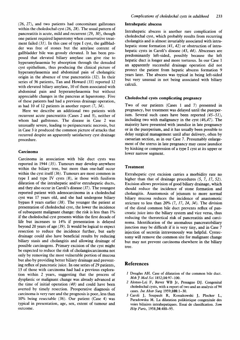
drained by cystojejunostomy Roux-en-Y. Her currentillness presented as an obscure pyrexia but was diagnosedas a hepatic abscess, which resolved after percutaneousdrainage under ultrasound control. The patient was thenreferred to the Hammersmith Hospital for definitivetreatment. Computed tomography (CT) now showedmild intrahepatic bile duct dilatation, gross common bileduct dilatation and residual signs of the hepatic abscess inSegment III. Percutaneous transhepatic cholangiography(PTC) (Fig. 1) confirmed intrahepatic dilatation anddemonstrated the choledochal cyst. Endoscopic retro-grade cholangiopancreatography (ERCP) showed con-
trast draining freely from the cyst into the Roux loop; thepancreatic duct was normal. Angiography was normal.At operation the choledochal cyst was found to taperproximally to a bile duct of 10 mm diameter at the originof the common hepatic duct. Distally the cyst was traceddown to a pinhole communication with the duodenum.The choledochal cyst was dissected off the firmly adher-ent head of the pancreas and excised. The existing Rouxloop was reanastomosed as a hepaticojejunostomy. Anoperative culture of cyst bile subsequently grew
Correspondence to: Professor R C N Williamson, Director ofSurgery, Royal Postgraduate Medical School, HammersmithHospital, London W12 ONN

230 N F G Hopkins et al.
Table I. Features of seven patients with choledochal cyst
Presenting featuresAge at Age at Cyst
presentation resection type PreviousPatient Sex (years) (years) (Todani) Jaundice Fever surgery Complications
1. KG F 26 35 I + + + Hepatic abscess2. LA M 49 51 I + + - Severe acute pancreatitis3. MB F Child 25 I + ? + Recurrent acute pancreatitis4. CR F 32 32 IV + - - Cholangiocarcinoma5. NK F 17 37 IV + + + Cholangitis, mucocele GB6. DD F 47 47 I + + - Cholangitis7. SJ F 28 28 I + - - Cholangitis
Clostridium welchii. Nevertheless the patient recoveredwell from the operation and a follow-up CT scan showedno residual abnormalities. The specimen showed noevidence of malignancy.
Case 2. Mr LA, aged 51 years, was involved in a roadtraffic accident 2 years before presentation when hesustained haemothorax, fracture-dislocation of the righthip and traumatic pancreatitis. Pancreatic trauma wasdiagnosed 4 days after injury on the basis of jaundice,fever, hyperamylasaemia and ultrasonic evidence of locu-lated fluid around the head of the pancreas. All symp-toms settled with conservative treatment, and serialsonography showed resolution of the pancreatic collec-tion within 5 weeks of the injury. He had a transientattack of abdominal pain 1 year later, and after afurther 6 months he was readmitted with abdominalpain, jaundice and renewed hyperamylasaemia. Ultra-sound scan showed a dilated biliary tree and a 2.7 cmcyst in the head of the pancreas. PTC showed a grosslydilated common bile duct consistent with a choledochalcyst (Fig. 2). Laparotomy revealed an inflamed gall-bladder without stones, a bile duct 5 cm in diameter
Figure 1. Case 1. Percutaneous transhepatic cholangiogramshowing the choledochal cyst with contrast draining into theRoux loop.
Figure 2. Case 2. Percutaneous transhepatic cholangiogramshowing the choledochal cyst and pancreatic duct.
tapering to a narrow waist just above the duodenum, and aresolving peripancreatic collection consistent with recentpancreatitis. The gallbladder and choledochal cyst wereresected, and biliary continuity was restored by hepatico-jejunostomy Roux-en-Y. Recovery was uncomplicated.Histological examination showed acute-on-chronic chole-cystitis and cholangitis.
Case 3. Miss MB, now aged 25 years, sustained epi"sodes of acute pancreatitis in childhood. At initialoperation, pancreatitis was confirmed and there was acholedochal cyst containing biliary mud. Cholecystec-tomy and cystoduodenostomy were performed withremoval of two small gallstones from the distal commonbile duct. Although the initial postoperative progress wassatisfactory, she suffered intermittent abdominal painradiating to the back, with three attacks of documentedacute pancreatitis when aged 19 years. On reinvestiga-tion, ERCP showed a short pancreatic duct arising fromthe narrow distal common bile duct, with a choledochalcyst behind the duodenum. The entire extrahepatic bileduct was dilated, but the intrahepatic ductal tree wasnormal. At operation the cystoduodenostomy was closedand the choledochal cyst was excised; the distal common

bile duct was dissected from the pancreas and ligated.Biliary drainage was re-established by hepaticojejunos-tomy Roux-en-Y. The pancreas appeared normal.Recovery was uneventful.
Case 4. Mrs CR, aged 32 years, presented with a
3-month history of severe epigastric pain radiating to theback and associated weight loss. She developed obstruc-tive jaundice 2 weeks before admission. A large epigastricmass was palpable. CT (Fig. 3) and ultrasound scans
showed a cystic mass, which was avascular on angio-graphy but displaced the superior mesenteric vessels tothe left. The pancreatic duct was irregular on ERCP. Atlaparotomy a large retroperitoneal tumour extended intothe transverse mesocolon and small bowel mesentery.Frozen section analysis showed undifferentiated carci-noma. A choledochal cyst, 15 cm in diameter, lay behindthe gallbladder displacing the duodenum. The cyst was
opened, and ascending cholangiography was performed todemonstrate dilated intrahepatic ducts. A 6 cm ulcerat-ing tumour obstructed the distal lumen of the cyst, andthis was continuous with the retroperitoneal mass.
Figure 3. Case 4. CT scan showing intrahepatic bile ductdilatation (above) and the cystic mass displacing structures tothe left (below).
Complications of choledochal cysts in adulthood 231
Subtotal resection of the cyst was performed, and aRoux loop was brought up for hepaticojejunostomy.Postoperative recovery was complicated by ascites,pleural effusions and generalised lymphadenopathy.Although chemotherapy was initiated 2 weeks later, thepatient developed duodenal obstruction and gastroenter-ostomy was required. She died of carcinomatosis 1 weeklater. Pathological examination of the resected specimenconfirmed the diagnosis of carcinoma arising within acholedochal cyst.
Case 5. Mrs NK, a 37-year-old lady from Greece,presented at the age of 17 years with jaundice. Alaparotomy was performed but accurate details are notavailable. The patient was told that a cystic structure hadbeen removed but not her gallbladder. For 20 years shewas well until she developed epigastric pains and fever(but no jaundice). Ultrasound showed a cystic structurecontaining sludge. CT scan showed some intrahepaticduct dilatation with marked aerobilia but no gallbladderand no stones. At ERCP pancreatography was normal,but there was a 4 mm opening in the proximal duodenumwhich communicated with the extrahepatic biliary tree(but not the gallbladder). PTC was performed to definethe biliary anatomy and confirmed a type IV choledochalcyst with a cystoduodenostomy. At operation the gall-bladder was thick-walled, chronically inflamed and con-tained mucus but no stones. There was a choledochalcyst, without stones, which was draining via a cystoduo-denostomy. The gallbladder and extrahepatic choledo-chal cyst were resected, the duodenum was repaired andbiliary drainage was achieved by hepaticojejunostomyRoux-en-Y. The pancreas appeared normal, although theserum amylase was mildly elevated at 424 IU/l on the daybefore operation. Histological examination of theresected tissue was benign.
Case 6. Mrs DD, aged 47 years, was admitted as anemergency with a 3-day history of right hypochondrialpain associated with attacks of shivering and sweating.There was no nausea or vomiting. Abdominal examina-tion revealed a somewhat obese abdomen with tender-ness and guarding in the right hypochondrium and apositive Murphy's sign. A working diagnosis of acutecholecystitis was made, and because of continued highspiking fever she was commenced on intravenous anti-biotics. Abdominal ultrasound the following day showeda very large choledochal cyst with marked intrahepaticductal dilatation and a normal gallbladder. PTC carriedout the same day confirmed this diagnosis. She remainedseverely pyrexial with continued abdominal tenderness,and urgent operation was undertaken on suspicion ofbiliary peritonitis following the PTC. At operation therewas only a little free bile in the abdomen. A type Icholedochal cyst was found extending from theconfluence of right and left hepatic ducts downwardsbehind the duodenum and head of the pancreas. Thegallbladder opened inxto the cyst through a very widecystic duct, and the right hepatic artery ran anterior tothe neck of the cyst below the confluence. The cyst was

232 N F G Hopkins et al.
mobilised along with the gallbladder, and the neck wastransected just below the level of this anomalous righthepatic artery. The cyst was then dissected down to thevery small orifice of an anomalous biliary-pancreaticductal junction behind the head of the pancreas andhere transected leaving a 1 cm rim of cyst wall aroundthe orifice, which was oversewn. HepaticojejunostomyRoux-en-Y was performed to the neck of the cyst.Postoperative recovery was uneventful, and pathologicalexamination showed no evidence of malignancy or dys-plasia.
Case 7. Mrs SJ, aged 28 years, presented with pre-eclampsia and jaundice at 32 weeks' gestation. Thehypertension proved difficult to control and she wasdelivered by emergency caesarian section. At operation alarge intra-abdominal mass was palpated. The patientremained jaundiced, and an abdominal CT scan showed amassive pseudocyst filling the upper abdomen. She wastransferred to Hammersmith Hospital, where on examin-ation she was deeply jaundiced (bilirubin 426,umol/l,alkaline phosphatase 1842 IU). Almost her entire abdo-men was filled with a large smooth cystic mass. PTCconfirmed the presence of a huge choledochal cyst, withenormously dilated intrahepatic ducts. Exploration wasundertaken 3 weeks after delivery, and the cyst wasfound to displace all of the upper abdominal structures.It was opened anteriorly, mobilised and completelyexcised except for an area 2 x 3 cm which was fused withthe posterior surface of the head of the pancreas. Thecyst was very thick-walled and the whole lininguniformly inflamed. The cyst edge was oversewn aroundthe remnant on the pancreas, but the ductal orifice couldnot be identified. An intravenous injection of 75 units ofsecretin produced a prompt flow of pancreatic juicefrom the very tiny orifice in the centre of this adherentcyst wall, which would not even permit the passage of thefinest probe. This area was underrun with a silk suture.The operation was completed by hepaticojejunostomy toa Roux-en-Y limb at the neck of the cyst just below thehepatic duct confluence. The patient recovered well andher bilirubin 2 weeks after operation was 58 [tmol/l andafter 4 weeks 29 [tmol/l. Histological examinationshowed no malignancy or dysplasia within the cyst.
Discussion
Classification
According to Todani's classification, type I cysts pre-dominate (82%), ie isolated 'aneurysmal' dilatation of theextrahepatic bile ducts. Type IV lesions, in which thereis a combination of intra- and extrahepatic cysts, areuncommon (9%) in some series (8), but commoner inthose in which detailed visualisation of the upper biliarytree has been obtained (9-13), suggesting underdiagno-sis. Five of our patients had a type I cyst and two a typeIV cyst.
Table II. Complications of choledochal cyst
Cholelithiasis (intra- and extrahepatic)CholangitisAcute pancreatitis (often recurrent)CholangiocarcinomaGallbladder carcinomaLiver abscessBiliary cirrhosisPortal hypertension
Causation
Several recent series have demonstrated an associationbetween an anomalous pancreaticobiliary ductal junction(APBDJ) and choledochal cysts. The abnormality takesthe form of an unusually long common channel(>20 mm) between the pancreaticobiliary junction andthe ampulla of Vater (14-17). Alternatively, it may beidentified by the angle between the ducts, which is lessacute than normal (18). The presence of amylase in thebile (19) suggests that an APBDJ facilitates reflux ofpancreatic juice, which may act as the injurious agentleading to cholangitis and ultimate carcinoma, notonly within the choledochal cyst but also in the gall-bladder (20-22). The corollary is that reflux of bile intothe pancreatic duct might conceivably account forpancreatitis.
Gallstones
Stone formation within the choledochal cyst (cystolithia-sis) is the most frequent complication of choledochal cyst(Table II), occurring in up to 70% of adult patients (7)and increasing in incidence with age (23). The calculi areoften soft and pigmented and are found throughout thebiliary tree, including the intrahepatic and pancreaticducts. Gallstones may complicate anastomotic stricturesafter cyst drainage. Although cystolithiasis might beexpected to predispose to cholangitis and acute pancreati-tis, symptoms of jaundice, abdominal pain and fever arejust as common in patients with choledochal cysts but nostones (23). It is common for stones to exist within thecholedochal cyst while the gallbladder remains acalcu-lous. However, gallbladder disease ultimately compli-cates most cases of choledochal cyst when the organ is notexcised as part of the treatment (24). Only one of ourseven patients developed gallstones, and she continuedwith recurrent acute pancreatitis after their removal.
Acute pancreatitis
This is a rare complication: in a review of the Japaneseliterature Yamaguchi (25) found only six cases in 1433patients. As a presenting feature of choledochal cyst it isreported in another six cases among Western series. Twoteenage patients had choledochocele (type III cyst)
Complications of choledochal cysts in adulthood 233
(26, 27), and two patients had concomitant gallstoneswithin the choledochal cyst (26, 28). The usual pattern ofpancreatitis is acute, mild and recurrent (29, 30), thoughone patient required laparotomy when conservative treat-ment failed (31). In this case of type I cyst, the gallblad-der was free of stones but the amylase content ofgallbladder bile was grossly elevated. It has been pro-posed that elevated biliary amylase can give rise tohyperamylasaemia by absorption through the denudedcyst epithelium, thus producing the clinical picture ofhyperamylasaemia and abdominal pain of cholangiticorigin in the absence of true pancreatitis (32). In theirseries of 36 patients, Tan and Howard (33) reported 12with elevated biliary amylase, 10 of them associated withabdominal pain and hyperamylasaemia but withoutappreciable changes in the pancreas at laparotomy. Fiveof these patients had had a previous drainage operation,as had 10 of 12 patients in another report (7, 34).Here we describe an additional two patients with
recurrent acute pancreatitis (Cases 2 and 3), neither ofwhom had gallstones. The disease in Case 2 wasunusually severe, leading to peripancreatic necrosis, butin Case 3 it produced the common picture of attacks thatrecurred despite an apparently satisfactory cyst drainageprocedure.
Carcinoma
Carcinoma in association with bile duct cysts wasreported in 1944 (35). Tumours may develop anywherewithin the biliary tree, but more than one-half occurwithin the cyst itself (36). Tumours are most common intype I and type IV cysts (8), ie those with fusiformdilatation of the intrahepatic and/or extrahepatic ducts,and they also occur in Caroli's disease (37). The youngestreported patient with adenocarcinoma in a choledochalcyst was 17 years old, and she had undergone biliarybypass 8 years earlier (38). The younger the patient atpresentation of choledochal cyst, the lower the incidenceof subsequent malignant change: the risk is less than 1%if the choledochal cyst presents within the first decade oflife but increases to 14% if presentation is delayedbeyond 20 years of age (39). It would be logical to expectresection to reduce the incidence further, but earlydrainage could also have beneficial results by reducingbiliary stasis and cholangitis and allowing drainage ofpossible carcinogens. Primary excision of the cyst mightbe expected to reduce the risk of cholangiocarcinoma notonly by removing the most vulnerable portion of mucosabut also by providing better biliary drainage and prevent-ing reflux of pancreatic juice. In one series of 29 patients,13 of those with carcinoma had had a previous explora-tion within 2 years, suggesting that the process ofdysplastic or malignant change was already advanced atthe time of initial operation (40) and could have beenaverted by timely resection. Preoperative diagnosis ofcarcinoma is very rare and the prognosis is poor, less than10% being resectable (36). Our patient (Case 4) wastypical in presentation, age, sex, extent of tumour andoutcome.
Intrahepatic abscess
Intrahepatic abscess is another rare complication ofcholedochal cyst, which probably results from recurringcholangitis and is almost invariably associated with intra-hepatic stone formation (41, 42) or obstruction of intra-hepatic cysts in Caroli's disease (43, 44). Abscesses arepredominantly left-sided, possibly because the lefthepatic duct is longer and more tortuous. In our Case 1an apparently successful drainage operation did notprotect the patient from hepatic abscess formation 9years later. The abscess was typical in being left-sidedbut very unusual in not being associated with biliarycalculi.
Choledochal cysts complicating pregnancy
Two of our patients (Cases 1 and 7) presented inpregnancy, but treatment was delayed until the puerper-ium. Several such cases have been reported (45-51),including two with malignancy in the cyst (46,47). Themajority have presented with jaundice in late pregnancyor in the puerperium, and it has usually been possible todelay surgical management until after delivery, often bycaesarian section, as in our Case 7. Presumably enlarge-ment of the uterus in late pregnancy may cause jaundiceby kinking or compression of a type I cyst at its upper orlower narrow segment.
Treatment
Extrahepatic cyst excision carries a morbidity rate nohigher than that of drainage procedures (5, 7, 17, 52).Excision allows provision of good biliary drainage, whichshould reduce the incidence of stone formation andcholangitis. Anastomosis of jejunum to more normalbiliary mucosa reduces the incidence of anastomoticstricture to less than 20% (7, 17, 24, 34). The divisionof the distal common bile duct prevents reflux of pan-creatic juice into the biliary system and vice versa, thusreducing the theroretical risk of pancreatitis and carci-noma. Identification of the anomalous pancreatobiliaryjunction may be difficult if it is very tiny, and in Case 7injection of secretin intravenously was helpful. Cystec-tomy will remove the common site for malignant changebut may not prevent carcinoma elsewhere in the biliarytree.
References
I Douglas AH. Case of dilatation of the common bile duct.Mth J Med Sci 1852;14:97-100.
2 Alonso-Lej F, Rever WB Jr, Pessagno DJ. Congenitalcholedochal cysts, with a report of two and an analysis of 94cases. Int Abstr Surg 1959;108: 1-30.
3 Caroli J, Soupault R, Kossakowski J, Plocker L,Paradowska M. La dilatation polikistique congenitale desvoies biliaires intrahepatiques. Essai de classification. SemHop Paris, 1958;34:488-95.

234 N F G Hopkins et al.
4 Todani T, Watanabe Y, Narusue M, Tabuchi K, OkajimaK. Congenital bile duct cysts. Classification, operativeprocedures and review of thirty-seven cases includingcancer arising from choledochal cyst. Am J Surg1977;134:263-9.
5 Flanigan DP. Biliary cysts. Ann Surg 1975;182:635-43.6 Powell CS, Sawyers JL, Reynolds VH. Management of
adult choledochal cysts. Ann Sur 1981;193:666-76.7 Nagorney DM, McIlrath DC, Adson MA. Choledochal
cysts in adults: clinical management. Surgery 1984;96:656-63.
8 Nagorney DM. Choledochal cysts in adult life. In:Blumgart LH, ed. Surgery of the Liver and Biliary Tract.London and Edinburgh: Churchill Livingstone, 1988:1003-12.
9 Glenn F, McSherry CK. Congenital segmental cystic dila-tation of the biliary ductal system. Ann Surg 1973;177:705-13.
10 Tsuchida Y, Ishida M. Dilatation of the intrahepatic bileducts in congenital cystic dilatation of the common bileduct. Surgery 1971;69:776-81.
11 Longmire WP, Mandiola SA, Gordan HC. Congenitalcystic disease of the liver and biliary system. Ann Surg1971;174:711-26.
12 Yue PCK. Choledochal cyst: a review of 18 cases. BrJ Surg1974;61:896-900.
13 Barros JL, Polo JR, Sanabia S, Garcia-Sabrido JL,Gomerz-Lorenzo FJ. Congenital cystic dilatation of theintrahepatic bile ducts (Caroli's disease): report of a caseand review of the literature. Surgery 1979;85:589-92.
14 Babbitt DP. Congenital choledochal cysts: a new etiologicalconcept based on anomalous relationships of the commonbile duct and pancreatic bulb. Ann Radiol 1969;12:231-5.
15 Babbitt DP, Starshak RJ, Clementt AR. Choledochal cyst:a concept of etiology. AJR 1973;119:57-62.
16 Jona JZ, Babbitt DP, Starshak RJ, LaPorta AJ, GlicklictsM, Cohen RD. Anatomic observations and etiologic andsurgical considerations in choledochal cyst. Jf Pediatr Surg1979;14:315-20.
17 Ono J, Sakoda K, Akita H. Surgical aspects of cysticdilatation of the bile ducts. An anomalous junction of thepancreaticobiliary tract in adults. Ann Surg 1982;195:203-8.
18 Todani T, Watanabe Y, Fujii T, Uemura S. Anomalousarrangement of the pancreaticobiliary ductal system inpatients with a choledochal cyst. AmJ Surg 1984;147:672-6.
19 Okada A, Oguchi Y, Kamata S, Ikeda Y, Kawashina Y,Saito R. Common channel syndrome-diagnosis with endo-scopic retrograde cholangiopancreatography and surgicalmanagement. Surgery 1983;93:634-42.
20 Yamauchi S, Koga A, Matsumoto S, Tanaka M, NakayamaF. Anomalous junction of pancreaticobiliary duct withoutcongenital choledochal cyst: a possible risk factor forgallbladder cancer. Am J Gastroenterol 1987;82:20-4.
21 Nagata E, Sakai K, Kinoshita H, Kobayashi Y. Therelation between carcinoma of the gallbladder and ananomalous connection between the choledochus and thepancreatic duct. Ann Surg r985;202: 182-90.
22 Nagata E, Sakai K, Kinoshita H, Hirohashi K. Choledochalcyst: complications of anomalous connection between thecholedochus and pancreatic duct and carcinoma of thebiliary tract. WorldJf Surg 1986;1O:102-10.
23 Matsumoto Y, Uchido K, Nakase A, Honjo I. Congenitalcystic dilatation of the common bile duct as a cause ofprimary bile duct stone. Am J Surg 1977;134:346-52.
24 Trout HH, Longmire WP. Long-term follow-up study ofpatients with congenital cystic dilatation of the common bileduct. Am J Surg 1971;121:68-86.
25 Yamaguchi M. Congenital choledochal cysts: Analysis of1433 patients in the Japanese literature. Am Jf Surg1980;140:653-7.
26 Jansen W. Intraduodenal cyst containing bile and stones:'choledochocele' in an accessory bile duct. Gastroenterology1970;58:397-401.
27 Stephens FO, Pauline GJ. Choledochocele: an unusual typeof choledochal cyst which presented as acute pancreatitis.Aust NZ J Surg 1966;36:124-7.
28 Kotner LM, McFadden JC. Pancreatitis and choledochalcyst with gallstones: an unusual case. J Arkansas Med Soc1975;71:299-301.
29 Karjoo M, Bishop HC, Barns P. Choledochal cyst pre-senting as recurrent pancreatitis. Pediatrics 1973;51:289-91.
30 Cuschieri A, Davis RS. Acute pancreatitis complicating acholedochal cyst. Br Med 7 1969;3:698.
31 Altman MS, Halls JM, Douglas AP, Penner IG.Choledochal cyst presenting as acute pancreatitis.Evaluation with endoscopic retrograde cholangiopancreato-graphy. Am J Gastroenterol 1978;70:514-19.
32 Stringel G, Filler RM. Fictitious pancreatitis in choledochalcyst. J Pediatr Surg 1982;17:359-61.
33 Tan KE, Howard ER. Choledochal cyst: a 14 year surgicalexperience with 36 patients. Br3J Surg 1988;75:892-5.
34 Rattner DW, Schapiro RH, Warshaw AL. Abnormalities ofthe pancreatic and biliary ducts in adult patients withcholedochal cysts. Arch Surg 1983;118: 1068-73.
35 Irwin ST, Marison JE. Congenital cyst of the common bileduct containing stones and undergoing cancerous change.Br J Surg 1944;32:319.
36 Flanigan DP. Biliary carcinoma associated with biliarycysts. Cancer 1977;40:880-3.
37 Dayton MT, Longmire WP, Tompkins RK. Caroli's dis-ease: a premalignant condition? Am J Surg 1983;145:41-8.
38 Fujiwara Y, Ohizumi T, Kakizaki G. A case of congenitalcholedochal cyst associated with carcinoma. J Pediatr Surg1976;11:587-8.
39 Voyles CR, Smadja C, Shands WC, Blumgart LH.Carcinoma in choledochal cysts. Age-related incidence.Arch Surg 1983;118:986-8.
40 Kagawa Y, Kashihara S, Kuramoto S. Carcinoma arising ina congenitally dilated biliary tract. Gastroenterology1978;74: 1286-94.
41 Adson MA, Nagorney DM. Hepatic resection for intra-hepatic ductal stones. Arch Surg 1982;117:611-16.
42 Nagase M, Hikasa Y, Soloway RD. Gallstones in WesternJapan: Factors affecting the prevalence of intrahepaticgallstones. Gastroenterolog 1980;78:684-90.
43 Mercadier M, Chigot JP, Clot JP, Langlois P, Lansiaux P.Caroli's disease. Jf Surg 1984;8:22-9.
44 Pridgen JE, Aust JB, McInnis WD. Primary intrahepaticgallstones. Arch Surg 1977;112: 1037-43.
45 Wijesundera P de S, Perera KS. Choledochal cyst compli-cating pregnancy. Sri Lanka J Surg 1985;87-90.
46 Binstock M, Sondek VK, Herd J et al. Adenocarcinoma in acholedochal cyst during pregnancy: a case report andguidelines for management. Surgerv 1988;103:588-92.
47 Austin RM, Sussman 5, McArdle CR, Kim D, Elboim C.Computed tomographic and ultrasound appearances of asolitary intrahepatic choledochal cyst. Glin Radiol 1986;37:149-50.

Complications of choledochal cysts in adulthood 235
48 Diamond T, Panesar KJ. Biliary peritonitis due to choledo-chal cyst presenting in late pregnancy. Ulster Med J1986;55: 190-2.
49 Angel JL, Knuppel RA, Trabin J. Choledochal cyst compli-cating a twin gestation. South Med J 1985;78:463-6.
50 Taylor TV, Brigg JK, Russell JG, Torrance B. Choledochalcyst of pregnancy. J Roy Coll Surg Edinb 1977;22:424-7.
S1 Kitahama A, Harkness SO, Moynihan PO, Webb WR. Alarge choledochal cyst with impending rupture post-partum. Br J Surg 1984;71:156.
52 Todani W, Watanabe Y, Mizuguchi T, Fujii T, Toki A.Hepaticoduodenostomy at the hepatic hilum after excisionof choledochal cyst. AmJI Surg 1981;142:584-7.
Received 9 November 1989
A new microsurgical suction switchKey words: Microsurgery, surgical instruments
Precise suction control is desirable but often diffi-cult to achieve during microsurgical procedures.The removal of small volumes of blood or secre-tions may be complicated by the adherence of solidmaterial to the suction tip and delicate tissues,sutures or prostheses can easily become displacedor damaged. A finger-operated cutout apertureincorporated in the suction tube will act as a ventand reduce the power of the sucker. However, itwill not usually overcome the problem of tissueadherence to a fine tip owing to residual surfacetension.
Footswitches have been designed to control suc-tion during microsurgery, but most require con-stant pressure which may be tedious and tiringduring a long procedure. Moreover, the surgeonmay simultaneously wish to activate a diathermy,motorised operating microscope, laser or drill usingfoot pedals.A compact foot operated suction switch has been
designed which overcomes many of these disadvan-tages, and is easy to use and maintain (Fig. 1). Itconsists of a polypropylene body enclosing a stain-less steel piston with 0-ring seals set on a non-slipbase. The few moving parts are easily removed forcleaning by unscrewing the base with a coin. Theswitch allows continuous suction after connectioninto the vacuum line. When required, pressure onthe pedal immediately stops the suction allowingprecise control. If a tissue pledget or prosthesisadheres to the sucker a sharp depression of thepedal produces a small pressure surge along thedistal line which is sufficient to overcome surfacetension at the working tip. This releases the mat-erial without damage or displacement and is afeature that has proved especially useful during themicromanipulation of grafts and prostheses.
..........T.- .............
2 3 4
Figure 1. The microsurgical footswitch.
The footswitch has applications in middle earreconstruction, endolaryngeal microsurgery, micro-vascular anastomoses, neurosurgical and ophthal-mological procedures. It is available from RimmerBrothers, 18 Aylesbury St, London ECIR ODD.
Malcolm Keene FRCSConsultant ENT Surgeon
Kathryn L Evans FRcsSenior Registrar, ENT
ENT Unit, St Bartholomew's HospitalLondon
B AshleySenior Technician
Mechanical Section of theDepartment of Medical ElectronicsSt Bartholomew's Hospital, London