Compliant vascular models 3D printed with the Stratasys ...
11
RESEARCH Open Access Compliant vascular models 3D printed with the Stratasys J750: a direct characterization of model distensibility using intravascular ultrasound Adam J. Sparks 1 , Cody M. Smith 1 , Ariana B. Allman 1 , Jillian L. Senko 1 , Karen M. Meess 1 , Richard W. Ducharme 1 , Michael E. Springer 1 , Muhammad Waqas 2 and Adnan H. Siddiqui 1,2,3* Abstract Purpose: The purpose of this study is to evaluate biomechanical accuracy of 3D printed anatomical vessels using a material jetting printer (J750, Stratasys, Rehovot, Israel) by measuring distensibility via intravascular ultrasound. Materials and methods: The test samples are 3D printed tubes to simulate arterial vessels (aorta, carotid artery, and coronary artery). Each vessel type is defined by design geometry of the vessel inner diameter and wall thickness. Vessel inner diameters are aorta = 30mm, carotid = 7mm, and coronary = 3mm. Vessel wall thickness are aorta = 3mm, carotid = 1.5mm, and coronary = 1mm. Each vessel type was printed in 3 different material options. Material options are user-selected from the J750 printer software graphical user interface as blood vessel wall anatomy elements in ‘compliant’, ‘slightly compliant’, and ‘rigid’ options. Three replicates of each vessel type were printed in each of the three selected material options, for a total of 27 models. The vessels were connected to a flow loop system where pressure was monitored via a pressure wire and cross-sectional area was measured with intravascular ultrasound (IVUS). Distensibility was calculated by comparing the % difference in cross-sectional area vs. pulse pressure to clinical literature values. Target clinical ranges for normal and diseased population distensibility are 10.3-44 % for the aorta, 5.1-10.1 % for carotid artery, and 0.5-6 % for coronary artery. Results: Aorta test vessels had the most clinically representative distensibility when printed in user-selected ‘compliant’ and ‘slightly compliant’ material. All aorta test vessels of ‘compliant’ material (n = 3) and 2 of 3 ‘slightly compliant’ vessels evaluated were within target range. Carotid vessels were most clinically represented in distensibility when printed in ‘compliant’ and ‘slightly compliant’ material. For carotid test vessels, 2 of 3 ‘compliant’ material samples and 1 of 3 ‘slightly compliant’ material samples were within target range. Coronary arteries were most clinically represented in distensibility when printed in ‘slightly compliant’ and ‘rigid’ material. For coronary test vessels, 1 of 3 ‘slightly compliant’ materials and 3 of 3 ‘rigid’ material samples fell within target range. © The Author(s). 2021 Open Access This article is licensed under a Creative Commons Attribution 4.0 International License, which permits use, sharing, adaptation, distribution and reproduction in any medium or format, as long as you give appropriate credit to the original author(s) and the source, provide a link to the Creative Commons licence, and indicate if changes were made. The images or other third party material in this article are included in the article's Creative Commons licence, unless indicated otherwise in a credit line to the material. If material is not included in the article's Creative Commons licence and your intended use is not permitted by statutory regulation or exceeds the permitted use, you will need to obtain permission directly from the copyright holder. To view a copy of this licence, visit http://creativecommons.org/licenses/by/4.0/. The Creative Commons Public Domain Dedication waiver (http://creativecommons.org/publicdomain/zero/1.0/) applies to the data made available in this article, unless otherwise stated in a credit line to the data. * Correspondence: [email protected] 1 The Jacobs Institute, Buffalo, New York, USA 2 Department of Neurosurgery, University at Buffalo, State University of New York, 100 High Street, Suite B4, Buffalo, NY 14203, USA Full list of author information is available at the end of the article Sparks et al. 3D Printing in Medicine (2021) 7:28 https://doi.org/10.1186/s41205-021-00114-8
Transcript of Compliant vascular models 3D printed with the Stratasys ...

RESEARCH Open Access
Compliant vascular models 3D printed withthe Stratasys J750: a direct characterizationof model distensibility using intravascularultrasoundAdam J. Sparks1, Cody M. Smith1, Ariana B. Allman1, Jillian L. Senko1, Karen M. Meess1, Richard W. Ducharme1,Michael E. Springer1, Muhammad Waqas2 and Adnan H. Siddiqui1,2,3*
Abstract
Purpose: The purpose of this study is to evaluate biomechanical accuracy of 3D printed anatomical vessels using amaterial jetting printer (J750, Stratasys, Rehovot, Israel) by measuring distensibility via intravascular ultrasound.
Materials and methods: The test samples are 3D printed tubes to simulate arterial vessels (aorta, carotid artery,and coronary artery). Each vessel type is defined by design geometry of the vessel inner diameter and wallthickness. Vessel inner diameters are aorta = 30mm, carotid = 7mm, and coronary = 3mm. Vessel wall thickness areaorta = 3mm, carotid = 1.5mm, and coronary = 1mm. Each vessel type was printed in 3 different material options.Material options are user-selected from the J750 printer software graphical user interface as blood vessel wallanatomy elements in ‘compliant’, ‘slightly compliant’, and ‘rigid’ options. Three replicates of each vessel type wereprinted in each of the three selected material options, for a total of 27 models. The vessels were connected to aflow loop system where pressure was monitored via a pressure wire and cross-sectional area was measured withintravascular ultrasound (IVUS). Distensibility was calculated by comparing the % difference in cross-sectional areavs. pulse pressure to clinical literature values. Target clinical ranges for normal and diseased population distensibilityare 10.3-44 % for the aorta, 5.1-10.1 % for carotid artery, and 0.5-6 % for coronary artery.
Results: Aorta test vessels had the most clinically representative distensibility when printed in user-selected‘compliant’ and ‘slightly compliant’ material. All aorta test vessels of ‘compliant’ material (n = 3) and 2 of 3 ‘slightlycompliant’ vessels evaluated were within target range. Carotid vessels were most clinically represented indistensibility when printed in ‘compliant’ and ‘slightly compliant’ material. For carotid test vessels, 2 of 3 ‘compliant’material samples and 1 of 3 ‘slightly compliant’ material samples were within target range. Coronary arteries weremost clinically represented in distensibility when printed in ‘slightly compliant’ and ‘rigid’ material. For coronary testvessels, 1 of 3 ‘slightly compliant’ materials and 3 of 3 ‘rigid’ material samples fell within target range.
© The Author(s). 2021 Open Access This article is licensed under a Creative Commons Attribution 4.0 International License,which permits use, sharing, adaptation, distribution and reproduction in any medium or format, as long as you giveappropriate credit to the original author(s) and the source, provide a link to the Creative Commons licence, and indicate ifchanges were made. The images or other third party material in this article are included in the article's Creative Commonslicence, unless indicated otherwise in a credit line to the material. If material is not included in the article's Creative Commonslicence and your intended use is not permitted by statutory regulation or exceeds the permitted use, you will need to obtainpermission directly from the copyright holder. To view a copy of this licence, visit http://creativecommons.org/licenses/by/4.0/.The Creative Commons Public Domain Dedication waiver (http://creativecommons.org/publicdomain/zero/1.0/) applies to thedata made available in this article, unless otherwise stated in a credit line to the data.
* Correspondence: [email protected] Jacobs Institute, Buffalo, New York, USA2Department of Neurosurgery, University at Buffalo, State University of NewYork, 100 High Street, Suite B4, Buffalo, NY 14203, USAFull list of author information is available at the end of the article
Sparks et al. 3D Printing in Medicine (2021) 7:28 https://doi.org/10.1186/s41205-021-00114-8

Conclusions: This study suggests that advancements in materials and 3D printing technology introduced with theJ750 Digital Anatomy 3D Printer can enable anatomical models with clinically relevant distensibility.
Keywords: 3D-Printing, Vasculature, Compliance, Intravascular Ultrasound, Distensibility
BackgroundCardiovascular disease is the number one cause of deathglobally, creating a demand for accelerated and well-informed endovascular device development [1]. There isan evolving need for accurate vascular models to supportthis rapid device development. Currently, there are vari-ous benchtop models including 2D rigid [2], 3D printed(3DP) rigid [3], 3DP compliant [4], silicone [5], and ex-vivoplatforms [6]. Although in-vivotesting is an essentialphase of device development, benchtop models offermore durable and cost effective solutions, include aquicker turnaround time for manufacture, and have alonger shelf life [7–9].Specifically, material jetting 3D-printing capabilities
are evolving within the realm of medicine to provide cli-nicians and engineers with life-like patient-specificmodels for medical device development, physician train-ing, surgical demonstration, and strategic proceduralplanning. These 3DP models can replicate patient-basedvessel geometry within 125-microns [10]. In addition,material jetted 3D printed models can mimic diseasestates such as calcifications or lesions and be printed invarious colors and material stiffnesses [11, 12]. To aid insimulating biomechanical properties of vessel walls, thevasculature may be printed using a variety of compliantmaterials. Material jetted 3D-printed anatomical modelscan be designed for use in a flow loop with physiologicalpressurized conditions for device testing under variousimaging modalities such as planar x-ray [13], ComputedTomography [14], Magnetic Resonance [15] and ultra-sound [16].In the material jetting 3D printing community, there
have been continuous efforts to replicate physiologicalcharacteristics in the 3D printed in-vitro models such asmaterial selection, vessel geometry, lubricity, and elasticproperties. Tabaczynski et al. characterized materialproperties of vascular models that best mimic healthyand diseased vessels. In addition, Tabaczynski’s otherwork has included characterizing vessel lubricity along amulti-material vessel path comprised of rigid reinforce-ments and compliant vessel wall which results in smoothand accurate device trackability within the vessel lumen[9, 11]. Furthermore, testing of different vessel wallthicknesses to vary vessel distensibility under a pressur-ized flow loop allows a more accurately represented in-vitrobenchtop system [9, 11]. Studies conducted using aJ750 3D Printer, prior to the release of the J750 DigitalAnatomy 3D Printer, indicated that preset materials may
not be sufficient to replicate the arterial wall withphysiological accuracy [9, 11]. The J750 printer can em-ploy multiple materials within a single printed model,which provides many options to vary mechanical proper-ties. Stratasys’ J750 Digital Anatomy 3D Printer materialsestablished six new preset blood vessel wall complianceoptions, thereby improving the ability to create vascularmodels with various biomechanical properties.Arterial distensibility is defined as an artery’s capacity
to expand in response to an increase in blood pressure[17]. Cross-sectional distensibility is calculated by therelative change in lumen area for a given pressurechange (ΔA/A ×ΔP, where ΔA is change in lumencross-sectional area between systole and diastole, A islumen cross-sectional area in diastole, and ΔP is localpulse pressure), expressed in units of %ΔA/100mmHg.Clinical values for distensibility are 10.3-44 % for aortaarterial vessels, 5.1-10.1 % for carotid arterial vessels, and0.5-6 % for coronary arterial vessels [17–19]. To enhancethe simulation of evaluating device performance by aclinician, vascular models that feature distensibility canbe a useful biomechanical attribute. To mimic nativedistensibility, 3D-printed vascular models’ materialsmust functionally replicate vessel change in cross-sectional area under physiological blood pressures [20].With the option of using J750 materials, there is a possi-bility to simulate clinically relevant distensibility in 3DParterial vessels. The opportunity to select vessel compli-ance material on the printer’s software graphic userinterface (GUI) could allow for simulation of a widerange of vessel distensibility and therefore a more accur-ate assessment of vascular devices.Clinically, physicians routinely use intravascular ultra-
sound (IVUS) to characterize vasculature such as innerlumen geometry and cross-sectional area [18, 21]. TheIVUS system outputs a cross sectional image which canbe measured, and along with measuring pulse pressure,distensibility can be calculated. IVUS catheters detectinner lumen diameter by propagating sound waves tothe blood vessel and computing the signal reflected fromthe walls. Although designed for use in native vascula-ture, 3D-printed material in a water flow loop provides acompatible in-vitro environment for performing mea-surements for this study. Using the same clinical IVUSequipment to measure 3D-printed vessels reduces errorof converting data from other measurement systems,providing a direct comparison to clinically reportedvalues.
Sparks et al. 3D Printing in Medicine (2021) 7:28 Page 2 of 11
In this study, we aim to evaluate the distensibility ofvarious 3D printed vessels using a J750 printer and com-pare with distensibility values of in-vivo vessels reportedin literature.
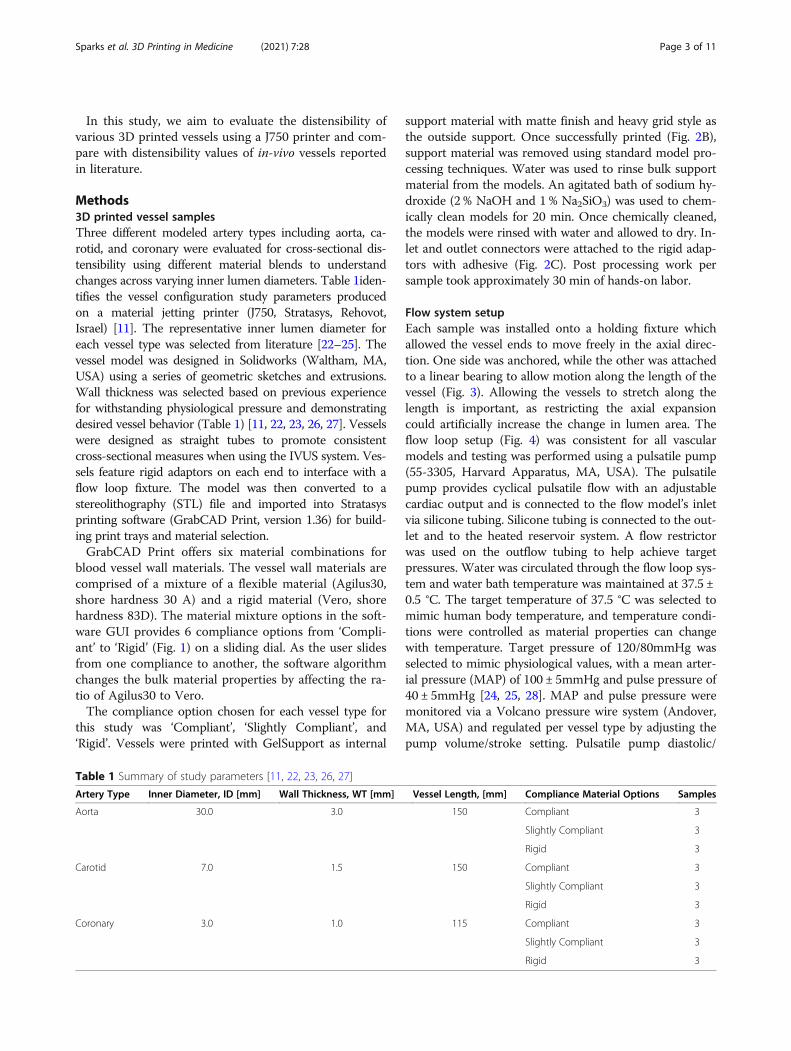
Methods3D printed vessel samplesThree different modeled artery types including aorta, ca-rotid, and coronary were evaluated for cross-sectional dis-tensibility using different material blends to understandchanges across varying inner lumen diameters. Table 1iden-tifies the vessel configuration study parameters producedon a material jetting printer (J750, Stratasys, Rehovot,Israel) [11]. The representative inner lumen diameter foreach vessel type was selected from literature [22–25]. Thevessel model was designed in Solidworks (Waltham, MA,USA) using a series of geometric sketches and extrusions.Wall thickness was selected based on previous experiencefor withstanding physiological pressure and demonstratingdesired vessel behavior (Table 1) [11, 22, 23, 26, 27]. Vesselswere designed as straight tubes to promote consistentcross-sectional measures when using the IVUS system. Ves-sels feature rigid adaptors on each end to interface with aflow loop fixture. The model was then converted to astereolithography (STL) file and imported into Stratasysprinting software (GrabCAD Print, version 1.36) for build-ing print trays and material selection.GrabCAD Print offers six material combinations for
blood vessel wall materials. The vessel wall materials arecomprised of a mixture of a flexible material (Agilus30,shore hardness 30 A) and a rigid material (Vero, shorehardness 83D). The material mixture options in the soft-ware GUI provides 6 compliance options from ‘Compli-ant’ to ‘Rigid’ (Fig. 1) on a sliding dial. As the user slidesfrom one compliance to another, the software algorithmchanges the bulk material properties by affecting the ra-tio of Agilus30 to Vero.The compliance option chosen for each vessel type for
this study was ‘Compliant’, ‘Slightly Compliant’, and‘Rigid’. Vessels were printed with GelSupport as internal
support material with matte finish and heavy grid style asthe outside support. Once successfully printed (Fig. 2B),support material was removed using standard model pro-cessing techniques. Water was used to rinse bulk supportmaterial from the models. An agitated bath of sodium hy-droxide (2 % NaOH and 1 % Na2SiO3) was used to chem-ically clean models for 20 min. Once chemically cleaned,the models were rinsed with water and allowed to dry. In-let and outlet connectors were attached to the rigid adap-tors with adhesive (Fig. 2C). Post processing work persample took approximately 30 min of hands-on labor.
Flow system setupEach sample was installed onto a holding fixture whichallowed the vessel ends to move freely in the axial direc-tion. One side was anchored, while the other was attachedto a linear bearing to allow motion along the length of thevessel (Fig. 3). Allowing the vessels to stretch along thelength is important, as restricting the axial expansioncould artificially increase the change in lumen area. Theflow loop setup (Fig. 4) was consistent for all vascularmodels and testing was performed using a pulsatile pump(55-3305, Harvard Apparatus, MA, USA). The pulsatilepump provides cyclical pulsatile flow with an adjustablecardiac output and is connected to the flow model’s inletvia silicone tubing. Silicone tubing is connected to the out-let and to the heated reservoir system. A flow restrictorwas used on the outflow tubing to help achieve targetpressures. Water was circulated through the flow loop sys-tem and water bath temperature was maintained at 37.5 ±0.5 °C. The target temperature of 37.5 °C was selected tomimic human body temperature, and temperature condi-tions were controlled as material properties can changewith temperature. Target pressure of 120/80mmHg wasselected to mimic physiological values, with a mean arter-ial pressure (MAP) of 100 ± 5mmHg and pulse pressure of40 ± 5mmHg [24, 25, 28]. MAP and pulse pressure weremonitored via a Volcano pressure wire system (Andover,MA, USA) and regulated per vessel type by adjusting thepump volume/stroke setting. Pulsatile pump diastolic/
Table 1 Summary of study parameters [11, 22, 23, 26, 27]
Artery Type Inner Diameter, ID [mm] Wall Thickness, WT [mm] Vessel Length, [mm] Compliance Material Options Samples
Aorta 30.0 3.0 150 Compliant 3
Slightly Compliant 3
Rigid 3
Carotid 7.0 1.5 150 Compliant 3
Slightly Compliant 3
Rigid 3
Coronary 3.0 1.0 115 Compliant 3
Slightly Compliant 3
Rigid 3
Sparks et al. 3D Printing in Medicine (2021) 7:28 Page 3 of 11

systolic phase ratio and rate remained constant at 35/65 %phase ratio, and 60 revolutions per minute, respectively[29, 30]. Air bubbles were eliminated via an in-line com-pliance chamber included in the inlet tubing. Volume ofwater in the compliance chamber was adjusted in con-junction with pump volume/stroke output until targetpressure conditions were met.
Distensibility testingIVUS image measurementsAn IVUS catheter specified for the corresponding innerlumen diameter was positioned at center-length withineach vessel. Philips (Andover, MA, USA) IVUS catheters(Eagle Eye Platinum ST or Visions PV8.2) were cross-sectionally centered with rail-assistance via an .035”
Fig. 1 J750 Graphic User Interface (GUI), GrabCAD Print – Model Settings. On the GUI, the user can choose model type, view the materials thatare in the printer at time of use, and select the desired anatomy type (anatomy family/element). These settings were the independent variable ofthe study in terms of vessel material selection and can be seen in the corresponding red boxes. The ‘attribute properties’ are the dependentvariable in terms of vessel material selection and can be chosen along the sliding dial as seen in the corresponding blue boxes
Fig. 2 Vessel Model Creation Process. A Vessel with inlet and outlet supports designed and assembled in Solidworks, ready to print.; B Modelsuccessfully printed, still in support, ready for post-processing; C Model ready for testing with inlet and outlet connectors attached
Sparks et al. 3D Printing in Medicine (2021) 7:28 Page 4 of 11

Terumo (Somerset, NJ, USA) guide wire (Glidewire)tracked through the entire sample. Once clinicallyrelevant hemodynamics were established, conditionswere held for 2 min before measurements began, withstability confirmed by unchanged MAP value between0 and 2 min. A 15 s IVUS reading was recorded.Pressure (MAP, Systolic, Diastolic, and Pulse Pres-sure) was also simultaneously recorded during thistime.The IVUS hardware captures videos of the pulsating
vessels at 12 to 30 frames per second. Using the lon-gitudinal view, the user visually identified locations ofminimum diameter, representing diastole and max-imum diameter representing systole for three separatecardiac cycles (Fig. 5). Cross-sectional area (mm2) wasthen measured from each image using the IVUS con-sole software.
Pressure wire measurementsPressure was captured continuously from the Volcanosystem at 60 Hz. The minimum, maximum, and MAPwere obtained from the captured pressure waveform(Fig. 6).
Target values & distensibility determinationDistensibility was calculated per equations below. Unitsare expressed as %ΔArea / 100mmHg:
Distensibility%ΔArea
100mmHg
� �¼
MaximumLumenArea mm2½ ��MinimumLumenArea mm2½ �MinimumLumenArea mm2½ �
PulsePressure mmHg½ � � 100
ð1Þ
PulsePressure mmHg½ � ¼ Systolic Maximumð ÞPressure mmHg½ �� Diastolic Minimumð ÞPressure mmHg½ �
ð2Þ
Distensibility results for each sample were then com-pared to the corresponding target clinical ranges re-ported in available literature (Table 2) [17–19].
ResultsTesting conditionsAll vessel test conditions achieved target MAP and pulsepressure. Table 3, below, identifies mean recorded MAPand pulse pressure for each tested vessel type.All vessel cross-sectional area measurements were success-
fully taken using the IVUS software (Fig. 7). The area mea-surements were then used for distensibility calculations.
Fig. 3 Test vessel fixture allowing axial movement: left side of vesselis fixed, right side of vessel is attached to a linear bearing
Fig. 4 Flow System Setup
Sparks et al. 3D Printing in Medicine (2021) 7:28 Page 5 of 11

DistensibilityNine samples of each vessel type were tested. Therewere three replicates of each of the three materials:‘compliant’, ‘semi compliant’, and ‘rigid’. Compliant ex-hibited the highest distensibility, whereas rigid corre-sponded with the lowest distensibility. The measureddistensibility values had a decreasing trend as materialsmoved from compliant to rigid (Fig. 8).
Aorta cross-sectional distensibilityFigure 8 (top graph) is an individual value plot display-ing calculated distensibility results per material typewhere target literature range is represented by the areabetween the 2 dotted red lines. ‘Compliant’ (3 of 3 sam-ples) and ‘slightly compliant’ (2 of 3 samples) fell withinthe target range for aorta cross sectional distensibility.
Carotid cross-sectional distensibilityAn individual value plot displaying calculated distensibil-ity results per material type where target literature rangeis represented by the area between the two dotted redlines (Fig. 8, middle graph). ‘Compliant’ (2 of 3 samples)and ‘slightly compliant’ (1 of 3 samples) vessels fellwithin the target range for carotid cross sectionaldistensibility.
Coronary cross-sectional distensibilityFigure 8 (bottom graph) consists of an individual valueplot displaying calculated distensibility results per mater-ial type where target literature range is represented bythe area between the 2 dotted red lines. ‘Slightly compli-ant’ (1 of 3 samples) and ‘rigid’ (3 of 3 samples) vesselsfell within the target range for coronary cross sectionaldistensibility.Based on the results of our study, recommended ma-
terial assignment and wall thickness have been identifiedfor the vessel types analyzed (Table 4).
DiscussionSimulation of endovascular intervention demands clin-ical relevancy, and material jetting 3D printing presentsa solution for mimicking vascular distensibility. For thefirst time, 3D printed vascular models on a J750 wereanalyzed using intravascular ultrasound and successfullydemonstrated the potential to accurately represent thedistensibility of human arteries.Target distensibility values were referenced from clin-
ical studies varying in patient disease-state, sex, and agerange. The use of IVUS allows for the capture of dy-namic response, which is the only way to capture thenon-linear response of the material in pulsatile physio-logical conditions. While static measurements may be
Fig. 5 IVUS In-line Digital Display layout shows the transverse view of an artery on the left and the corresponding time-lapse longitudinal viewon the right. The yellow bar in the longitudinal view is a cursor control defining the time of the transverse view, with time increasing as theyellow bar moves down. The arrow in the center of the transverse view is a cursor control for defining the angular orientation of the longitudinalview. The diastole and systole positions are identified by the widest and narrowest points within the longitudinal view
Sparks et al. 3D Printing in Medicine (2021) 7:28 Page 6 of 11

more stable, it introduces error related to how the ma-terial responds dynamically due to its elastic properties.A vessel at static pressure may not have the same cross-sectional area as a vessel in physiological pulsatile flowat that same pressure. The clinical data was captured dy-namically, therefore the test conditions were matched tomake the most direct comparison.By integrating controlled mechanical properties to vas-
cular anatomical models, the utility of these models canbe expanded to perform meaningful endovascular devicetesting in known physiological conditions. Recognizingthat both material and geometrical factors contribute tothe overall model properties, there is significant
opportunity to improve the clinical accuracy of thesemodels. In the endovascular space, devices traversethrough the blood vessels to reach target locations andperform specific functions. The distensibility of a bloodvessel is a key factor in endovascular device performancemetrics such as the ability to track a device to a desiredlocation, how a stent maintains its position within an ar-tery when deployed, or how a device fills an aneurysm.This feasibility study is limited by a small sample size,
simplified anatomical test geometries, and limited bloodvessel locations. There are other features important tovascular models that were not explored. The focus ofthis study is distensibility, but other properties such aslubricity, clarity, and toughness are also important fea-tures for vascular models.Further characterization testing could increase the un-
derstanding of the relationships of wall thickness andvessel diameter to distensibility, which could inform thecreation of a look-up chart to select parameters to reacha target distensibility. Further research into materialmodulation, including voxel-based or multi-layer de-signs, will contribute to the ability to design arteriesrepresenting more specific populations i.e., healthy vs.diseased, old vs. young.The results of this study indicate the J750 provides an
appropriate range of arterial distensibility to fit researchand clinical needs in 3D printed vascular models.
ConclusionsWhen simulating arteries for treatment planning, edu-cation, and product testing, the distensibility of arter-ies is important in understanding how the artery willmove as internal and external forces are applied. Ar-teries are dynamic structures that expand as a resultof internal blood pressures. This study suggests theJ750 and its associated materials can create arterialmodels that are biomechanically at, or close to, targetphysiological values representing a generalized popu-lation of healthy and diseased vessels. The realism ofmodels produced by the J750 can provide tremendousvalue by enabling use of models with accurate disten-sibility in simulations. In addition, this study demon-strates that it is both feasible and appropriate toutilize IVUS as an appropriate method to characterizedistensibility in vascular models.
Table 2 Clinical target distensibility per artery type
ArteryType
Target Distensibility [ %ΔArea / 100mmHg]
Population
Aorta 10.3–44.0 [18] 57 ± 11 year (standarddeviation), normal & diseased
Carotid 5.1–10.1 [27] 21–75 year, normal &hypertensive
Coronary 0.5–6.0 [26] 50–85 year, healthy & diseased
Table 3 Hemodynamics across all material types during testing
Artery Type Mean MAP [mmHg] Mean Pulse Pressure [mmHg]
Aorta (n = 9) 99.6 ± 0.7 40.1 ± 0.7
Carotid (n = 9) 99.2 ± 0.9 40.2 ± 1.5
Coronary (n = 9) 98.6 ± 1.9 40.4 ± 1.8
Results reported as Mean ± Standard Deviation
Fig. 6 Pressure wire display layout shows the pressure value over time.The vertical line is a cursor finding the time of the pressure data point
Sparks et al. 3D Printing in Medicine (2021) 7:28 Page 7 of 11
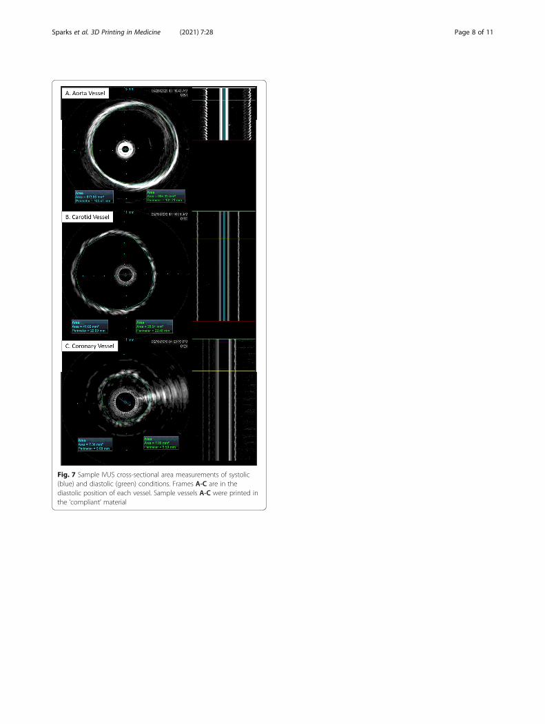
Fig. 7 Sample IVUS cross-sectional area measurements of systolic(blue) and diastolic (green) conditions. Frames A-C are in thediastolic position of each vessel. Sample vessels A-C were printed inthe ‘compliant’ material
Sparks et al. 3D Printing in Medicine (2021) 7:28 Page 8 of 11
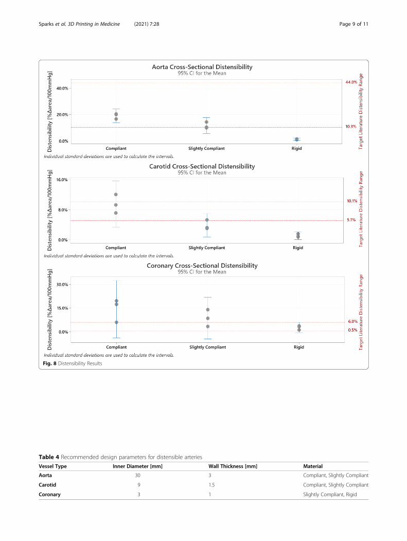
Fig. 8 Distensibility Results
Table 4 Recommended design parameters for distensible arteries
Vessel Type Inner Diameter [mm] Wall Thickness [mm] Material
Aorta 30 3 Compliant, Slightly Compliant
Carotid 9 1.5 Compliant, Slightly Compliant
Coronary 3 1 Slightly Compliant, Rigid
Sparks et al. 3D Printing in Medicine (2021) 7:28 Page 9 of 11
Abbreviations3DP: 3D Printing; GUI: Graphic User Interface; IVUS: Intravascular Ultrasound;MAP: Mean Arterial Pressure; STL: STereoLithography
AcknowledgementsAcknowledgement to Joe Affronte for providing guidance and instructionon using the IVUS and pressure system.
Authors' contributionsTest samples were prepared by AS and CS. AS and CS executed testingperformed data collection. IVUS measurements were performed by CS.Results were analyzed by CS, AS, RD, and MS. AA, JS, KM and MWcontributed to writing the manuscript. All authors read and approved thefinal manuscript.
FundingThis study was partially supported by the Jacobs Institute and the New YorkEmpire State Development Grant.
Availability of data and materialsThe data that support the findings of this study are available from TheJacobs Institute, but restrictions apply to the availability of these data, whichwere used under license for the current study, and so are not publiclyavailable. Data are however available from the authors upon reasonablerequest and with permission of The Jacobs Institute.
Declarations
Ethics approval and consent to participateNot applicable.
Consent for publicationNot applicable.
Competing interestsThe authors declare that they have no competing interests.
Author details1The Jacobs Institute, Buffalo, New York, USA. 2Department of Neurosurgery,University at Buffalo, State University of New York, 100 High Street, Suite B4,Buffalo, NY 14203, USA. 3Canon Stroke and Vascular Research Center,University at Buffalo, State University of New York, Buffalo, New York, USA.
Received: 14 September 2020 Accepted: 3 July 2021
References1. Benjamin Emelia J, Muntner P, Alonso A, et al. Heart disease and stroke
statistics—2019 update: a report from the American heart association.Circulation. 2019;139(10):e56-28. https://doi.org/10.1161/CIR.0000000000000659.
2. Lee S, Lee S, Kim S, et al. Fabrication and characterization of a magneticdrilling actuator for navigation in a three-dimensional phantom vascularnetwork. Sci Rep. 2018;8 https://doi.org/10.1038/s41598-018-22110-5.
3. Costa PF, Albers HJ, Linssen JEA, et al. Mimicking arterial thrombosis in a3D-printed microfluidic in vitro vascular model based on computedtomography angiography data. Lab on a chip. 2017;17(16):2785–92. https://doi.org/10.1039/c7lc00202e.
4. Meess K. Development of Additive Manufactured Abdominal AorticAneurysm Phantoms For Minimally Invasive Endovascular Image GuidedProcedures. Order No. 13885963 ed. State University of New York at Buffalo;2019. ProQuest, https://www.proquest.com/docview/2305528338?accountid=14169.
5. Kono K, Shintani A, Okada H, Terada T. Preoperative simulations ofendovascular treatment for a cerebral aneurysm using a patient-specificvascular silicone model. Neurol Med Chir. 2013;53(5):347–51. https://doi.org/10.2176/nmc.53.347.
6. McClure RS, Fedak PWM, Commentary. Using ex vivo modeling to validatetechnical innovations in cardiac surgery. J Thoracic Cardiovasc Surg. 2019;158(2):404–5. https://doi.org/10.1016/j.jtcvs.2019.02.010.
7. Shepard LM, Sommer KN, Angel E, et al. Initial evaluation of three-dimensionally printed patient-specific coronary phantoms for CT-FFRsoftware validation. J Med Imaging. 2019;6(2):021603. https://doi.org/10.1117/1.JMI.6.2.021603.
8. Chepelev L, Wake N, Ryan J, et al. Radiological Society of North America(RSNA) 3D printing Special Interest Group (SIG): guidelines for medical 3Dprinting and appropriateness for clinical scenarios. 3D Print Med. 2018;4(1):11. https://doi.org/10.1186/s41205-018-0030-y.
9. Tabaczynski J, Stoll T, Shepard L, et al. Use of patient specific 3D printed(3DP) neurovascular phantoms for mechanical assessment of devices usedin image guided minimally invasive procedures. Proc SPIE Int Soc Opt Eng.2018;10579:105790K. https://doi.org/10.1117/12.2293370.
10. Ionita CN, Mokin M, Varble N, et al. Challenges and limitations of patient-specific vascular phantom fabrication using 3D Polyjet printing. Proc SPIE IntSoc Opt Eng. 2014;13:90380M. https://doi.org/10.1117/12.2042266.
11. Tabaczynski J. Mechanical Assessment of 3D Printed Patient SpecificPhantoms for Simulation of Minimally Invasive Image Guided Procedures.Order No. 10823110 ed. State University of New York at Buffalo; 2018.ProQuest, https://www.proquest.com/docview/2057213064?accountid=14169.
12. Mogali SR, Yeong WY, Tan HKJ, et al. Evaluation by medical students of theeducational value of multi-material and multi-colored three-dimensionalprinted models of the upper limb for anatomical education. Anat Sci Educ.2018;11(1):54–64. https://doi.org/10.1002/ase.1703.
13. Nagesh SVS, Russ M, Ionita CN, Bednarek D, Rudin S. Use of patient specific3D printed neurovascular phantoms to evaluate the clinical utility of a highresolution x-ray imager. Proc SPIE Int Soc Opt Eng. 2017;10137. https://doi.org/10.1117/12.2254390.
14. Shepard LM, Sommer KN, Angel E, et al. CT investigation of patient-specificphantoms with coronary artery disease. Proc SPIE Int Soc Opt Eng. 2018;10573:105731V. https://doi.org/10.1117/12.2292918.
15. Mitsouras D, Lee TC, Liacouras P, et al. Three-dimensional printing of MRI‐visible phantoms and MR image‐guided therapy simulation. Magneticresonance in medicine. 2017;77(2):613–22.
16. Poulin E, Gardi L, Fenster A, Pouliot J, Beaulieu L. Towards real-time 3Dultrasound planning and personalized 3D printing for breast HDRbrachytherapy treatment. Radiother Oncol. 2015;114(3):335–8. https://doi.org/10.1016/j.radonc.2015.02.007.
17. Laurent S, Caviezel B, Beck L, et al. Carotid artery distensibility anddistending pressure in hypertensive humans. Hypertension. 1994;23(6 Pt 2):878–83. https://doi.org/10.1161/01.hyp.23.6.878.
18. Tobey D, Reynolds T, Kopchok G, Donayre C, Khoynezhad A, White R. InVivo Assessment of Ascending & Arch Aortic Compliance. Ann Vasc Surg.2018;08/01:270. https://doi.org/10.1016/j.avsg.2018.05.003.
19. Alfonso F, Macaya C, Goicolea J, et al. Determinants of coronary compliancein patients with coronary artery disease: an intravascular ultrasound study. JAm Coll Cardiol. 1994;23(4):879–84. https://doi.org/10.1016/0735-1097(94)90632-7.
20. Whincup PH, Gilg JA, Donald AE, et al. Arterial distensibility in adolescents -the influence of adiposity, the metabolic syndrome, and classic risk factors.Circulation. 2005;112(12):1789–97. https://doi.org/10.1161/CIRCULATIONAHA.104.532663.
21. Robinson M, Scheuermann-Freestone M, Leeson P, et al. Uncomplicatedobesity is associated with abnormal aortic function assessed bycardiovascular magnetic resonance. J Cardiovasc Magnet Reson. 2008;02/13:10. https://doi.org/10.1186/1532-429X-10-10.
22. Sommer K, Izzo RL, Shepard L, et al. Design optimization for accurate flowsimulations in 3d printed vascular phantoms derived from computedtomography angiography. Proc SPIE Int Soc Opt Eng. 2017;10138. https://doi.org/10.1117/12.2253711.
23. Russ M, apos, Hara R, et al. Treatment planning for image-guidedneuro-vascular interventions using patient-specific 3D printed phantoms.Proc SPIE Int Soc Opt Eng. 2015;9417:941726. https://doi.org/10.1117/12.2081997.
24. Vriz O, Aboyans V, Minisini R, et al. Reference values of one-point carotidstiffness parameters determined by carotid echo-tracking and brachial pulsepressure in a large population of healthy subjects. Hypertens Res. 2017;40(7):685–95. https://doi.org/10.1038/hr.2017.24.
25. Shaw JA, Kingwell BA, Walton AS, et al. Determinants of coronary arterycompliance in subjects with and without angiographic coronary artery disease. JAm Coll Cardiol. 2002;39(10):1637. https://doi.org/10.1016/S0735-1097(02)01842-9.
Sparks et al. 3D Printing in Medicine (2021) 7:28 Page 10 of 11

26. Meess KM, Izzo RL, Dryjski ML, et al. 3D printed abdominal aortic aneurysmphantom for image guided surgical planning with a patient specificfenestrated endovascular graft system. Proc SPIE Int Soc Opt Eng. 2017;10138:101380P-101380P – 14. https://doi.org/10.1117/12.2253902.
27. Allman AB, Shepard LM, Podgorsak AR, Rava RA, Ionita CN. Controlledcompliancy of 3D printed vascular patient specific phantoms. Proc SPIE IntSoc Opt Eng. 2019;10954:109540C. https://doi.org/10.1117/12.2512528.
28. Williams MJ, Stewart RA, Low CJ, Wilkins GT. Assessment of the mechanicalproperties of coronary arteries using intravascular ultrasound: an in vivostudy. Int J Card Imaging. 1999;15(4):287–94. https://doi.org/10.1023/a:1006279228534.
29. Peelukhana SV, Wang Y, Berwick Z, et al. Role of pulse pressure andgeometry of primary entry tear in acute type B dissection propagation. AnnBiomed Eng. 2017;45(3):592–603. https://doi.org/10.1007/s10439-016-1705-4.
30. Ostchega Y. Resting pulse rate reference data for children, adolescents, andadults: United States, 1999–2008 / by Yechiam Ostchega [and four others].National health statistics reports; number 41. U.S. Department of Health andHuman Services, Centers for Disease Control and Prevention, NationalCenter for Health Statistics; 2011.
Publisher’s NoteSpringer Nature remains neutral with regard to jurisdictional claims inpublished maps and institutional affiliations.
Sparks et al. 3D Printing in Medicine (2021) 7:28 Page 11 of 11