Complex Liver Resections for Colorectal...
6
Case Report Complex Liver Resections for Colorectal Metastases: Are They Safe in the Low-Volume, Resource-Poor Caribbean Setting? Shamir O. Cawich, Dexter A. W. Thomas, Chunilal Ramjit, Roderick Bhagan, and Vijay Naraynsingh Department of Clinical Surgical Sciences, University of the West Indies, St. Augustine Campus, St. Augustine, Trinidad and Tobago Correspondence should be addressed to Shamir O. Cawich; [email protected] Received 13 December 2014; Accepted 21 January 2015 Academic Editor: Gregorio Santori Copyright © 2015 Shamir O. Cawich et al. is is an open access article distributed under the Creative Commons Attribution License, which permits unrestricted use, distribution, and reproduction in any medium, provided the original work is properly cited. Introduction. Although many authorities suggest that major liver resections should only be carried out in high-volume specialized centres, many patients in the Caribbean do not have access to these health care systems. Presentation of a Case. A 50-year-old woman with a solitary colorectal metastasis invading the inferior vena cava underwent an extended leſt hepatectomy with caval resection and reconstruction. Several technical maneuvers were utilized that were suited to the resource-poor environment. Conclusion. We suggest that good outcomes can still be attained in the resource-poor, low-volume centres once dedicated and appropriately trained teams are available. 1. Introduction Over the past 2-3 decades, liver resections have become accepted as feasible and safe in dedicated high-volume cen- tres with service centralization [1]. However, in the Caribbean several geographic [2], political [3], and logistic [4] factors limit service centralization. In addition, surgeons in the Caribbean setting have to adapt and improvise to deliver care in these resource-poor health care systems [2–5]. Caribbean surgeons, therefore, treat relatively small numbers of patients without service centralization in resource-poor health care systems. Although some authorities have argued that major liver resections should not be carried out in these circumstances [1], most of these patients cannot afford care in developed countries with service centralization and well-supported health care systems. We argue that a high standard of care can still be delivered in these less than ideal circumstances once dedicated, trained health care teams perform these operations, even at low volumes. 2. Presentation of a Case A 50-year-old woman with no comorbidities presented 2 years aſter a sigmoid colectomy for colorectal carcinoma. Histology revealed moderately differentiated adenocarci- noma with 5/12 nodes positive for metastatic disease. Cir- cumferential and longitudinal margins were microscopically clear of disease. She completed adjuvant systemic therapy with Xeloda (capecitabine). During surveillance, metastatic liver disease was detected. ere was a single 2.5cm metastatic deposit in segment IVa that involved leſt hepatic vein (LHV) and middle hepatic vein (MHV) and encroached on the inferior vena cava (IVC) anteriorly (Figures 1 and 2). Second line systemic therapy with IROX (irinotecan and oxaliplatin) was commenced but resulted in only minimal tumour regression. Although there was still a suspicion of IVC involvement aſter preoperative systemic therapy (Figure 3), she was taken to the operating theatre for exploration. e abdomen was accessed through an upper midline incision with transverse extension. No metastatic deposits were noted in the peritoneal cavity. e anterior layers of the coronary ligaments were incised to expose the hepatocaval junction (Figure 4). e metastatic deposit was not visible on inspection of the liver surface but intraoperative ultrasound confirmed the solitary deposit at segment IVa involving the MHV and LHV. Ultrasound also confirmed that the RHV and Hindawi Publishing Corporation Case Reports in Surgery Volume 2015, Article ID 570968, 6 pages http://dx.doi.org/10.1155/2015/570968
Transcript of Complex Liver Resections for Colorectal...

Case ReportComplex Liver Resections for Colorectal Metastases: Are TheySafe in the Low-Volume, Resource-Poor Caribbean Setting?
Shamir O. Cawich, Dexter A. W. Thomas, Chunilal Ramjit,Roderick Bhagan, and Vijay Naraynsingh
Department of Clinical Surgical Sciences, University of the West Indies, St. Augustine Campus, St. Augustine, Trinidad and Tobago
Correspondence should be addressed to Shamir O. Cawich; [email protected]
Received 13 December 2014; Accepted 21 January 2015
Academic Editor: Gregorio Santori
Copyright © 2015 Shamir O. Cawich et al. This is an open access article distributed under the Creative Commons AttributionLicense, which permits unrestricted use, distribution, and reproduction in any medium, provided the original work is properlycited.
Introduction. Although many authorities suggest that major liver resections should only be carried out in high-volume specializedcentres,many patients in theCaribbean do not have access to these health care systems. Presentation of a Case. A 50-year-oldwomanwith a solitary colorectal metastasis invading the inferior vena cava underwent an extended left hepatectomy with caval resectionand reconstruction. Several technical maneuvers were utilized that were suited to the resource-poor environment. Conclusion. Wesuggest that good outcomes can still be attained in the resource-poor, low-volume centres once dedicated and appropriately trainedteams are available.
1. Introduction
Over the past 2-3 decades, liver resections have becomeaccepted as feasible and safe in dedicated high-volume cen-tres with service centralization [1]. However, in theCaribbeanseveral geographic [2], political [3], and logistic [4] factorslimit service centralization. In addition, surgeons in theCaribbean setting have to adapt and improvise to deliver carein these resource-poor health care systems [2–5]. Caribbeansurgeons, therefore, treat relatively small numbers of patientswithout service centralization in resource-poor health caresystems.
Although some authorities have argued that major liverresections should not be carried out in these circumstances[1], most of these patients cannot afford care in developedcountries with service centralization and well-supportedhealth care systems. We argue that a high standard of carecan still be delivered in these less than ideal circumstancesonce dedicated, trained health care teams perform theseoperations, even at low volumes.
2. Presentation of a Case
A 50-year-old woman with no comorbidities presented 2years after a sigmoid colectomy for colorectal carcinoma.
Histology revealed moderately differentiated adenocarci-noma with 5/12 nodes positive for metastatic disease. Cir-cumferential and longitudinal margins were microscopicallyclear of disease. She completed adjuvant systemic therapywith Xeloda (capecitabine).
During surveillance,metastatic liver diseasewas detected.There was a single 2.5 cm metastatic deposit in segment IVathat involved left hepatic vein (LHV) and middle hepaticvein (MHV) and encroached on the inferior vena cava (IVC)anteriorly (Figures 1 and 2). Second line systemic therapywith IROX (irinotecan and oxaliplatin) was commenced butresulted in only minimal tumour regression. Although therewas still a suspicion of IVC involvement after preoperativesystemic therapy (Figure 3), she was taken to the operatingtheatre for exploration.
The abdomen was accessed through an upper midlineincision with transverse extension. No metastatic depositswere noted in the peritoneal cavity. The anterior layers of thecoronary ligaments were incised to expose the hepatocavaljunction (Figure 4).Themetastatic deposit was not visible oninspection of the liver surface but intraoperative ultrasoundconfirmed the solitary deposit at segment IVa involving theMHVandLHV.Ultrasound also confirmed that the RHVand
Hindawi Publishing CorporationCase Reports in SurgeryVolume 2015, Article ID 570968, 6 pageshttp://dx.doi.org/10.1155/2015/570968

2 Case Reports in Surgery
Figure 1: Axial CT images showing the IVC (V) receiving the LHV,MHV, and RHV. The solitary metastatic deposit (∗) is seen wedgedbetween MHV and LHV and encroaches on the IVC. On axialcuts, the metastatic deposit is juxtaposed to, but does not appear to,involve the RHV.
Figure 2: Coronal and sagittal reconstructed CT images showingthe IVC (V) receiving theMHV.The solitarymetastatic deposit (∗) isseen in segment IVa at the junction of the hepatic-caval confluence.
hepatocaval junctionwere uninvolved and the right hemiliverwas free of metastases.
In preparation for a hangingmaneuver, a curved vascularclamp was used for cephalad dissection between the MHVand RHV (Figure 4). Caudal dissection then followed overthe avascular plane on the anterior IVC surface (Figure 5),preserving the inferior RHV. A large nasogastric tube wasused to develop the plane and connect the planes frombelow. Traction on the nasogastric tube passed between RHVand MHV allowed us to perform the hanging manoeuvre(Figure 6).
The left coronary and triangular ligaments were theninterrupted to expose the tumour posteriorly. A caudatelobectomy was performed because tumour extended intothis lobe. The tumour also invaded the IVC at the hepa-tocaval junction on the left side. At this point, the hilarstructures were controlled and divided in preparation forparenchymal transection. Anterior parenchymal transectionwas performed, skeletonizing and preserving the RHV at itsentry into the IVC (Figure 7). This allowed the metastaticdeposit to be approached from the right. In order to achieve
Figure 3: Coronal CT images marked with yellow arrows that pointto the area of involvement of the retrohepatic IVC at the superiorborder of the liver. This area corresponds to the metastatic depositon the axial images in Figure 1.
a R0 resection, the IVC was temporarily clamped and a 2 cmsegment of IVC was resected and reconstructed longitudi-nally (Figure 8). After reconstruction, there was narrowing ofthe IVC to about 70% of its normal diameter. Intraoperativeblood loss was 650mLs and no transfusion was required.Thepatient spent 36 hours in the intensive care unit and had anuneventful postoperative course. She was discharged fromhospital at day 7 after the operation.
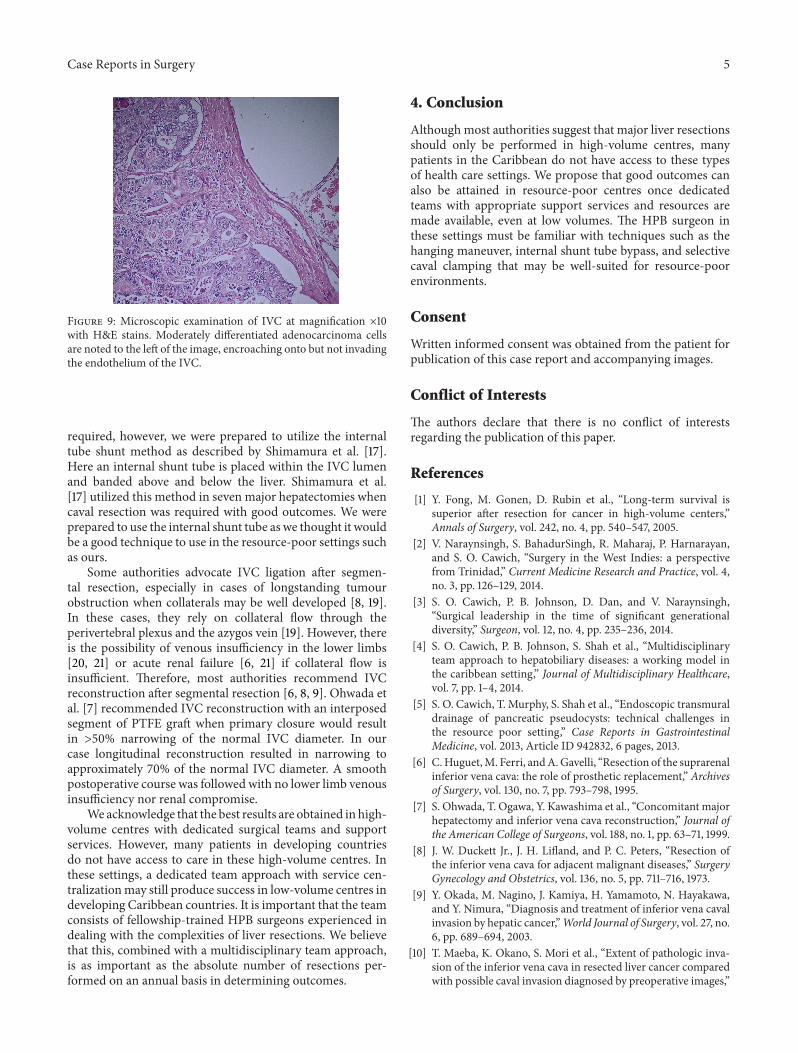
Pathologic examination of the specimen confirmed thepresence of a 2.5 cm tumour deposit within the liver at thejunction of LHV and MHV encroaching onto the IVC. Onmicroscopy the moderately differentiated adenocarcinomacells were noted to invade the IVC but had not yet penetratedthe endothelium (Figure 9). All resection margins were clearwith minimummargins of 0.5mm at the IVC wall.
3. Discussion
It is now well accepted that major liver resections canbe performed with acceptable morbidity and mortality inhigh-volume centers with dedicated hepatopancreatobiliary(HPB) teams and specialized supportive services [1]. In thesesettings, HPB surgeons have become quite aggressive in theirpursuit of R0 resections, even in the face of tumour involvingmajor structures such as the IVC.
Although major hepatectomy combined with IVC resec-tion and reconstruction has now become feasible as apotentially curative resection for hepatic malignancy [6–10], most authorities suggest that these complex proceduresshould only be performed in high-volume centres [1]. In theCaribbean, however, this is not possible. Although there areapproximately 6,500,000 persons living in the AnglophoneCaribbean, they are scattered across many small island stateseach with small individual populations and few practitioners[4]. With three HPB surgical teams, Trinidad and Tobagohas the largest HPB surgical service in the English speakingCaribbean.The HPB surgery service at Port of Spain GeneralHospital performs an average of 10 major liver resectionseach year, not qualifying as a high-volume service (>25resections annually) according to international definitions

Case Reports in Surgery 3
(a) (b)
Figure 4:The hepatocaval junction is exposed in (a), allowing dissection of the space betweenMHV and RHVwith a curved vascular clampin (b).
Figure 5: Segments III (S3) and IVb (S4b) of the liver are elevated toallow access to the IVC caudally. This allows access to the avascularplane on the IVC surface. A large vascular clamp bluntly dissects theavascular space in a cephalad direction as indicated by the brokenyellow arrow. An inferior right hepatic vein has been identified andpreserved.
Figure 6: A nasogastric tube is used to execute the hangingmaneuver to facilitate anterior parenchymal transection right downon to the IVC at the junction between RHV and MHV identified tobe tumour-free on intraoperative ultrasound.
[1]. Nevertheless, we have overcome this challenge by creatinga dedicated specialist unit to treat HPB diseases with a mul-tidisciplinary team approach [4]. The HPB team, staffed byone fellowship-trained HPB surgeon and two surgical houseofficers, has access to a general operating room with sharedequipment and postoperative support from a multipurposeICU and two gastroenterologists.
Although we performed IVC resection to achieve clearmargins, histologic examination revealed that tumour didnot penetrate the IVC endothelium. It is well recognizedthat IVC invasion is difficult to predict in preoperativeimaging because this is a low-pressure vessel that can easilybe compressed, especially by a large metastatic deposit [9,10]. The metastatic deposit in this case was only 2.5 cm indiameter and so we maintained a suspicion that this mayhave been due to invasion rather than compression. However,the absence of histologic IVC invasion reinforces the factthat it is difficult to distinguish invasion and compression onpreoperative imaging.
Maeba et al. [10] reported that 40% of patients withconfirmed IVC invasion on preoperative imaging (includingCT, MRI, and cavography) actually had histologic evidenceof tumor invading the IVC. Despite using standard radio-logic criteria (longitudinal compression > 50mm, transversecompression extending > 50% of the circumference, lesionsprotruding into the IVC lumen, or the presence of welldeveloped collaterals), it was difficult for them to accuratelypredict IVC invasion on imaging [10]. Transesophagealendoscopic ultrasonographymay assist in distinguishing IVCinvasion versus compression [11], but it was unavailable in ourresource-poor setting.
In this case, we used the anterior parenchymal transectiontechnique popularized by surgeons in Hong Kong [12–14].Only by using this anterior transection technique werewe able to approach the hepatocaval junction to achieve

4 Case Reports in Surgery
(a) (b)
Figure 7: (a)The parenchyma was transected right down to the IVC and allowed the RHV to be preserved in order to maintain outflow fromthe future liver remnant.
(a) (b)
Figure 8: Anterior parenchymal transection allowed the metastasis to be approached from the right side, confirming IVC involvement. Theleft liver is now completely mobilized except at the point at which the metastatic deposit invades the cava. In (b) the left liver was completelyexcised and the IVC reconstructed (arrow).
control from both sides of the tumour, thereby completinga safe controlled resection of the IVC with minimal bloodloss. There are few disadvantages to anterior transection,including uncontrollable bleeding from vessels deep in theparenchyma and difficulty elevating the liver for effectivemanual compression. To circumvent these, we employed thehangingmaneuver.The original hangingmaneuver describedby Belghiti et al. [15] in 2001 involved the passage of atape in the avascular plane along the anterior surface of theIVC to suspend the liver. This serves to facilitate anteriorparenchymal transection, bringing better control of bleeding
from vessels deep within the parenhyma and a lower risk oftumour rupture [16]. In addition it guided the direction ofanterior transection [16] that was indispensible in this casewith metastatic deposits so close to the RHV.
To avoid the resultant haemodynamic consequences dur-ing IVC resection, many authorities advocate venovenousbypass [11, 17, 18]. An intraoperative decision was taken toproceedwithout bypass since selective clamping of the IVC asdescribed by Togo et al. [11] allowedmaintained flow throughthe native IVC. This maintained flow through both hepaticand systemic circulations [11]. If venovenous bypass was
Case Reports in Surgery 5
Figure 9: Microscopic examination of IVC at magnification ×10with H&E stains. Moderately differentiated adenocarcinoma cellsare noted to the left of the image, encroaching onto but not invadingthe endothelium of the IVC.
required, however, we were prepared to utilize the internaltube shunt method as described by Shimamura et al. [17].Here an internal shunt tube is placed within the IVC lumenand banded above and below the liver. Shimamura et al.[17] utilized this method in seven major hepatectomies whencaval resection was required with good outcomes. We wereprepared to use the internal shunt tube as we thought it wouldbe a good technique to use in the resource-poor settings suchas ours.
Some authorities advocate IVC ligation after segmen-tal resection, especially in cases of longstanding tumourobstruction when collaterals may be well developed [8, 19].In these cases, they rely on collateral flow through theperivertebral plexus and the azygos vein [19]. However, thereis the possibility of venous insufficiency in the lower limbs[20, 21] or acute renal failure [6, 21] if collateral flow isinsufficient. Therefore, most authorities recommend IVCreconstruction after segmental resection [6, 8, 9]. Ohwada etal. [7] recommended IVC reconstruction with an interposedsegment of PTFE graft when primary closure would resultin >50% narrowing of the normal IVC diameter. In ourcase longitudinal reconstruction resulted in narrowing toapproximately 70% of the normal IVC diameter. A smoothpostoperative course was followed with no lower limb venousinsufficiency nor renal compromise.
We acknowledge that the best results are obtained in high-volume centres with dedicated surgical teams and supportservices. However, many patients in developing countriesdo not have access to care in these high-volume centres. Inthese settings, a dedicated team approach with service cen-tralizationmay still produce success in low-volume centres indeveloping Caribbean countries. It is important that the teamconsists of fellowship-trained HPB surgeons experienced indealing with the complexities of liver resections. We believethat this, combined with a multidisciplinary team approach,is as important as the absolute number of resections per-formed on an annual basis in determining outcomes.
4. Conclusion
Althoughmost authorities suggest that major liver resectionsshould only be performed in high-volume centres, manypatients in the Caribbean do not have access to these typesof health care settings. We propose that good outcomes canalso be attained in resource-poor centres once dedicatedteams with appropriate support services and resources aremade available, even at low volumes. The HPB surgeon inthese settings must be familiar with techniques such as thehanging maneuver, internal shunt tube bypass, and selectivecaval clamping that may be well-suited for resource-poorenvironments.
Consent
Written informed consent was obtained from the patient forpublication of this case report and accompanying images.
Conflict of Interests
The authors declare that there is no conflict of interestsregarding the publication of this paper.
References
[1] Y. Fong, M. Gonen, D. Rubin et al., “Long-term survival issuperior after resection for cancer in high-volume centers,”Annals of Surgery, vol. 242, no. 4, pp. 540–547, 2005.
[2] V. Naraynsingh, S. BahadurSingh, R. Maharaj, P. Harnarayan,and S. O. Cawich, “Surgery in the West Indies: a perspectivefrom Trinidad,” Current Medicine Research and Practice, vol. 4,no. 3, pp. 126–129, 2014.
[3] S. O. Cawich, P. B. Johnson, D. Dan, and V. Naraynsingh,“Surgical leadership in the time of significant generationaldiversity,” Surgeon, vol. 12, no. 4, pp. 235–236, 2014.
[4] S. O. Cawich, P. B. Johnson, S. Shah et al., “Multidisciplinaryteam approach to hepatobiliary diseases: a working model inthe caribbean setting,” Journal of Multidisciplinary Healthcare,vol. 7, pp. 1–4, 2014.
[5] S. O. Cawich, T. Murphy, S. Shah et al., “Endoscopic transmuraldrainage of pancreatic pseudocysts: technical challenges inthe resource poor setting,” Case Reports in GastrointestinalMedicine, vol. 2013, Article ID 942832, 6 pages, 2013.
[6] C.Huguet,M. Ferri, andA.Gavelli, “Resection of the suprarenalinferior vena cava: the role of prosthetic replacement,” Archivesof Surgery, vol. 130, no. 7, pp. 793–798, 1995.
[7] S. Ohwada, T. Ogawa, Y. Kawashima et al., “Concomitant majorhepatectomy and inferior vena cava reconstruction,” Journal ofthe American College of Surgeons, vol. 188, no. 1, pp. 63–71, 1999.
[8] J. W. Duckett Jr., J. H. Lifland, and P. C. Peters, “Resection ofthe inferior vena cava for adjacent malignant diseases,” SurgeryGynecology and Obstetrics, vol. 136, no. 5, pp. 711–716, 1973.
[9] Y. Okada, M. Nagino, J. Kamiya, H. Yamamoto, N. Hayakawa,and Y. Nimura, “Diagnosis and treatment of inferior vena cavalinvasion by hepatic cancer,”World Journal of Surgery, vol. 27, no.6, pp. 689–694, 2003.
[10] T. Maeba, K. Okano, S. Mori et al., “Extent of pathologic inva-sion of the inferior vena cava in resected liver cancer comparedwith possible caval invasion diagnosed by preoperative images,”

6 Case Reports in Surgery
Journal of Hepato-Biliary-Pancreatic Surgery, vol. 7, no. 3, pp.299–305, 2000.
[11] S. Togo, H. Shimada, K. Tanaka et al., “Management of malig-nant tumor with intracaval extension by selective clamping ofIVC,” Hepato-Gastroenterology, vol. 43, no. 11, pp. 1165–1171,1996.
[12] C.-L. Liu, S.-T. Fan, C.-M. Lo, R. T.-P. Poon, and J. Wong,“Anterior approach for major right hepatic resection for largehepatocellular carcinoma,” Annals of Surgery, vol. 232, no. 1, pp.25–31, 2000.
[13] E. C. S. Lai, S.-T. Fan, C.-M. Lo, K.-M. Chu, and C.-L. Liu,“Anterior approach for difficultmajor right hepatectomy,”WorldJournal of Surgery, vol. 20, no. 3, pp. 314–318, 1996.
[14] C. L. Liu, S. T. Fan, S. T. Cheung, C. M. Lo, I. O. Ng, andJ. Wong, “Anterior approach versus conventional approachright hepatic resection for large hepatocellular carcinoma: aprospective randomized controlled study,” Annals of Surgery,vol. 244, no. 2, pp. 194–203, 2006.
[15] J. Belghiti, O. A. Guevara, R. Noun, P. F. Saldinger, and R.Kianmanesh, “Liver hanging maneuver: a safe approach toright hepatectomy without liver mobilization,” Journal of theAmerican College of Surgeons, vol. 193, no. 1, pp. 109–111, 2001.
[16] G. Liddo, E. Buc, G. Nagarajan, M. Hidaka, S. Dokmak, and J.Belghiti, “The liver hanging manoeuvre,”HPB, vol. 11, no. 4, pp.296–305, 2009.
[17] Y. Shimamura, P. Gunven, M. Ishii, and H. Hasegawa, “Juxta-caval liver resectionswith the use of an internal IVC shunt tube,”HPB Surgery, vol. 2, no. 2, pp. 121–127, 1990.
[18] T. Kin, Y. Nakajima, H. Kanehiro et al., “Comparison ofhemodynamic changes in two veno-venous bypass techniquesmodified at the portal cannulation site,” Journal of Hepato-Biliary-Pancreatic Surgery, vol. 5, no. 1, pp. 93–96, 1998.
[19] R. Miyata, M. Shimazu, S. Kawachi et al., “Left trisegmen-tectomy and combined resection of the inferior vena cava,without reconstruction, for giant cystadenocarcinoma of theliver,” Journal of Hepato-Biliary-Pancreatic Surgery, vol. 12, no.3, pp. 272–276, 2005.
[20] C. Huguet and A. Gavelli, “Total vascular exclusion for liverresection,” Hepato-Gastroenterology, vol. 45, no. 20, pp. 368–369, 1998.
[21] Y. Yamaoka, K. Ozawa, K. Kumada et al., “Total vascularexclusion for hepatic resection in cirrhotic patients: applicationof venovenous bypass,” Archives of Surgery, vol. 127, no. 3, pp.276–280, 1992.