Complete Head Neck Exam 2016 Mercado - entpa.org ENT for... · Complete Head & Neck Exam Learning...
25
1/7/2016 1 Complete Head & Neck Exam Jose C. Mercado, PA‐C, MMS, DFAAPA [email protected] Orlando, FL March 30‐April 2, 2016 Complete Head & Neck Exam Financial Disclosure – no financial relationship related to this lecture. Sixth Annual ENT for the PA‐C |March 30‐April 2, 2016 | Orlando, FL Diagnosis @ a Glance Mercado 2011© Mercado 2011© Mercado 2011© Mercado 2011© Mercado 2011© Mercado 2014©
Transcript of Complete Head Neck Exam 2016 Mercado - entpa.org ENT for... · Complete Head & Neck Exam Learning...

1/7/2016
1
Complete Head & Neck ExamJose C. Mercado, PA‐C, MMS, DFAAPA
Orlando, FLMarch 30‐April 2, 2016
Complete Head & Neck Exam
Financial Disclosure – no financial relationship related to this lecture.
Sixth Annual ENT for the PA‐C |March 30‐April 2, 2016 | Orlando, FL
Diagnosis @ a Glance
Mercado 2011© Mercado 2011© Mercado 2011©
Mercado 2011© Mercado 2011©Mercado 2014©
1/7/2016
2
Complete Head & Neck ExamLearning Objectives
• Use of proper equipment• Discuss efficient and thorough exam
– Systematic approach to H&N exam
• Anatomy ‐ Visual reference normal versus normal variants versus abnormal physical findings– Head– Ears*– Nose (Sinuses)*– Mouth*– Neck* (Inflammatory, Neoplastic, & Congenital)
• Lymph nodes• Thyroid gland*• Neoplasms*
• Exam pearls / general considerations
*Foundation for additional lectures covered separately in program
Additional Resources
The ENT Exam Video Series℠ depicts how to perform a thorough examination of the ear, oral cavity, face, nose, neck, nasopharnyx, and larynx. Images and video of normal anatomy, normal variances, and common abnormalities have been added to enhance the learning experience.
Episode 1: The Ear ExamEpisode 2: The Oral Cavity and Neck Exam Episode 3: The Face and Nose ExamEpisode 4: The Nasopharynx and Larynx Exam
http://www.entnet.org/EducationAndResearch/The‐ENT‐EXAM.cfm
Examination of the Head and Neck
• The head and neck exam is not a single, fixed sequence. There are infinite approaches and sequences. Find which works best for you!
• Repetitive, sequential and systematic approach is best to avoid missing a diagnosis!
• Don’t be afraid/embarrassed to ask patient about abnormalities.
• Different portions are included depending on the examiner and the situation.
• Neurological exam will not be discussed.• AAO-HNS Video Sequential Demonstration

1/7/2016
3
Equipment Needed• An Otoscope
• Nasal Speculum• Tongue Blades
• Cotton TippedApplicators• Guaze
Mercado 2011 ®
Equipment Needed• Examination Gloves (latex/synthetic)
• Bright light (Head light / mirror)
• Lacrimal Probe Dilator
• Ruler
• Fiberoptic endoscope
Mercado 2011©
History
• A quick word about history….head & neck abnormalities may present at any age and the differential diagnosis is broad for both benign and malignant processes.
• Metastatic disease to the cervical lymph nodes is the most common type of cancer with 85% arising from an upper aerodigestive tract primary, 10% from infraclavicular tumors and 5% are unknown primaries.
• Key information like age, location, duration, personal habits and contributing factors are all an important part of a thorough head & neck exam because they let the examiner focus on areas of concern.
Pain, infection, prior cancer, exposure to TB and animals

1/7/2016
4
History• Duration
– Days - <7 days = inflammatory– Years - >7 years = congenital– Weeks - Months = malignant
• Contributing Factors– Age– Hoarseness of Voice– Weight loss– Hemoptysis– Dysphagia / Odynophagia– Respiratory distress / Shortness of Breath– Halitosis
• Pain– Otalgia; very important sign, the vagus nerve has cutaneous
intervention in ear and pharynx,– Odynophagia. Pain on swallowing may be a sign of possible throat
cancer from lateral pharyngeal wall and tonsillar region.
Head & NeckAnatomy
Nasopharynx
Oropharynx
Hypopharynx
Examination of Head
• Look for scars, lumps, rashes, hair loss, or other lesions. Measure all lesions!
• Look for facial asymmetry, involuntary movements, or edema.
• Palpate to identify anyareas of tenderness ordeformity.
• Again, don’t be afraid or embarrassed to touch patient and ask aboutabnormalities. Mercado 2011©
1/7/2016
5
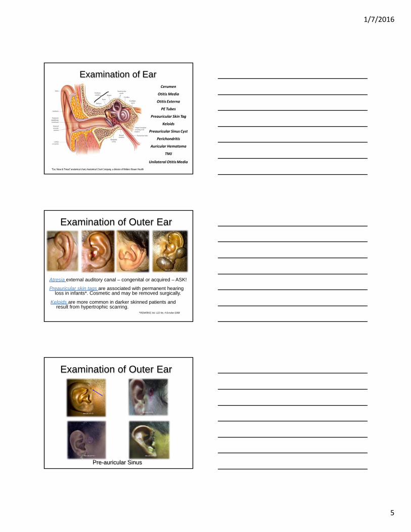
Examination of Ear
“Ear, Nose & Throat” anatomical chart, Anatomical Chart Company, a division of Wolters Kluwer Health
Cerumen
Otitis Media
Otitis Externa
PE Tubes
Preauricular Skin Tag
Keloids
Preauricular Sinus Cyst
Perichondritis
Auricular Hematoma
TMJ
Unilateral OtitisMedia
Examination of Outer Ear
Mercado 2011©Mercado 2011© Mercado 2011© Mercado 2011©
Atresia external auditory canal – congenital or acquired – ASK!
Preauricular skin tags are associated with permanent hearing loss in infants*. Cosmetic and may be removed surgically.
Keloids are more common in darker skinned patients and result from hypertrophic scarring.
*PEDIATRICS Vol. 122 No. 4 October2008
Pre-auricular Sinus
Mercado 2011©
Mercado 2011©Mercado 2011©
Mercado 2011©
Examination of Outer Ear

1/7/2016
6
Examination of Outer EarPerichondritis – an infection of the skin
and tissue surrounding the cartilage of the outer ear. Usually painful & sometimes caused by trauma– MCC Pseudomonas– TX Cephalosporin, or
Fluroquinolone (I&D only iffluctuance) Mercado 2011®
Mercado 2011®
Auricular Hematoma- associated with trauma. (cauliflower ear)MCC Staph. Aureus, Pseudomonas TX I/D, Cephalosporin, or QuinoloneTrauma to ear lobe when an earring is torn free
Examination of Outer Ear
Symptoms may include:Ear pain Jaw popping/ clicking
Locking of the jaw or Trismus
Temple/cheek pain worse chewing
Frequent head/neck aches
Treatment aimed at breaking the cycle of inflammation.Nonsteroidal anti-inflamatories (NSAID)Mild muscle relaxantsDecreased mastication – avoid gum chewing.Warm compress to jaw joint Relaxation and stress reduction
Temporal Mandibular Joint Dysfunction (TMJ) inflammation of the jaw joint that may present as an “ear ache” or “headache”. This is a cycle of inflammation that causes referred pain to the ear and head. Pain is reproducible with mouth opening and physical exam is otherwise normal without evidence of infection.
Mercado 2011©
Examination of Middle EarInspect and palpate the auricles and
mastoid process for tendernessand deformity.
Pull the ear upwards and backwards to straighten the canal before inserting otoscope.
Insert the otoscope to a point just beyond the protective hairs in the ear canal. Use the largest speculum that will fit comfortably.
Anchor otoscope - hold the otoscope with your thumb and fingers so that your handmakes contact with the patient. Mercado 2011©
1/7/2016
7
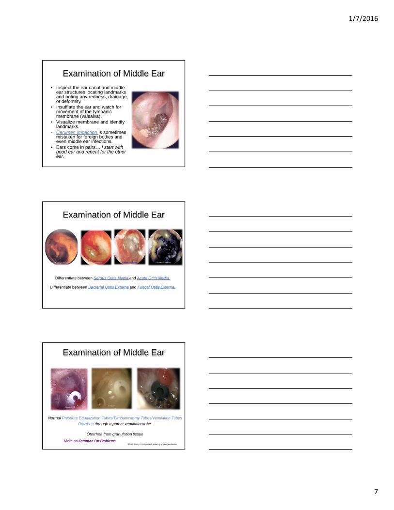
Examination of Middle Ear
• Inspect the ear canal and middleear structures locating landmarks and noting any redness, drainage, or deformity.
• Insufflate the ear and watch for movement of the tympanic membrane (valsalva).
• Visualize membrane and identify landmarks.
• Cerumen impaction is sometimes mistaken for foreign bodies and even middle ear infections.
• Ears come in pairs… I start with good ear and repeat for the other ear.
Mercado 2011©
Examination of Middle Ear
Color Atlas of Ear DiseaseA Color Atlas of E.N.T. Diagnosis Color Atlas of Ear DiseaseColor Atlas of Ear Disease
Differentiate between Serous Otitis Media and Acute Otitis Media.
Differentiate between Bacterial Otitis Externa and Fungal Otitis Externa.
Mercado 2011®
Examination of Middle Ear
F. Telischi*F. Telischi*
Normal Pressure Equalization Tubes/Tympanostomy Tubes/Ventilation Tubes
Otorrhea through a patent ventilation tube.
Otorrhea from granulation tissue
More on Common Ear Problems*Photo courtesy Dr. Fred Telischi, University of Miami, EarInstitute
1/7/2016
8
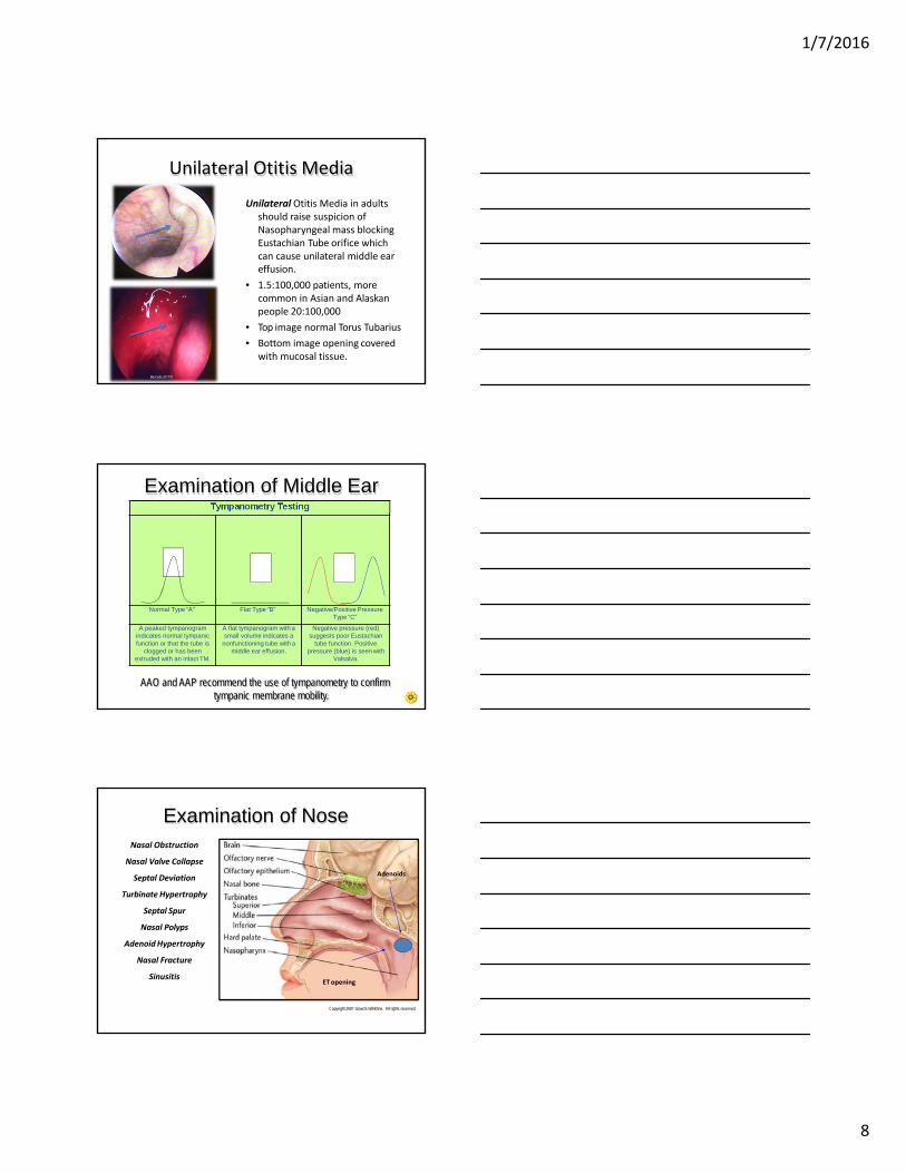
Unilateral Otitis Media
Unilateral Otitis Media in adults should raise suspicion of Nasopharyngeal mass blocking Eustachian Tube orifice which can cause unilateral middle eareffusion.
• 1.5:100,000 patients, more common in Asian and Alaskan people 20:100,000
• Top image normal Torus Tubarius
• Bottom image opening covered with mucosal tissue.
Mercado 2011©
Mercado 2011©
AAO and AAP recommend the use of tympanometry to confirm tympanic membrane mobility.
Tympanometry Testing
Normal Type “A” Flat Type “B” Negative/Positive Pressure Type “C”
A peaked tympanogram indicates normal tympanic function or that the tube is
clogged or has been extruded with an intact TM.
A flat tympanogram with a small volume indicates a
nonfunctioning tube with a middle ear effusion.
Negative pressure (red) suggests poor Eustachian
tube function. Positive pressure (blue) is seen with
Valsalva.
Examination of Middle Ear
Examination of Nose
Copyright 2001 GlaxoSmithKline. All rights reserved.
Adenoids
ETopening
Nasal Obstruction
Nasal Valve Collapse
Septal Deviation
Turbinate Hypertrophy
Septal Spur
Nasal Polyps
AdenoidHypertrophy
Nasal Fracture
Sinusitis

1/7/2016
9
Examination of Nose• Examine the nose using
bright light and nasal speculum.
• Tilt the patient's head back slightly. Ask them to hold their breath for the next few seconds.
• Insert the nasal speculum into the nostril, avoiding contact with the septum.
• Inspect the visible nasal structures and note any swelling, redness, drainage, or deformity.
• Repeat for the other side. Mercado 2011©
Dermatologic manifestations Saddle nose deformity secondary to large septal perforation
Examination of Nose
Mercado 2014©
Mercado 2014©
Examination of Nasal Airway• Nasal breathing can be
influenced by the nasal valve, septum and turbinates.
• The examination of the nasal valve should be performed duringinspiration.
• Nasal valve collapse occurs due to weakened nasal side walls.
• Septal Deviation, either congenital or acquired may also cause nasal obstruction.
Bilateral nasal valve collapse
Septal deviation

1/7/2016
10
Examination of Nasal Airway
Fiberoptic Nasal endoscopy is an invaluable method to evaluate the upper airway.
Diagnostic Nasal Endoscopy Can Be Performed with Rigid (0 or 30 degree) or Flexible Endoscopes
Turbinate hypertrophy
Turbinate Hypertrophy –congenital, inflammatory (chemical or environmental)
Septal Spur – congenital or trauma
Septal spur
Nasal Polyps are secondary to chronic mucosal inflammation. Distinguish from turbinates.– Treatment is aimed at eliminate cause, i.e, allergen/irritant.– Oral/Topical steriods– Surgical removal
Examination of Nasal Airway
Mercado 2011© Mercado 2014©
Examination of Nasal AirwayAdenoids are described
based on % of obstruction• No obstruction• Partial (percentage)
obstruction• Complete obstruction• Lateral X-Ray• Dental Mirror• Endoscopic view of the
nasopharynx showing adenoidal obstruction of the choana.
http://icarus.med.utoronto.ca/carr/atlas/atlasoutline.htm

1/7/2016
11
Sinus/Headaches• Diagnosis made by history and physical NOT diagnostic test.• Differential diagnosis depends on Location and Duration of symptoms.• Modifying/contributing factors.• Plain x-rays (waters views) good initial screening to confirm diagnosis.• CT Scan sinus coronal views– gold standard.• Culture & Sensitivity – only AFTER empiric therapy FAILS.• ENT – advantage of direct visualization via Fiberoptic endoscopy.
Mercado 2011© Pus OMC
More on Sinus Disease Panel
Examination of MouthOral Malocclusion
Gingivitis / Dental AbnormalitiesFissured Tongue
Ankyloglossia Geographic Tongue
Hairy Tongue Glossitis
Salivary Gland Disease Torus
RanulaOral Ulceration / Leukoplakia
PapilomaBifid Uvula
Cleft Lip/PalateTonsillar Hypertrophy Mercado 2011©
Examination of MouthUse a tongue blade and a bright light source to inspect inside of mouth.
Make special note of tonsil size, buccal folds, lateral tongue, floor of mouth and dentition.
If abnormalities are discovered, use a gloved finger to palpate the anterior structures and floor of the mouth.
Inspect the posterior oropharynx by depressing the tongue and asking the patient to say "Ah." Note any tonsilar enlargement, redness, or discharge.
Mercado 2011©Mercado 2011© Mercado 2011©

1/7/2016
12
Examination of Mouth• Palpate base of tongue.
– Tongue is anchored to floor of oral cavity posteriorly, and by frenulum anteriorly
– Dorsal surface covered by thick mucosa that supports the filiform papillae.
– Lingual tonsillar tissue• Pay special attention to the lateral boarders of the tongue.• Use a piece of gauze to hold tongue while you inspect
lateral boarders
Mercado 2011© Mercado 2011©Mercado 2011©
• Important to have patient remove dentures and visualize/palpate entire mouth.
Examination of Mouth
Mercado2014©Mercado 20114©
Examination of Mouth
Tonsillar Hypertrophy -Enlarged tonsils thatobstruct airway
• Gag reflex may make tonsils appear larger -
• Panting without use of a tongue blade can relax posterior pharynx and provide better visualization especially in children.
Mercado 2011©

1/7/2016
13
Examination of MouthTonsils are subjectively graded on a scale from 0 to 4,
4 being the largest, 0 - Not visible1- <25% of transverse oropharyngeal space2 - 25 to 49% of transverse oropharyngeal space3- 50 to 74% of transverse oropharyngeal space 4 - >75% of transverse oropharyngeal space
Mercado 2011©
Mercado 2011©
Mercado 2011©
Mercado 2011©
Examination of Mouth
Tonsillitis• Streptococcus pyogenes*
(group Abeta-hemolytic)• Streptococcus pneumoniae• M. catarrhalis• Staphylococcus aureus
* Most common pathogen
Asymmetry should raise suspicion of malignancy
Tonsillar concretions may cause halitosis
Mercado 2014©Mercado 2014©Mercado 2014©Mercado2014©
Mercado 2014©
Examination of Mouth
Post-Op Tonsillectomy
Normal grey and white eschar over tonsillar fossa (7-10 days).
Mercado 2011©
Mercado 2011©
Mercado 2011©
Mercado 2011©
1/7/2016
14
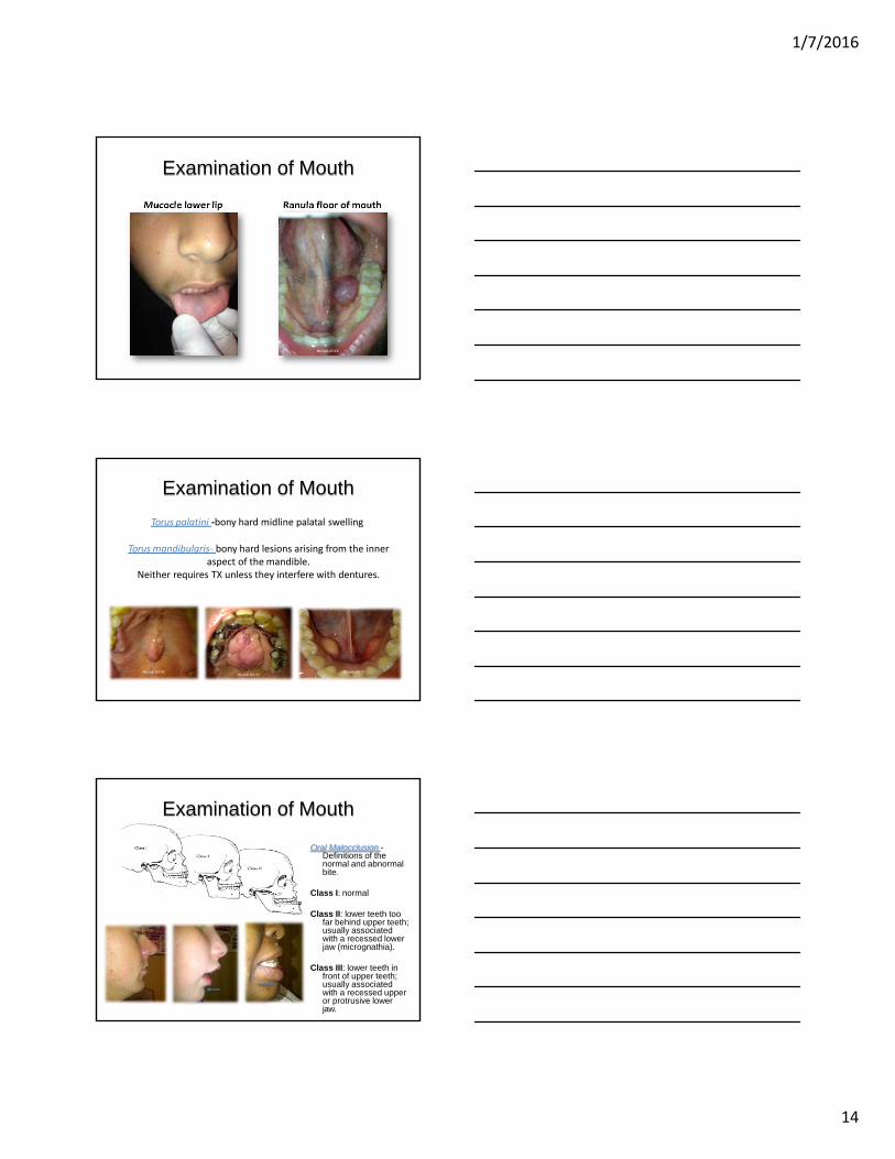
Mucocle lower lip Ranula floor of mouth
Examination of Mouth
Mercado 2014© Mercado 2014©
Examination of Mouth
Torus palatini ‐bony hard midline palatal swelling
Torus mandibularis‐ bony hard lesions arising from the inner aspect of the mandible.
Neither requires TX unless they interfere with dentures.
Mercado 2011©Mercado 2011©
Mercado 2011©
Examination of Mouth
Oral Malocclusion -Definitions of the normal and abnormal bite.
Class I: normal
Class II: lower teeth too far behind upper teeth; usually associated with a recessed lower jaw (micrognathia).
Class III: lower teeth in front of upper teeth; usually associated with a recessed upper or protrusive lower jaw.

1/7/2016
15
Examination of Teeth
Teeth and gums 32 adult teeth: 4 incisors, 2 canines, 4 premolars, 6 molars in each jaw.
Gingiva attach to the alveolar tissue and normally cover the root and neck of each tooth.
Gingivitis -Mild vs. Chronic. Remove dentures and partials
Color Atlas of Common Oral DiseasesvColor Atlas of Common OralDiseases
Examination of Teeth
MCC Staph. aureus, Strep. pyogenes TXRemoval of tooth, I & D Clindamycin orUnasyn IV
Mercado 2011© Mercado 2011©
Mercado 2011©
Mercado 2011©
Peridontal Disease/ Abscess
Cervical Cellulitis‐ Lateral neck edema secondary to dental abscess in lower molars
Ludwig’s Angina‐Midline “woody” tenderness
Examination of MouthThe incidence of bifid uvula in the general
population has been reported as 1 in 76 individuals
Consider submucous cleft palate. Total Adenoidectomy is contraindicated because of velopharyngeal insufficiency
Partial adenoidectomy recommended because leaves a midline mass of adenoid tissue for velopharyngeal closure (closing the nasopharyngeal space).
Over time, patients with submucous clefts who have not undergone adenoid removal, can develop hypernasality as the natural process of involution (atrophy) of the adenoid mass occurs. Where this does not occur over time, the palate has adapted to the gradual change in the architecture of the pharynx. By contrast, a dramatic change in the diameter of the pharynx with adenoidectomy does notpermit the shorter submucous cleft palate to adapt.
Mercado 2011©

1/7/2016
16
Mercado 2011 © Mercado 2011 © Mercado 2011©
Ankyloglossia (tongue ties) –Short lingual frenulum Interferes with speech when the tongue cannot be fully extended to the lower lip or touch the palate.
Short Labial Frenulum attaches to the center of the upper lip and between the upper two front teeth. This can cause a large gap and gum recession by pulling the gums off the bone. A labial frenectomy removes the labial frenulum.Orthodontic patients often have this procedure done to assist with closing a front tooth gap.
TX Frenulectomy or Lingual Lysis
Examination of Mouth
Examination of Mouth
Fissured tongue - benign condition in which tongue appears “cracked” Common in Down’s Syndrome No TX
Stomatitis – viral inflammation, chemo therapy agents, think connective tissue disorders – rheumatology evaluation.
Mercado 2011®Mercado 2011® Mercado 2012®
Examination of Mouth
• Benign inflammatory glossitis (Geographic Tongue) benign condition thought to be exacerbated by stress, Nutritional deficiency, or Heredity.
• Pattern CHANGES• No TX
Mercado 2011© Mercado 2011© Mercado 2011©
1/7/2016
17
Examination of Mouth
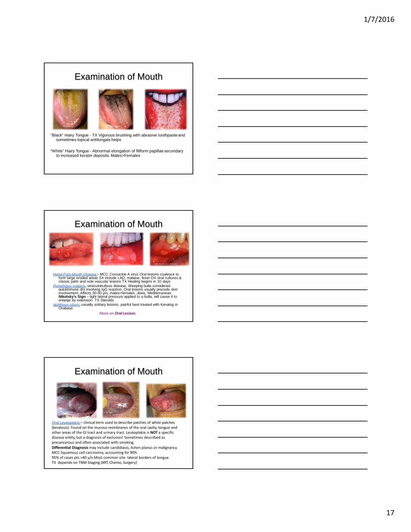
“Black” Hairy Tongue - TX Vigorous brushing with abrasive toothpasteand sometimes topical antifungals helps
“White” Hairy Tongue - Abnormal elongation of filiform papillaesecondary to increased keratin deposits. Males>Females
A ColoAr ACtolalosroAftEla.sNo.Tf.E.N.T.Mercado 2011© Mercado 2011©
Examination of Mouth
Color Atlas of Common OralDiseases Color Atlas of Common OralDiseasesColor Atlas of Common OralDiseases
Hand-Foot-Mouth Disease - MCC Coxsackie A virus Oral lesions coalesce to form large eroded areas SX include LAD, malaise, fever DX viral cultures & classic palm and sole vascular lesions TX Healing begins in 10 days
Pemphigus vulgaris -vesiculobullous disease, Weeping bulla considered autoimmune d/o involving IgG reaction, Oral lesions usually precede skin involvement, Affects 30-50 y/o, males=females ,Jews, Mediterranean.Nikolsky’s Sign – light lateral pressure applied to a bulla, will cause it to enlarge by extension. TX Steroids
Aphthous ulcers -usually solitary lesions, painful best treated with Kenalog in Orabase
More on Oral Lesions
Examination of Mouth
Oral Leukoplakia – clinical term used to describe patches of white patches (keratosis). Found on the mucous membranes of the oral cavity, tongue and other areas of the GI tract and urinary tract. Leukoplakia is NOT a specific disease entity, but a diagnosis of exclusion! Sometimes described as precancerous and often associated with smoking.Differential Diagnosis may include candidiasis, lichen planus or malignancy. MCC Squamous cell carcinoma, accounting for 90%95% of cases pts >40 y/o Most common site‐ lateral borders of tongue TX depends on TNM Staging (XRT, Chemo, Surgery)
Mercado 2011© Mercado 2011©Mercado 2011©

1/7/2016
18
Salivary Glands
25%
75%
50%
Examination of Salivary GlandsTwo paired salivary ducts enter the
oral cavity
– Wharton's ducts, from the submandibular glands, open on each side of the tongue's frenulum
– Stensen's ducts, from the parotid glands, open onto the buccal mucosa across from the second molar of the upper jaw.
– The sublingual gland drains through a number of smaller, not readily visible ducts (Ducts of Rivinus).
Mercado 2011®
Mercado 2011®
Examination of Salivary Glands
Sialadenitis - Inflammation of the salivary glands from nonspecific bacterial infection or blockedexcretory ducts. MCC Staph aureus. TX cephalosporin.Dilation, sialogoues
• The parotid is the most commonly affected gland with invasion of bacteria from the oral cavity.
Mercado 2011©
Mercado 2011©
More on Salivary GlandDisease
1/7/2016
19
Examination of Salivary Glands
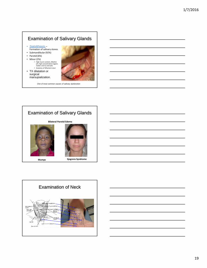
• Sialolithiasis –Formation of salivary stones
• Submandibular (92%)
• Parotid (6%)
• Minor (2%)• High mucin content, Alkaline
pH, high concentration organic matter and Ca and salts
• Anatomy of Wharton’s duct
• TX dilatation or surgical marsupialization.
One of most common causes of salivary dysfunction
Bilateral Parotid Edema
Examination of Salivary Glands
Mumps Sjogrens Syndrome
Mercado 2011©
Examination of Neck
Mercado 2011©

1/7/2016
20
Examination of Neck• Although frequently benign, a cervical mass is the
presenting symptom in 12 to 15% of patients with head & neck cancer.
• Majority of neck masses in patients over 40 years of age are malignant.
• Metastatic disease to the cervical lymph nodes is the most common type of cancer with 85% arising from an upper aerodigestive tract primary, 10% from infraclavicular tumors and 5% are unknown primaries.
• Differential diagnosis of neck abnormalities is based onlocation, duration and modifying factors and includeCongenital, Inflammatory, and Neoplastic etiologies
• Location– Triangles of Neck – anterior and posterior neck triangles.– Neck Level description of anatomic location level I-VII
Examination of NeckAnterior Triangle: Bordered by mandibles and
SCM• Anterior Cervical (both superficial and deep):
Nodes that lie both on top of and beneath the sternocleidomastoid muscles (SCM) on either side of the neck, from the angle of the jaw to the top of the clavicle. They can be easily identified by asking the patient to turn their head into your hand while you provide resistance.
• Drainage: The internal structures of the throat as well as part of the posterior pharynx, tonsils, and thyroid gland.
Posterior Triangle: Bordered by anterior margin of trapezius, posterior margin of the SCM, and superior margin of the clavicle.
• Posterior Cervical: Extend in a line posterior to the SCMs but in front of the trapezius, from the level of the mastoid bone to the clavicle.
• Drainage: The skin on the back of the head. Also frequently enlarged during upper respiratory infections (e.g. mononucleosis).
Anterior
Posterior
Mercado 2011©
Examination of Neck• Level I: Contains the nodes of the submental
and submandibular triangles, defined inferiorly by the diagastric muscles.
• Level II: Contains the upper jugular nodes from the base of skull to hyoid bone.
• Level III: Contains the middle jugular nodes from the hyoid bone to the inferior edge of the cricoid cartilage.
• Level IV: Contains the low jugular nodes from the cricoid cartilage to the clavicle.
• Level V: Contains the nodes of the posterior triangle that is bounded anteriorly by the sternocleidomastoid muscle and posteriorly by the trapezius.
• Level VI: Contains the nodes of the anteriorcentral compartment from the hyoid bone tothe manubrium with lateral boundaries beingthe carotid arteries
• Level VII: Contains the superior mediastinalnodes from the level of the superior edge ofthe manubrium to the innominate vein.

1/7/2016
21
Examination of Neck•••
Inspect the neck for asymmetry, scars, or other lesions. Identify antatomic landmarks.
Bimanual palpation is essential to evaluate floor of mouth, base of tongue and submandibular gland
Mercado 2011©Mercado 2011©
Inflammatory Neck MassesInflammatory neck masses usually present with erythema, induration, and tenderness.
Most common inflammatory neck lesion is lymphadenitis secondary to Staphyloccusor Streptococcus.
Cat Scratch Disease – MCC Bartonella. Localized skin lesions w/ LAD. TX macrolide (Biaxin).
Mononucleosis – MCC Epstein-Barr Virus. Generalized LAD, fatigue, fever, splenomegaly.
Scrofula - tuberculosis of the neck, or more precisely, a cervical tuberculouslymphadenopathy. Common in immunocompromised patients. About 95% of the scrofula cases in adults are caused by Mycobacterium tuberculosis
Sebaceous Cyst Insect Bite
Mercado 2011©Mercado 2011©
Scrofula
Mercado 2011©
Neoplastic Neck Masses
• Neoplastic lesions may be either primary or metastatic lesions.• Skin cancer and melanoma of scalp often metastasize to parotid.• Although the primary site for most tumors which metastasize to the
cervical lymph nodes are upper aerodigestive, tumors like testicular, lung, breast, and gastrointestinal may also metastasize to neck.
.
Mercado 2011© Mercado 2011©
More on Head & Neck Surgical Techniques
1/7/2016
22
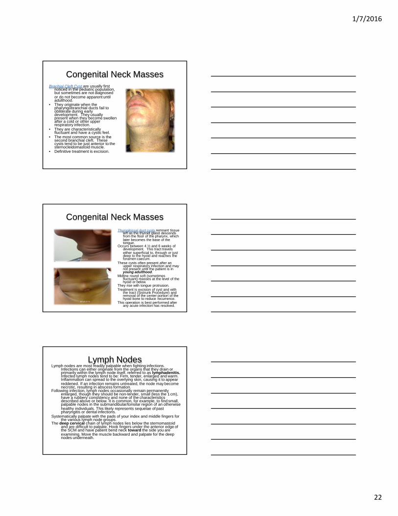
Congenital Neck MassesBrachial Cleft Cyst are usually first
noticed in the pediatric population,but sometimes are not diagnosedor do not become apparent until adulthood.
• They originate when the pharyngobranchial ducts fail to obliterate during early development. They usually present when they become swollen after a cold or other upper respiratory infection.
• They are characteristically fluctuant and have a cystic feel.
• The most common source is the second branchial cleft. These cysts tend to be just anterior to the sternocleidomastoid muscle.
• Definitive treatment is excision.Mercado 2011©
Congenital Neck MassesThyroglossal duct cysts remnant tissue
left as the thyroid gland descendsfrom the floor of the pharynx, whichlater becomes the base of the tongue.
Occurs between 4 ½ and 6 weeks ofdevelopment. This tract travelseither superficial to, through or just deep to the hyoid and reaches theforamen caecum.
These cysts often present after an upper respiratory infection and may not present until the patient is in young adulthood.
Midline round soft (sometimes fluctuant) masses at the level of the hyoid or below.
They rise with tongue protrusion.Treatment is excision of cyst and with
the tract (Sistrunk Procedure) and removal of the center portion of the hyoid bone to reduce recurrence.
This operation is best performed after any acute infection has resolved.
Mercado 2011©
Mercado 2011©
Lymph NodesLymph nodes are most readily palpable when fighting infections.
Infections can either originate from the organs that they drain orprimarily within the lymph node itself, referred to as lymphadenitis.Infected lymph nodes tend to be: Firm, tender, enlarged and warm.Inflammation can spread to the overlying skin, causing it to appearreddened. If an infection remains untreated, the node may become necrotic, resulting in abscess formation.
Following infection, lymph nodes occasionally remain permanentlyenlarged, though they should be non-tender, small (less the 1 cm),have a rubbery consistency and none of the characteristicsdescribed above or below. It is common, for example, to find small,palpable nodes in the submandibular/tonsilar region of an otherwisehealthy individuals. This likely represents sequelae of past pharyngitis or dental infections.
Systematically palpate with the pads of your index and middle fingers for the various lymph node groups.
The deep cervical chain of lymph nodes lies below the sternomastoidand are difficult to palpate. Hook fingers under the anterior edge ofthe SCM and have patient bend neck toward the side you areexamining. Move the muscle backward and palpate for the deep nodes underneath.

1/7/2016
23
Lymph Nodes
• Malignancies may also involve the lymph nodes, either primarily (e.g. lymphoma) or as a site of metastasis. In either case, these nodes are generally: Firm, non-tender, matted (i.e. stuck to each other), fixed (i.e. not freely mobile but rather stuck down to underlying tissue), and increase in size over time.
• Diffuse, bilateral involvement suggests a systemic malignancy (e.g. lymphoma) while those limited to a specific anatomic region are more likely associated with a local problem. Enlargement of nodes located only on the right side of the neck in the anterior cervical chain, for example, would be consistent with a squamous cell carcinoma, frequently associated with an intra-oral primary cancer.
• Knowledge of which nodes drain specific areas will help you search efficiently. Note the size and location of any palpable nodes and whether they were soft or hard, non-tender or tender, and mobile or fixed.
Lymph Nodes
Tonsilar
Preauricular
Postauricular
Occipital
Submandibular
Submental
AnteriorCervicalPosterior Cervical
Supraclavicular
1.Preauricular ‐ In front of the ear2. Postauricular ‐ Behind the ear3.Tonsillar ‐ At the angle of the jaw Drainage: The tonsilar and posterior pharyngeal regions.4. Occipital ‐ At the base of the skull5.Posterior Cervical side and back of neck above and behind SCM Drainage: The skin on the back of the head. Also frequently enlarged during upper respiratory infections (e.g. mononucleosis).6.Supraclavicular ‐ In the angle of the sternomastoid and the clavicle Drainage: Part of the throacic cavity, abdomen.7.Submental ‐ Under the jaw in the midline Drainage: The teeth and intra‐oral cavity.8.Submandibular ‐ Under the jaw on the side Drainage: The structures in the floor of the mouth.9.Anterior Cervical (both superficial and deep): Nodes that lie both on top of and beneath the SCM on either side of the neck, from the angle of the jaw to the top of the clavicle.
Mercado 2011©
Thyroid GlandThe thyroid gland is shaped like a two-inch bow tie and is
located in the front of the neck and below the larynx. It is the largest gland in the neck. Its main function is to secrete key hormones that regulate metabolism and other functions such as body heat and bone growth.
It is estimated that 13 million Americans have a disorder of the thyroid. Women are eight times more likely than men to develop a disorder.
Tumors in the thyroid gland are usually benign, but can still cause serious health problems. Benign or malignant, thyroid tumors are often best treated with surgery.Malignant tumors are usually curable when caught early.
. More on Head & Neck Surgical Techniques

1/7/2016
24
Thyroid Gland• Palpate from behind, Identify the cricoid cartilage with the fingers of both hands.• Move downward two or three tracheal rings while palpating for the isthmus, then
move laterally from the midline while palpating for the lobes of the thyroid.• When patient swallows, the gland is felt beneath your fingers while the larynx
rises and falls. Note the size, symmetry, and position of the lobes, as well as the presence of any nodules. The normal gland is often not palpable.
• If the gland feels firm, is it attached to the adjacent structures (i.e. fixed to underlying tissue.. consistent with malignancy) or freely mobile (i.e. moves up and down with swallowing)?
• If there is concern of malignancy, a careful lymph node exam (described above) is important as this is the most common site of spread.
Mercado 2011© Mercado 2011© Mercado 2011©
Summary Examination of the Head and Neck
• The head and neck exam is not a single, fixed sequence. There are infinite approaches and sequences. Find which works best for you!
• Repetitive, sequential and systematic approach is best to avoid missing a diagnosis!
• Don’t be afraid/embarrassed to ask patient about abnormalities.
• Complete all portions of exam before moving on.• Knowledge of anatomy and a good history will
narrow differential diagnosis.
Congenital Neoplastic
Malignant
Neoplastic
Benign
Inflammatory
Brachial Cleft Cyst
Dermoid Cyst
Thyroglossal Duct Cyst
Laryngocele
Ectopic Thyroid Tissue
Metastasis
Thyroid
Lymphoma
Salivary Gland Tumor
Hemangioma
Goiter
Lymphangioma
Salivary Gland Tumor
Lipoma
Lymphadenitis
Deep Neck Abscess
Cat Scratch Disease
TB/ Scrofula
Sarcoidosis
Sialadentitis
Differential Diagnosis Head & Neck
1/7/2016
25
Bibliography• Bull T.R., A Color Atlas of E.N.T. Diagnosis 2nd
Edition Hazel Books, England 1992• Langlais R.P., Miller C.S., Color Atlas of Common
Oral Diseases, Lea & Febiger, USA 1990• Fairbanks D.N., Pocket Guide to Antimicrobial
Therapy in Otolaryngology – Head and Neck Surgery 10th Edition, AAO-HNS, USA2001
• 2000 Clinical Indicators Compendium, AAO-HNS, 2000
• Barbara Bates' A Guide to Physical Examination and History Taking, Sixth Edition , published by Lippincott in 1995
• Chole RA, Forsen JW, Color Atlas of Ear Disease, 2nd Edition, BC Decker, 2002
Thank You
American Academy of Otolaryngology Head & Neck Surgery (AAO)www.entnet.org
Society of Physician Assistants in Otorhinolaryngology – Head & Neck Surgery (SPAO‐
HNS)Scholarship
www.entpa.org
No animals or models were harmed in the making of this presentation