Complementary imaging of the nuclear dynamics in laser-excited diatomic …€¦ · diatomic...
13
This content has been downloaded from IOPscience. Please scroll down to see the full text. Download details: This content was downloaded by: thummuwe IP Address: 129.130.106.65 This content was downloaded on 10/06/2014 at 19:29 Please note that terms and conditions apply. Complementary imaging of the nuclear dynamics in laser-excited diatomic molecular ions in the time and frequency domains View the table of contents for this issue, or go to the journal homepage for more 2014 J. Phys. B: At. Mol. Opt. Phys. 47 124003 (http://iopscience.iop.org/0953-4075/47/12/124003) Home Search Collections Journals About Contact us My IOPscience
Transcript of Complementary imaging of the nuclear dynamics in laser-excited diatomic …€¦ · diatomic...

This content has been downloaded from IOPscience. Please scroll down to see the full text.
Download details:
This content was downloaded by: thummuwe
IP Address: 129.130.106.65
This content was downloaded on 10/06/2014 at 19:29
Please note that terms and conditions apply.
Complementary imaging of the nuclear dynamics in laser-excited diatomic molecular ions in
the time and frequency domains
View the table of contents for this issue, or go to the journal homepage for more
2014 J. Phys. B: At. Mol. Opt. Phys. 47 124003
(http://iopscience.iop.org/0953-4075/47/12/124003)
Home Search Collections Journals About Contact us My IOPscience

Journal of Physics B: Atomic, Molecular and Optical Physics
J. Phys. B: At. Mol. Opt. Phys. 47 (2014) 124003 (12pp) doi:10.1088/0953-4075/47/12/124003
Review Article
Complementary imaging of the nucleardynamics in laser-excited diatomicmolecular ions in the time and frequencydomains
M Magrakvelidze1, A Kramer2, K Bartschat2 and U Thumm1
1 J.R. Macdonald Laboratory, Department of Physics, Kansas State University, Manhattan, KS 66506,USA2 Department of Physics and Astronomy, Drake University, Des Moines, IA 50311, USA
E-mail: [email protected]
Received 12 December 2013, revised 14 February 2014Accepted for publication 21 February 2014Published 10 June 2014
AbstractExperimentally, the bound and dissociative nuclear dynamics in small molecular ions can beresolved in time by using intense ultrashort pump in combination with delayed probe laserpulses. We discuss the modelling of related pump–probe-delay-dependent fragmentkinetic-energy-release (KER) spectra for the laser-induced dissociative ionization of selecteddiatomic molecules and show how the quantum-mechanical simulation of measured KERspectra—in both the time domain and as a function of the beat frequency between molecularvibrational levels—reveals dissociation pathways and the characteristics of initially occupiedmolecular potential curves.
Keywords: kinetic energy release, molecular dynamics, pump–probe, molecular ion, diatomicmolecule, quantum beat, strong field physics
(Some figures may appear in colour only in the online journal)
1. Introduction
Over the past decades, advances in laser technology ingenerating ultrashort and intense laser pulses have enabled thetime-resolved imaging of the nuclear wave-packet dynamicsof diatomic molecules by employing intense pump and probelaser pulses with pulse durations that are significantly shorterthan the molecular vibration period [1–5]. In particular, thedynamics of the wave-packet motion for processes suchas dissociation and Coulomb explosion are investigatedexperimentally and theoretically by analysing kinetic-energy-release (KER) spectra as functions of the adjustable pump–probe delay in both the smallest diatomic molecules, H2 andD2 [3–8], as well as in heavier diatomic molecules, such asO2, N2, CO [9–12], and noble-gas dimers [13, 14]. Morerecently, these time-resolved investigations of the nuclear
dynamics in small molecules were extended from the infrared(IR) spectral range to the extreme ultraviolet (XUV) domainin XUV-pump–XUV-probe experiments at free-electron laserfacilities [15–17].
In a typical pump–probe experiment the pump pulseionizes the neutral molecule while a delayed probe pulsedissociates the molecular ion, thereby revealing the nucleardynamics in the bound and dissociating molecular ions inKER spectra. For instance, immediately following the singleionization of a D2 molecule in an intense few femtosecond (fs)pump pulse, a nuclear vibrational wave packet is launched andpropagated in the lowest 1sσ g adiabatic potential curve of themolecular ion (figure 1). Experimentally, the KER spectrumis measured as a function of the pump–probe delay, fromwhich the internuclear distance (R)-dependent probabilitydensity of the vibrational wave packet can be reconstructed by
0953-4075/14/124003+12$33.00 1 © 2014 IOP Publishing Ltd Printed in the UK

J. Phys. B: At. Mol. Opt. Phys. 47 (2014) 124003 Review Article
Figure 1. Schematic of the pump–probe process allowing for thetime-resolved detection of the nuclear dynamics in deuteriummolecular ions. The pump-laser pulse launches a nuclear vibrationalwave packet from the electronic and vibrational ground state of theneutral molecule onto the D2
+ (1sσ g) potential curve by ionizingD2 and starts the molecular clock. After a variable time delay, anintense short probe pulse can cause fragmentation by Coulombexplosion, leading to fragmentation along the repulsive 1/RCoulomb potential curve and allowing for the detection of thefragment kinetic energy distribution.
‘Coulomb-explosion imaging’ [7, 18]. The vibrational wavepacket can be described as a coherent superposition ofstationary vibrational states in the 1sσ g adiabatic potentialof the molecular cation, which dephase over time due tothe anharmonicity of the potential curve [5, 19, 20]. Thiscollapse of the wave packet is followed by full and partialrevivals at the revival period Trev and at fractions of Trev [21],respectively. At these revivals, the localized periodic motionof the wave packet reemerges as the stationary vibrationalstates interfere in such a way that either their original relativephases in the initially localized wave packet are approximatelyrestored (full revivals) or a fraction of the original relativephases is restored (partial revivals). For heavier molecules,such as O2, a large number of adiabatic potential curves ofthe molecular ion can typically be populated in the initialionization by the pump pulse. Consequently, tracing adiabaticstates that are involved in the pump-laser-induced nucleardynamics in heavier molecular ions is significantly morecomplicated than in H2
+ (and its isotopes). A noticeableexception, however, is found in the weakly (van-der-Waals)bound noble-gas dimers. Here, single ionization by the pumppulse results in molecular ions with a comparatively simpleelectronic structure consisting of either four (not includingfine structure) or six (including fine structure) adiabatic states[13, 14].
In this paper we review the time-resolved imagingof the nuclear dynamics in diatomic molecules as afunction of the pump–probe delay in comparison with analternative and complementary analysis of the same basicKER spectra. The alternative analysis is based on the Fourier
transformation of the time- and internuclear-separation-dependent wavefunction probability density or, equivalently,on the Fourier transformation of the delay-dependent KERspectra. This frequency-domain analysis allows for theextraction of the distribution of stationary vibrational statesand the mapping of the nuclear potential curves of themolecular cation [5, 7, 8]. The method was applied indissociative ionization calculations for the D2
+ molecule [5–8]and later extended to heavier molecules [10, 11]. It wasvalidated in a proof-of-principles experiment for D2
+ [5].Throughout this publication we neglect molecular
rotation and discuss numerical models that assume molecularalignment along the polarization direction of linearly polarizedlaser pulses. The neglect of molecular rotation is justifiedif the delay between pump and probe pulse is significantlyshorter than the rotational period Trot of the parent moleculeand molecular ion [1–5, 9, 10, 12–16, 20]. The assumptionof molecular alignment is valid, even though the gaseoustarget molecules are randomly oriented in the vast majorityof pump–probe experiments performed to date, as thecoincident detection of molecular fragments allows for the‘post selection’ after laser-induced dissociation of a subset ofaligned molecules. For pump–probe delays that are of the orderof or larger than Trot, the vibrational and rotational moleculardynamics needs to be taken into account. For the modellingand interpretation of ro-vibrational KER spectra we referto [6].
We first briefly review the modelling and analysis ofKER spectra in the time domain (section 2.1) and thensupplement the discussion of pump–probe-delay-dependentKER spectra by their analysis in the frequency domain(section 2.2). In section 3 we discuss numerical examplesfor the nuclear vibrational motion in D2
+, O2+, and noble-
gas dimer molecular ions in the time and frequency domainsand relate characteristic features that are best identified ineither one or the other domain to measured KER spectra. Weconclude with a brief summary and outlook in section 4. Weuse atomic units (au) throughout this work unless indicatedotherwise.
2. Modelling the laser-induced nuclear dynamicsand KER spectra
2.1. Time-domain analysis
We model the nuclear dynamics in the pump–probe sequence(figure 1), where the pump pulse ionizes the neutral diatomicmolecules and a delayed probe pulse destructively images thenuclear motion in the molecular ion. We assume a delay τ
between the temporal centres of the two pulses. The laserelectric field is modelled as a cosine wave within a Gaussianenvelope and is linearly polarized along the internuclear axis.In all numerical results discussed in section 3 we assumethat the neutral diatomic molecules were singly ionized by anintense short laser pulse. We approximate the quantum statesof the resulting molecular ion as adiabatic Born–Oppenheimer(BO) states and truncate the BO expansion. We outline ournumerical model by including only the lowest and the first
2

J. Phys. B: At. Mol. Opt. Phys. 47 (2014) 124003 Review Article
excited adiabatic state. However, the following description caneasily be extended to include more than two terms in the BOexpansion. Neglecting molecular rotation, the wavefunction ofthe diatomic molecular ion is then given by
�(�r, R; t) = 1√2
[�1(R, t)χ1(�r, R, t) + �2(R, t)χ2(�r, R, t)],
(1)
where �1 and �2 are nuclear wavefunctions, χ1 and χ2
the ground and first excited BO electronic states, and �rcollectively denotes all electronic position vectors. For thesimplest molecular ion, H2
+, the indices 1 and 2 correspond tothe adiabatic states 1sσ g and 2pσ u.
We write the ground-state component of the initial nuclearvibrational wave packet as a superposition of stationaryvibrational eigenstates {φμ} in the electronic ground-statepotential V1(R), with (in general complex) amplitudes {aμ}and assume that the excited state component is initially notoccupied,
�1(R, 0) =∑
μ
aμφμ(R), �2(R, 0) = 0. (2)
The most elementary way to model the ionization processassumes instant ionization of the neutral parent molecule andemploys real-valued amplitudes {aμ} in the Franck–Condon(FC) approximation [7, 8, 22]. The FC amplitudes {aμ} canbe obtained numerically by imaginary-time propagation of atrial function on the ground-state BO potential curve of theneutral molecule to generate the ground-state wavefunction�0(R, t = 0) of the neutral parent molecule, followed byprojection onto stationary vibrational states of the molecularion, {aμ = 〈φμ|�0(R, t = 0)〉}. A more realistic and laser-pulse-parameter-dependent approximation for modelling theinitial ionization of the neutral molecule is based on molecularAmmosov–Delone–Krainov (ADK) ionization rates (see [13]for details).
The bound and dissociating nuclear motion of themolecular ion can be described by projecting the time-dependent Schrodinger equation on the adiabatic electronicstates χ1 and χ2. For electronic states χ1 and χ2 with well-defined parity this leads to coupled equations
i∂
∂t
(�1(R, t)�2(R, t)
)=
(TR + V1(R) d12(R)E(t − τ )
d12(R)E(t − τ ) TR + V2(R)
)
×(
�1(R, t)�2(R, t)
)(3)
for the time evolution of the nuclear wavefunction components,where μ is the reduced mass of the nuclei, TR = − 1
2μ∂2
∂R2 is thenuclear kinetic energy operator, and V1(R) and V2(R) denotethe lowest two BO potential curves of the molecular ion. Thedipole coupling between the two adiabatic electronic statesin the presence of laser fields is defined as d12 = 〈�1|�r|�2〉.We numerically solve equation (3) using the Crank–Nicolsonfinite-difference method [8, 23].
Prior to the pump pulse, the two states in equation (3)are decoupled, and the nuclear wavefunction �1 evolvesas a bound nuclear wave packet on the adiabatic potentialcurve V1, undergoing characteristic cycles of dephasing andrevival [8, 18]. The amplitudes {aμ} in equation (2) remain
time-independent between the assumed instantaneous initialionization by the pump pulse at t = 0 and the onset of the probepulse [7]. During this time interval, the nuclear probabilitydensity can be written as
ρ(R, t) =∫
dr|�(�r, R; t)|2 = |�1(R, t)|2 + |�2(R, t)|2
=∑
μ
|aμ|2|φμ(R)|2 +∑μ �=ν
a∗μaνe−i(Eν−Eμ)tφ∗
μ(R)φν (R).
(4)
The diagonal term (first term in the second line) is timeindependent and gives a large incoherent background to thewavefunction probability density. We subtract this diagonalcontribution from the probability density to eliminate thedominant incoherent static terms in equation (4).
2.2. Frequency-domain analysis
Quantum-beat (QB) spectra (also referred to as ‘powerspectra’) are obtained by Fourier transforming the nuclearprobability density as a function of the propagation time,separately for each internuclear distance R. Each of the QBfrequencies can be linked to the contributing vibrational energylevels of the potential curve, and the derivative of the molecularpotential energy curve can be mapped from the power spectra[8].
We Fourier transform the coherent (time-dependent) termsin equation (4) over the finite time T and take the square of theresult to obtain the power spectrum
P(R, ω; T ) =∣∣∣∣∣∣
N∑μ,ν=0
a∗μaνφ
∗μ(R)φν (R)δT ( ωμ,ν − ω)
∣∣∣∣∣∣2
, (5)
where the broadened δ-function is defined as
δT (�) ≡ 1
2π
∫ T
0dtei�t = 1
πei�T/2 sin(�T/2)
�(6)
and centred at the QB energies ωμ,ν = ων − ωμ. It isbroadened due to the Fourier transformation over a finitetime interval. Equation (6) applies to free propagation ofthe nuclear vibrational wave packet only. If the action ofthe probe laser pulse is to be included, the power spectrummust be calculated numerically and the maximal time T isassociated with the interval over which pump–probe delaysare sampled. In the limit of large sampling times, δT becomesidentical to the usual Dirac δ-function, and the power spectrumP(R,ω; ∞) reproduces the QB spectrum at infinite resolution.Further details on the properties and interpretation ofP(R,ω; T) can be found in [8].
For simulating the KER spectra, we numericallypropagate the coupled equations (3) for a sufficiently longtime T (several hundreds of fs up to a few ps), includingthe field-free propagation of the nuclear wave packets afterthe action of the probe pulse. This allows us to separate thebound and dissociating parts of the nuclear motion by selectingan internuclear cutoff distance R1 that provides an effectiverange for the bound nuclear motion. The bound motion ofthe wave packet remains restricted to distances R < R1, whilethe probability current associated with the dissociation of the
3

J. Phys. B: At. Mol. Opt. Phys. 47 (2014) 124003 Review Article
Figure 2. Probability density ρ(R, t) (logarithmic colour/greyscale)for a two-state calculation including the I(1/2)u and II(1/2)g statesof Kr2
+ dipole-coupled at the fixed pump–probe delay τ = 140 fs inthe electric field of a 60 fs, 1014 W cm−2, 800 nm probe pulse.
molecular ion is associated with R > R1. As an example,figure 2 shows the probability density of Kr2
+ in a 60 fs(FWHM in field strength), 1014 W cm−2, 800 nm field, ata fixed pump–probe pulse delay of τ = 140 fs [14]. The boundand dissociative portions of the wave packet are clearly visible,and a cutoff distance of R1 = 10 is suitable. In calculating KERspectra, we ensure that our numerical results are stable withregard to variations of R1 over a reasonable range of distancesand converged in all numerical parameters (numerical grid sizeRmax, equidistant grid spacing R, and number of equidistanttime steps t). The numerical parameters for the nuclear wave-packet evolution shown in figure 2 are Rmax = 100, R = 0.01,and t = 1.
Fourier transformation of the dissociating portions of thenuclear wave packets over the interval [R1, Rmax] yields themomentum representations of the dissociating wave packets[10, 11]
�dissi (P, T ) =
∫ Rmax
R1
dR�dissi (R, T ) e−iPR, i = 1, 2. (7)
The distribution of fragment KERs is now given by
Cdiss(E, τ ) ∝ ∣∣�diss1 (P, T )
∣∣2 + ∣∣�diss2 (P, T )
∣∣2, (8)
where E = P2
2M is the kinetic energy per fragment. Aftersubtracting the large incoherent static contribution
Cdissincoh(E ) = 1
T
∫ T
0dτCdiss(E, τ ) (9)
from Cdiss, we obtain the power spectrum as a function of theQB frequency f = ω/2π ,
Pdiss(E, f ) =∣∣∣∣∫ T
0dτCdiss
coh (E, τ ) e−i2π f τ
∣∣∣∣2
. (10)
Example power spectra for various diatomic molecules willbe discussed in section 3 below. These examples are basedon ‘snapshots’ of the nuclear probability density equation (4)taken at time t = τ + 50 fs, i.e. 50 fs after the end of theGaussian probe pulse. In numerical tests, we found that furtherincreasing the propagation time does not noticeably change thenumerical results discussed in this publication.
We note that the ‘virtual detector’ (VD) method isan efficient alternative computational scheme for extractingmomentum distributions from numerical position-spacewavefunctions without propagating the wave packet over alarge numerical grid. This method consists in calculatingmomentum expectation values of the fragment momenta overa small position-space interval at the edge of the numerical gridat every time step and combines fragment momenta that fallinto small momentum bins into a histogram. This histogram isthen converted into a KER spectrum [11, 24]. For all numericalexamples shown below, the two methods, equation (10) and theVD approach, provide indistinguishable results on the graphshown in this work.
3. Numerical results for the nuclear dynamics andKER spectra
In this section, we discuss probability densities, power spectra,and nuclear KER spectra for selected diatomic molecules inthe strong laser fields.
3.1. D2 molecules
Starting with the simple molecular ion D2+, we present
the wave-packet evolution on the 1sσ g adiabatic potentialcurve in the time and frequency domains. The anharmonicityof the potential curve causes the wave packet to dephase.This dephasing is followed by periodic wavefunction revivals[10, 21]. Features of this vibrational revival phenomenon giveinformation about the energetic spacing and nodal structureof the relevant vibrational levels, the initial wave packet, andthe nuclear vibrational dynamics. Figure 3 shows probabilitydensities as a function of propagation time and internucleardistance R, along with the corresponding power spectra. Thevibrational oscillation period in D2
+ is about 20 fs and thefull revival time is approximately 590 fs (figure 3(a)). Thepower spectrum in figure 3(b) shows distinct quantum beatsbelow 50 THz. Each vertical ‘QB line’ has a nodal structurealong the R axis that reveals the combined nodal structureof the two beating vibrational levels (cf equation (5)). Forexample, the quantum-beat frequency line at 47 THz hasone node, indicating beating between the first and secondvibrational eigenstates in the 1sσ g adiabatic electronic stateof D2
+. In addition, the contour of the 1sσ g potential curve(more accurately stated, of the derivative of the 1sσ g potentialcurve [8]) is clearly visible in the power spectrum (figure 3(b)).The large yields in figure 3(b) with the closest spacing in QBfrequencies extending up to about 48 THz correspond to QBsbetween successive vibrational levels ν and ν+1. The sequenceof fainter lines separated by about twice the frequency of the(ν, ν+1) series of vertical lines is due to QBs between the nextnearest vibration levels ν and ν+2. The faintest vertical linescorrespond to the (ν, ν+3) and higher QB series.
The ‘Floquet picture’ is commonly used for explainingmolecular dynamics in laser pulses [23, 25]. In this picture,due to the presence of an oscillating electric field, eigenstatesof the free molecule evolve as so-called field-dressedstates (figure 4(a)). These field-dressed states are separated
4

J. Phys. B: At. Mol. Opt. Phys. 47 (2014) 124003 Review Article
(a) (b)
Figure 3. (a) Evolution of the nuclear probability density and (b) power spectrum (logarithmic colour/greyscale) for field-free propagationof the D2
+ molecular ion on the 1sσ g adiabatic potential curve.
(a) (b)
Figure 4. (a) Field-dressed adiabatic potential curves (1sσ g and 2pσ u) for the D2+ molecular ion in an 800 nm, 1013 W cm−2, 20 fs probe
laser pulse. The adiabatic potential curves are adapted as described in footnote 3. (b) Power spectrum (logarithmic colour/greyscale) of thefield-dressed molecular ion for a delay range of T = 5 ps.
energetically by a photon energy (thin black lines in figure 4(a)corresponding to one-photon field-dressed states). Couplingof the nuclear motion and the electric field leads to avoidedcrossings of Floquet states. Avoided crossings of the 1sσ g
and 2pσ u states of D2+ are separated by an odd number
of photons (1ω, 3ω, . . . ), due to parity conservation (thickblue lines in figure 4(a)). Depending on the laser intensity,the more energetic upper field-dressed potential curve mayform a potential well near the avoided crossing point in whichmolecular probability density can be trapped. This ‘bond-hardening (BH) well’ changes its shape with increasing laserintensity and is centred at the internuclear distance R = 4.8in the power spectra in figures 4(a) and (b). This spectrumis calculated for 20 fs, 800 nm, 1013 W cm−2 peak intensityprobe pulses and distorted compared to the field-free spectrumin figure 3(b). It maps the field-dressed 1sσ g and 2pσ u potentialcurves. Several features, including bond softening (BS) belowthe avoided crossing and BH [7, 26], can be recognized andwere studied for different probe-laser parameters in [7].
Figure 5 summarizes the measured and calculated protonenergy as a function of the pump–probe delay in thedissociation process D2
+ → D+ + D and correspondingpower spectra. The measured wave-packet oscillation period of∼20 fs in the 1sσ g state and revival time of ∼580 fs infigure 5(a) are reproduced in the calculation (figure 5(c)).A fractional revival is visible near delays of 290 fs. The
3 References for adiabatic potential curves and dipole coupling elements ofnoble gas dimers are given in [13].
monotonously decreasing line in figure 5(a) starting at 2 eVand ∼40 fs delay is due to ‘delayed dissociation’ [20], i.e.partial dissociative ionization by the pump pulse followed byionization of the remaining bound part of the wave packet bythe probe pulse. This process is not included in the simulatedKERs in figure 5(c). Power spectra for the dissociation ofD2
+ are shown in figures 5(b) and (d) as functions of theinternuclear distance R and the QB frequency for 10 fs, 800 nm,3 × 1014 W cm−2 laser pulses [5].
The inset in figure 5(b) displays the measured temporalprofile of the pump and probe pulses used in the experiment.The superimposed white contour lines in figure 5(b) shownumerical results that were computed for this experimentalprobe-pulse profile. The oscillation periods and revival timescalculated in figure 5(c) without including the probe pulseagree with the measured values in figure 5(a). The comparisonof the power spectra in figures 4(b) and 5(b) reveals similarQB frequencies for the propagation of the D2
+ nuclear wavepacket with and without the presence of the probe laserpulse. This allows us to use parameters extracted from field-free propagation calculations as a guide for the analysis ofmeasured spectra, in order to identify transient adiabatic statesrelevant to the dynamics of diatomic molecular ions with alarge number of adiabatic electronic states.
3.2. O2 molecules
For heavier diatomic molecules such as O2+, the vibrational
nuclear dynamics is more complicated than in D2+ due
5
J. Phys. B: At. Mol. Opt. Phys. 47 (2014) 124003 Review Article
(a)
(c)
(b)
(d)
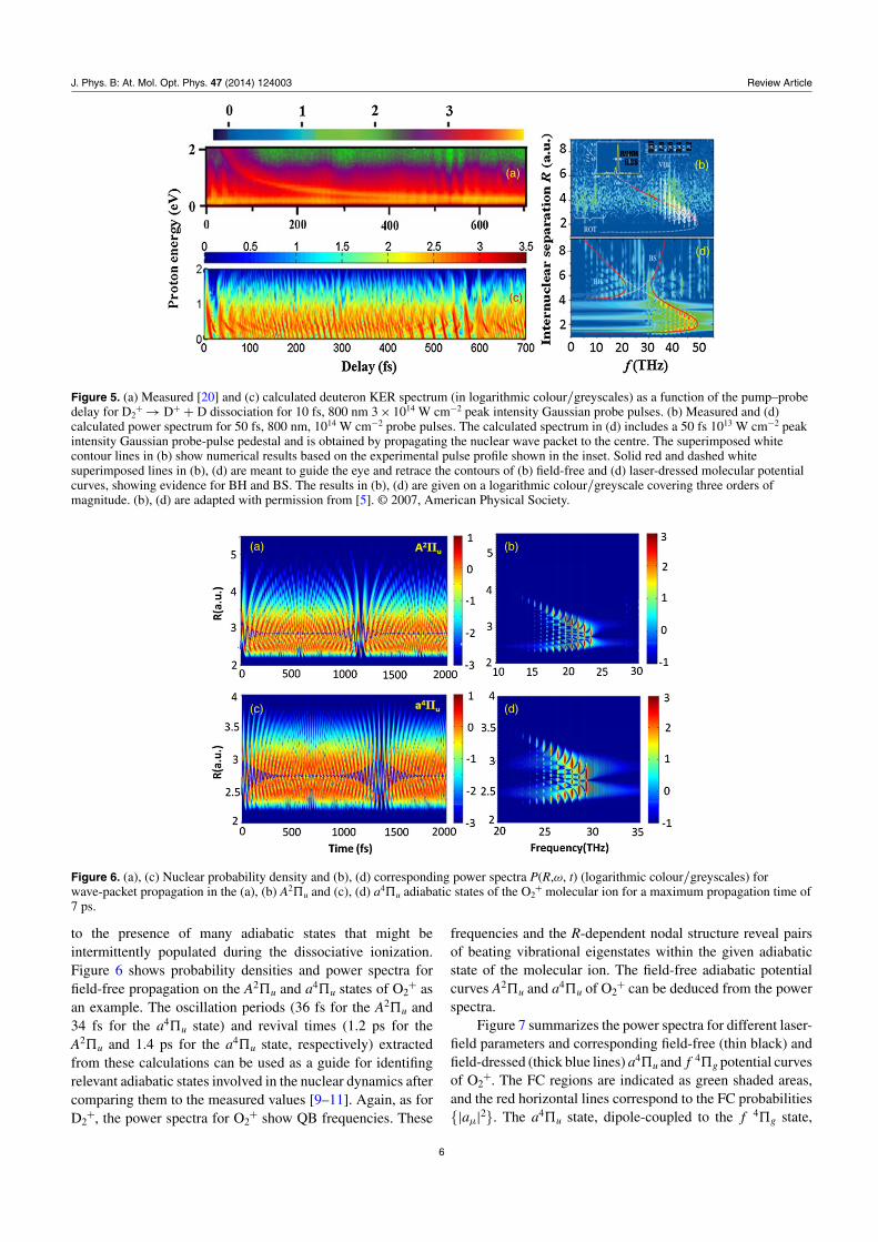
Figure 5. (a) Measured [20] and (c) calculated deuteron KER spectrum (in logarithmic colour/greyscales) as a function of the pump–probedelay for D2
+ → D+ + D dissociation for 10 fs, 800 nm 3 × 10¹⁴ W cm−2 peak intensity Gaussian probe pulses. (b) Measured and (d)calculated power spectrum for 50 fs, 800 nm, 1014 W cm−2 probe pulses. The calculated spectrum in (d) includes a 50 fs 1013 W cm−2 peakintensity Gaussian probe-pulse pedestal and is obtained by propagating the nuclear wave packet to the centre. The superimposed whitecontour lines in (b) show numerical results based on the experimental pulse profile shown in the inset. Solid red and dashed whitesuperimposed lines in (b), (d) are meant to guide the eye and retrace the contours of (b) field-free and (d) laser-dressed molecular potentialcurves, showing evidence for BH and BS. The results in (b), (d) are given on a logarithmic colour/greyscale covering three orders ofmagnitude. (b), (d) are adapted with permission from [5]. © 2007, American Physical Society.
(a) (b)
(c) (d)
Figure 6. (a), (c) Nuclear probability density and (b), (d) corresponding power spectra P(R,ω, t) (logarithmic colour/greyscales) forwave-packet propagation in the (a), (b) A2�u and (c), (d) a4�u adiabatic states of the O2
+ molecular ion for a maximum propagation time of7 ps.
to the presence of many adiabatic states that might beintermittently populated during the dissociative ionization.Figure 6 shows probability densities and power spectra forfield-free propagation on the A2�u and a4�u states of O2
+ asan example. The oscillation periods (36 fs for the A2�u and34 fs for the a4�u state) and revival times (1.2 ps for theA2�u and 1.4 ps for the a4�u state, respectively) extractedfrom these calculations can be used as a guide for identifingrelevant adiabatic states involved in the nuclear dynamics aftercomparing them to the measured values [9–11]. Again, as forD2
+, the power spectra for O2+ show QB frequencies. These
frequencies and the R-dependent nodal structure reveal pairsof beating vibrational eigenstates within the given adiabaticstate of the molecular ion. The field-free adiabatic potentialcurves A2�u and a4�u of O2
+ can be deduced from the powerspectra.
Figure 7 summarizes the power spectra for different laser-field parameters and corresponding field-free (thin black) andfield-dressed (thick blue lines) a4�u and f 4�g potential curvesof O2
+. The FC regions are indicated as green shaded areas,and the red horizontal lines correspond to the FC probabilities{|aμ|2}. The a4�u state, dipole-coupled to the f 4�g state,
6

J. Phys. B: At. Mol. Opt. Phys. 47 (2014) 124003 Review Article
(a) (b) (c) (d)
(e) (f) (g) (h)
Figure 7. (a)–(d) Power spectra from calculations simulating the propagation of vibrational wave packets in the a4�u and f 4�g adiabaticstates of the O2
+ molecular ion (logarithmic colour/greyscale) that are dipole-coupled by the electric field of the probe pulse. The probepulses have a length of 20 fs (FWHM in field strength), wavelengths (a), (b) 1400 nm, (c) 800 nm, (d) 400 nm, and peak intensities (a)1012 W cm−2, (b), (c), (d) 1014 W cm−2. The delay range T covers 12 ps. (e)–(h) Corresponding field-dressed adiabatic potentials, based onadiabatic potential curves and dipole coupling elements from the footnote (see footnote 3). Green shaded areas indicate the FC region, andthe red horizontal lines correspond to the FC probabilities {|aμ|2} at the corresponding stationary vibrational-state energies {ωμ}.
(a) (b)
(c) (d)
Figure 8. (a) (b) Measured [10, 11] and (c), (d) calculated KER spectra for O2+ → O+ + O dissociation in 10 fs 800 nm, 4 × 10¹⁴ W cm−2
peak intensity laser pulses. The calculated KERs in (c), (d) include a 100 fs, 800 nm, 5 × 10¹¹ W cm−2 peak intensity Gaussian pedestal.(a), (c) KERs as a function of the pump–probe delay. (b), (d) Corresponding power spectra (propagated 800 fs past the Gaussian 10 fs pulse)for a delay range of T = 2 ps. Adapted with permission from [10]. © 2011, American Physical Society.
results in power spectra that are quite different for all of thecases shown.
The effects of the probe-pulse electric field on themolecular potential curve become clear by comparingfigures 7(a) and (b). As the pulse intensity increases, thedressed potential curves widen. This effect is further enhanced
by increasing the pulse frequency in figures 7(c) and (d).For different photon energies and peak intensities, the 1ω
crossings of the f 4�g and a4�u states occur at differentinternuclear distances, and the corresponding BH wells arelocated at different internuclear distances R = 3.5 (figure 7(e)),3.1 (figure 7(f)), 3.2 figure (7(g)), and 3.0 (figure 7(h)).
7

J. Phys. B: At. Mol. Opt. Phys. 47 (2014) 124003 Review Article
(a) (b) (c) (d) (e)
(f) (g) (h) (i)
Figure 9. Adiabatic potential curves for the noble-gas dimer cations. (a)–(e) Potential curves for He2+ to Xe2
+ without including spin–orbitcoupling (no fine-structure splitting). (f)–(i) Potential curves for Ne2
+ to Xe2+ including spin–orbit coupling (see footnote 3). Adapted with
permission from [14]. © 2013, American Physical Society.
Figures 7(d) and (h) show the power spectra for 400 nm probepulses and the corresponding field-dressed potential curves.Compared to the 800 nm spectra in figures 7(c) and (g) thesepower spectra differ substantially. For 800 nm probe pulses,most of the wave packet is trapped in the a4�u potential well,whereas for 400 nm probe pulses a significant portion of thenuclear probability density appears to be absent due to BSdissociation.
Figure 8 compares measured and calculated KER spectraas a function of pump–probe delay and QB frequencies forthe O2
+ → O+ + O dissociation process with 10 fs width,800 nm, 4 × 10¹⁴ W cm−2 peak intensity probe pulses. Detailson measurements and calculations are given in [10, 11]. TheKER spectra display energy bands separated by 0.1 eV andoscillations with a period of about 33 fs. The horizontal energybands correspond to KERs from the vibrational energy levelsof the a4�u adiabatic state starting from the ν = 10 vibrationallevel that is energetically just above the dissociative limit ofthe a4�u and f 4�g states. The oscillatory structure along thepump–probe delay axis is consistent with the wave-packetoscillation period in the a4�u state. The above observations
allow us to conclude that the a4�u and f 4�g states are the maincontributors in the O2
+ dissociation dynamics. In addition tothe main 4 × 10¹⁴ W cm−2 pulse, a weak 5 × 10¹¹ W cm−2
pedestal with 100 fs pulse length was added in the numericalsimulation to yield the energy-band structure. If no pedestalwere added, this energy-dependent structure in the KERspectra would be absent.
Typically, several intermediate states contribute to thesame KER. To identify electronic states that contribute tothe experimentally-observed molecular dynamics, we derivethe following scheme from the preceding discussion. Wefirst calculate time-dependent probability density spectraand quantum-beat frequencies for each adiabatic molecularpotential curve independently and compare respectivecalculated KER spectra—in both the time and QB frequencydomain—with available experimental data, in order to assessthe relevance of the selected adiabatic potential curve. Havingidentified the most relevant adiabatic electronic states, weinclude laser-induced dipole couplings between two (or more)preselected adiabatic electronic states and again compare theresulting KER spectra with experimental results. Should the
8

J. Phys. B: At. Mol. Opt. Phys. 47 (2014) 124003 Review Article
comparison not be satisfactory, we modify the set of relevantstates, possibly including additional adiabatic states to thecoupled-state calculation [10, 11].
3.3. Noble-gas dimers
In comparison to O2, noble-gas dimers feature a greatlyreduced number of relevant adiabatic dimer cation states[13]. Figure 9 shows adiabatic potential curves of the noble-gas dimers without (panels a–e) and with (panels f–i)spin–orbit couplings (see footnote 3). Figure 10 exhibitsprobability densities and corresponding power spectra forthe nuclear vibrational wave-packet propagation on theadiabatic 2�u
+ potentials of the noble-gas dimer ions. Theoscillation frequency, noticeable in the probability densityspectra (left panels), decreases with the mass of the dimer.For heavier dimers, the wave packet moves more slowly onthe 2�u
+ state, resulting in larger oscillation periods. Thenumber of oscillations completed before vibrational dephasingincreases with the dimer mass and the quantum revival timeare an order of magnitude larger for Xe2
+ than for He2+ [13].
The QB frequencies are spaced more densely as the massof the dimer increases (figures 10(b)–(j)). This is due tothe fact that for heavier dimers the potential wells of the2�u
+ adiabatic potential curves are deeper and accommodate alarger number of vibrational states than in lighter dimers. Thenuclear dynamics in laser-excited noble-gas dimer cations thusincreasingly resembles classical nuclear motion for growingdimer mass.
Figure 11 summarizes the power spectra andcorresponding field-dressed potential curves for Ne2
+ in1012 W cm−2, 200 fs probe laser pulses with wavelengthsof 1400 nm (figures 11(a) and (c)) and 800 nm (figures 11(b)and (d)). The outer classical turning points of the adiabaticfield-dressed potential are mapped better for 800 nm pulsesin figure 11(b) than for 1400 nm pulses (figure 11(a)), sincethe 800 nm BH well is deep enough to trap a significant partof the wave packet (cf figures 11(c) and (d)). For 800 nmwavelength pulses, the one-photon crossing appears at smallerinternuclear distances than for 1400 nm pulses. Figures 11(c)and (d) indicate that the majority of the wave packet can betrapped by the BH well at internuclear distances between 4and 7 au; this is corroborated by figures 11(a) and (b) as themajority of the wave packet yield is located in that same range.
Figure 12 summarizes the power spectra andcorresponding field-dressed potential curves for Ar2
+ in1012 W cm−2, 80 fs probe laser pulses with a wavelength of1400 nm. The one-photon BH well is mapped comparativelybetter for 2�u
+ and 2�g+ potential curves in figure 12(a) than
the shallower BH well of the field-dressed I(1/2)u and II(1/2)g
curves in figure 12(b).To investigate the effect of fine-structure splitting on the
nuclear motion we compare calculated power spectra P(R,ω,τ )based on the adiabatic potential curves I(1/2)u and II(1/2)g
including spin–orbit interactions with the results based on theadiabatic potential curves 2�u
+ and 2�g+ that were computed
without including spin–orbit interactions for Ar2+ (figure 12),
Kr2+ (figure 13), and Xe2
+ (figure 14). With increasing
(a) (b)
(c) (d)
(e) (f)
(g) (h)
(i) (j)
Figure 10. Nuclear probability densities (left) and correspondingpower spectra (right) (logarithmic colour/greyscale) for thewave-packet propagation of nuclear vibrational wave packets on theadiabatic 2�u
+ potential curves for (a), (b) He2+, (c), (d) Ne2
+, (e),(f) Ar2
+, (g), (h) Kr2+, and (i), (j) Xe2
+.
dimer mass, spin–orbit coupling becomes more relevant, andthe I(1/2)u and II(1/2)g fine-structure splitting increases.This causes the BH well in Kr2
+ (figure 13(d)) to becomeshallower than in Ar2
+ (figure 12(d)). For the dipole-coupled
9

J. Phys. B: At. Mol. Opt. Phys. 47 (2014) 124003 Review Article
(a) (b)
(c) (d)
Figure 11. (a), (b) Power spectra (logarithmic colour/greyscale) and (c), (d) field-dressed potential curves for Ne2+ and 200 fs,
1012 W cm−2 probe laser pulses with wavelengths of (a), (c) 1400 nm and (b), (d) 800 nm. Simulations based on dipole coupled 2�u+ and
2�g+ potential curves, not including fine-structure splitting. (c), (d) Field-dressed potential curves (thick blue curves) and corresponding
field-free 2�u+ and 2�g
+ adiabatic potential curves (thin black lines). Green-shaded areas indicate the FC region. Red horizontal lines showthe FC probabilities {|aμ|2} at the corresponding stationary vibrational-state energies {ωμ}.
(a) (b)
(c) (d)
Figure 12. (a), (b) Power spectra (logarithmic colour/greyscale) and (c), (d) field-dressed potential curves for Ar2+ in 80 fs, 1012 W cm−2,
1400 nm probe laser pulses. (a) Simulations based on dipole coupled 2�u+ and 2�g
+ potential curves, not including fine-structure splitting.(b) Simulations based on the dipole-coupled I(1/2)u and II(1/2)g potential curves, including fine-structure splitting. (c) Field-dressedpotential curves (thick blue curves) and corresponding field-free 2�u
+ and 2�g+ potential curves (thin black lines). (d) Field-dressed
potential curves (thick blue curves) and corresponding field-free I(1/2)u and II(1/2)g adiabatic potential curves (thin black lines).Green-shaded areas indicate the FC region. Red horizontal lines correspond to the FC probabilities {|aμ|2} at the corresponding stationaryvibrational-state energies {ωμ}.
10

J. Phys. B: At. Mol. Opt. Phys. 47 (2014) 124003 Review Article
(a) (b)
(c) (d)
Figure 13. Same as figure 12 for Kr2+.
(a) (b)
(c) (d)
Figure 14. Same as figure 13 for Xe2+ and 400 fs probe pulses.
2�u+ and 2�g
+ states, the BH wells for Ar2+ (figure 12(c))
and Kr2+ (figure 13(c)) are deeper compared to the I(1/2)u and
II(1/2)g states (figures 12(d) and 13(d)). For Xe2+, the fine-
structure splitting exceeds the photon energy at 1400 nm andthe I(1/2)u and II(1/2)g adiabatic potential curve one-photoncrossings are absent (figure 14(d)).
4. Summary and outlook
We studied the bound and dissociative nuclear motion ofvibrationally excited diatomic molecular ions in the timeand frequency domains. While the time-domain KER spectra
display oscillation periods, revival times, and the nuclear-probability-density evolution, QB imaging in the frequencydomain complements time-domain investigations of thenuclear dynamics by revealing (i) QB frequencies and thenodal structure of vibrational states within a given adiabaticmolecular potential curve and (ii) laser-electric-field-dressedmolecular potential curves. QB frequencies, wave-packetoscillation periods, and revival times are characteristics thatindicate which adiabatic states of a photo-excited diatomicmolecular ion are transiently populated during its boundor dissociative motion. Identifying these relevant potentials’curves by the combined analysis of experimentally-measured
11

J. Phys. B: At. Mol. Opt. Phys. 47 (2014) 124003 Review Article
KER spectra in the time and frequency domains, in comparisonwith simulated KER spectra, provides a powerful tool foranalysing the nuclear dynamics in diatomic molecules andfor identifying nuclear dissociation (and reaction) pathways.Due to the weak binding of noble-gas dimer cations anda small number of relevant electronic states, noble-gasdimers, in particular, are ideal targets for modelling thenuclear dissociation dynamics, allowing for the scrutiny ofdetails such as the existence and relevance of transient BHstates in field-dressed molecular potential curves. To identifystates relevant in the dissociation dynamics, we discussed ascheme in which wave-packet propagation calculations are firstperformed separately for individual adiabatic potential curves,allowing for the selection of relevant adiabatic electronicstates by identifying characteristic features (such as revivaltimes, oscillation periods and quantum-beat frequencies) thatthe simulated time- and frequency-domain spectra have incommon with measured KER spectra. Finally, after selectingthe closest matches to the measured spectra, we carried outmore complex calculations including the dipole coupling ofthese preselected states in the laser pulses. The QB imagingtechnique allows for the direct mapping of adiabatic nuclearpotential curves and provides the distribution of stationarystates that superimpose to form the nuclear vibrational wavepacket that is launched by the molecular ionization in the pumppulse. In principle, this technique can be used to determine theinternuclear-distance-dependent wavefunction of the nuclearwave packets (up to an overall phase) from measured KERspectra [8].
Acknowledgments
This work was supported by the National Science Foundationunder grant no PHY-1068140 (KB), grant no PHY-1068752(MM, UT), grant no PHY-1157044 (AK), and the Division ofChemical Sciences, Office of Basic Energy Sciences, Officeof Science, US Department of Energy.
References
[1] Ullrich J, Rudenko A and Moshammer R 2012 Annu. Rev.Phys. Chem. 63 635
[2] Calvert C, Bryan W, Newell W and Williams I 2010 Phys.Rep. 491 1
[3] Ergler T, Rudenko A, Feuerstein B, Zrost K, Schroter C D,Moshammer R and Ullrich J 2005 Phys. Rev. Lett.95 093001
[4] Alnaser A S et al 2005 Phys. Rev. A 72 030702(R)[5] Feuerstein B, Ergler T, Rudenko A, Zrost K, Schroter C D,
Moshammer R, Ullrich J, Niederhausen T and Thumm U2007 Phys. Rev. Lett. 99 153002
[6] Winter M, Schmidt R and Thumm U 2008 New J. Phys.12 023020
Winter M, Schmidt R and Thumm U 2009 Phys. Rev. A80 031401 and references therein
[7] Magrakvelidze M, He F, Niederhausen T, Litvinyuk I Vand Thumm U 2009 Phys. Rev. A 79 033410
[8] Thumm U, Niederhausen T and Feuerstein B 2008 Phys. Rev.A 77 063401
[9] De S, Bocharova I A, Magrakvelidze M, Ray D, Cao W,Bergues B, Thumm U, Kling M F, Litvinyuk I Vand Cocke C L 2010 Phys. Rev. A 82 013408
[10] De S et al 2011 Phys. Rev. A 84 043410[11] Magrakvelidze M, Aikens C M and Thumm U 2012 Phys. Rev.
A 86 023402[12] Bocharova I A, Alnaser A S, Thumm U, Niederhausen T,
Ray D, Cocke C L and Litvinyuk I V 2011 Phys. Rev. A83 013417
[13] Wu J, Magrakvelidze M, Vredenborg A, Schmidt L, Ph H,Jahnke T, Czasch A, Dorner R and Thumm U 2013 Phys.Rev. Lett. 110 033005 and references therein
[14] Magrakvelidze M and Thumm U 2013 Phys. Rev. A 88 013413[15] Jiang Y H et al 2010 Phys. Rev. A 82 041403(R)[16] Magrakvelidze M et al 2012 Phys. Rev. A 86 013415[17] Feldhaus J, Krikunova M, Meyer M, Moller T, Moshammer R,
Rudenko A, Tschentschler T and Ullrich J 2013 J. Phys. B:At. Mol. Opt. Phys. 46 164002
[18] Feuerstein B and Thumm U 2003 Phys. Rev. A 67 063408[19] Ergler T, Rudenko A, Feuerstein B, Zrost K, Schroter C D,
Moshammer R and Ullrich J 2006 Phys. Rev. Lett.97 193001
[20] Rudenko A, Ergler T, Feuerstein B, Zrost K, Schroter C D,Moshammer R and Ullrich J 2006 Chem. Phys. 329 193
[20a] Rudenko A, Ergler T, Feuerstein B, Zrost K, Schroter C D,Moshammer R and Ullrich J 2007 J. Phys.: Conf. Ser.88 012050
[21] Robinett R W 2004 Phys. Rep. 392 1[22] Marian C M, Marian R, Peyerimhoff S D, Hess B A,
Buenker R J and Seger G 1982 Mol. Phys. 46 779[23] Bransden B H and Joachain C J 2003 Physics of Atoms and
Molecules 2nd edn (London: Prentice-Hall)[24] Feuerstein B and Thumm U 2003 J. Phys. B: At. Mol. Opt.
Phys. 36 707[25] Barone S R, Narcowich M A and Narcowich F J 1977 Phys.
Rev. A 15 1109[26] Bucksbaum P H, Zavriyev A, Muller H G
and Schumacher D W 1990 Phys. Rev. Lett. 64 1883
12