Complement in the brain - Lund University Publications · 1.2 Complement in the brain ......
46
Complement in the brain Veerhuis, Robert; Nielsen, Henrietta; Tenner, Andrea J. Published in: Molecular Immunology DOI: 10.1016/j.molimm.2011.04.003 2011 Link to publication Citation for published version (APA): Veerhuis, R., Nielsen, H., & Tenner, A. J. (2011). Complement in the brain. Molecular Immunology, 48(14), 1592-1603. DOI: 10.1016/j.molimm.2011.04.003 General rights Copyright and moral rights for the publications made accessible in the public portal are retained by the authors and/or other copyright owners and it is a condition of accessing publications that users recognise and abide by the legal requirements associated with these rights. • Users may download and print one copy of any publication from the public portal for the purpose of private study or research. • You may not further distribute the material or use it for any profit-making activity or commercial gain • You may freely distribute the URL identifying the publication in the public portal Take down policy If you believe that this document breaches copyright please contact us providing details, and we will remove access to the work immediately and investigate your claim.
Transcript of Complement in the brain - Lund University Publications · 1.2 Complement in the brain ......

LUND UNIVERSITY
PO Box 117221 00 Lund+46 46-222 00 00
Complement in the brain
Veerhuis, Robert; Nielsen, Henrietta; Tenner, Andrea J.
Published in:Molecular Immunology
DOI:10.1016/j.molimm.2011.04.003
2011
Link to publication
Citation for published version (APA):Veerhuis, R., Nielsen, H., & Tenner, A. J. (2011). Complement in the brain. Molecular Immunology, 48(14),1592-1603. DOI: 10.1016/j.molimm.2011.04.003
General rightsCopyright and moral rights for the publications made accessible in the public portal are retained by the authorsand/or other copyright owners and it is a condition of accessing publications that users recognise and abide by thelegal requirements associated with these rights.
• Users may download and print one copy of any publication from the public portal for the purpose of private studyor research. • You may not further distribute the material or use it for any profit-making activity or commercial gain • You may freely distribute the URL identifying the publication in the public portalTake down policyIf you believe that this document breaches copyright please contact us providing details, and we will removeaccess to the work immediately and investigate your claim.
1
Complement in the Brain
Robert Veerhuisa, Henrietta M. Nielsen
b, and Andrea J. Tenner
c
aDepts of Clinical Chemistry, Pathology, Psychiatry and Alzheimer Center, VU
University Medical Center, Amsterdam, The Netherlands
bDept of Clinical Sciences Malmö, Molecular Memory Research Unit, Lund University,
The Wallenberg Lab 2nd
floor, Skåne University Hospital entrance 46, Malmö, Sweden
c Depts of Molecular Biology and Biochemistry and Neurobiology and Behavior, Institute
for Immunology, UCI MIND, University of California, Irvine, USA
Corresponding Author:
Andrea J. Tenner, Ph.D.
University of California, Irvine
3205 McGaugh Hall
Irvine, CA. 92697-3900
949-824-3268
FAX 949-824-8551

2
ABSTRACT
The brain is considered to be an immune privileged site, because the blood-brain barrier limits
entry of blood borne cells and proteins into the central nervous system (CNS). As a result, the
detection and clearance of invading microorganisms and senescent cells as well as surplus
neurotransmitters, aged and glycated proteins, in order to maintain a healthy environment for
neuronal and glial cells, is largely confined to the innate immune system. In recent years it has
become clear that many factors of innate immunity are expressed throughout the brain. Neuronal
and glial cells express Toll like receptors as well as complement receptors, and virtually all
complement components can be locally produced in the brain, often in response to injury or
developmental cues. However, as inflammatory reactions could interfere with proper functioning
of the brain, tight and fine tuned regulatory mechanisms are warranted. In age related diseases,
such as Alzheimer‟s disease (AD), accumulating amyloid proteins elicit complement activation
and a local, chronic inflammatory response that leads to attraction and activation of glial cells
that, under such activation conditions, can produce neurotoxic substances, including pro-
inflammatory cytokines and oxygen radicals. This process may be exacerbated by a disturbed
balance between complement activators and complement regulatory proteins such as occurs in
AD, as the local synthesis of these proteins is differentially regulated by pro-inflammatory
cytokines. Much knowledge about the role of complement in neurodegenerative diseases has
been derived from animal studies with transgenic overexpressing or knockout mice for specific
complement factors or receptors. These studies have provided insight into the potential
therapeutic use of complement regulators and complement receptor antagonists in chronic
neurodegenerative diseases as well as in acute conditions, such as stroke. Interestingly, recent
animal studies have also indicated that complement activation products are involved in brain
development and synapse formation. Not only are these findings important for the understanding
of how brain development and neural network formation is organized, it may also give insights
into the role of complement in processes of neurodegeneration and neuroprotection in the injured

3
or aged and diseased adult central nervous system, and thus aid in identifying novel and specific
targets for therapeutic intervention.
Keywords: Complement; brain; neurons; glia; neurodegeneration; neuroprotection

4
1. INTRODUCTION
1.1 Complement
Complement (C) is a major component of innate immunity, recognizing danger, as well as
discriminating self from non-self (Ricklin et al., 2010). The C system is best known for its role in
the recognition and killing of pathogenic microbes. Activation of the C system, which consists of
over 30 soluble and cell-associated factors, can occur through three pathways, each triggered by
different types of agents. All three pathways lead to the assembly of C3 convertases that, in turn,
can cleave C3 resulting in formation of C3b and C3a activation products. The larger C3b
fragment, a major effector molecule of the C system, acts as an opsonin and, in addition, together
with other factors can assemble the C5 convertase, which enables further activation of the C
cascade ultimately leading to generation of the chemotactic C5a fragment and the formation of
the terminal complement complex C5b-9, also called membrane attack complex (Figure 1). Thus,
when the cascade is fully activated, C leads to assembly of the membrane attack complex (C5b-9;
MAC) and lysis of invading microorganisms. However, if C5b-9 production is excessive or
targeted to host cells, C5b-9 can induce host cell death. On the other hand, some C activation
products also facilitate the generation of adaptive immune responses (Carroll, 2004;Erdei et al.,
2009), while other components contribute to the control of autoimmunity during the clearance of
apoptotic cells (Sjoberg et al., 2009;Fraser et al., 2009;van Kooten et al., 2008). Interestingly,
sublytic amounts of C5b-9 on host cells may cause an influx of extracellular calcium that leads to
activation and/or proliferation of the cells and resistance to induction of apoptosis (Cole and
Morgan, 2003), further illustrating the diverse functions of this ancient pathway.
Binding of C1, a Ca2+-
dependent complex of the recognition unit C1q and a tetramer of the
proenzymes C1r and C1s, to an activator is the initial event in classical pathway (CP) activation
of C. Activators can be immune complexes, certain microbes, apoptotic cells, and other specific
protein motifs, such as amyloid in a fibrillar beta sheet structure found in the plaques in brain
from Alzheimer‟s disease (AD) patients.
5
Mannose-binding lectin (MBL) and ficolins (Ficolin-1,-2 and -3) bind to mannan and other
carbohydrate moieties or acetylated moieties on microorganisms or dying cells initiating C
activation, through the lectin pathway (LP). MBL and ficolins share homology with C1q and, like
C1q, are associated with proenzymes, MBL-associated serine proteases (MASPs).
The alternative pathway (AP) is initiated by spontaneous hydrolysis of the internal thioester
within C3, resulting in C3b-like C3 (“tick-over”) or by recruitment of C3 by properdin bound to
specific targets (Kemper and Hourcade, 2008). A range of microbial and also eukaryotic cell
surfaces with a low sialic acid content allow AP C activation, whereas the inactivation of C3b by
the C regulatory proteins (Creg) factor H (fH) and factor I (fI) is more efficient on surfaces rich in
sialic acid (AUSTEN and Fearon, 1979). C3b generated by the CP or MP activation pathways can
enlist the alternative pathway components, thereby amplifying the amount of downstream
complement cascade events (Figure 1).
1.2 Complement in the brain
The brains of organisms with a well developed central nervous system are shielded by the blood-
brain-barrier (BBB) with tight junction formations at three principal barrier sites i) the BBB
formed by endothelial cells in the cerebral capillaries ii) the arachnoid barrier formed by the
arachnoid multi-layered epithelium and iii) the blood-CSF barrier formed by the CSF-secreting
choroid plexus epithelium. The integrity properties are further defined by BBB-associated cells
including pericytes and astrocytes. Together these brain barriers efficiently prevent infiltration of
circulating immune cells, such as B- and T lymphocytes, and minimize influx of plasma proteins
as well as neuroexcitatory and neurotoxic substances from the blood (reviewed in (Abbott et al.,
2010)). The functions of immune surveillance and differentiation between “self” and “nonself” in
non-CNS tissue, provided by neutrophils, dendritic cells, macrophages and natural killer cells in
the periphery, are in the CNS attributed to resident glial cells including astrocytes, microglia,
oligodendrocytes, and NG2 chondroitin sulphate (NG2) and platelet-derived growth factor-α
6
receptor (PDGFα) positive oligodendrocyte precursor cells (NG2+PDGFα
+ OPCs) ((Butt et al.,
2005) and reviewed in (Dong and Benveniste, 2001;Griffiths et al., 2009)). Much of our
understanding of the occurrence and role of complement in the CNS derives from studies into the
pathogenic mechanisms involved in various diseases affecting the brain. In a variety of CNS
diseases, including bacterial meningitis, transmissible spongiform encephalopathies (TSE; prion
disease), stroke and in more chronic conditions such as multiple sclerosis (MS), amyotrophic
lateral sclerosis (ALS), Huntington‟s (HD) and Parkinson‟s disease (PD), AD, as well as age-
related macular degeneration (AMD), C contributes to the inflammatory process (Woodruff et al.,
2008;Bonifati and Kishore, 2007). In this review we discuss the findings that have led to insight
on the role of C in acute and chronic brain disorders and, importantly, its role in the normal
homeostasis and brain functioning, as more recent studies (Stevens et al., 2007) indicate that C is
also involved in brain development and synapse pruning.
2. Local Synthesis of Complement and Complement Regulators in the Brain
The liver is the main source of C proteins, but although the BBB is not absolute and some
macromolecules can by-pass the barriers by use of extracellular routes (Broadwell and Sofroniew,
1993) most C proteins are unlikely to penetrate the brain parenchyma unless the BBB integrity is
disrupted. Therefore, local synthesis of C components by resident cells in the brain is crucial to
appropriate functions of the local defense system. Local synthesis of C proteins in the brain was
suggested after identification of C in human brain tissue. In situ hybridization studies (Lampert-
Etchells et al., 1993;Rozovsky et al., 1994;Veerhuis et al., 1998) confirmed that C factors are all
locally produced and that the presence of C is not merely due to leakage of plasma proteins
because of BBB damage.
2.1 Glial cells

7
Astrocytes are the most numerous cell type in the human brain and are dedicated to
various functions such as regulation of synaptogenesis, metabolic support and control of the
homeostatic system including regulation of extracellular ion concentrations and
neurotransmitters, especially glutamate, and regulation of brain water homeostasis (Verkhratsky
and Parpura, 2010). Microglia, the macrophages of the brain, are scattered throughout the brain
tissue at a density of about 6x10⁶ cells per mm
3 and, when in a “resting” state are highly dynamic
and estimated to completely scan the brain parenchyma once every few hours (Nimmerjahn et al.,
2005). Microglia as well as astrocytes are considered CNS immune effector cells and are able to
produce cytokines and chemokines as well as to phagocytose up targets upon stimulation (Dong
and Benveniste, 2001;Nielsen et al., 2010;Familian et al., 2007;Nielsen et al., 2009;Fraser et al.,
2010;Bohlson et al., 2007;Yang et al., 2010;Watabe et al., 1989). Another group of macroglial
cells are the oligodendrocytes, which are responsible for myelinating axons in the CNS.
More than two decades ago Levi-Strauss and Mallat reported that primary cultures of
murine astrocytes were capable of producing C components of the AP (Levi-Strauss and Mallat,
1987). Extensive research during the 1990‟s, initially performed in several human astrocyte-
derived tumor cell lines (118MG, T193, T98G), demonstrated the expression of components of
the CP and also of terminal C components (Gasque et al., 1995;Gasque et al., 1993) by human
glial cells. In further studies with cell lines and also primary cells isolated from mouse and adult
human brain (Walker and McGeer, 1992;Veerhuis et al., 1999;Walker et al., 1995;Veerhuis et al.,
1998) convincing evidence was obtained for local synthesis of most C components of both the
classical and alternative pathway, including Cregs, by human astrocytes and microglia (Table 1).
What cell type that is responsible for local production of fluid-phase C inhibitor C4b binding
protein (C4bp) has yet to be determined in the brain as detectable levels of secreted C4bp
appeared to be absent in cultures of primary human astrocytes as well as several cell lines (Trouw
et al., 2008). Further, results from in vitro studies on primary human microglia and astrocytes
suggest that synthesis of several C components, C1 subcomponents C1s and C1r, C3, C4 and C1-
8
inh can be modulated by various factors like pro-inflammatory cytokines but as well as by the
AD-related amyloid-β peptide (Aβ) perhaps via TLR stimulation (Veerhuis et al., 1999). The
same study, in support of earlier investigations, also suggested that microglia, but not astrocytes,
are a significant source of locally secreted C1q in the brain (Lampert-Etchells et al.,
1993;Veerhuis et al., 1999).
Results from initial studies on rodent oligodendrocytes suggested that these cells are
vulnerable to C lysis due to a deficiency of C inhibitor expression (Wren and Noble,
1989;Piddlesden et al., 1994). Indeed, oligodendrocytes are susceptible to C attack which is
particularly evident in multiple sclerosis (MS) (Schwab and McGeer, 2002). In one study, adult
primary human oligodendrocyte cultures were found to produce only a limited Creg repertoire,
which suggests that a relative deficiency in Creg expression may render oligodendrocytes
sensitive to C damage in MS (Scolding et al., 1998). Interestingly, oligodendrocytes seem to not
only be susceptible to C attack but also to themselves be a source of a large number of C proteins
including C1q, C1s, C4, C2, C3, C5, C6, C7, C8 and C9 (Hosokawa et al., 2003). Little is known
about C expression in various other cells in the CNS. However, initial studies suggest that
primary human pericytes in vitro produce C1q (Verbeek et al., 1999) and that cultured endothelial
cells from human brain microvessels produce soluble regulators fH and C1-inh and components
of both the classical (C4), and the alternative (fB) complement pathway (Vastag et al., 1998).
Ependymal cells, ciliated epithelial cells that line the lumen of the brain ventricular system,
express Cregs CD59 and at a low level CD55, but no CD46 or CD35, however in inflammatory
conditions (meningitis) CD46 and CD35 are highly expressed on epithelial cells of the
ependymal lining, as well as in the choroid plexus (Canova et al., 2006). To what extent these
cell types contribute to the levels of C factors in the brain parenchyma and of the CSF is
unknown. Thus, future studies extending the knowledge on C expression in various cells of the
CNS are warranted.

9
2.2 Neurons
Neuronal cells have long been considered innocent victims of C activation in neurodegenerative
conditions, as a result of activation of C factors that had passed the BBB or that had been
synthesized by activated glial cells. Robust activated complement system with C5b-9 insertion
can lead to lysis and death of a targeted cell, a process which can be prevented by appropriate
expression of complement regulators. However, neuronal cells were also found to be capable of
de novo synthesis of complement factors both in vivo and in vitro. Neuronal mRNA expression
of C1q, C2, C3, C4, C5, C6, C7, C8 and C9 was minimally detected using in situ hybridization in
the temporal cortex and hippocampus in post mortem control brain tissue, with increased
expression in AD tissue. The strongest signals were recorded over pyramidal neurons (Shen et
al., 1997). Neuronal expression of C1-Inh was detectable in brain tissue from postmortem AD
and control subjects as well as in the neuroblastoma cell line SK-N-SH (Veerhuis et al., 1998). In
vitro expression of C1-Inh could only be upregulated by treatment with interferon γ (Veerhuis et
al., 1998;Veerhuis et al., 1999), whereas expression of the fluid-phase regulators, MCP and
CD59 in human neuroblastoma cell lines could be modulated by treatment with pro-inflammatory
cytokines (Gasque et al., 1996). Extending those in vitro studies, Fontaine and colleagues
showed that the above mentioned neuroblastoma cell lines and the human neuroblastoma cell
lines SH-SY5Y and KELLY were able to express a complete set of C proteins and further
suggested that the rate of synthesis was cell differentiation-dependent (Thomas et al., 2000).
Interestingly, primary fetal human neurons in vitro were shown to spontaneously and independent
of antibody activate the CP, possibly by expressing a molecule with affinity for C1q, leading to
assembly of the cytolytic C5b-9 on their membranes. Limited neuronal expression of Cregs MCP
and CD59, and lack of DAF and CR1 expression was suggested to underlie this vulnerability to
complement damage (Singhrao et al., 2000). CD59 has been shown crucial to protection of for
example NT2-N neurons (human NT2 cell line differentiated into post-mitotic neurons) against C
attacks (Pedersen et al., 2007) and current strategies aiming at increasing neuronal protection
10
against C include attempts of upregulation of Cregs like CD59. For example, the CD59
expression-regulating neural-restrictive silencer factor (REST) protected neurons from C-
mediated lysis by a five-fold upregulation of CD59 expression in neuronal cultures (Kolev et al.,
2010). Whether modulation of neuronal production of Cregs is a successful neuroprotective
strategy remains to be elucidated as recent in vivo studies suggest that C activation products,
including the anaphylatoxins C3a and C5a and sublytic levels of the MAC, may in fact have
several neuroprotective functions ((Osaka et al., 1999;Tocco et al., 1997;O'Barr et al., 2001;Van
Beek et al., 2003) and reviewed in (Woodruff et al., 2010).
3. The role of complement in normal CNS
Similar to other proteins that are part of the immune system, such as proinflammatory
cytokines (e.g., TNFα, IL-6) and proteins of the adaptive immune system (e.g. major
histocompatibility complex class I [MHCI] molecules and MHCI-binding immunoreceptors and
their components (e.g., PIRB, Ly49, DAP12, CD3δ) (for a review see (Boulanger, 2009)), C
factors are now thought to also have nonimmune functions in the brain. Complement proteins
were found to promote proliferation and regeneration in various tissues (reviewed in (Ricklin et
al., 2010)) and may exert similar functions in the CNS, as neuronal stem cells differentiate and
migrate in response to C. C3a-C3aR interactions were found to be a positive regulator of adult
neurogenesis (Bogestal et al., 2007;Shinjyo et al., 2009).
Recent studies have also shown that C activation products can modulate synapse
formation during brain development (Stevens et al., 2007;Chu et al., 2010). Neurons isolated
from the developing eye were found to express high levels of C1q mRNA. Using C1q and C3
knock-out mice, it was shown that whereas relay neurons in wild type (wt) mice are innervated by
one or two axons, relay neurons in C deficient (C1q -/- , C3 -/-) mice have four or more
functional inputs. This lead to the conclusion that C1q and C3 may tag synapses for elimination,
leading to remodelling of synaptic connections in the developing visual system (Stevens et al.,

11
2007). In a subsequent study, a complete genetic deficiency of C1q resulted in enhanced circuitry
that led to epileptogenesis in mouse models (Chu et al., 2010). C1q, both alone and in
conjunction with C3, can facilitate microglial clearance of misfolded proteins, apoptotic neurons
and damaged cells such as neuronal blebs (Fraser et al., 2010;Trouw et al., 2008) and modulate
cytokine profiles to subdue potentially neurotoxic inflammatory gene expression (Fraser et al.,
2010). Thus, depending on the timing and local environment, the C cascade can facilitate proper
neuronal development or accelerate chronic inflammatory response contributing to
neurodegeneration (see below).
Proteins related to C1q, cerebellins (Cbln) (Yuzaki, 2010) and C1ql (Bolliger et al.,
2011) have been found to be expressed in the cerebellum, as well as other brain regions of
developing and mature brain. Cbln members may serve as regulators of synapse development and
synaptic plasticity through regulation of the post synaptic endocytosis pathway of AMPA
receptors (Yuzaki, 2008), and C1ql proteins have recently been shown to interact with a neuronal
surface receptor BAI3 also involved in regulation of synapse formation and/or maintenance
(Bolliger et al., 2011). As C1q is expressed by neurons in the hippocampus and temporal cortex
((Afagh et al., 1996;Rozovsky et al., 1994) and Veerhuis, unpublished, and reviewed in
(Alexander et al., 2008)) of injured brain, it is tempting to speculate that C1q in the neocortex
may serve a function similar to that of the Cbln proteins in the cerebellum. In addition, in vitro
C1q enhances neuronal survival and is neuroprotective in response to certain toxic agents, such
as fibrillar amyloid and serum amyloid P (Pisalyaput and Tenner, 2008). Whether these BAI3-
C1ql interactions are influenced by C1q itself (which has been shown to influence neuron
survival and neurite outgrowth in vitro (Benoit and Tenner, 2011;Pisalyaput and Tenner, 2008))
remains to be seen. Interestingly, half of more than 50 genes encoding putative Cregs predicted
in the mouse genome, are expressed in the CNS, consistent with at least some of the
uncharacterized C control protein domain (CCP)-bearing proteins in mammals may be involved
in synapse organization (Gendrel et al., 2009).

12
4. Complement during acute brain injury
Acute brain injuries including infections, brain trauma, ischemic and hemorrhagic stroke and
subsequent reperfusion injuries are to date associated with a limited repertoire of effective
treatments and high morbidity. Neurodegeneration and death in these acute conditions can be via
necrosis or via apoptosis. For example, apoptosis was recently shown to be dominant in the peri-
infarct area after ischemic stroke in humans (Sairanen et al., 2006). Most likely neuronal death
following acute conditions occurs via a combination of both necrosis and apoptosis.
4.1 Brain infections
Various pathogens including bacteria, virus and fungi can invade the CNS and cause life-
threatening diseases. In the immune privileged brain C functionality can be crucial to fight off
and kill invading microbes. However, C activation and regulation needs to be delicately balanced
as excessive C activation might be detrimental to bystander cells. Intriguingly, several CNS
invading microorganisms have developed mechanisms to avoid the destructive actions of C and
in fact even to use C to their advantage. One of these mechanisms is mimicry of human Cregs
which enables control and down-regulation of C activation against invading microbes (Cooper
and Nemerow, 1989). Further strategies to circumvent C include the use of membrane-bound C
receptors and Cregs to enter the host cell and acquisition of Cregs during budding from the
membranes of the host cell or by binding to soluble Cregs (reviewed in (Speth et al., 2002)). For
example the meningitis causing bacteria Neisseria meningitidis invades the CNS through the
nasopharyngeal mucosa and uses the membrane bound Creg CD46 which interacts with bacterial
pili, to cross the blood-brain-barrier (Johansson et al., 2003). Also, gram-negative Escherichia
coli K1 avoids C killing by binding to C4bp and promoting degradation of C3b and C4b (Wooster
et al., 2006). Similar to bacteria, several virus strains have developed protective strategies to
avoid C (recently reviewed in (Stoermer and Morrison, 2011)) . The herpes virus Epstein-Barr

13
(EBV), which can cause encephalitis and aseptic meningitis, uses CR2 for viral entry by binding
to the receptor at the same location as the C3 fragment C3dg (Carel et al., 1989). HIV-1,
detectable in the brains of >85% patients who died with AIDS (Johnson et al., 1996), acquires
Cregs CD46, CD55 and CD59 upon budding from the host cell (Frank et al., 1996) and binding
to soluble Creg fH (Stoiber et al., 1995) thereby avoiding C mediated lysis of the virion particles.
In addition, recent studies indicate that CNS invading fungi also have developed C evasion
mechanisms (reviewed in Speth and colleagues (Speth et al., 2008)). Although resident brain cells
including astrocytes, neurons, oligodendrocytes, but to a lesser extent microglia, produce highly
increased levels of C1q, C4 and C3 in response to fungus infection, as in the case of cerebral
aspergillosis, fungal hyphae can limit surface deposition of C3 and thereby interfere with C-
mediated phagocytosis of this pathogen (Rambach et al., 2008;Speth et al., 2008). Taken
together, these examples illustrate that C plays various roles in brain infections and that the C
evasion strategies by microbial pathogens invading the CNS may be a target for therapeutic
intervention.
4.2 Trauma, stroke and reperfusion injuries
The diverse roles played by the C system in acute brain disorders are not fully elucidated,
however a growing body of evidence suggests an important role in secondary brain damage
(Stahel et al., 1998). During some conditions of acute brain damage the BBB integrity is
disrupted allowing influx of plasma proteins, including C proteins, and immune cells from the
periphery, whereas in others CNS injuries C synthesis is induced by CNS insults (including
oxidative stress). Activation of the CP in human brain following traumatic brain injury has been
shown by increased immunoreactivity for C1q, C3b, C3d and C5b-9in the immediate vicinity of
neurons in the penumbra area of the cerebral contusion (Bellander et al., 2001). Further, C3
mRNA and upregulation of clusterin was found in the penumbra, indicating local de novo
synthesis of C and Cregs following injury. The authors suggested that an unknown component,
14
possibly in the debris from injured neurons or myelin breakdown products, might be able to
trigger C activation and formation of the following brain contusions. Investigation of brain tissue
of patients with acute brain ischaemia or ischaemic stroke further revealed deposition of C1q,
C3c and C4d in all ischaemic lesions, further supporting activation of the CP. In necrotic zones
of the brains from the same patients, C9, C-reactive protein and IgM were found. The possibility
of uncontrolled C activation following ischaemic insults, which might be harmful was underlined
by the findings of virtually absent CD59 and CD55 in ischaemic lesions (Pedersen et al., 2009).
Consistent with a detrimental role of a fully activated C cascade, in an animal model of traumatic
brain injury the C5a receptor antagonist (CD88-specific) was shown to reduce disease activity
(Sewell et al., 2004) suggesting that the generation of the chemotactic C5a activation fragment
contributes to the detrimental consequences of C activation in this model.
4.3 Role of C5a in Neuroinflammation
The role of the activation fragment C5a in the brain has recently been reviewed
(Woodruff et al., 2010) and thus will not be extensively discussed here. However, it should be
noted that seemingly contradictory results of the influence of the C component C5 on
inflammation have been reported. In contrast to the detrimental effect of C5a mentioned above
in the traumatic brain injury models (and below in chronic neurodegenerative disorders), C5a,
when given with kainic acid intraventricularly or 24 hours prior to glutamate treatment in
neuronal mouse cultures, was shown to be neuroprotective against glutamate mediated caspase-3
activation (Osaka et al., 1999). It was subsequently hypothesized that the C5a mediated
protection may be dependent on the modulation of Ca2+
and MAP-kinase activity (Mukherjee and
Pasinetti, 2000;Mukherjee and Pasinetti, 2001). In other systems, C5a, as well as C3a, provided
direct neuroprotection (Van Beek et al., 2001;Mukherjee and Pasinetti, 2001;O'Barr et al., 2001);
however, these were cell lines and/or neurons perhaps at different stages of maturation which
may align with the studies of Fontaine and colleagues in newborn rat brain. These researchers

15
demonstrate that in the developing cerebellar cortex brain, C5aR stimulation triggered increased
BrdU incorporation by granule neurons, and a C3aR agonist promoted migration of cells to their
proper location (Jauneau et al., 2006;Benard et al., 2004;Benard et al., 2008). However, since
defects in cerebellum have not been reported in C3, C3aR, C5 or C5aR deficient animals, further
study will be necessary to determine if these systems are redundant, residual or involved in
facilitating survival and development during infection. The underlying basis for the differences in
outcome due to C5a/C3a engagement of their receptors are likely different differentiation states
of the cells and/or the cell signaling resulting from mixed cell interaction. Another example of
the complexity of these responses is the report that C5a (but not C3a) upregulates expression of
microglial (but not astrocyte) glutamate receptor (GLT-1) which should provide increased
glutamate uptake and thus protect neurons in the environment against glutamate toxicity
(Humayun et al., 2009). Since there are two C5aR (CD88 and C5L2) and there are suggestions
that these receptors may “cooperate” with other receptors (reviewed in (Klos et al., 2009), it is
possible that a diverse, but precise, set of responses to a changing environment could be
orchestrated depending on the repertoire of interacting receptors available in the sensing cell.
Clearly a systematic approach using carefully characterized reagents with defined cells and
differentiation states is needed to clarify these pathways and identify potential targets for
therapeutic interventions.
5. Complement during chronic conditions of brain injury
Substantial advances in understanding the effects of C in the brain comes from research in
neurodegenerative diseases (ND) and subsequent studies with animal models for ND including C
knock outs or genetically manipulated animals over expressing certain C factors bred to AD
mouse models. Here we will focus on only a few diseases that demonstrate some of the
mechanisms of disease acceleration and begin to provide insight on potential therapeutic targets.
Recent reviews of the pathogenesis of age related macular degeneration (Charbel et al., 2010), as

16
well as contributions of C to systemic lupus erythematosus and spinal cord injury (Alexander et
al., 2008), and prion disease (Veerhuis et al., 2005) provide additional examples of a substantial
role of C in neurodegenerative disease.
5.1 Complement in AD
Characteristic neuropathological changes seen in AD brain include synaptic and neuronal
loss, neurofibrillary tangles (NFTs), extracellular senile plaques composed of amyloid (Aß)
protein deposits and evidence of inflammatory events (Querfurth and LaFerla, 2010). The
relative contributions of these pathological markers to the cognitive dysfunction in AD remains
controversial, but results from studies in both AD patients and transgenic mouse models of AD
make it likely that multiple, overlapping processes contribute to the ultimate cognitive loss in
this disorder. Evidence of neuroinflammation as a substantial component in the development of
AD has been accumulating since the 1990‟s and immune activation in the brain has been
identified as a potential target for therapeutic intervention ((Craft et al., 2006;Hu et al., 2007)
and reviewed in (Shaftel et al., 2008;Eikelenboom et al., 2011)), although the presence of
beneficial as well as detrimental effects requires care in selection of targets (Lucin and Wyss-
Coray, 2009;Gasparini et al., 2005).
The association of C factors with amyloid deposits in Alzheimer‟s disease (AD) was first
described in immunohistochemical studies in the early „80s (Eikelenboom and Stam, 1982;Ishii
and Haga, 1984). The development of monoclonal antibodies improved the specific detection of
C activation products and the use of component specific knockout mice to validate antibodies
used in mouse models of neurodegeneration further strengthened the validity of the reports of C
component association with fibrillar amyloid containing plaques, thus providing stronger
evidence that amyloid plaques do activate complement in vivo, and suggesting a role for C in AD
pathophysiology (Eikelenboom et al., 1989;Fonseca et al., 2004b;Rogers et al., 1992;McGeer et
al., 1989).
17
Interestingly, when different neuropathological Braak stages, representing different stages
of disease progression (from control to severe AD), are compared, C factors C1q, C4d and C3d
were found in early AD stages in plaques, but later C factors such as C5b-9 were absent or much
less prominent. In later AD stages along with more prominent immunostaining for C1q, C4d and
C3d, and some C5b-9 is seen in neuritic plaques and on neurofibrillary tangles (NFT) (Fonseca
et al., 2004a;Veerhuis et al., 2003;Webster et al., 1997;Zanjani et al., 2005;Veerhuis et al., 1995),
suggesting a major role for CP activation and C3. In a post mortem study comparing young,
middle aged and old Down syndrome (DS) cases, as a temporal model for studying the
development of AD, similar results were obtained as in AD brain (Head et al., 2001;Stoltzner et
al., 2000). The observed prominent presence of earlier activation products and relative absence
of C5b-9 (Stoltzner et al., 2000;Zhan et al., 1995;Eikelenboom and Veerhuis, 1996;Zanjani et al.,
2005) is in line with the results from a mouse study comparing APP23 Tg mice and wild type
mice (Reichwald et al., 2009), and with in vitro data, showing lower than expected levels of C5b-
9 upon activation of the C cascade by Aß (Cadman and Puttfarcken, 1997). Alternatively, the
C5b-9 may be cleared since it associates either with membranes, clusterin or vitronectin (“S
Protein”) (Itagaki et al., 1994;McGeer et al., 1992;Verbeek et al., 1998) rather than becoming
covalently linked to the more long lived plaque as occurs with C4b/d and C3b/d.
Strong immunostaining for C1q and C activation products C4b/c/d and C3b/c/d is observed
in the majority of highly fibrillar, dense-cored and primitive neuritic plaques in the temporal
cortex of AD cases (Loeffler et al., 2008;Veerhuis et al., 1996) and in mouse models of AD
and/or neurodegeneration (Fan et al., 2007;Zhou et al., 2008;Loeffler et al., 2008) (Figure 2). In
contrast, the C1 subcomponents C1r and C1s are only occasionally observed in neuritic plaques
in AD (Veerhuis et al., 1996), undoubtedly due to their dissociation from the activator-bound C1q
by C1-Inh (Ziccardi and Cooper, 1979). Some positive C immunostaining has been seen in
thioflavine-negative, cognitively normal brains, (Lue et al., 2001;Zanjani et al., 2005) but to a far
lesser extent (Zhan et al., 1995). Further indications that C1q binding to Aß depends on the
18
degree of Aß fibril formation, came from an immunohistochemical study in a preclinical familial
AD case with only diffuse Aß plaques, where in contrast to advanced AD cases, immunostaining
for C1q was only seen in neurons (Fonseca et al., 2004a). In vitro studies in which interactions of
purified C1q and synthetic Aß peptides were investigated (Tacnet-Delorme et al., 2001;Snyder et
al., 1994;Velazquez et al., 1997), validated the activation of C by beta sheet amyloid fibrils and
identified candidate amino acids on both the amyloid and the C1q molecule that are involved in
the interaction (Velazquez et al., 1997;Jiang et al., 1994;Tacnet-Delorme et al., 2001).
Predominantly CP C activation products were found to co-localize with most cerebral Aß
deposits in AD brain, as well as extracellular neuronal tangles, although AP components have
been found associated with amyloid plaques in both human AD ((Strohmeyer et al., 2000) and
reviewed in (Veerhuis, 2011)) and in murine models of AD (Fonseca et al., 2011). Additional in
vitro studies have shown that Aß can activate C via the AP pathway ((Bradt et al., 1998),
reviewed in (Alexander et al., 2008;Veerhuis, 2011)). In addition, in C1q-/- AD mouse models,
while there was essentially no CP deposition, cleaved C3 products and properdin were
prominently present on the fibrillar amyloid plaques (Zhou et al., 2008;Fonseca et al., 2011).
The observed presence of CP products up to iC3b, and limited further C activation (C5b-9
and AP amplification loop) seen in AD, could be due to the presence of Cregs fH (Strohmeyer et
al., 2002) and C4bp (Trouw et al., 2008;Zhan et al., 1995) that accumulate in Aß deposits
associated with C activation and covalently bound C4b and C3b. Factor H and C4bp enhance the
conversion of C3b into iC3b, thereby preventing further C activation and enhancing Aß uptake
by microglia via CR3 and CR4 (Sjoberg et al., 2009;Strohmeyer et al., 2002). Clusters of
activated microglia that express the ß2-integrin C receptors CR3 and CR4, can be found
surrounding fibrillar amyloid plaques (Rozemuller et al., 1989;Akiyama and McGeer,
1990;Kobayashi et al., 1998) suggesting that these phagocytes may be trying to ingest
complement-tagged plaque material.

19
However, clearly the role of the C system in AD pathogenesis and progression is complex, as
in animal models both C-dependent detrimental and protective effects have been observed. When
AD mouse models were made C3 deficient or overexpressing Crry, pathology was enhanced
relative to the C3 sufficient mice or to mice with normal levels of Crry suggesting a protective
contribution of C3 (Wyss-Coray et al., 2002;Maier et al., 2008). Enhanced pathology in these
mice was likely due to the loss of the opsonic effect of C3b for amyloid and/or cellular debris.
This protective role of early components of C is also consistent with the recent report
demonstrating a correlation between the induction of C1q and C3 and the suppression of Aß
deposition in the TgCRND8 AD mouse model (Chakrabarty et al., 2010). However, deletion of
C1q in the Tg2576 and APPPS1 models of AD suggested a detrimental role for C activation since
the Tg2576C1q-/- and APPPS1C1q-/- mice showed less reactive glia surrounding plaques and
increased synaptophysin than the C1q-sufficient Tg2576 or APPPS1 (Fonseca et al., 2004b). The
protection given by the lack of C1q was substantial (~50%) but not complete, suggesting that the
AP and/or other non C mediated events contribute to the inflammatory reaction around the
plaques. Consistent with a role for C in contributing to the rate of progression of the disease, the
development of pathology was accelerated in the 3xTg AD mouse model on BUB background (a
strain with higher serum hemolytic activity in vitro) (Fonseca et al., 2011) .
Perhaps the most compelling evidence that cleavage of C5 plays a substantial detrimental
role in AD progression was the decrease in pathology and the suppression of behavioral deficits
in AD mice treated with a C5a receptor antagonist, PMX205 (Fonseca et al., 2009). It has been
demonstrated that receptors for C5a are expressed in the brain (recently reviewed in (Klos et al.,
2009)) and that CNS cells do respond to C5a (Sayah et al., 2003). The genetic deficiency of C5
has been shown to be one of a limited number of genetic differences that are associated with
decreased amyloid deposition in DBA/2J mice vs. C57Bl6 mice transgenic for the human APP
gene (Ryman et al., 2008) and more recently, the AD mouse model 3xTg was shown to lack
pathology when crossed onto the C5-deficient FVB strain for 6 generations (Morrissette, 2009).

20
Recent studies have demonstrated that C5a-C5aR signaling synergizes with other receptor
signaling, including TLR (Zhang et al., 2007) and P2Y6 (Flaherty et al., 2008), in multiple
tissues including the brain. Some of these receptors, specifically TLR2 and TLR4 (Jana et al.,
2008;Jin et al., 2008;Udan et al., 2008) have been shown to mediate detrimental effects of ß-
amyloid. Thus, in addition to recruiting glia to the site of plaque deposition, inflammation
generated in response to Aß interactions (or other proinflammatory signals) with receptors on
C5a-recruited glial cells (Tahara et al., 2006) may be significantly enhanced by the binding of
C5a to C5aR, thus accelerating pathology and/or neuronal dysfunction. Thus, developing
inhibitors of C5a activation of myeloid cells, such as receptor antagonists of C5a, should be
further investigated as a therapeutic strategy in human AD as this would specifically inhibit the
detrimental consequences of complete activation of the C cascade but leave the beneficial
effects of C1q and C3 intact.
Finally, recent reports of the potential polymorphisms in CR1 and clusterin associated
with human AD also suggests a point of control of C activation (Lambert et al., 2009). While
CR1 is a critical regulator of C3 convertase activity in humans, it is expressed predominantly in
the periphery. Clusterin (Apo J) is a soluble inhibitor highly expressed in brain. How these
regulators influence disease progression remains to be investigated.
5.2 Complement in other dementias and neurodegenerative diseases
Although in Parkinson‟s disease (PD) C activation was described to be associated with
Lewy bodies (the intraneuronal inclusion bodies consisting of aggregates of α-synuclein) as well
as axonal spheroids in the substantia nigra (Yamada et al., 1992), no indications for C activation
were found in another study, investigating cortical Lewy bodies in the cingulated gyrus
(Rozemuller et al., 2000). Whether this is due to region specific forms of α-synuclein, or to
regional differences in expression of C proteins and Cregs, remains to be determined. In Pick‟s
disease neuronal inclusions can be found in the frontal and temporal cortex. Pick‟s bodies
21
consisting of filaments of tau protein and can evoke inflammatory reactions and C activation.
Complement activation products including C1q, C4, C2, C3, C5, C6, C8, but not C9 or C5b-9
were seen co-localized with astrocytes, cytoplasmic ballooned neurons and Pick bodies. Fluid
phase Cregs vitronectin and clusterin, as well as the membrane bound regulatory protein CD59,
but not other Cregs as CR1, DAF and MCP, were also found at these sites, suggesting sufficient
protection to TCC mediated cell lysis. The intracellular localization of many C factors in
ballooned neurons and the Pick bodies, was suggested to be caused by the internalization C
targeted cell membranes (Singhrao et al., 1996). While animal models of these diseases are far
from perfect, the reports of disease reduction (pathology and in some cases behavior) by
treatment with C5a receptor antagonist of models of amyotrophic lateral sclerosis (ALS)
(Woodruff et al., 2008) and Huntington-like neurodegeneration (Woodruff et al., 2006) warrant
further investigation.
In other neurodegenerative diseases such as in familial British and Danish chromosome 13
dementia cases, termed ABri and ADan respectively, amyloid deposits were immunopositive for
CP activation products C1q, C4d and also C5b-9 (Rostagno et al., 2002). When aggregated
synthetic ABri and ADan, the peptides that form cerebral amyloid deposits in chromosome 13
dementia, were incubated with human serum, sC5b-9 was generated of which 25% could be
attributed to AP activation (Rostagno et al., 2002). Taken together these findings suggest that C
activation can be a general reaction to a number of proteins of different etiology that form highly
fibrillar aggregates with specific motifs that interact with and activate the C cascade (Velazquez
et al., 1997). Whether the neurtoxicity is the direct (primary) result of the aggregates or the result
from the secretion of neurotoxic factors by glial cells activated by the fibrillar protein deposits or
both, remains to be determined.
5.3 Multiple sclerosis

22
Multiple sclerosis (MS) is a chronic demyelinating disease of the central nervous system (CNS),
resulting in progressive loss of motor and sensory function. Focal areas (lesions or plaques) of
myelin and partial axonal loss within the CNS parenchyma are hallmarks of MS (Bo et al., 2003).
Deposition of C and IgG in white matter MS lesions was reported by a number of groups. While
the diffuse presence of C activation products probably results from leakage through a damaged
BBB (Gay and Esiri, 1991), C activation products (C1q, C3d, and C5b-9) and IgG are also
detectable in capillary walls in active MS lesions, and, although less consistently, on myelin
sheaths (Lumsden, 1971) and on degraded myelin as well as in microglia /macrophages
containing myelin (Compston et al., 1989;Brink et al., 2005;Barnett et al., 2009;Storch et al.,
1998) , all of which is consistent with the possible involvement of C in myelin degradation in
MS. Based on observed differences in occurrence of C deposition between cases, a classification
with 4 pathological subtypes of MS was proposed (Lucchinetti et al., 2000). However in
subsequent studies, the heterogeneous presence of IgG and of C activation products was found to
be due to different stages in evolution of lesions within cases, rather than heterogeneity between
cases (Barnett et al., 2009;Breij et al., 2008;Barnett and Prineas, 2004).
C activation in MS probably is lesion and location dependent. In viturally all white
matter lesions C3d and C4d were prominently present on myelin sheath and C3d, C1q and C5b-9
are on disrupted myelin and in macrophage / microglia, astrocytes and vessel walls (Compston et
al., 1989;Brink et al., 2005;Prineas et al., 2001;Breij et al., 2008). C3d and C4d probably are
covalently bound to the myelin, in contrast to other factors that are rapidly turned over, which
may explain the inability to detect C1q and C5b-9 on myelin sheaths in many studies (Prineas et
al., 2001;Brink et al., 2005;Compston et al., 1989;Breij et al., 2008). In mixed white and grey
matter lesions much less frequent C activation was seen, with C3d and C4d on myelin sheaths on
the border of the lesions, and C3d in blood vessel walls only. Moreover, in pure grey matter
lesions the extent of C deposition was found to be extremely low (Brink et al., 2005). While not
much is known concerning the source of C proteins in MS lesions, enhanced expression of

23
mRNAs for C1q and to a lesser extent C3 in MS lesions demonstrates again that in response to
injury at least part of the C proteins in areas of active demyelination are produced locally.
Astrocytes in all lesion areas were immunopositive for C proteins, but C immunoreactive myeloid
cells were restricted to inflammatory demyelinating areas, suggesting that macrophages are
responsible for enhanced local production of C1q and C3 (Lock et al., 2002;Breij et al.,
2008;Brink et al., 2005). Additional roles for complement uncovered in the murine model for
MS, experimental autoimmune encephalomyelitis (EAE) have been recently reviewed by
Alexander and colleagues (Alexander et al., 2008).
5.4 Disturbed protease / protease inhibitor balance in AD and other ND
Neurons and astrocytes express a number of serine protease inhibitors, including C1-Inh
(Veerhuis et al., 1998), thrombin inhibitors, such as protease nexin 1 (PN-1) (Choi et al., 1995)
and inhibitors of plasminogen activation (including PAI-1 (Soeda et al., 2008)), neuroserpin
(Osterwalder et al., 1998) and alpha2-macroglobulin (α2M) (Bauer et al., 1991) However, in
neurodegenerative diseases like AD, expression levels of several regulatory proteins including
C1-Inh (Veerhuis et al., 1998;Yasojima et al., 1999a) and AP Cregs fH and fI (Strohmeyer et
al., 2000) remain low or are decreased (PN-1) (Choi et al., 1995) which may lead to uncontrolled
actions of the proteases. Functions of some regulatory proteins can be taken over by others, as
many proteases and protease inhibitors act in the C, the coagulation, the kallikrein-kinin, as well
as in fibrinolytic systems. Examples are α2M, which regulates thrombin, plasmin and kallikrein
activation, PN-1 which inhibits thrombin and also forms complexes with activated C1s (Van
Nostrand et al., 1988), and especially C1-Inh, that except for being the only known physiological
regulator of C1 activation, it is a major inhibitor of MASP2 of the lectin pathway and of the
contact system of coagulation (kallikrein-kinin system) (Beinrohr et al., 2008). In AD Aß can
initiate the C cascades and the kallikrein-kinin system (Bergamaschini et al., 1998). Therefore,
Aß-induced activation of one system may lead to a disturbed protease - protease inhibitor balance
24
in another system, especially when simultaneously the synthesis of the proteases (thrombin,
C1s,C1r, and other C factors) increase, as is seen in AD (Yasojima et al., 1999b;Veerhuis et al.,
1999). Such disturbed protease – protease inhibitor balances may then initiate subsequent steps in
neurodegenerative processes in AD, including APP metabolism, maintenance of BBB integrity
and neuritic outgrowth. In an attempt toward a therapeutic strategy, administration of C1-Inh
was found to restrict infarct size in experimental models (Storini et al., 2005). A recombinant
form was shown to have a much wider time window of efficacy compared to plasma purified C1-
Inh when applied in transient and permanent cerebral ischemia studies in mice. This difference
probably is due to the selective binding of the recombinant protein to MBL (Gesuete et al., 2009).
However, getting C1Inh into the brain in cases of an intact BBB is currently problematic.
Another approach is to enhance C1-Inh functioning with low molecular weight heparin, which
was found to be effective in reducing Aß plaque load, as well as to reduce the number of
activated astrocytes and activation of C and contact systems in an AD model (Bergamaschini et
al., 2004)
Summary and Future Directions
In summary, various cell types in the CNS were shown to synthesize C factors, and the
synthesis rates of many factors increase during development and within the injured brain. Data
from human immunohistochemistry, animal models of diseases, and in vitro studies suggest that
the role of C in AD is complex, with evidence for both detrimental and beneficial functions,
presumably dependent on location, timing, and environmental signals. The potential disease
associated polymorphisms of C factors also suggests that control of C activation may have
substantial effect on the rate of progression of neurodegenerative diseases. As a result, with
precise understanding of the interrelationships between these processes in the CNS in health and
disease, C proteins and Cregs can be targeted for therapeutic intervention. The use of inhibitors
of selective events downstream of potentially beneficial C cascade events would avoid interfering

25
with these beneficial consequences of C activation (Fonseca et al., 2009). Some therapeutic
approaches utilizing large recombinant molecules may work only when the BBB is compromised,
but small molecule drugs, such as known receptor antagonists and low molecular weight heparin,
are candidates for chronic disorders that may maintain an intact BBB. Indeed, while challenges
of specificity and balance of multiple coincident cascades cannot be over emphasized, the
cocktail approach of both promoting beneficial effects and preventing detrimental activities is an
attractive and realistic goal for developing treatments for human neurological disorders.
.
Acknowledgements
The work in the authors‟ lab reported here was supported by NIH grants NS35144 and AG 00538
(AT), Stchting Dioraphte, Hersenstichting Nederland and Internationale Stichting Alzheimer
Onderzoek (06-517) (RV) and Demensfonden and Alzheimerfonden (HN). The authors thank
Dr. Maria I. Fonseca (University of California, Irvine) for Figure 2.
26
Figure Legends
Figure 1: Complement activation and regulation.
Binding of the C1 macromolecule to the immune complexes, DNA, SAP and Aß can initiate the
CP, binding of mannose-binding lectin (MBL) or ficolins, complexed with a homodimer of
MASP2, to carbohydrates (on bacterial cell walls) or attachment of spontaneously hydrolyzed C3
via active thio-ester to permissive surfaces or to properdin bound to an activating surface,
generates a C3 convertase (C4b2a or C3bBb), and subsequently C5 convertases (C4b2a3b or
C3bBb3b). Soluble and membrane-bound complement inhibitors regulate C activation. The
soluble inhibitors C1-Inhibitor regulates activated C1, while factor I (fI) and C4b-binding protein
(C4bp) control activation at the C4 and C3 level of the CP and LP, and fI together with factor H
(fH) at the C3 and C5 convertase level of the AP. In addition, the membrane bound inhibitors
CD35 and CD46 act as co-factors for fI, and CD55, decay accelerating factor (DAF) that
accelerates the decay of C3 convertases. The fluid phase regulators vitronectin and clusterin and
the membrane bound regulator CD59 can prevent formation of the C5b-9 complex on host cell
membranes. [Adapted from (Veerhuis, 2011).]
Figure 2: Complement proteins C1q and C3 are associated with plaque structures in
human AD and in transgenic mouse models of AD.
C1q immunostaining (brown) in hippocampus of an AD case (90 years old) (top left) and in
cortex of 20 mo Tg2576 (bottom left) using anti human (Dako) and anti mouse (1151) C1q
antibodies respectively. Activated C3 immunostaining in frontal cortex of an 68 year old AD case
(Dako, red, top right) and in cortex of an 18m Tg2576 (brown, bottom right) using an anti human
C3d and an anti mouse C3b/iC3b/C3c (2/11, Hycult) antibody respectively. Scale bar: 50 um.
Photomicrographs courtesy of Dr. M.I. Fonseca, UC, Irvine.

27
Reference List
1. Abbott,N.J., Patabendige,A.A., Dolman,D.E., Yusof,S.R., Begley,D.J., 2010.
Structure and function of the blood-brain barrier. Neurobiol. Dis. 37, 13-25.
2. Afagh,A., Cummings,B.J., Cribbs,D.H., Cotman,C.W., Tenner,A.J., 1996.
Localization and cell association of C1q in Alzheimer's disease brain. Exp.
Neurol. 138, 22-32.
3. Akiyama,H., McGeer,P.L., 1990. Brain microglia constitutively express B-2
integrins. J. Neuroimmunol. 30, 81-93.
4. Alexander,J.J., Anderson,A.J., Barnum,S.R., Stevens,B., Tenner,A.J., 2008. The
complement cascade: Yin-Yang in neuroinflammation--neuro-protection and -
degeneration. J Neurochem. 107, 1169-1187.
5. AUSTEN,K.F., Fearon,D.T., 1979. A molecular basis of activation of the
alternative pathway of human complement. Adv. Exp Med Biol 120B, 3-17.
6. Barnett,M.H., Parratt,J.D., Cho,E.S., Prineas,J.W., 2009. Immunoglobulins and
complement in postmortem multiple sclerosis tissue. Ann Neurol 65, 32-46.
7. Barnett,M.H., Prineas,J.W., 2004. Relapsing and remitting multiple sclerosis:
pathology of the newly forming lesion. Ann. Neurol. 55, 458-468.
8. Bauer,J., Strauss,S., Schreiter-Gasser,U., Ganter,U., Schlegel,P., Witt,I., Yolk,B.,
Berger,M., 1991. Interleukin-6 and alpha-2-macroglobulin indicate an acute-
phase state in Alzheimer's disease cortices. FEBS Lett. 285, 111-114.
9. Beinrohr,L., Dobo,J., Zavodszky,P., Gal,P., 2008. C1, MBL-MASPs and C1-
inhibitor: novel approaches for targeting complement-mediated inflammation.
Trends Mol. Med. 14, 511-521.
10. Bellander,B.M., Singhrao,S.K., Ohlsson,M., Mattsson,P., Svensson,M., 2001.
Complement activation in the human brain after traumatic head injury. J
Neurotrauma 18, 1295-1311.
11. Benard,M., Gonzalez,B.J., Schouft,M.T., Falluel-Morel,A., Vaudry,D., Chan,P.,
Vaudry,H., Fontaine,M., 2004. Characterization of C3a and C5a receptors in rat
cerebellar granule neurons during maturation. Neuroprotective effect of C5a
against apoptotic cell death. J Biol Chem 279, 43487-43496.
12. Benard,M., Raoult,E., Vaudry,D., Leprince,J., Falluel-Morel,A., Gonzalez,B.J.,
Galas,L., Vaudry,H., Fontaine,M., 2008. Role of complement anaphylatoxin
28
receptors (C3aR, C5aR) in the development of the rat cerebellum. Mol. Immunol.
45, 3767-3774.
13. Benoit,M.E., Tenner,A.J., 2011. Complement Protein C1q-Mediated
Neuroprotection Is Correlated with Regulation of Neuronal Gene and MicroRNA
Expression. J Neurosci. 31, 3459-3469.
14. Bergamaschini,L., Parnetti,L., Pareyson,D., Canziani,S., Cugno,M., Agostoni,A.,
1998. Activation of the contact system in cerebrospinal fluid of patients with
Alzheimer disease. Alzheimer Dis. Assoc. Disord. 12, 102-108.
15. Bergamaschini,L., Rossi,E., Storini,C., Pizzimenti,S., Distaso,M., Perego,C., De
Luigi,A., Vergani,C., De Simoni,M.G., 2004. Peripheral treatment with
enoxaparin, a low molecular weight heparin, reduces plaques and beta-amyloid
accumulation in a mouse model of Alzheimer's disease. J Neurosci. 24, 4181-
4186.
16. Bo,L., Vedeler,C.A., Nyland,H.I., Trapp,B.D., Mork,S.J., 2003. Subpial
demyelination in the cerebral cortex of multiple sclerosis patients. J Neuropathol.
Exp Neurol. 62, 723-732.
17. Bogestal,Y.R., Barnum,S.R., Smith,P.L., Mattisson,V., Pekny,M., Pekna,M.,
2007. Signaling through C5aR is not involved in basal neurogenesis. J Neurosci
Res. 85, 2892-2897.
18. Bohlson,S.S., Fraser,D.A., Tenner,A.J., 2007. Complement proteins C1q and
MBL are pattern recognition molecules that signal immediate and long-term
protective immune functions. Mol Immunol 44, 33-43.
19. Bolliger,M.F., Martinelli,D.C., Sudhof,T.C., 2011. The cell-adhesion G protein-
coupled receptor BAI3 is a high-affinity receptor for C1q-like proteins. Proc Natl
Acad Sci U S A 108, 2534-2539.
20. Bonifati,D.M., Kishore,U., 2007. Role of complement in neurodegeneration and
neuroinflammation. Mol Immunol 44, 999-1010.
21. Boulanger,L.M., 2009. Immune proteins in brain development and synaptic
plasticity. Neuron 64, 93-109.
22. Bradt,B.M., Kolb,W.P., Cooper,N.R., 1998. Complement-dependent
proinflammatory properties of the Alzheimer's disease beta-peptide. J Exp Med
188, 431-438.
23. Breij,E.C., Brink,B.P., Veerhuis,R., van den Berg,C., Vloet,R., Yan,R.,
Dijkstra,C.D., Van der Valk,P., Bo,L., 2008. Homogeneity of active
demyelinating lesions in established multiple sclerosis. Ann. Neurol. 63, 16-25.

29
24. Brink,B.P., Veerhuis,R., Breij,E.C., Van der Valk,P., Dijkstra,C.D., Bo,L., 2005.
The pathology of multiple sclerosis is location-dependent: no significant
complement activation is detected in purely cortical lesions. J. Neuropathol. Exp.
Neurol. 64, 147-155.
25. Broadwell,R.D., Sofroniew,M.V., 1993. Serum proteins bypass the blood-brain
fluid barriers for extracellular entry to the central nervous system. Exp Neurol.
120, 245-263.
26. Butt,A.M., Hamilton,N., Hubbard,P., Pugh,M., Ibrahim,M., 2005. Synantocytes:
the fifth element. J Anat. 207, 695-706.
27. Cadman,E.D., Puttfarcken,P.S., 1997. Beta-amyloid peptides initiate the
complement cascade without producing a comparable effect on the terminal
pathway in vitro. Exp Neurol. 146, 388-394.
28. Canova,C., Neal,J.W., Gasque,P., 2006. Expression of innate immune
complement regulators on brain epithelial cells during human bacterial
meningitis. J. Neuroinflammation. 3, 22.
29. Carel,J.C., Frazier,B., Ley,T.J., Holers,V.M., 1989. Analysis of epitope
expression and the functional repertoire of recombinant complement receptor 2
(CR2/CD21) in mouse and human cells. j immunol 143, 923-930.
30. Carroll,M.C., 2004. The complement system in regulation of adaptive immunity.
Nat. Immunol 5, 981-986.
31. Chakrabarty,P., Ceballos-Diaz,C., Beccard,A., Janus,C., Dickson,D., Golde,T.E.,
Das,P., 2010. IFN-gamma promotes complement expression and attenuates
amyloid plaque deposition in amyloid beta precursor protein transgenic mice. J.
Immunol. 184, 5333-5343.
32. Charbel,I.P., Victor,C.N., Scholl,H.P., 2010. The significance of the complement
system for the pathogenesis of age-related macular degeneration - current
evidence and translation into clinical application. Graefes Arch. Clin Exp
Ophthalmol.
33. Choi,B.H., Kim,R.C., Vaughan,P.J., Lau,A., Van Nostrand,W.E., Cotman,C.W.,
Cunningham,D.D., 1995. Decreases in protease nexins in Alzheimer's disease
brain. Neurobiol. Aging 16, 557-562.
34. Chu,Y., Jin,X., Parada,I., Pesic,A., Stevens,B., Barres,B., Prince,D.A., 2010.
Enhanced synaptic connectivity and epilepsy in C1q knockout mice. Proc. Natl.
Acad. Sci. U. S. A 107, 7975-7980.
35. Cole,D.S., Morgan,B.P., 2003. Beyond lysis: how complement influences cell
fate. Clin Sci (Lond) 104, 455-466.

30
36. Compston,D.A.S., Morgan,B.P., Campbell,A.K., Wilkins,P., Cole,G.,
Thomas,N.D., Jasani,B., 1989. Immunocytochemical localization of the terminal
complement complex in multiple sclerosis. Neuropathol. Appl. Neurobiol. 15,
307-316.
37. Cooper,N.R., Nemerow,G.R., 1989. Complement and infectious agents: A tale of
disguise and deception. Comple. and Inflam. 6, 249-258.
38. Craft,J.M., Watterson,D.M., Van Eldik,L.J., 2006. Human amyloid beta-induced
neuroinflammation is an early event in neurodegeneration. Glia 53, 484-490.
39. Dong,Y., Benveniste,E.N., 2001. Immune function of astrocytes. Glia 36, 180-
190.
40. Eikelenboom,P., Hack,C.E., Rozemuller,J.M., Stam,F.C., 1989. Complement
activation in amyloid plaques in Alzheimer's dementia. Virchows Archiv B Cell
Pathol. 56, 259-262.
41. Eikelenboom,P., Stam,F.C., 1982. Immunoglobulins and complement factors in
senile plaques. An immunoperoxidase study. Acta Neuropathol. 57, 239-242.
42. Eikelenboom,P., Veerhuis,R., 1996. The role of complement and activated
microglia in the pathogenesis of Alzheimer's disease. Neurobiol. Aging 17, 673-
680.
43. Eikelenboom,P., Veerhuis,R., Van Exel,E., Hoozemans,J.J., Rozemuller,A.J., van
Gool,W.A., 2011. The Early Involvement of the Innate Immunity in the
Pathogenesis of Alzheimer's Disease: Neuropathological, Epidemiological and
Genetic Evidence. Curr Alzheimer Res.
44. Erdei,A., Isaak,A., Torok,K., Sandor,N., Kremlitzka,M., Prechl,J., Bajtay,Z.,
2009. Expression and role of CR1 and CR2 on B and T lymphocytes under
physiological and autoimmune conditions. Mol Immunol 46, 2767-2773.
45. Familian,A., Eikelenboom,P., Veerhuis,R., 2007. Minocycline does not affect
amyloid beta phagocytosis by human microglial cells. Neurosci. Lett. 416, 87-91.
46. Fan,R., DeFilippis,K., Van Nostrand,W.E., 2007. Induction of complement
proteins in a mouse model for cerebral microvascular A beta deposition. J.
Neuroinflammation. 4, 22.
47. Flaherty,P., Radhakrishnan,M.L., Dinh,T., Rebres,R.A., Roach,T.I., Jordan,M.I.,
Arkin,A.P., 2008. A dual receptor crosstalk model of G-protein-coupled signal
transduction. PLoS. Comput. Biol 4, e1000185.
48. Fonseca,M.I., Ager,R.R., Chu,S.H., Yazan,O., Sanderson,S.D., LaFerla,F.M.,
Taylor,S.M., Woodruff,T.M., Tenner,A.J., 2009. Treatment with a C5aR
31
antagonist decreases pathology and enhances behavioral performance in murine
models of Alzheimer's disease. J. Immunol 183, 1375-1383.
49. Fonseca,M.I., Chu,S.H., Berci,A.M., Benoit,M.E., Peters,D.G., Kimura,Y.,
Tenner,A.J., 2011. Contribution of complement activation pathways to
neuropathology differs among mouse models of Alzheimer's disease. J
Neuroinflammation. 8, 4.
50. Fonseca,M.I., Kawas,C.H., Troncoso,J.C., Tenner,A.J., 2004a. Neuronal
localization of C1q in preclinical Alzheimer's disease. Neurobiol. Dis. 15, 40-46.
51. Fonseca,M.I., Zhou,J., Botto,M., Tenner,A.J., 2004b. Absence of C1q leads to
less neuropathology in transgenic mouse models of Alzheimer's disease. J
Neurosci. 24, 6457-6465.
52. Frank,I., Stoiber,H., Godar,S., Stockinger,H., Steindl,F., Katinger,H.W.,
Dierich,M.P., 1996. Acquisition of host cell-surface-derived molecules by HIV-1.
AIDS 10, 1611-1620.
53. Fraser,D.A., Laust,A.K., Nelson,E.L., Tenner,A.J., 2009. C1q differentially
modulates phagocytosis and cytokine responses during ingestion of apoptotic
cells by human monocytes, macrophages, and dendritic cells. J. Immunol. 183,
6175-6185.
54. Fraser,D.A., Pisalyaput,K., Tenner,A.J., 2010. C1q enhances microglial clearance
of apoptotic neurons and neuronal blebs, and modulates subsequent inflammatory
cytokine production. J Neurochem. 112, 733-743.
55. Gasparini,L., Ongini,E., Wilcock,D., Morgan,D., 2005. Activity of flurbiprofen
and chemically related anti-inflammatory drugs in models of Alzheimer's disease.
Brain Res. Brain Res. Rev. 48, 400-408.
56. Gasque,P., Fontaine,M., Morgan,B.P., 1995. Complement expression in human
brain. Biosynthesis of terminal pathway components and regulators in human
glial cells and cell lines. J. Immunol. 154, 4726-4733.
57. Gasque,P., Ischenko,A., Legoedec,J., Mauger,C., Schouft,M.T., Fontaine,M.,
1993. Expression of the complement classical pathway by human glioma in
culture. A model for complement expression by nerve cells. J Biol. Chem. 268,
25068-25074.
58. Gasque,P., Thomas,A., Fontaine,M., Morgan,B.P., 1996. Complement activation
on human neuroblastoma cell lines in vitro: route of activation and expression of
functional complement regulatory proteins. J Neuroimmunol. 66, 29-40.
59. Gay,D., Esiri,M., 1991. Blood-brain barrier damage in acute multiple sclerosis
plaques. An immunocytological study. Brain 114 ( Pt 1B), 557-572.
32
60. Gendrel,M., Rapti,G., Richmond,J.E., Bessereau,J.L., 2009. A secreted
complement-control-related protein ensures acetylcholine receptor clustering.
Nature 461, 992-996.
61. Gesuete,R., Storini,C., Fantin,A., Stravalaci,M., Zanier,E.R., Orsini,F.,
Vietsch,H., Mannesse,M.L., Ziere,B., Gobbi,M., De Simoni,M.G., 2009.
Recombinant C1 inhibitor in brain ischemic injury. Ann. Neurol. 66, 332-342.
62. Griffiths,M.R., Gasque,P., Neal,J.W., 2009. The multiple roles of the innate
immune system in the regulation of apoptosis and inflammation in the brain. J
Neuropathol. Exp Neurol 68, 217-226.
63. Head,E., Azizeh,B.Y., Lott,I.T., Tenner,A.J., Cotman,C.W., Cribbs,D.H., 2001.
Complement association with neurons and beta-amyloid deposition in the brains
of aged individuals with Down Syndrome. Neurobiol. Dis 8, 252-265.
64. Hosokawa,M., Klegeris,A., Maguire,J., McGeer,P.L., 2003. Expression of
complement messenger RNAs and proteins by human oligodendroglial cells. Glia
42, 417-423.
65. Hu,W., Ranaivo,H.R., Roy,S.M., Behanna,H.A., Wing,L.K., Munoz,L., Guo,L.,
Van Eldik,L.J., Watterson,D.M., 2007. Development of a novel therapeutic
suppressor of brain proinflammatory cytokine up-regulation that attenuates
synaptic dysfunction and behavioral deficits. Bioorg. Med Chem Lett. 17, 414-
418.
66. Humayun,S., Gohar,M., Volkening,K., Moisse,K., Leystra-Lantz,C., Mepham,J.,
McLean,J., Strong,M.J., 2009. The complement factor C5a receptor is
upregulated in NFL-/- mouse motor neurons. J Neuroimmunol.
67. Ishii,T., Haga,S., 1984. Immuno-electron-microscopic localization of complement
in amyloid fibrils of senile plaques. Acta Neuropathol. 63, 296-300.
68. Itagaki,S., Akiyama,H., Saito,H., McGeer,P.L., 1994. Ultrastructural localization
of complement membrane attack complex (MAC)-like immunoreactivity in brains
of patients with Alzheimer's disease. Brain Res. 645, 78-84.
69. Jana,M., Palencia,C.A., Pahan,K., 2008. Fibrillar amyloid-beta peptides activate
microglia via TLR2: implications for Alzheimer's disease. J Immunol. 181, 7254-
7262.
70. Jauneau,A.C., Ischenko,A., Chatagner,A., Benard,M., Chan,P., Schouft,M.T.,
Patte,C., Vaudry,H., Fontaine,M., 2006. Interleukin 1b and anaphylatoxins exert a
synergistic effect on NGF expression by astrocytes. J Neuroinflammation. 3, 8.
71. Jiang,H., Burdick,D., Glabe,C.G., Cotman,C.W., Tenner,A.J., 1994. -amyloid
activates complement by binding to a specific region of the collagen-like domain
of the C1q A chain. J. Immunol. 152, 5050-5059.

33
72. Jin,J.J., Kim,H.D., Maxwell,J.A., Li,L., Fukuchi,K., 2008. Toll-like receptor 4-
dependent upregulation of cytokines in a transgenic mouse model of Alzheimer's
disease. J Neuroinflammation. 5, 23.
73. Johansson,L., Rytkonen,A., Bergman,P., Albiger,B., Kallstrom,H., Hokfelt,T.,
Agerberth,B., Cattaneo,R., Jonsson,A.B., 2003. CD46 in meningococcal disease.
Science 301, 373-375.
74. Johnson,R.T., Glass,J.D., McArthur,J.C., Chesebro,B.W., 1996. Quantitation of
human immunodeficiency virus in brains of demented and nondemented patients
with acquired immunodeficiency syndrome. Ann. Neurol. 39, 392-395.
75. Kemper,C., Hourcade,D.E., 2008. Properdin: New roles in pattern recognition and
target clearance. Mol. Immunol 45, 4048-4056.
76. Klos,A., Tenner,A.J., Johswich,K.O., Ager,R.R., Reis,E.S., Kohl,J., 2009. The
role of the anaphylatoxins in health and disease. Mol. Immunol 46, 2753-2766.
77. Kobayashi,K., Muramori,F., Aoki,T., Hayashi,M., Miyazu,K., Fukutani,Y.,
mukai,m., Koshino,F., 1998. KP-1 is a marker for extraneuronal neurofibrillary
tangles and senile plaques in Alzheimer diseased brains. Dement. Geriatr. Cogn
Disord. 9, 13-19.
78. Kolev,M.V., Ruseva,M.M., Morgan,B.P., Donev,R.M., 2010. Targeting neural-
restrictive silencer factor sensitizes tumor cells to antibody-based cancer
immunotherapy in vitro via multiple mechanisms. j immunol 184, 6035-6042.
79. Lambert,J.C., Heath,S., Even,G., Campion,D., Sleegers,K., Hiltunen,M.,
Combarros,O., Zelenika,D., Bullido,M.J., Tavernier,B., Letenneur,L., Bettens,K.,
Berr,C., Pasquier,F., Fievet,N., Barberger-Gateau,P., Engelborghs,S., De,D.P.,
Mateo,I., Franck,A., Helisalmi,S., Porcellini,E., Hanon,O., de Pancorbo,M.M.,
Lendon,C., Dufouil,C., Jaillard,C., Leveillard,T., Alvarez,V., Bosco,P.,
Mancuso,M., Panza,F., Nacmias,B., Bossu,P., Piccardi,P., Annoni,G., Seripa,D.,
Galimberti,D., Hannequin,D., Licastro,F., Soininen,H., Ritchie,K., Blanche,H.,
Dartigues,J.F., Tzourio,C., Gut,I., Van,B.C., Alperovitch,A., Lathrop,M.,
Amouyel,P., 2009. Genome-wide association study identifies variants at CLU and
CR1 associated with Alzheimer's disease. Nat Genet. 41, 1094-1099.
80. Lampert-Etchells,M., Pasinetti,G.M., Finch,C.E., Johnson,S.A., 1993. Regional
localization of cells containing complement C1q and C4 mRNAs in the frontal
cortex during Alzheimer's disease. Neurodegeneration 2, 111-121.
81. Levi-Strauss,M., Mallat,M., 1987. Primary cultures of murine astrocytes produce
C3 and Factor B, two components of the alternative pathway of complement
activation. J. Immunol. 139, 2361-2366.
82. Lock,C., Hermans,G., Pedotti,R., Brendolan,A., Schadt,E., Garren,H., Langer-
Gould,A., Strober,S., Cannella,B., Allard,J., Klonowski,P., Austin,A., Lad,N.,
34
Kaminski,N., Galli,S.J., Oksenberg,J.R., Raine,C.S., Heller,R., Steinman,L.,
2002. Gene-microarray analysis of multiple sclerosis lesions yields new targets
validated in autoimmune encephalomyelitis. Nat Med 8, 500-508.
83. Loeffler,D.A., Camp,D.M., Bennett,D.A., 2008. Plaque complement activation
and cognitive loss in Alzheimer's disease. J Neuroinflammation. 5, 9.
84. Lucchinetti,C., Bruck,W., Parisi,J., Scheithauer,B., Rodriguez,M., Lassmann,H.,
2000. Heterogeneity of multiple sclerosis lesions: implications for the
pathogenesis of demyelination. Ann. Neurol. 47, 707-717.
85. Lucin,K.M., Wyss-Coray,T., 2009. Immune activation in brain aging and
neurodegeneration: too much or too little? Neuron 64, 110-122.
86. Lue,L.F., Rydel,R., Brigham,E.F., Yang,L.B., Hampel,H., Murphy,G.M., Jr.,
Brachova,L., Yan,S.D., Walker,D.G., Shen,Y., Rogers,J., 2001. Inflammatory
repertoire of Alzheimer's disease and nondemented elderly microglia in vitro. Glia
35, 72-79.
87. Lumsden,C.E., 1971. The immunogenesis of the multiple sclerosis plaque. Brain
Res. 28, 365-390.
88. Maier,M., Peng,Y., Jiang,L., Seabrook,T.J., Carroll,M.C., Lemere,C.A., 2008.
Complement C3 deficiency leads to accelerated amyloid beta plaque deposition
and neurodegeneration and modulation of the microglia/macrophage phenotype in
amyloid precursor protein transgenic mice. J. Neurosci. 28, 6333-6341.
89. McGeer,P.L., Akiyama,H., Itagaki,S., McGeer,E.G., 1989. Activation of the
classical complement pathway in brain tissue of Alzheimer patients. Neurosci.
Lett. 107, 341-346.
90. McGeer,P.L., Kawamata,T., Walker,D.G., 1992. Distribution of clusterin in
Alzheimer brain tissue. Brain Res. 579, 337-341.
91. Morrissette,D.A. Effects of mouse genetic background strain on Alzheimer-like
pathology and behavior in the triple transgenic mouse model of Alzheimer
Disease. 2009. University of California, Irvine.
Ref Type: Thesis/Dissertation
92. Mukherjee,P., Pasinetti,G.M., 2000. The role of complement anaphylatoxin C5a
in neurodegeneration: implications in Alzheimer's disease. J. Neuroimmunol. 105,
124-130.
93. Mukherjee,P., Pasinetti,G.M., 2001. Complement anaphylatoxin C5a
neuroprotects through mitogen-activated protein kinase-dependent inhibition of
caspase 3. J. Neurochem. 77, 43-49.

35
94. Nielsen,H.M., Mulder,S.D., Belien,J.A., Musters,R.J., Eikelenboom,P.,
Veerhuis,R., 2010. Astrocytic A beta 1-42 uptake is determined by A beta-
aggregation state and the presence of amyloid-associated proteins. Glia 58, 1235-
1246.
95. Nielsen,H.M., Veerhuis,R., Holmqvist,B., Janciauskiene,S., 2009. Binding and
uptake of A beta1-42 by primary human astrocytes in vitro. Glia 57, 978-988.
96. Nimmerjahn,A., Kirchhoff,F., Helmchen,F., 2005. Resting microglial cells are
highly dynamic surveillants of brain parenchyma in vivo. Science 308, 1314-
1318.
97. O'Barr,S.A., Caguioa,J., Gruol,D., Perkins,G., Ember,J.A., Hugli,T., Cooper,N.R.,
2001. Neuronal expression of a functional receptor for the C5a complement
activation fragment. J. Immunol. 166, 4154-4162.
98. Osaka,H., Mukherjee,P., Aisen,P.S., Pasinetti,G.M., 1999. Complement-derived
anaphylatoxin C5a protects against glutamate-mediated neurotoxicity. J Cell
Biochem 73, 303-311.
99. Osterwalder,T., Cinelli,P., Baici,A., Pennella,A., Krueger,S.R., Schrimpf,S.P.,
Meins,M., Sonderegger,P., 1998. The axonally secreted serine proteinase
inhibitor, neuroserpin, inhibits plasminogen activators and plasmin but not
thrombin. J Biol Chem 273, 2312-2321.
100. Pedersen,E.D., Aass,H.C., Rootwelt,T., Fung,M., Lambris,J.D., Mollnes,T.E.,
2007. CD59 efficiently protects human NT2-N neurons against complement-
mediated damage. Scand. J. Immunol. 66, 345-351.
101. Pedersen,E.D., Loberg,E.M., Vege,E., Daha,M.R., Maehlen,J., Mollnes,T.E.,
2009. In situ deposition of complement in human acute brain ischaemia. Scand. J
Immunol 69, 555-562.
102. Piddlesden,S.J., Storch,M.K., Hibbs,M., Freeman,A.M., Lassmann,H.,
Morgan,B.P., 1994. Soluble recombinant complement receptor 1 inhibits
inflammation and demyelination in antibody-mediated demyelinating
experimental allergic encephalomyelitis. j immunol 152, 5477-5484.
103. Pisalyaput,K., Tenner,A.J., 2008. Complement component C1q inhibits beta-
amyloid- and serum amyloid P-induced neurotoxicity via caspase- and calpain-
independent mechanisms. J. Neurochem. 104, 696-707.
104. Prineas,J.W., Kwon,E.E., Cho,E.S., Sharer,L.R., Barnett,M.H., Oleszak,E.L.,
Hoffman,B., Morgan,B.P., 2001. Immunopathology of secondary-progressive
multiple sclerosis. Ann. Neurol. 50, 646-657.
105. Querfurth,H.W., LaFerla,F.M., 2010. Alzheimer's disease. N. Engl. J Med 362,
329-344.

36
106. Rambach,G., Maier,H., Vago,G., Mohsenipour,I., Lass-Florl,C., Defant,A.,
Wurzner,R., Dierich,M.P., Speth,C., 2008. Complement induction and
complement evasion in patients with cerebral aspergillosis. Microbes. Infect. 10,
1567-1576.
107. Reichwald,J., Danner,S., Wiederhold,K.H., Staufenbiel,M., 2009. Expression of
complement system components during aging and amyloid deposition in APP
transgenic mice. J Neuroinflammation. 6, 35.
108. Ricklin,D., Hajishengallis,G., Yang,K., Lambris,J.D., 2010. Complement: a key
system for immune surveillance and homeostasis. Nat Immunol 11, 785-797.
109. Rogers,J., Cooper,N.R., Webster,S., Schultz,J., McGeer,P.L., Styren,S.D.,
Civin,W.H., Brachova,L., Bradt,B., Ward,P., Lieberburg,I., 1992. Complement
activation by beta-amyloid in Alzheimer disease. Proc. Natl. Acad. Sci. 89,
10016-10020.
110. Rostagno,A., Revesz,T., Lashley,T., Tomidokoro,Y., Magnotti,L.,
Braendgaard,H., Plant,G., Bojsen-Moller,M., Holton,J., Frangione,B., Ghiso,J.,
2002. Complement activation in chromosome 13 dementias. Similarities with
Alzheimer's disease. J. Biol. Chem. 277, 49782-49790.
111. Rozemuller,A.J., Eikelenboom,P., Theeuwes,J.W., Jansen Steur,E.N., de
Vos,R.A., 2000. Activated microglial cells and complement factors are unrelated
to cortical Lewy bodies. Acta Neuropathol. 100, 701-708.
112. Rozemuller,J.M., Eikelenboom,P., Stam,F.C., Beyreuther,K., Masters,C.L., 1989.
A4 protein in Alzheimer's disease: primary and secondary cellular events in
extracellular amyloid deposition. J. of Neuropathology and Experimental
Neurology 48, 674-691.
113. Rozovsky,I., Morgan,T.E., Willoughby,D.A., Dugichi-Djordjevich,M.M.,
Pasinetti,G.M., Johnson,S.A., Finch,C.E., 1994. Selective expression of clusterin
(SGP-2) and complement C1qB and C4 during responses to neurotoxins in vivo
and in vitro. Neuroscience 62, 741-758.
114. Ryman,D., Gao,Y., Lamb,B.T., 2008. Genetic loci modulating amyloid-beta
levels in a mouse model of Alzheimer's disease. Neurobiol. Aging 29, 1190-1198.
115. Sairanen,T., Karjalainen-Lindsberg,M.L., Paetau,A., Ijas,P., Lindsberg,P.J., 2006.
Apoptosis dominant in the periinfarct area of human ischaemic stroke--a possible
target of antiapoptotic treatments. Brain 129, 189-199.
116. Sayah,S., Jauneau,A.C., Patte,C., Tonon,M.C., Vaudry,H., Fontaine,M., 2003.
Two different transduction pathways are activated by C3a and C5a
anaphylatoxins on astrocytes. Brain Res. Mol. Brain Res. 112, 53-60.
37
117. Schwab,C., McGeer,P.L., 2002. Complement activated C4d immunoreactive
oligodendrocytes delineate small cortical plaques in multiple sclerosis. Exp.
Neurol. 174, 81-88.
118. Scolding,N.J., Morgan,B.P., Compston,D.A., 1998. The expression of
complement regulatory proteins by adult human oligodendrocytes. J
Neuroimmunol. 84, 69-75.
119. Sewell,D.L., Nacewicz,B., Liu,F., Macvilay,S., Erdei,A., Lambris,J.D.,
Sandor,M., Fabry,Z., 2004. Complement C3 and C5 play critical roles in
traumatic brain cryoinjury: blocking effects on neutrophil extravasation by C5a
receptor antagonist. J Neuroimmunol. 155, 55-63.
120. Shaftel,S.S., Griffin,W.S., O'Banion,M.K., 2008. The role of interleukin-1 in
neuroinflammation and Alzheimer disease: an evolving perspective. J
Neuroinflammation. 5, 7.
121. Shen,Y., Li,R., McGeer,E.G., McGeer,P.L., 1997. Neuronal expression of
mRNAs for complement proteins of the classical pathway in Alzheimer brain.
Brain Res. 769, 391-395.
122. Shinjyo,N., Stahlberg,A., Dragunow,M., Pekny,M., Pekna,M., 2009.
Complement-derived anaphylatoxin C3a regulates in vitro differentiation and
migration of neural progenitor cells. Stem Cells 27, 2824-2832.
123. Singhrao,S.K., Neal,J.W., Gasque,P., Morgan,B.P., Newman,G.R., 1996. Role of
complement in the aetiology of Pick's disease? J Neuropathol. Exp Neurol. 55,
578-593.
124. Singhrao,S.K., Neal,J.W., Rushmere,N.K., Morgan,B.P., Gasque,P., 2000.
Spontaneous classical pathway activation and deficiency of membrane regulators
render human neurons susceptible to complement lysis. Am. J Pathol. 157, 905-
918.
125. Sjoberg,A.P., Trouw,L.A., Blom,A.M., 2009. Complement activation and
inhibition: a delicate balance. Trends Immunol 30, 83-90.
126. Snyder,S.W., Wang,G.T., Barrett,L., Ladror,U.S., Casuto,D., Lee,C.M.,
Krafft,G.A., Holzman,R.B., Holzman,T.F., 1994. Complement C1q does not bind
monomeric -amyloid. Exp. Neurol. 128, 136-142.
127. Soeda,S., Koyanagi,S., Kuramoto,Y., Kimura,M., Oda,M., Kozako,T.,
Hayashida,S., Shimeno,H., 2008. Anti-apoptotic roles of plasminogen activator
inhibitor-1 as a neurotrophic factor in the central nervous system. Thromb.
Haemost. 100, 1014-1020.
128. Speth,C., Dierich,M.P., Gasque,P., 2002. Neuroinvasion by pathogens: a key role
of the complement system. Mol. Immunol 38, 669-679.
38
129. Speth,C., Rambach,G., Wurzner,R., Lass-Florl,C., 2008. Complement and fungal
pathogens: an update. Mycoses 51, 477-496.
130. Stahel,P.F., Morganti-Kossmann,M.C., Kossmann,T., 1998. The role of the
complement system in traumatic brain injury. Brain Res. Brain Res. Rev 27, 243-
256.
131. Stevens,B., Allen,N.J., Vazquez,L.E., Howell,G.R., Christopherson,K.S.,
Nouri,N., Micheva,K.D., Mehalow,A.K., Huberman,A.D., Stafford,B., Sher,A.,
Litke,A.M., Lambris,J.D., Smith,S.J., John,S.W., Barres,B.A., 2007. The classical
complement cascade mediates CNS synapse elimination. Cell 131, 1164-1178.
132. Stoermer,K.A., Morrison,T.E., 2011. Complement and viral pathogenesis.
Virology 411, 362-373.
133. Stoiber,H., Ebenbichler,C., Schneider,R., Janatova,J., Dierich,M.P., 1995.
Interaction of several complement proteins with gp120 and gp41, the two
envelope glycoproteins of HIV-1. AIDS 9, 19-26.
134. Stoltzner,S.E., Grenfell,T.J., Mori,C., Wisniewski,K.E., Wisniewski,T.M.,
Selkoe,D.J., Lemere,C.A., 2000. Temporal accrual of complement proteins in
amyloid plaques in Down's syndrome with Alzheimer's disease. Am. J. Pathol.
156, 489-499.
135. Storch,M.K., Piddlesden,S., Haltia,M., Iivanainen,M., Morgan,P., Lassmann,H.,
1998. Multiple sclerosis: in situ evidence for antibody- and complement-mediated
demyelination. Ann. Neurol. 43, 465-471.
136. Storini,C., Rossi,E., Marrella,V., Distaso,M., Veerhuis,R., Vergani,C.,
Bergamaschini,L., De Simoni,M.G., 2005. C1-inhibitor protects against brain
ischemia-reperfusion injury via inhibition of cell recruitment and inflammation.
Neurobiol. Dis. 19, 10-17.
137. Strohmeyer,R., Ramirez,M., Cole,G.J., Mueller,K., Rogers,J., 2002. Association
of factor H of the alternative pathway of complement with agrin and complement
receptor 3 in the Alzheimer's disease brain. J Neuroimmunol. 131, 135-146.
138. Strohmeyer,R., Shen,Y., Rogers,J., 2000. Detection of complement alternative
pathway mRNA and proteins in the Alzheimer's disease brain. Brain Res. Mol.
Brain Res. 81, 7-18.
139. Tacnet-Delorme,P., Chevallier,S., Arlaud,G.J., 2001. Beta-amyloid fibrils activate
the C1 complex of complement under physiological conditions: evidence for a
binding site for A beta on the C1q globular regions. J. Immunol. 167, 6374-6381.
140. Tahara,K., Kim,H.D., Jin,J.J., Maxwell,J.A., Li,L., Fukuchi,K., 2006. Role of toll-
like receptor signalling in Abeta uptake and clearance. Brain 129, 3006-3019.

39
141. Thomas,A., Gasque,P., Vaudry,D., Gonzalez,B., Fontaine,M., 2000. Expression
of a complete and functional complement system by human neuronal cells in
vitro. Int. Immunol 12, 1015-1023.
142. Tocco,G., Musleh,W., Sakhi,S., Schreiber,S.S., Baudry,M., Pasinetti,G.M., 1997.
Complement and glutamate neurotoxicity. Genotypic influences of C5 in a mouse
model of hippocampal neurodegeneration. Mol. Chem. Neuropathol. 31, 289-300.
143. Trouw,L.A., Nielsen,H.M., Minthon,L., Londos,E., Landberg,G., Veerhuis,R.,
Janciauskiene,S., Blom,A.M., 2008. C4b-binding protein in Alzheimer's disease:
binding to Abeta1-42 and to dead cells. Mol Immunol 45, 3649-3660.
144. Udan,M.L., Ajit,D., Crouse,N.R., Nichols,M.R., 2008. Toll-like receptors 2 and 4
mediate Abeta(1-42) activation of the innate immune response in a human
monocytic cell line. J Neurochem. 104, 524-533.
145. Van Beek,J., Elward,K., Gasque,P., 2003. Activation of complement in the central
nervous system: roles in neurodegeneration and neuroprotection. Ann. N. Y.
Acad. Sci. 992, 56-71.
146. Van Beek,J., Nicole,O., Ali,C., Ischenko,A., MacKenzie,E.T., Buisson,A.,
Fontaine,M., 2001. Complement anaphylatoxin C3a is selectively protective
against NMDA-induced neuronal cell death. NeuroReport 12, 289-293.
147. van Kooten,C., Fiore,N., Trouw,L.A., Csomor,E., Xu,W., Castellano,G.,
Daha,M.R., Gelderman,K.A., 2008. Complement production and regulation by
dendritic cells: molecular switches between tolerance and immunity. Mol.
Immunol. 45, 4064-4072.
148. Van Nostrand,W.E., McKay,L.D., Baker,J.B., Cunningham,D.D., 1988.
Functional and structural similarities between protease nexin I and C1 inhibitor. J
Biol Chem 263, 3979-3983.
149. Vastag,M., Skopal,J., Kramer,J., Kolev,K., Voko,Z., Csonka,E., Machovich,R.,
Nagy,Z., 1998. Endothelial cells cultured from human brain microvessels produce
complement proteins factor H, factor B, C1 inhibitor, and C4. Immunobiology
199, 5-13.
150. Veerhuis,R., 2011. Histological and Direct Evidence for the Role of Complement
in the Neuroinflammation of AD. Curr Alzheimer Res. 8, 34-58.
151. Veerhuis,R., Boshuizen,R.S., Familian,A., 2005. Amyloid associated proteins in
Alzheimer's and prion disease. Curr. Drug Targets. CNS. Neurol. Disord. 4, 235-
248.
152. Veerhuis,R., Janssen,I., De Groot,C.J., Van Muiswinkel,F.L., Hack,C.E.,
Eikelenboom,P., 1999. Cytokines associated with amyloid plaques in Alzheimer's

40
disease brain stimulate human glial and neuronal cell cultures to secrete early
complement proteins, but not C1-inhibitor. Exp Neurol. 160, 289-299.
153. Veerhuis,R., Janssen,I., Hack,C.E., Eikelenboom,P., 1996. Early complement
components in Alzheimer's disease brains. Acta Neuropathol. (Berl. ) 91, 53-60.
154. Veerhuis,R., Janssen,I., Hoozemans,J.J.M., De Groot,C.J.A., Hack,C.E.,
Eikelenboom,P., 1998. Complement c1-inhibitor expression in Alzheimer's
disease. Acta Neuropathol. (Berl. ) 96, 287-296.
155. Veerhuis,R., Van Breemen,M.J., Hoozemans,J.M., Morbin,M., Ouladhadj,J.,
Tagliavini,F., Eikelenboom,P., 2003. Amyloid beta plaque-associated proteins
C1q and SAP enhance the Abeta(1-42 )peptide-induced cytokine secretion by
adult human microglia in vitro. Acta Neuropathol. (Berl) 105, 135-144.
156. Veerhuis,R., Van der Valk,P., Janssen,I., Zhan,S.S., VanNostrand,W.E.,
Eikelenboom,P., 1995. Complement activation in amyloid plaques in Alzheimer's
disease brains does not proceed further than C3. Virchows Arch. 426, 603-610.
157. Velazquez,P., Cribbs,D.H., Poulos,T.L., Tenner,A.J., 1997. Aspartate residue 7 in
amyloid beta-protein is critical for classical complement pathway activation:
implications for Alzheimer's disease pathogenesis. Nat Med 3, 77-79.
158. Verbeek,M.M., Otte-Holler,I., Ruiter,D.J., de Waal,R.M., 1999. Human brain
pericytes as a model system to study the pathogenesis of cerebrovascular
amyloidosis in Alzheimer's disease. Cell Mol Biol (Noisy. -le-grand) 45, 37-46.
159. Verbeek,M.M., Otte-Höller,I., Veerhuis,R., Ruiter,D.J., De Waal,R.M.W., 1998.
Distribution of A-associated proteins in cerebrovascular amyloid of Alzheimer's
disease. Acta Neuropathol. (Berl. ) 96, 628-636.
160. Verkhratsky,A., Parpura,V., 2010. Recent advances in (patho)physiology of
astroglia. Acta Pharmacol. Sin. 31, 1044-1054.
161. Walker,D.G., Kim,S.U., McGeer,P.L., 1995. Complement and cytokine gene
expression in cultured microglial derived from postmortem human brains. J.
Neurosci. Res. 40, 478-493.
162. Walker,D.G., McGeer,P.L., 1992. Complement gene expression in human brain:
comparison between normal and Alzheimer disease cases. Brain Res. Mol. Brain
Res. 14, 109-116.
163. Watabe,K., Osborne,D., Kim,S.U., 1989. Phagocytic activity of human adult
astrocytes and oligodendrocytes in culture. J Neuropathol. Exp Neurol. 48, 499-
506.
164. Webster,S., Lue,L.F., Brachova,L., Tenner,A.J., McGeer,P.L., Terai,K.,
Walker,D.G., Bradt,B., Cooper,N.R., Rogers,J., 1997. Molecular and cellular
41
characterization of the membrane attack complex, C5b-9, in Alzheimer's disease.
Neurobiol. Aging 18, 415-421.
165. Woodruff,T.M., Ager,R.R., Tenner,A.J., Noakes,P.G., Taylor,S.M., 2010. The
role of the complement system and the activation fragment C5a in the central
nervous system. Neuromolecular. Med 12, 179-192.
166. Woodruff,T.M., Costantini,K.J., Crane,J.W., Atkin,J.D., Monk,P.N., Taylor,S.M.,
Noakes,P.G., 2008. The complement factor C5a contributes to pathology in a rat
model of amyotrophic lateral sclerosis. J Immunol. 181, 8727-8734.
167. Woodruff,T.M., Crane,J.W., Proctor,L.M., Buller,K.M., Shek,A.B., de,V.K.,
Pollitt,S., Williams,H.M., Shiels,I.A., Monk,P.N., Taylor,S.M., 2006. Therapeutic
activity of C5a receptor antagonists in a rat model of neurodegeneration. FASEB
J 20, 1407-1417.
168. Wooster,D.G., Maruvada,R., Blom,A.M., Prasadarao,N.V., 2006. Logarithmic
phase Escherichia coli K1 efficiently avoids serum killing by promoting C4bp-
mediated C3b and C4b degradation. Immunology 117, 482-493.
169. Wren,D.R., Noble,M., 1989. Oligodendrocytes and oligodendrocyte/type-2
astrocyte progenitor cells of adult rats specifically susceptible to the effects of
complement in absence of antibody. Proc. Natl. Acad. Sci. USA 86, 9025-9029.
170. Wyss-Coray,T., Yan,F., Lin,A.H., Lambris,J.D., Alexander,J.J., Quigg,R.J.,
Masliah,E., 2002. Prominent neurodegeneration and increased plaque formation
in complement-inhibited Alzheimer's mice. Proc. Natl. Acad. Sci. U. S. A 99,
10837-10842.
171. Yamada,T., McGeer,P.L., McGeer,E.G., 1992. Lewy bodies in Parkinson's
disease are recognized by antibodies to complement proteins. Acta Neuropathol.
84, 100-104.
172. Yang,I., Han,S.J., Kaur,G., Crane,C., Parsa,A.T., 2010. The role of microglia in
central nervous system immunity and glioma immunology. J Clin Neurosci. 17, 6-
10.
173. Yasojima,K., McGeer,E.G., McGeer,P.L., 1999a. Complement regulators C1
inhibitor and CD59 do not significantly inhibit complement activation in
Alzheimer disease. Brain Res. 833, 297-301.
174. Yasojima,K., Schwab,C., McGeer,E.G., McGeer,P.L., 1999b. Up-regulated
production and activation of the complement system in Alzheimer's disease brain.
Amer. J. Path. 154, 927-936.
175. Yuzaki,M., 2008. Cbln and C1q family proteins: new transneuronal cytokines.
Cell Mol. Life Sci 65, 1698-1705.

42
176. Yuzaki,M., 2010. Synapse formation and maintenance by C1q family proteins: a
new class of secreted synapse organizers. Eur. J Neurosci. 32, 191-197.
177. Zanjani,H., Finch,C.E., Kemper,C., Atkinson,J., McKeel,D., Morris,J.C.,
Price,J.L., 2005. Complement activation in very early Alzheimer disease.
Alzheimer Dis. Assoc. Disord. 19, 55-66.
178. Zhan,S.S., Veerhuis,R., Kamphorst,W., Eikelenboom,P., 1995. Distribution of
beta amyloid associated proteins in plaques in Alzheimer's disease and in the non-
demented elderly. Neurodegeneration. 4, 291-297.
179. Zhang,X., Kimura,Y., Fang,C., Zhou,L., Sfyroera,G., Lambris,J.D., Wetsel,R.A.,
Miwa,T., Song,W.C., 2007. Regulation of Toll-like receptor-mediated
inflammatory response by complement in vivo. Blood 110, 228-236.
180. Zhou,J., Fonseca,M.I., Pisalyaput,K., Tenner,A.J., 2008. Complement C3 and C4
expression in C1q sufficient and deficient mouse models of Alzheimer's disease.
J. Neurochem. 106, 2080-2092.
181. Ziccardi,R.J., Cooper,N.R., 1979. Active Disassembly of the First Complement
Component, C1, by C1 Inactivator. J. Immunol. 123, 788-792.
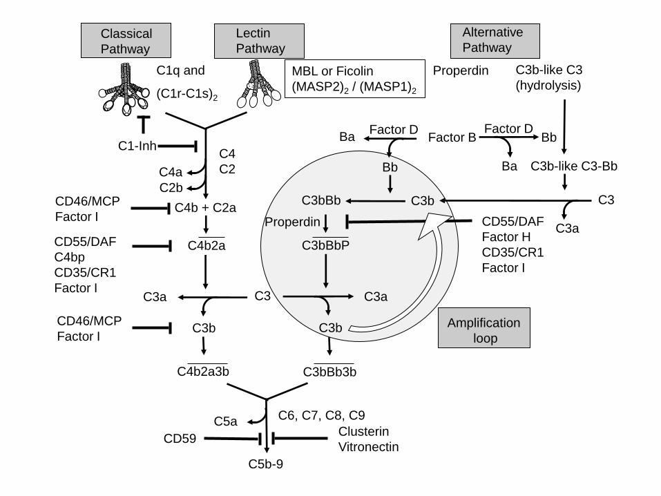
Classical
Pathway
C1q and
(C1r-C1s)2
Lectin
Pathway
MBL or Ficolin
(MASP2)2 / (MASP1)2
C1-Inh
C4a
C4b2a
C2b
C4b + C2a CD46/MCP
Factor I
CD55/DAF
C4bp
CD35/CR1
Factor I
C3b
C3a C3
C4b2a3b
CD46/MCP
Factor I
C4
C2
C3bBbP
C3b
C3bBb3b
C5a
C5b-9
CD59
C6, C7, C8, C9
Clusterin
Vitronectin
C3a
Alternative
Pathway
C3b-like C3
(hydrolysis) Properdin
Factor D
Ba C3b-like C3-Bb
C3bBb
C3a
C3
Bb Factor B Factor D
Bb
Ba
Properdin
C3b
Amplification
loop
CD55/DAF
Factor H
CD35/CR1
Factor I

AD
C1q
Tg2576 Tg2576C1q
C3
AD
C3

Table 1. Expression of C proteins by human astrocytes and microglia1
Complement Components
Cell Type Classical Alternative Terminal C-receptors C-regulators
Soluble Membrane-bound
Astrocytes C1q,C1r, C1s
C2, C3, C4
C3, fB, fD C5, C6,
C7, C8,
C9
C1qR, CR2,
C3aR, C5aR
C1-inh, fH, fI,
clusterin
CD59, DAF, MCP,
CR1
Microglia C1q, C1r,
C1s, C2, C3,
C4
C3 C1qR, CR3,
C3aR, CR4,
C5aR,
C1-inh CD59, CR1
1 C components in the brain are inducible, most notably in response to injury. Recently reviewed in (Woodruff et al., 2010).


![From Stem Cells to Oligodendrocytes: Prospects for Brain ... · detemlined "stem" state for self replenishment [23]. The best studied of these are stem cells in bone marrow, and hematopoietic](https://static.fdocuments.net/doc/165x107/5fdbb6da6b15bc4fa25f733e/from-stem-cells-to-oligodendrocytes-prospects-for-brain-detemlined-stem.jpg)