
CD47-17, Rev. 1 Control de enfermedades crónicas 11 Sep.06 ...
HAL Id: inserm-01755551https://www.hal.inserm.fr/inserm-01755551
Submitted on 30 Mar 2018
HAL is a multi-disciplinary open accessarchive for the deposit and dissemination of sci-entific research documents, whether they are pub-lished or not. The documents may come fromteaching and research institutions in France orabroad, or from public or private research centers.
L’archive ouverte pluridisciplinaire HAL, estdestinée au dépôt et à la diffusion de documentsscientifiques de niveau recherche, publiés ou non,émanant des établissements d’enseignement et derecherche français ou étrangers, des laboratoirespublics ou privés.
Complement Factor H Inhibits CD47-MediatedResolution of Inflammation
Bertrand Calippe, Sébastien Augustin, Fanny Beguier, HugoCharles-Messance, Lucie Poupel, Jean-Baptiste Conart, Shulong Hu, Sophie
Lavalette, Alexandre Fauvet, Julie Rayes, et al.
To cite this version:Bertrand Calippe, Sébastien Augustin, Fanny Beguier, Hugo Charles-Messance, Lucie Poupel, et al..Complement Factor H Inhibits CD47-Mediated Resolution of Inflammation. Immunity, Elsevier, 2017,46 (2), pp.261 - 272. �10.1016/j.immuni.2017.01.006�. �inserm-01755551�
1
Complement factor H inhibits CD47‐mediated resolution of inflammation
Bertrand Calippe1*, Sebastien Augustin1*, Fanny Beguier1, Hugo Charles Messance1,
Lucie Poupel2, Jean-Baptiste Conart1, Shulong Hu1, Sophie Lavalette1, Alexandre Fauvet1, Julie Rayes3, Olivier Levy1, William Raoul1, Catherine Fitting10, Thomas Denèfle8, Matthew C Pickering4, Claire Harris5, Sylvie Jorieux6, Patrick M. Sullivan7, José-Alain Sahel 1, Philippe Karoyan8, Przemyslaw Sapieha9, Xavier Guillonneau1, Emmanuel L. Gautier2, Florian Sennlaub1†
1Institut de la Vision, 17 rue Moreau, Sorbonne Universités, UPMC Univ Paris 06, INSERM,
CNRS, 75012 Paris, France. 2Sorbonne Universités, UPMC Univ Paris 06, INSERM UMR_S 1166, Faculté de médecine
Pitié-Salpétrière, 91 Boulevard de l’hôpital, 75013, Paris, France. 3Université Paris Descartes, Unité Mixte de Recherche en Santé 872, Centre de Recherche des
Cordeliers, Paris, France; 4Centre for Complement and Inflammation Research, Department of Medicine, Imperial
College, London W12 0NN, UK. 5Institute of Infection and Immunity, Cardiff University, Cardiff, UK. 6LFB Biotechnologies, Lille, France. 7Department of Medicine, Centers for Aging and Geriatric Research Education and Clinical
Center, Durham Veteran Affairs Medical Center, Duke University, Durham, North Carolina 27710.
8Laboratoire des Biomolécules, UMR 7203 and FR 2769, Sorbonne Universités, Université Pierre et Marie Curie, Paris, France; Centre National de la Recherche Scientifique, UMR 7203, Paris, France; Département de Chimie, École Normale Supérieure, Paris, France.
9Department of Ophthalmology, Maisonneuve-Rosemont Hospital Research Centre, University of Montreal, Quebec, Canada.
10Unité Cytokines & Inflammation, Département Infection et Epidémiologie, Institut Pasteur, 75015 Paris, France.
*these authors have contributed equally
†Correspondence should be addressed to Dr Florian Sennlaub, Inserm, UMR_S 968,
Institut de la Vision, Paris, F-75012, France. Tel: (33) 1 53 46 26 93,
Email: [email protected].
2
ABSTRACT Age-related macular degeneration (AMD) is a highly heritable major cause of blindness
characterized by subretinal inflammation. Of all genetic factors, variants of Complement factor
H (CFH) are associated with greatest linkage to AMD. Using loss of function genetics and
orthologous models of AMD, we provide mechanistic evidence that deficiency in CFH
completely prevents pathogenic subretinal accumulation of mononuclear phagocytes (MP) and
accelerates resolution of inflammation. We show that MP-persistence arises secondary to
binding of CFH to CD11b/CD18, which obstructs physiologically-occurring thrombospsondin-
1 (TSP-1)-CD47-mediated elimination of MPs from the subretinal space. The AMD-associated
CFH402H isoform markedly increased this inhibitory effect on microglial cells, indicating a
causal link to disease etiology. Our findings were confirmed in acute sterile peritonitis.
Pharmacological activation of CD47 accelerated resolution of both subretinal and peritoneal
inflammation, which may be exploited in the therapy for chronic inflammatory diseases,
including AMD.
3
INTRODUCTION Age-related Macular Degeneration (AMD) is a highly heritable neuroinflammatory disorder
characterized by sizeable deposits of lipoproteinaceous debris called soft drusen (early AMD),
choroidal neovascularisation (wet AMD, late form), or by an extending lesion of the retinal
pigment epithelium (RPE) and photoreceptors (geographic atrophy, GA, late form) (Ambati et
al., 2013; Sarks, 1976). Early AMD affects more than 150 million people worldwide and 10
million patients suffer from late AMD (Wong et al., 2014). AMD is strongly associated with
common and rare variants of Complement factor H (CFH), indicating its causal implication in
the pathogenesis (Fritsche et al., 2013; Fritsche et al., 2016). The understanding of its mode of
action and more generally the origins of AMD remain unclear.
CFH is an abundant soluble plasma factor composed of 20 Short Consensus Repeat domains
(SCR) (Kopp et al., 2012) with important roles in inflammation (Kopp et al., 2012; Mihlan et
al., 2009), coagulation (Rayes et al., 2014; Vaziri-Sani et al., 2005) and as an antioxidant (Shaw
et al., 2012; Weismann et al., 2011). The SCR7 domain of CFH binds to glycosaminoglycans
(GAG) on cell surfaces where it inhibits complement activation (Kopp et al., 2012). This
domain additionally allows CFH to bind to myeloid cells via the integrin CD11b/CD18
(Complement 3 Receptor, Mac-1) (DiScipio et al., 1998; Losse et al., 2010), supporting myeloid
cell adhesion (DiScipio et al., 1998) and migration (Losse et al., 2010) as well as the
phagocytosis of microbes (Losse et al., 2010) and cell debris (Kang et al., 2012). At the
inflammatory site, CFH is strongly secreted by mononuclear phagocytes (MP), such as
microglial cells (MC) and macrophages (Mφ) (Gautier et al., 2012; Luo et al., 2011; Schlaf et
al., 2001), adding to the exudated plasma CFH and CFH produced by certain stromal cells such
as the retinal pigment epithelium (RPE) in the eye (Anderson et al., 2010).
The substitution of histidine 402 for tyrosine (Y402H) in the SCR7 domain accounts for
the major part of the genetic risk of AMD (Fritsche et al., 2014), but is also strongly associated
with other conditions such as smoking-associated lung cancer (Zhang et al., 2012) and increased
4
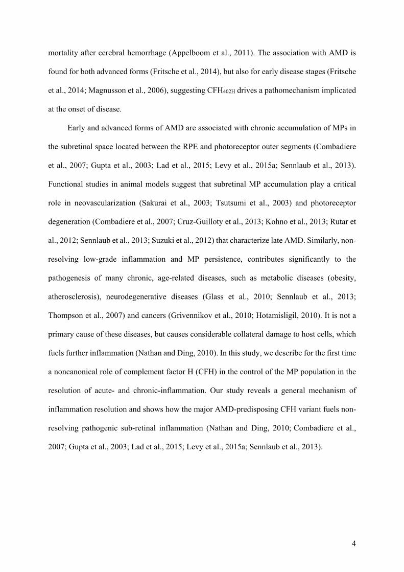
mortality after cerebral hemorrhage (Appelboom et al., 2011). The association with AMD is
found for both advanced forms (Fritsche et al., 2014), but also for early disease stages (Fritsche
et al., 2014; Magnusson et al., 2006), suggesting CFH402H drives a pathomechanism implicated
at the onset of disease.
Early and advanced forms of AMD are associated with chronic accumulation of MPs in
the subretinal space located between the RPE and photoreceptor outer segments (Combadiere
et al., 2007; Gupta et al., 2003; Lad et al., 2015; Levy et al., 2015a; Sennlaub et al., 2013).
Functional studies in animal models suggest that subretinal MP accumulation play a critical
role in neovascularization (Sakurai et al., 2003; Tsutsumi et al., 2003) and photoreceptor
degeneration (Combadiere et al., 2007; Cruz-Guilloty et al., 2013; Kohno et al., 2013; Rutar et
al., 2012; Sennlaub et al., 2013; Suzuki et al., 2012) that characterize late AMD. Similarly, non-
resolving low-grade inflammation and MP persistence, contributes significantly to the
pathogenesis of many chronic, age-related diseases, such as metabolic diseases (obesity,
atherosclerosis), neurodegenerative diseases (Glass et al., 2010; Sennlaub et al., 2013;
Thompson et al., 2007) and cancers (Grivennikov et al., 2010; Hotamisligil, 2010). It is not a
primary cause of these diseases, but causes considerable collateral damage to host cells, which
fuels further inflammation (Nathan and Ding, 2010). In this study, we describe for the first time
a noncanonical role of complement factor H (CFH) in the control of the MP population in the
resolution of acute- and chronic-inflammation. Our study reveals a general mechanism of
inflammation resolution and shows how the major AMD-predisposing CFH variant fuels non-
resolving pathogenic sub-retinal inflammation (Nathan and Ding, 2010; Combadiere et al.,
2007; Gupta et al., 2003; Lad et al., 2015; Levy et al., 2015a; Sennlaub et al., 2013).
5
RESULTS
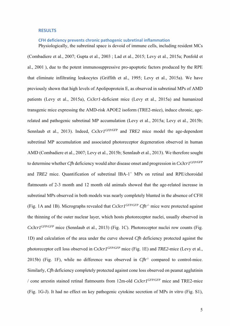
CFH deficiency prevents chronic pathogenic subretinal inflammation Physiologically, the subretinal space is devoid of immune cells, including resident MCs
(Combadiere et al., 2007; Gupta et al., 2003 ; Lad et al., 2015; Levy et al., 2015a; Penfold et
al., 2001 ), due to the potent immunosuppressive pro-apoptotic factors produced by the RPE
that eliminate infiltrating leukocytes (Griffith et al., 1995; Levy et al., 2015a). We have
previously shown that high levels of Apolipoprotein E, as observed in subretinal MPs of AMD
patients (Levy et al., 2015a), Cx3cr1-deficient mice (Levy et al., 2015a) and humanized
transgenic mice expressing the AMD-risk APOE2 isoform (TRE2-mice), induce chronic, age-
related and pathogenic subretinal MP accumulation (Levy et al., 2015a; Levy et al., 2015b;
Sennlaub et al., 2013). Indeed, Cx3cr1GFP/GFP and TRE2 mice model the age-dependent
subretinal MP accumulation and associated photoreceptor degeneration observed in human
AMD (Combadiere et al., 2007; Levy et al., 2015b; Sennlaub et al., 2013). We therefore sought
to determine whether Cfh deficiency would alter disease onset and progression in Cx3cr1GFP/GFP
and TRE2 mice. Quantification of subretinal IBA-1+ MPs on retinal and RPE/choroidal
flatmounts of 2-3 month and 12 month old animals showed that the age-related increase in
subretinal MPs observed in both models was nearly completely blunted in the absence of CFH
(Fig. 1A and 1B). Micrographs revealed that Cx3cr1GFP/GFP Cfh-/- mice were protected against
the thinning of the outer nuclear layer, which hosts photoreceptor nuclei, usually observed in
Cx3cr1GFP/GFP mice (Sennlaub et al., 2013) (Fig. 1C). Photoreceptor nuclei row counts (Fig.
1D) and calculation of the area under the curve showed Cfh deficiency protected against the
photoreceptor cell loss observed in Cx3cr1GFP/GFP mice (Fig. 1E) and TRE2-mice (Levy et al.,
2015b) (Fig. 1F), while no difference was observed in Cfh-/- compared to control-mice.
Similarly, Cfh deficiency completely protected against cone loss observed on peanut agglutinin
/ cone arrestin stained retinal flatmounts from 12m-old Cx3cr1GFP/GFP mice and TRE2-mice
(Fig. 1G-J). It had no effect on key pathogenic cytokine secretion of MPs in vitro (Fig. S1),
6
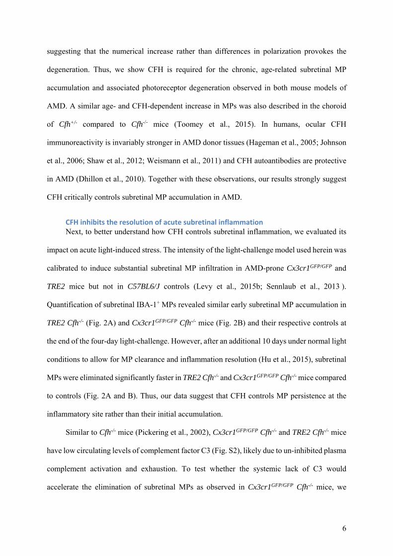
suggesting that the numerical increase rather than differences in polarization provokes the
degeneration. Thus, we show CFH is required for the chronic, age-related subretinal MP
accumulation and associated photoreceptor degeneration observed in both mouse models of
AMD. A similar age- and CFH-dependent increase in MPs was also described in the choroid
of Cfh+/- compared to Cfh-/- mice (Toomey et al., 2015). In humans, ocular CFH
immunoreactivity is invariably stronger in AMD donor tissues (Hageman et al., 2005; Johnson
et al., 2006; Shaw et al., 2012; Weismann et al., 2011) and CFH autoantibodies are protective
in AMD (Dhillon et al., 2010). Together with these observations, our results strongly suggest
CFH critically controls subretinal MP accumulation in AMD.
CFH inhibits the resolution of acute subretinal inflammation Next, to better understand how CFH controls subretinal inflammation, we evaluated its
impact on acute light-induced stress. The intensity of the light-challenge model used herein was
calibrated to induce substantial subretinal MP infiltration in AMD-prone Cx3cr1GFP/GFP and
TRE2 mice but not in C57BL6/J controls (Levy et al., 2015b; Sennlaub et al., 2013 ).
Quantification of subretinal IBA-1+ MPs revealed similar early subretinal MP accumulation in
TRE2 Cfh-/- (Fig. 2A) and Cx3cr1GFP/GFP Cfh-/- mice (Fig. 2B) and their respective controls at
the end of the four-day light-challenge. However, after an additional 10 days under normal light
conditions to allow for MP clearance and inflammation resolution (Hu et al., 2015), subretinal
MPs were eliminated significantly faster in TRE2 Cfh-/- and Cx3cr1GFP/GFP Cfh-/- mice compared
to controls (Fig. 2A and B). Thus, our data suggest that CFH controls MP persistence at the
inflammatory site rather than their initial accumulation.
Similar to Cfh-/- mice (Pickering et al., 2002), Cx3cr1GFP/GFP Cfh-/- and TRE2 Cfh-/- mice
have low circulating levels of complement factor C3 (Fig. S2), likely due to un-inhibited plasma
complement activation and exhaustion. To test whether the systemic lack of C3 would
accelerate the elimination of subretinal MPs as observed in Cx3cr1GFP/GFP Cfh-/- mice, we
7
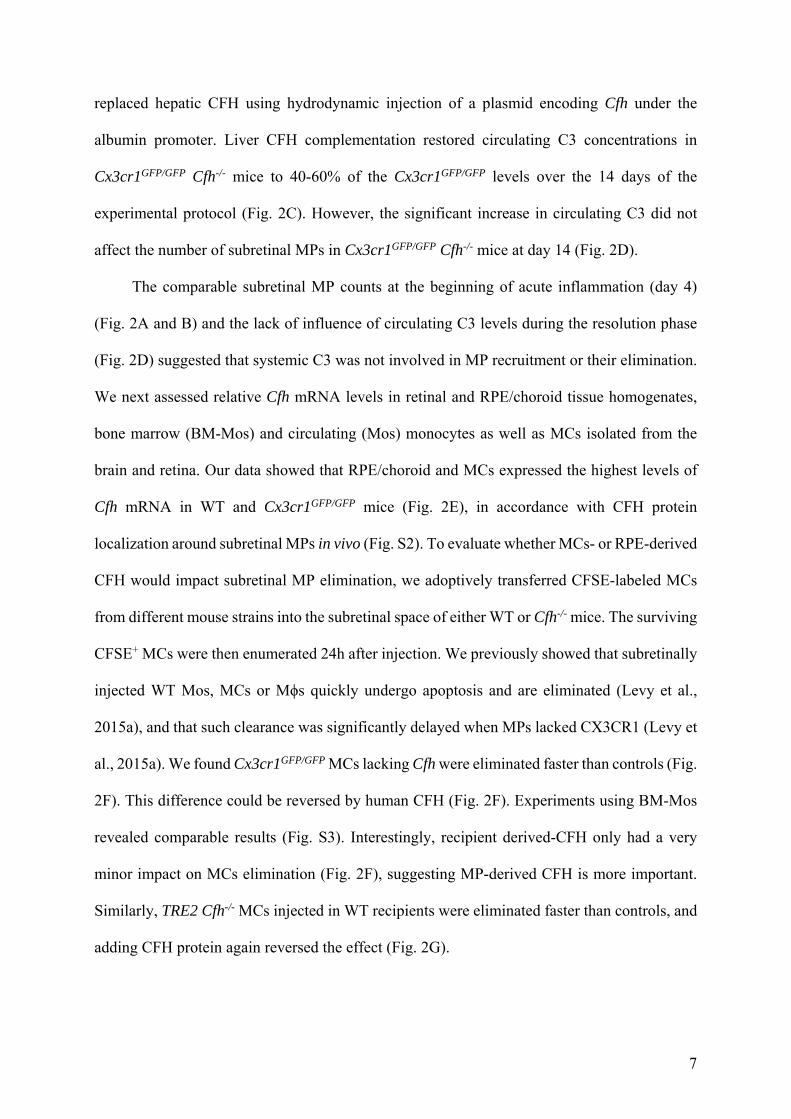
replaced hepatic CFH using hydrodynamic injection of a plasmid encoding Cfh under the
albumin promoter. Liver CFH complementation restored circulating C3 concentrations in
Cx3cr1GFP/GFP Cfh-/- mice to 40-60% of the Cx3cr1GFP/GFP levels over the 14 days of the
experimental protocol (Fig. 2C). However, the significant increase in circulating C3 did not
affect the number of subretinal MPs in Cx3cr1GFP/GFP Cfh-/- mice at day 14 (Fig. 2D).
The comparable subretinal MP counts at the beginning of acute inflammation (day 4)
(Fig. 2A and B) and the lack of influence of circulating C3 levels during the resolution phase
(Fig. 2D) suggested that systemic C3 was not involved in MP recruitment or their elimination.
We next assessed relative Cfh mRNA levels in retinal and RPE/choroid tissue homogenates,
bone marrow (BM-Mos) and circulating (Mos) monocytes as well as MCs isolated from the
brain and retina. Our data showed that RPE/choroid and MCs expressed the highest levels of
Cfh mRNA in WT and Cx3cr1GFP/GFP mice (Fig. 2E), in accordance with CFH protein
localization around subretinal MPs in vivo (Fig. S2). To evaluate whether MCs- or RPE-derived
CFH would impact subretinal MP elimination, we adoptively transferred CFSE-labeled MCs
from different mouse strains into the subretinal space of either WT or Cfh-/- mice. The surviving
CFSE+ MCs were then enumerated 24h after injection. We previously showed that subretinally
injected WT Mos, MCs or Mϕs quickly undergo apoptosis and are eliminated (Levy et al.,
2015a), and that such clearance was significantly delayed when MPs lacked CX3CR1 (Levy et
al., 2015a). We found Cx3cr1GFP/GFP MCs lacking Cfh were eliminated faster than controls (Fig.
2F). This difference could be reversed by human CFH (Fig. 2F). Experiments using BM-Mos
revealed comparable results (Fig. S3). Interestingly, recipient derived-CFH only had a very
minor impact on MCs elimination (Fig. 2F), suggesting MP-derived CFH is more important.
Similarly, TRE2 Cfh-/- MCs injected in WT recipients were eliminated faster than controls, and
adding CFH protein again reversed the effect (Fig. 2G).
8
In summary, our data show that CFH does not influence initial MP recruitment but
inhibits MP elimination during inflammation resolution. They also demonstrate that RPE- and
liver-derived CFH have little influence on this phenotype. Indeed, MP-derived CFH is
sufficient to inhibit MP clearance. Similar to our experiments, the observation that recipient,
but not liver Cfh genotype, confers the AMD-risk in liver transplant patients (Khandhadia et
al., 2013) suggests that plasma CFH is not involved in AMD pathogenesis and points to the
importance of MP-derived CFH in the disease.
CFH binding to CD11b/CD18 inhibits TSP‐1/CD47 mediated MP elimination CFH can act as a cofactor of complement factor I (CFI) to cleave C3b into iC3b, thereby
inactivating the C3 convertase that is formed by C3b and activated complement factor B (CFB),
while opsonizing apoptotic bodies with increased iC3b (Martin and Blom, 2016). However, the
absence of C3 or activated C3 fragments in subretinal MPs of Cx3cr1GFP/GFP or Cx3cr1GFP/GFP
Cfh-/- mice (Fig. S2) and low levels of C3 mRNA in MCs and undetectable transcription of Cfi
mRNA in MCs and BM-Mos (Fig. S4) and make a significant implication of locally produced
C3, C3b, or iC3b in CFH-mediated inhibition of MP elimination unlikely.
CFH also binds directly to the integrin CD11b/CD18 (DiScipio et al., 1998; Kang et al.,
2012; Losse et al., 2010) that is strongly expressed in MPs with no detectable differences in our
different mouse strains (Fig. S5). Flow cytometry analysis showed fluorescently labeled CFH
binds dose-dependently to MCs (Fig. 3A) and BM-Mos isolated from Cx3cr1GFP/GFP Cfh-/- mice
(Fig. 3B). Pre-incubation with an anti-CD11b antibody inhibited CFH binding (Fig. 3B), as
previously demonstrated for neutrophils (DiScipio et al., 1998). As shown above (Fig. 2F),
adding CFH to Cx3cr1GFP/GFP Cfh-/- MCs transferred subretinally in WT recipients delayed their
elimination. This effect was no longer observed when transferred MPs were treated with the
anti-CD11b antibody (Fig. 3C). The antibody also significantly accelerated the elimination of
CFH-competent Cx3cr1GFP/GFP MCs, contrary to an anti-C3b/iC3b/C3c antibody that inhibits
9
complement-induced hemolysis (clone 3/26 (Mastellos et al., 2004), Fig. 3C). These results
show that CFH binding to the CD11b/CD18 integrin complex is necessary to inhibit MC
elimination and suggest a C3-independent, non-canonical role of CFH in the mechanism.
CD11b/CD18 co-localizes with the integrin-associated protein (IAP, CD47) in lipid-rafts
(Pfeiffer et al., 2001), a thrombospsondin 1 (THBS1, TSP-1) receptor known to potentiate
FasL-induced endothelial cell and T cell death (Manna et al., 2005; Quesada et al., 2005). We
therefore investigated whether CFH binding to CD11b/CD18 could limit CD47 activation and
impair MP elimination in Cx3cr1GFP/GFP and TRE2 mice (the expression level of TSP-1 and
CD47 and the plasma TSP-1 levels were similar in our different mouse strains, Fig. S5). First,
proximity ligation assay revealed numerous CD11b/CD47 complexes on Cx3cr1GFP/GFP Cfh-/-
MCs (Fig. 3D). We next analyzed the role of CD47 in subretinal MC clearance in adoptive
transfer experiments. Subretinally injected CFSE-labeled MCs from WT, Thbs1-/- or Cd47-/-
donors into WT recipients revealed MCs lacking Thbs1 or Cd47 have slower elimination rates
than WT MCs (Fig. 3E). Co-injected recombinant TSP-1 accelerated the elimination of WT
MCs, reversed the phenotype of Thbs1-deficient MCs but had no effect on MCs lacking Cd47
(Fig. 3E), suggesting the interaction of TSP1 with CD47 mediates MC elimination. Moreover,
analysis of Thbs1-/- and Cd47-/- mice revealed a significant age-related (Fig. 3F) and light-
induced (Fig. 3G) increase in subretinal MPs in these strains as compared to controls, but not
in mice lacking Cd36, an alternative TSP-1 receptor. Overall, our results point to TSP-1/CD47
signaling as central in the maintenance of subretinal immunosuppression, and likely explains
the previously reported increased and prolonged subretinal inflammation observed in Thbs1-/-
mice (Chen et al., 2012; Ng et al., 2009; Wang et al., 2012). Interestingly, binding of TSP-1 to
CD36 that mediates endothelial cell apoptosis (Jimenez et al., 2000) and is necessary for latent
TGF-β activation (Yehualaeshet et al., 1999) had no significant influence on subretinal MP
accumulation, as observed here in Cd36-/- mice.
10
Taking into account the opposing effects of TSP1 and CFH, we next evaluated their
interaction in MP elimination. Using our adoptive transfer assay, we found a TSP-1 blocking
antibody reversed the accelerated elimination of Cx3cr1GFP/GFP Cfh-/- MCs compared to controls
(Fig. 3H). Furthermore, the faster elimination rate observed after supplementation of
recombinant TSP-1 to Cx3cr1GFP/GFP Cfh-/- MCs was completely lost when purified CFH was
concomitantly added (Fig. 3I).
Next, we used the laser-injury model to test whether CFH binding to CD11b or CD47
activation can accelerate inflammation resolution in CFH-competent Cx3cr1GFP/GFP mice.
Using this model, we take advantage of the fact that laser burn induces a thinning of the retina
above the impact, facilitating diffusion of intravitreally-injected molecules to the subretinal
space. Our results showed the intravitreal injection at the height of laser-induced subretinal
inflammation (d4 and d7) of recombinant TSP-1 (Fig. 3J), CD47-activating peptide PKHB1 (a
derivate of the 4N1K CD47-agonist peptide with improved pharmacological properties
(Martinez-Torres et al., 2015), Fig. 3K), or the anti-CD11b antibody (that blocks CFH binding
to CD11b, Fig. 3L) all significantly accelerated subretinal MP elimination as compared to their
controls.
Taken together, our data show that CFH binding to the integrin CD11b/CD18 inhibits
subretinal MC elimination. We demonstrate that CD47 co-localizes with CD11b/CD18 on MPs
and mediates the physiological role of TSP-1 in subretinal MP elimination. The observation
that TSP-1 blockade reversed the effect of CFH deficiency and that recombinant CFH blocks
the effect of TSP-1 on subretinal MP elimination strongly suggests that CFH binding to
CD11b/CD18 interferes with TSP-1/CD47 signaling. We show that TSP-1 and more
specifically CD47 activation efficiently accelerates MP elimination similar to Cfh-deficiency.
11
CFH inhibits the resolution of acute sterile peritonitis Non-resolving inflammation contributes significantly to the pathogenesis of many
chronic, age-related diseases (Nathan and Ding, 2010). To test whether CFH influences
inflammation resolution in other pathological contexts, we used a model of acute thioglycollate-
induced peritonitis, characterized by an early accumulation of neutrophils, followed by
recruited monocyte-derived inflammatory macrophages (recMφ), both experiencing an
apoptosis-driven elimination at different kinetics (Gautier et al., 2013). Analysis of the ImmGen
dataset (Gautier et al., 2012) (GenBank no. GSE37448) shows Cfh mRNA levels in
thioglycollate-elicited peritoneal recMφ is approximately doubled compared to circulating
blood Ly-6C+ Mo, from which they derive (Fig. 4A). Thus, recMφs likely participate to local
CFH in peritonitis, in addition to extravasated plasma CFH. Quantification of recruited CD115+
F4/80+ ICAM2lo recMφs in Cfh-/- mice and controls (Fig. 4B), revealed a robust and similar
early accumulation 1 day after peritonitis induction (Fig. 4C). At day 3 however, while the
numbers of recMφs continued to rise in WT mice, they had receded by 50% in Cfh-/- mice (Fig.
4C). This observation was similar to the accelerated elimination of subretinal MPs observed in
light-challenged TRE2 Cfh-/- and Cx3cr1GFP/GFP Cfh-/- mice (Fig. 2A and B). Human
recombinant CFH injected into the peritoneal cavity of Cfh-/- mice at day 1 significantly
inhibited the enhanced clearance of recMφs observed in this strain at day 2 as compared to heat-
denatured CFH (Fig. 4D), akin to the effect we observed in subretinally injected Cx3cr1GFP/GFP
Cfh-/- MCs (Fig. 2F). In addition, similar to our results in MCs (Fig. 3D), a proximity ligation
assay revealed numerous and specific CD11b/CD47 complexes in WT recMφ retrieved at day
1 (Fig. 4E). Finally, a single intra-peritoneal injection of recombinant TSP-1 or the CD47-
specific activating peptide PKHB1 at day 1 significantly accelerated the elimination of recMφs
as observed at day 2 (Fig. 4F).
In summary, our results show that CFH inhibits recMφ elimination in sterile peritonitis
confirming findings for infiltrating MPs in the subretinal space. The observation that
12
CD11b/CD47 complexes are present on peritoneal recMφ and that CD47 activation accelerates
recMφ elimination during peritonitis strongly suggests that CFH inhibits CD47-dependent
inflammation resolution similarly in both the eye and the peritoneum.
The CFH402H variant inhibits subretinal MC elimination more potently than the common CFHY402 The substitution of histidine 402 for tyrosine (Y402H) in CFH sequence accounts for a major
part of the genetic risk of AMD. To test whether the Y402H polymorphism influenced the
elimination of distinct MPs differently, we transferred CFSE-labeled Cx3cr1GFP/GFP Cfh-/- MCs
or BM-Mos to the subretinal space together with purified CFHY402 or the disease associated
CFH402H to WT mice. CFSE+ cell counts after 24h revealed that both isoforms inhibited
clearance of BM-Mos (Fig. 5A) and MCs (Fig. 5B) compared to cells injected without CFH.
However, while CFHY402 and CFH402H had similar effects on BM-Mo elimination (Fig. 5A),
CFH402H limited MC clearance by 37% compared to CFHY402 (Fig. 5B). In addition,
recombinant CFHY402 reversed the accelerated elimination rate observed when subretinally
injected Cx3cr1GFP/GFP Cfh-/- MCs were treated with recombinant TSP-1, and recombinant
CFH402H was 50% more potent at inhibiting this phenomenon (Fig. 5C).
Taken together our results show that CFH402H was significantly more potent to inhibit
the subretinal elimination of certain MPs, such as MCs. The CFH402H variant might thereby
spur non-resolving inflammation under the retina, and thus explain its association with early
and late AMD (Fritsche et al., 2014; Magnusson et al., 2006) where subretinal MPs
accumulate(Combadiere et al., 2007; Gupta et al., 2003; Lad et al., 2015; Levy et al., 2015a;
Sennlaub et al., 2013).
DISCUSSION We report the previously unknown ability of CFH to favor subretinal MP accumulation in mice
developing chronic, non-resolving, age-related inflammation in the eye. We extended this
finding to inflammation resolution in general by using models of acute inflammation in the
13
peritoneum and the eye. Our work supports the long-standing association between CFH variant
and AMD, and we uncovered the mechanisms by which CFH impacts MP infiltration,
independently from its action in the complement cascade.
The subretinal space is prone to MP infiltration due to light-induced oxidative stress and
high metabolic activity (Combadiere et al., 2007; Ng and Streilein, 2001; Suzuki et al., 2012),
and this is physiologically counterbalanced by the expression of immune-suppressive factors
by the RPE (Griffith et al., 1995; Levy et al., 2015a). We show that CFH favors MP
accumulation by inhibiting their TSP-1/CD47-mediated elimination. Remarkably, Thbs1-/- and
Cd47-/- mice develop age-related subretinal inflammation under normal living conditions. This
can possibly be explained by the sensitizing role of TSP-1 on FasL-induced cell death (Manna
et al., 2005; Quesada et al., 2005) as FasL expression by the RPE is necessary to prevent
subretinal MP accumulation (Griffith et al., 1995; Levy et al., 2015a).
CFH is expressed at high levels in MCs and Mo-derived Mφs in peritonitis and we
confirmed a step-wise binding interaction between CFH and CD11b/CD18 (Gautier et al., 2012;
Luo et al., 2011; Schlaf et al., 2001) and CD11b and CD47 (Pfeiffer et al., 2001) on MPs. CFH
ligation to CD11b/CD18 might thereby sterically hinder the ligation of TSP-1 to CD47 , and
increase MP lifespan. Complement activation or CFH-dependent production of iC3b, an
alternative ligand of CD11b/CD18, did not play a detectable role in the process, as
C3b/iC3b/C3c fragments were not found in the subretinal space and CFI (necessary to produce
iC3b) is not significantly transcribed in MPs. A specific antibody that inhibits C3-dependent
hemolysis (Mastellos et al., 2004) did not accelerate MP elimination.
In addition, we found that the AMD-associated CFH402H variant has an increased capacity
to inhibit the elimination of certain MP populations, such as MCs, strengthening the causal link
to AMD etiology. Although CFH402H affinity is decreased to certain GAG species, it is higher
in particular to GAG sulfates (Clark et al., 2006). GAG sulfate profiles differ greatly between
14
MPs and different microenvironments. For example, keratan sulfate proteoglycans (KPSG) are
strongly present on ramified brain MCs but not on blood Mos (Wilms et al., 1999). The
differential expression of GAG sulfates, such as KPSG, on MCs (Wilms et al., 1999) might
explain why CFH402H differentially influences MC but not BM-Mos elimination. Subretinal
MPs originating from infiltrating Mo and MCs (Sennlaub et al., 2013) invariably express KPSG
strongly (Combadiere et al., 2007; Ng and Streilein, 2001; Ng et al., 2009). This is also the case
in spinal cord injury 53, but not in autoimmune neuritis (Jones and Tuszynski, 2002; Matsui et
al., 2013). CFH402H might therefore have a particularly strong influence on subretinal
inflammation observed in AMD but not necessarily in other chronic inflammatory diseases. In
human evolution, the limited elimination of MPs and the increased inflammatory reaction
associated with CFH402H might have increased survival to certain infectious diseases, leading
to its high frequency as observed today in certain populations. In AMD, CFH402H might be
pathogenic as it fuels subretinal inflammation and leads to chronic inflammation, which add to
its decreased capacity to inhibit oxidative stress (Shaw et al., 2012; Weismann et al., 2011) and
to bind to Bruchs membrane (Clark et al., 2010) that protects the RPE against uncontrolled
complement activation (Coffey et al., 2007; Toomey et al., 2015).
Current anti-inflammatory therapies, such as steroids, non-steroidal anti-inflammatroy
drugs (NSAID), or immunosuppressive drugs (Ciclosporin) can have paradoxical effects on
macrophage function. They increase proinflammatory mediators (high-dose steroids) (Lim et
al., 2007), upregulate toll-like receptors on macrophages (Ciclosporin) (Tedesco and Haragsim,
2012) and prolong macrophage infiltration (NSAID) (Gilroy et al., 1999), which might explain
their lack of therapeutic effect in AMD. Our findings introduce a new strategy to directly induce
the elimination of resilient macrophage accumulation in AMD, and possibly other conditions,
by pharmacological activation of CD47.

15
Acknowledgments: This work was supported by grants from INSERM, ANR Geno 2009 (R09099DS), ANR
MACLEAR (ANR-15-CE14-0015-01), LABEX LIFESENSES [ANR-10-LABX-65]
supported by the ANR (Investissements d'Avenir programme [ANR-11-IDEX-0004-02]),
Carnot, and ERC starting Grant (ERC-2007 St.G. 210345), the Association de Prévoyance
Santé de ALLIANZ, and a generous donation by Doris and Michael Bunte.

16
Materials and Methods:
Animals Cfh-/- and targeted replacement mice that express human APOE isoforms (TRE2, TRE3)
were generous gifts from Mathew Pickering and Patrick Sullivan, respectively. Cx3cr1GFP/GFP,
Thbs1-/-, Cd47-/-, and Cd36-/- mice were purchased (Charles River Laboratories, Jackson
laboratories) and Cx3cr1GFP/GFP Cfh-/- and TRE2 Cfh-/- mouse strains were generated in-house.
All mouse strains were either negative or backcrossed to become negative (Thbs1-/-) for the
Pde6brd1, Gnat2cpfl3, and Crb1rd8 mutations that can lead to AMD-like features (Mattapallil et
al., 2012). Mice were housed under specific pathogen-free condition, in a 12h/12h light/dark
(100 lux) cycle with no additional cover in the cage, and with water and normal chow diet
available ad libitum. All experimental protocols and procedures were approved by the local
animal care ethics committee, “Comité d’éthique en expérimentation animale Charles Darwin”
(N° Ce5/2010/011, Ce5/2010/044, Ce5/2011/033).
Choroidal and retinal flatmounts for mononuclear phagocyte and cone quantification Eyes from male and female mice (as we never noticed a sex-dependent difference in
subretinal MP accumulation in the past) (Combadiere et al., 2007; Hu et al., 2015; Levy et al.,
2015a; Levy et al., 2015b; Sennlaub et al., 2013) were enucleated, fixed for 30 min in 4% PFA
and sectioned at the limbus while the cornea and lens were discarded. The retinas were carefully
peeled from the RPE/choroid/sclera. Retina and choroid were incubated with Peanut agglutinin
Alexa 594 (Thermofisher; 1:50), anti-IBA-1 antibody (Wako chemicals; 1:400), , and anti-
mouse cone-arrestin antibody (Millipore, #AB15282; 1:10 000) followed by secondary anti-
rabbit antibody coupled to Alexa 488 and Alexa 647 (Thermo Fisher; 1:500) and nuclear
staining using Hoechst. Choroids and retinas were flatmounted and viewed with a fluorescence
microscope DM5500B (Leica). IBA-1+ cells were counted on whole RPE/choroidal flatmounts
17
and on the outer segment side of the retina. PA+cone arrestin+ cells were counted on oriented
retinal flatmounts in the central and peripheral retina.
Histology and Immunohistochemistry Eyes were fixed in 0.5% glutaraldehyde, 4% PFA for 2h, dehydrated and mounted in
Historesin (Leica). Oriented sections (5µm) crossing the inferior pole, optic nerve and superior
pole were cut and stained with toluidin blue. Rows of nuclei in the outer nuclear layer (ONL)
were counted at different distances from the optic nerve (Sennlaub et al., 2013). For
immunohistochemistry, eyes from 4d light-challenged mice were fixed for 2h in 4% PFA,
incubated in 30% sucrose overnight at 4°C, embedded in OCT and sectioned (10μm), and
stained with anti-C3 antibody (clone 11H9 Hycult biotech; 1:50), anti-C3b/iC3b/C3c antibody
(clone 3/26 Hycult biotech; 1:50), and anti-CFH antibody (ab8842 Abcam; 1:100) and
appropriate secondary antibodies and Hoechst nuclear stain.
Light challenge model Two- to three-months old male and female mice were adapted to darkness for 6 hours,
pupils dilated daily and exposed to green LED light (starting at 2AM, 4500 Lux, JP Vezon
Equipements) for 4 days and subsequently kept in cyclic 12h/12h normal facility conditions as
previously described (Sennlaub et al., 2013). MP count was assessed at the end of light exposure
(day 4) or 10 days later (day 14).
Hydrodynamic injection Murine Complement Factor H was cloned into pLIVE vector (Mirus) using NheI and
SacII restriction sites, so that, its expression was under the control of a mouse minimal albumin
promoter. The construct was sequenced and the plasmid amplified with endotoxin-free
Megaprep kits (Qiagen). Control groups were injected with the empty pLIVE plasmid and test
groups with pLIVE expressing murine CFH. 100 µg of plasmid diluted in NaCl 0.9% were
injected per mouse. The volume injected in the venous tail was 10% of the body weight (Rayes
et al., 2010), which leads to robust transfection of the plasmid in the liver. Four days later, mice
18
were exposed to the light challenge model and 50μl of blood were taken (mandibular vein) at
day 0, 4 and 14 to quantify plasma C3 concentration by ELISA (Innovative Research).
Monocyte and microglial cell preparations, analysis, and culture Blood samples were collected for determination of plasma C3 (Innovative Research) and
TSP-1 (Elabscience) levels by ELISA. Bone marrow monocytes, circulating monocytes ,
central nervous MC and retinal MC were purified. Mos were isolated by negative selection
(EasySep Mouse Enrichment Kit, Stemcell Technologies). MCs were prepared from
dissociated PBS-perfused brains or retinas (Neural Dissociation Kit, Miltenyi Biotec). After
dissociation, 70µm filtered cell suspensions were washed and myelin was eliminated Percoll
density gradient centrifugation. Then cells labeled with anti-CD11b microbeads (clone
M1/70.15.11.5, Miltenyi Biotec) were purified by MS Columns (Miltenyi Biotec) and washed.
No serum was used in any step of the purification to avoid cell contamination with serum
derived CFH. The cells were used for adoptive transfer experiments, analyzed by RT-qPCR or
cultured for 24h in serum free DMEM, in presence of recombinant human APOE3 (Interchim;
10µg/mL), and finally their supernatants were analyzed by cytokine multiplex array
(MILLIPLEX MAP Mouse Cytokine/Chemokine Magnetic Bead Panel, Merck Millipore).
Subretinal adoptive mononuclear phagocyte transfer and clearance (Levy et al., 2015a)
Brain MCs, of the indicated male mouse strains were sorted as described above, labeled
in 10μM CFSE (Thermo Fisher Scientific), washed and resuspended in PBS. They express the
molecules of interest similarly to retinal MCs (Fig. S4, S6, and S7). 12000 cells (in 4µL) were
injected in the subretinal space of anesthetized WT or Cfh-/- male mice (10-14 weeks old) using
glass microcapillaries (Eppendorf) and a microinjector. A hole was pierced with the glass
capillary prior to the injection in order to avoid intra-ocular pressure increase and to allow
retinal detachment with 4µl of solution. The subretinal injection was verified by fundoscopy.
In specific experiments, cells were co-injected with purified commercial human CFH
19
(500μg/ml; Tecomedical), purified native CFHY402 and CFH402H (500μg/ml, obtained from
Claire Harris), recombinant CFH (500μg/ml, LFB Biotechnologies, Lille), recombinant human
TSP-1 (10µg/ml, R&D Systems), anti-CD11b antibody (10µg/ml, clone 5C6 Abd Serotec),
anti-C3b/iC3b/C3c antibody (10µg/ml, clone 3/26 Hycult biotech), isotype control rat IgG2
(10µg/ml, Invivogen), anti-TSP-1 antibody (10µg/ml, clone A4.1, Thermo Fisher), and isotype
control mouse IgM (10µg/ml, Invivogen). Eyes were enucleated after 24 hours, fixed 30
minutes in PFA 4% and counterstained with Hoechst nuclear stain. Eyes with hemorrhages
were discarded. CFSE+ cells in the subretinal space were quantified on flatmounts on the RPE
side of the retina and on the apical side of the RPE.
Thioglycollate induced peritonitis and flow cytometry
Intraperitoneal injections of 0.5 ml of 3% (wt/vol) thioglycollate (T 9032 Sigma Aldrich) broth
were used to elicit peritonitis in male Cfh-/- mice and wildtype C57BL6/J controls. Where
indicated, mice undergoing peritonitis were injected at day 1 with 100μl of PBS containing
native or heat inactivated (1h at 96°C) purified commercial human CFH (500μg/ml;
Tecomedical), recombinant human TSP-1 (50µg/ml, R&D Systems), the CD47-activating
peptide PKHB1 (500µM) or the 4NGG control peptide (500µM), and exudate cells were
analyzed at the indicated days.
At the indicated times, peritoneal lavage was performed after injecting 4 ml PBS containing
1% BSA and 0.5mM EDTA. Cells were labeled with antibodies directed against CD115 (clone
AFS98), F4/80 (clone BM8), Gr-1 (anti-Ly6C and Ly-6G, clone RB6-8C5), ICAM-2 (clone
3C4), CD11b (M1/70) and MHC-II (clone M5/114.15.2). Antibodies were purchased from
eBioscience. Cells suspensions were run on a BD Fortessa flow cytometer (BD biosciences)
and analysis was performed using Flowjo software (TreeStar)
20
CFH binding assay by flow cytometry Human CFH was conjugated to Cyanin 5.5 (Abcam conjugation kits). MCs from
Cx3cr1GFP/GFP Cfh-/- were stained 30 minutes with Cy5.5-conjugated human CFH in PBS at
indicated concentrations at 37°C and washed two times before acquisition on a BD Fortessa
flow cytometer (BD biosciences). Analysis was performed using Flowjo software (TreeStar).
Briefly, doublets were eliminated with FSC-H, FSC-W, SSC-H and SSC-W and microglial
cells were identified as GFPhigh cells. Bone marrow monocytes were blocked with anti
CD16/CD32 antibodies (Seroblock, AbD Serotec) for 15 minutes, preincubated with control
IgG or anti-CD11b antibody (clone 5C6, Abd Serotec) at 10µg/ml, and incubated with
hCFH::Cy3 (with or without preincubation of IgG and anti CD11b 10µg/ml), were labeled with
rat anti-CD115-PE (Abd Serotec), rat anti-Ly-6G AlexaFluor 700 (BD Biosciences) to be
defined as GFP+, CD115+, Ly-6G- cells. Note that the anti-CD11b clone M1/70.15.11.5 used
for MC purification does not interfere with CFH binding as shown in Fig. 3A and the fact that
recombinant CFH is capable to reverse the accelerated elimination of Cx3cr1GFP/GFP Cfh-/- MCs
and TRE2Cfh-/- MCs sorted using clone M1/70 (Fig. 2). This difference is likely due to the fact
clone M1/70 recognizes a distinct epitope of CD11b compared to the 5C6 clone (Rosen and
Gordon, 1987).
Laser‐injury model Laser-coagulations were performed on male mice with an 532nm ophtalmological laser
mounted on an operating microscope (Vitra Laser, 532nm, 450 mW, 50ms, and 250μm).
Intravitreal injections of 2μl of PBS, recombinant human TSP-1 (10µg/ml), the 4NGG control
peptide (200µM) and the PKHB1 CD47-activating peptide (200µM), anti-CD11b antibody
(50µg/ml, clone 5C6 Abd Serotec), isotype control rat IgG2 (50µg/ml, Invivogen), were
performed at day 4 and 7 using glass capillaries (Eppendorf) and a microinjector, and mice
were sacrificed at day 10. RPE and retinal flatmounts were stained and quantified as previously
described (Sennlaub et al., 2013) using polyclonal rabbit anti-IBA-1 (Wako) and rat anti-mouse
21
CD102 (clone 3C4, BD Biosciences) appropriate secondary antibodies and counterstained with
Hoechst if indicated. Preparations were observed under a fluorescence microscope (DM5500,
Leica) and IBA-1+ MPs on the RPE were counted in a diameter of 500μm around the CD102+
neovascularizations.
CD11b‐CD47 proximity ligation assay Mouse peritoneal exudate cells (PECs) were elicited by i.p. injection of 2 ml 3 %
thioglycollate (T9032, Sigma) into 10 weeks old male C57BL/6J and Cd47-/- mice. After 1 day,
PECs were isolated by flushing of the peritoneum with ice-cold PBS. Mφs were negatively
selected by magnetic sorting following the protocol suggested by the manufacturer (EasySep
Mouse Monocyte Enrichment Kit, Stemcell Technologies), then resuspended in X-VIVO 15
medium (Lonza) and plated in Lab-Tek® Chamber Slide™ (Nunc®). Brain Cx3Cr1GFP/GFP Cfh-
/- MCs were prepared as described above, resuspended in X-VIVO 15 medium (Lonza) and
plated in Lab-Tek® Chamber Slide™ similarly. After 2 h at 37 °C in a 5 % CO2 atmosphere,
cells were rinsed with PBS, fixed 10 minutes in 4% paraformaldehyde solution, rinsed and
permeabilized by incubating cells 10 minutes in 0.1% Triton solution in PBS. Duolink® PLA
assay was performed following the manufacturer’s instructions (Sigma-Aldrich). In brief, rabbit
anti-CD11b (ab75476, Abcam; 1:1000) and goat anti-CD47 (AF1866, R&D Systems; 1:1000)
were incubated overnight at 4°C. Afterwards, anti-rabbit and anti-mouse oligonucleotides-
labeled secondary antibodies (PLA probes) were incubated, followed by a ligase and
polymerase reaction to amplify the signal. Images were taken on an
Olympus FLUOVIEW FV1000 confocal laser-scanning microscope at imaging facility of the
Institut de la Vision.
Statistical analysis Sample sizes for our experiments were determined according to our previous
studies(Combadiere et al., 2007; Hu et al., 2015; Levy et al., 2015a; Levy et al., 2015b;
Sennlaub et al., 2013). Severe hemorrhage secondary to subretinal injection interferes with MP

22
clearance and was used as exclusion criteria. Graph Pad 6 (GraphPad Software) was used for
data analysis and graphic representation. All values are reported as mean ± SEM. Statistical
analysis and variance analysis was performed by one-way ANOVA followed by Bonferroni
post-test (for multiple comparison) or Mann-Whitney U-test (2-group comparison) among
means depending on the experimental design. The n and P-values are indicated in the figure
legends.
23
References Ambati, J.,Atkinson, J.P., andGelfand,B.D. (2013). Immunologyofage‐relatedmaculardegeneration.NatRevImmunol13,438‐451.Anderson,D.H.,Radeke,M.J.,Gallo,N.B.,Chapin,E.A.,Johnson,P.T.,Curletti,C.R.,Hancox,L.S.,Hu,J.,Ebright,J.N.,Malek,G.,etal.(2010).Thepivotalroleofthecomplementsysteminagingandage‐relatedmaculardegeneration:hypothesisre‐visited.ProgRetinEyeRes29,95‐112.Appelboom,G.,Piazza,M.,Hwang,B.Y.,Bruce,S.,Smith,S.,Bratt,A.,Bagiella,E.,Badjatia,N.,Mayer,S.,andSanderConnolly,E.(2011).ComplementFactorHY402Hpolymorphismisassociatedwithan increasedriskofmortalityafter intracerebralhemorrhage. JClinNeurosci18,1439‐1443.Chen, M., Copland, D.A., Zhao, J., Liu, J., Forrester, J.V., Dick, A.D., and Xu, H. (2012).Persistent inflammation subverts thrombospondin‐1‐induced regulation of retinalangiogenesisandisdrivenbyCCR2ligation.AmJPathol180,235‐245.Clark,S.J.,Higman,V.A.,Mulloy,B.,Perkins,S.J.,Lea,S.M.,Sim,R.B.,andDay,A.J.(2006).His‐384allotypicvariantof factorHassociatedwithage‐relatedmaculardegenerationhas different heparin binding properties from the non‐disease‐associated form. J BiolChem281,24713‐24720.Clark, S.J., Perveen,R.,Hakobyan, S.,Morgan,B.P., Sim,R.B., Bishop, P.N., andDay,A.J.(2010). Impaired binding of the age‐related macular degeneration‐associatedcomplementfactorH402HallotypetoBruch'smembraneinhumanretina.JBiolChem285,30192‐30202.Coffey,P.J.,Gias,C.,McDermott,C.J.,Lundh,P.,Pickering,M.C.,Sethi,C.,Bird,A.,Fitzke,F.W.,Maass,A., Chen, L.L.,etal. (2007). Complement factorHdeficiency in agedmicecausesretinalabnormalitiesandvisualdysfunction.ProcNatlAcadSciUSA104,16651‐16656.Combadiere, C., Feumi, C., Raoul, W., Keller, N., Rodero, M., Pezard, A., Lavalette, S.,Houssier,M.,Jonet,L.,Picard,E.,etal.(2007).CX3CR1‐dependentsubretinalmicrogliacellaccumulationisassociatedwithcardinalfeaturesofage‐relatedmaculardegeneration.JClinInvest117,2920‐2928.Cruz‐Guilloty,F.,Saeed,A.M.,Echegaray,J.J.,Duffort,S.,Ballmick,A.,Tan,Y.,Betancourt,M.,Viteri,E.,Ramkhellawan,G.C.,Ewald,E.,etal.(2013).Infiltrationofproinflammatorym1macrophagesintotheouterretinaprecedesdamageinamousemodelofage‐relatedmaculardegeneration.IntJInflam2013,503725.Dhillon, B., Wright, A.F., Tufail, A., Pappworth, I., Hayward, C., Moore, I., Strain, L.,Kavanagh, D., Barlow, P.N., Herbert, A.P., et al. (2010). Complement factor hautoantibodies and age‐related macular degeneration. Invest Ophthalmol Vis Sci 51,5858‐5863.DiScipio, R.G., Daffern, P.J., Schraufstatter, I.U., and Sriramarao, P. (1998). Humanpolymorphonuclear leukocytes adhere to complement factorH through an interactionthatinvolvesalphaMbeta2(CD11b/CD18).JImmunol160,4057‐4066.Eandi,C.M.,CharlesMessance,H.,Augustin,S.,Dominguez,E.,Lavalette,S.,Forster,V.,Hu,S.J.,Siquieros,L.,Craft,C.M.,Sahel,J.A.,etal.(2016).SubretinalmononuclearphagocytesinduceconesegmentlossviaIL‐1beta.eLife5.Fritsche,L.G.,Chen,W.,Schu,M.,Yaspan,B.L.,Yu,Y.,Thorleifsson,G.,Zack,D.J.,Arakawa,S.,Cipriani,V.,Ripke,S.,etal.(2013).Sevennewlociassociatedwithage‐relatedmaculardegeneration.NatGenet45,433‐439,439e431‐432.
24
Fritsche, L.G., Fariss, R.N., Stambolian, D., Abecasis, G.R., Curcio, C.A., and Swaroop, A.(2014).Age‐relatedmaculardegeneration:geneticsandbiologycomingtogether.AnnuRevGenomicsHumGenet15,151‐171.Fritsche,L.G.,Igl,W.,Bailey,J.N.,Grassmann,F.,Sengupta,S.,Bragg‐Gresham,J.L.,Burdon,K.P.,Hebbring, S.J.,Wen, C., Gorski,M.,etal. (2016).A large genome‐wide associationstudyofage‐relatedmaculardegenerationhighlightscontributionsofrareandcommonvariants.NatGenet48,134‐143.Gautier,E.L., Ivanov,S.,Lesnik,P.,andRandolph,G.J. (2013).Localapoptosismediatesclearanceofmacrophagesfromresolvinginflammationinmice.Blood122,2714‐2722.Gautier,E.L.,Shay,T.,Miller,J.,Greter,M.,Jakubzick,C.,Ivanov,S.,Helft,J.,Chow,A.,Elpek,K.G.,Gordonov,S.,etal.(2012).Gene‐expressionprofilesandtranscriptionalregulatorypathways that underlie the identity and diversity of mouse tissue macrophages. NatImmunol13,1118‐1128.Gilroy,D.W.,Colville‐Nash,P.R.,Willis,D.,Chivers,J.,Paul‐Clark,M.J.,andWilloughby,D.A.(1999). Inducible cyclooxygenasemay have anti‐inflammatory properties. NatMed 5,698‐701.Glass, C.K., Saijo, K., Winner, B., Marchetto, M.C., and Gage, F.H. (2010). Mechanismsunderlyinginflammationinneurodegeneration.Cell140,918‐934.Griffith,T.S.,Brunner,T.,Fletcher,S.M.,Green,D.R.,andFerguson,T.A.(1995).Fasligand‐inducedapoptosisasamechanismofimmuneprivilege.Science270,1189‐1192.Grivennikov,S.I.,Greten,F.R.,andKarin,M.(2010).Immunity,inflammation,andcancer.Cell140,883‐899.Gupta,N., Brown,K.E., andMilam,A.H. (2003). Activatedmicroglia in human retinitispigmentosa,late‐onsetretinaldegeneration,andage‐relatedmaculardegeneration.ExpEyeRes76,463‐471.Hageman, G.S., Anderson, D.H., Johnson, L.V., Hancox, L.S., Taiber, A.J., Hardisty, L.I.,Hageman, J.L., Stockman, H.A., Borchardt, J.D., Gehrs, K.M., et al. (2005). A commonhaplotype in the complement regulatory gene factor H (HF1/CFH) predisposesindividualstoage‐relatedmaculardegeneration.ProcNatlAcadSciUSA102,7227‐7232.Hotamisligil, G.S. (2010). Endoplasmic reticulum stress and the inflammatory basis ofmetabolicdisease.Cell140,900‐917.Hu,S.J.,Calippe,B.,Lavalette,S.,Roubeix,C.,Montassar,F.,Housset,M.,Levy,O.,Delarasse,C., Paques, M., Sahel, J.A., et al. (2015). Upregulation of P2RX7 in Cx3cr1‐DeficientMononuclear Phagocytes Leads to Increased Interleukin‐1beta Secretion andPhotoreceptorNeurodegeneration.JNeurosci35,6987‐6996.Jimenez, B., Volpert, O.V., Crawford, S.E., Febbraio, M., Silverstein, R.L., and Bouck, N.(2000). Signals leading to apoptosis‐dependent inhibition of neovascularization bythrombospondin‐1.NatMed6,41‐48.Johnson,P.T.,Betts,K.E.,Radeke,M.J.,Hageman,G.S.,Anderson,D.H.,and Johnson,L.V.(2006). Individuals homozygous for the age‐related macular degeneration risk‐conferringvariantofcomplementfactorHhaveelevatedlevelsofCRPinthechoroid.ProcNatlAcadSciUSA103,17456‐17461.Jones,L.L.,andTuszynski,M.H.(2002).Spinalcord injuryelicitsexpressionofkeratansulfate proteoglycans by macrophages, reactive microglia, and oligodendrocyteprogenitors.JNeurosci22,4611‐4624.Kang,Y.H.,Urban,B.C.,Sim,R.B.,andKishore,U. (2012).HumancomplementFactorHmodulatesC1q‐mediatedphagocytosisofapoptoticcells.Immunobiology217,455‐464.Khandhadia,S.,Hakobyan,S.,Heng,L.Z.,Gibson,J.,Adams,D.H.,Alexander,G.J.,Gibson,J.M.,Martin,K.R.,Menon,G.,Nash,K.,etal.(2013).Age‐relatedMacularDegenerationand
25
Modification of Systemic Complement Factor H Production Through LiverTransplantation.Ophthalmology.Kohno,H.,Chen,Y.,Kevany,B.M.,Pearlman,E.,Miyagi,M.,Maeda,T.,Palczewski,K.,andMaeda, A. (2013). Photoreceptor Proteins Initiate Microglial Activation via Toll‐likeReceptor4inRetinalDegenerationMediatedbyAll‐trans‐retinal.JBiolChem288,15326‐15341.Kopp, A., Hebecker, M., Svobodova, E., and Jozsi, M. (2012). Factor h: a complementregulatorinhealthanddisease,andamediatorofcellularinteractions.Biomolecules2,46‐75.Lad,E.M.,Cousins,S.W.,VanArnam,J.S.,andProia,A.D.(2015).AbundanceofinfiltratingCD163+ cells in the retina of postmortem eyeswith dry and neovascular age‐relatedmaculardegeneration.GraefesArchClinExpOphthalmol.Levy,O.,Calippe,B.,Lavalette,S.,Hu,S.J.,Raoul,W.,Dominguez,E.,Housset,M.,Paques,M., Sahel, J.A., Bemelmans, A.P., et al. (2015a). Apolipoprotein E promotes subretinalmononuclear phagocyte survival and chronic inflammation in age‐related maculardegeneration.EMBOMolMed7,211‐226.Levy,O.,Lavalette,S.,Hu,S.J.,Housset,M.,Raoul,W.,Eandi,C.,Sahel,J.A.,Sullivan,P.M.,Guillonneau,X.,andSennlaub,F.(2015b).APOE‐isoformscontrolpathogenicsubretinalinflammationinagerelatedmaculardegeneration.JNeurosci35,13568–13576.Lim, H.Y., Muller, N., Herold, M.J., van den Brandt, J., and Reichardt, H.M. (2007).Glucocorticoids exert opposing effects on macrophage function dependent on theirconcentration.Immunology122,47‐53.Losse,J.,Zipfel,P.F.,andJozsi,M.(2010).FactorHandfactorH‐relatedprotein1bindtohumanneutrophilsviacomplementreceptor3,mediateattachmenttoCandidaalbicans,andenhanceneutrophilantimicrobialactivity.JImmunol184,912‐921.Luo,C.,Chen,M.,andXu,H.(2011).Complementgeneexpressionandregulationinmouseretinaandretinalpigmentepithelium/choroid.MolVis17,1588‐1597.Magnusson,K.P.,Duan,S.,Sigurdsson,H.,Petursson,H.,Yang,Z.,Zhao,Y.,Bernstein,P.S.,Ge, J., Jonasson,F., Stefansson,E.,etal. (2006).CFHY402Hconferssimilar riskof softdrusenandbothformsofadvancedAMD.PLoSMed3,e5.Manna, P.P., Dimitry, J., Oldenborg, P.A., and Frazier, W.A. (2005). CD47 augmentsFas/CD95‐mediatedapoptosis.JBiolChem280,29637‐29644.Martin, M., and Blom, A.M. (2016). Complement in removal of the dead ‐ balancinginflammation.Immunologicalreviews274,218‐232.Martinez‐Torres, A.C., Quiney, C., Attout, T., Boullet, H., Herbi, L., Vela, L., Barbier, S.,Chateau, D., Chapiro, E., Nguyen‐Khac, F., et al. (2015). CD47 agonist peptides induceprogrammed cell death in refractory chronic lymphocytic leukemia B cells viaPLCgamma1activation:evidencefrommiceandhumans.PLoSMed12,e1001796.Mastellos, D., Prechl, J., Laszlo, G., Papp, K., Olah, E., Argyropoulos, E., Franchini, S.,Tudoran, R., Markiewski, M., Lambris, J.D., and Erdei, A. (2004). Novel monoclonalantibodiesagainstmouseC3interferingwithcomplementactivation:descriptionoffinespecificityandapplicationstovariousimmunoassays.Molecularimmunology40,1213‐1221.Mathis, T., Housset, M., Eandi, C., Beguier, F., Touhami, S., Reichman, S., Augustin, S.,Gondouin,P.,Sahel,J.A.,Kodjikian,L.,etal.(2016).ActivatedmonocytesresisteliminationbyretinalpigmentepitheliumanddownregulatetheirOTX2expressionviaTNF‐alpha.AgingCell.Matsui,H.,Ohgomori,T.,Natori,T.,Miyamoto,K.,Kusunoki,S.,Sakamoto,K.,Ishiguro,N.,Imagama, S., and Kadomatsu, K. (2013). Keratan sulfate expression in microglia is
26
diminished in the spinal cord in experimental autoimmune neuritis. Cell Death Dis4,e946.Mattapallil,M.J.,Wawrousek,E.F.,Chan,C.C.,Zhao,H.,Roychoudhury,J.,Ferguson,T.A.,andCaspi,R.R.(2012).Therd8mutationoftheCrb1geneispresentinvendorlinesofC57BL/6N mice and embryonic stem cells, and confounds ocular induced mutantphenotypes.InvestOphthalmolVisSci53,2921‐2927.Mihlan,M., Stippa, S., Jozsi,M., and Zipfel, P.F. (2009).Monomeric CRP contributes tocomplementcontrolinfluidphaseandoncellularsurfacesandincreasesphagocytosisbyrecruitingfactorH.CellDeathDiffer16,1630‐1640.Nathan,C.,andDing,A.(2010).Nonresolvinginflammation.Cell140,871‐882.Ng,T.F.,andStreilein,J.W.(2001).Light‐inducedmigrationofretinalmicrogliaintothesubretinalspace.InvestOphthalmolVisSci42,3301‐3310.Ng, T.F., Turpie, B., and Masli, S. (2009). Thrombospondin‐1‐mediated regulation ofmicrogliaactivationafterretinalinjury.InvestOphthalmolVisSci50,5472‐5478.Penfold, P.L., Madigan, M.C., Gillies, M.C., and Provis, J.M. (2001). Immunological andaetiologicalaspectsofmaculardegeneration.ProgRetinEyeRes20,385‐414.Pfeiffer,A.,Bottcher,A.,Orso,E.,Kapinsky,M.,Nagy,P.,Bodnar,A.,Spreitzer,I.,Liebisch,G.,Drobnik,W.,Gempel,K.,etal. (2001).Lipopolysaccharideandceramidedocking toCD14provokesligand‐specificreceptorclusteringinrafts.EurJImmunol31,3153‐3164.Pickering,M.C.,Cook,H.T.,Warren,J.,Bygrave,A.E.,Moss,J.,Walport,M.J.,andBotto,M.(2002).UncontrolledC3activationcausesmembranoproliferativeglomerulonephritisinmicedeficientincomplementfactorH.NatGenet31,424‐428.Quesada,A.J.,Nelius,T.,Yap,R.,Zaichuk,T.A.,Alfranca,A.,Filleur,S.,Volpert,O.V.,andRedondo, J.M. (2005). In vivo upregulation of CD95 and CD95L causes synergisticinhibitionofangiogenesisbyTSP1peptideandmetronomicdoxorubicintreatment.CellDeathDiffer12,649‐658.Rayes,J.,Hollestelle,M.J.,Legendre,P.,Marx,I.,deGroot,P.G.,Christophe,O.D.,Lenting,P.J.,andDenis,C.V.(2010).MutationandADAMTS13‐dependentmodulationofdiseaseseverityinamousemodelforvonWillebranddiseasetype2B.Blood115,4870‐4877.Rayes,J.,Roumenina,L.T.,Dimitrov,J.D.,Repesse,Y.,Ing,M.,Christophe,O.,Jokiranta,T.S.,Halbwachs‐Mecarelli, L., Borel‐Derlon, A., Kaveri, S.V., et al. (2014). The interactionbetween factor H and VWF increases factor H cofactor activity and regulates VWFprothromboticstatus.Blood123,121‐125.Rosen,H.,andGordon,S.(1987).Monoclonalantibodytothemurinetype3complementreceptor inhibits adhesion of myelomonocytic cells in vitro and inflammatory cellrecruitmentinvivo.JExpMed166,1685‐1701.Rutar,M.,Natoli,R.,andProvis,J.M.(2012).SmallinterferingRNA‐mediatedsuppressionof Ccl2 in Muller cells attenuates microglial recruitment and photoreceptor deathfollowingretinaldegeneration.JNeuroinflammation9,221.Sakurai,E.,Anand,A.,Ambati,B.K.,vanRooijen,N.,andAmbati, J. (2003).Macrophagedepletioninhibitsexperimentalchoroidalneovascularization.InvestOphthalmolVisSci44,3578‐3585.Sarks,S.H.(1976).Ageinganddegenerationinthemacularregion:aclinico‐pathologicalstudy.BrJOphthalmol60,324‐341.Schlaf,G.,Demberg,T.,Beisel,N.,Schieferdecker,H.L.,andGotze,O.(2001).ExpressionandregulationofcomplementfactorsHandIinratandhumancells:somecriticalnotes.Molecularimmunology38,231‐239.Sennlaub, F., Auvynet, C., Calippe, B., Lavalette, S., Poupel, L., Hu, S.J., Dominguez, E.,Camelo,S.,Levy,O.,Guyon,E.,etal.(2013).CCR2(+)monocytesinfiltrateatrophiclesions
27
inage‐relatedmaculardiseaseandmediatephotoreceptordegenerationinexperimentalsubretinalinflammationinCx3cr1deficientmice.EMBOMolMed5,1775‐1793.Shaw,P.X.,Zhang,L.,Zhang,M.,Du,H.,Zhao,L.,Lee,C.,Grob,S.,Lim,S.L.,Hughes,G.,Lee,J., et al. (2012). Complement factor H genotypes impact risk of age‐related maculardegenerationbyinteractionwithoxidizedphospholipids.ProcNatlAcadSciUSA109,13757‐13762.Suzuki,M.,Tsujikawa,M.,Itabe,H.,Du,Z.J.,Xie,P.,Matsumura,N.,Fu,X.,Zhang,R.,Sonoda,K.H., Egashira,K.,etal. (2012). Chronic photo‐oxidative stress and subsequentMCP‐1activationascausativefactorsforage‐relatedmaculardegeneration.JCellSci125,2407‐2415.Tedesco,D.,andHaragsim,L.(2012).Cyclosporine:areview.JTransplant2012,230386.Thompson,C.L.,Klein,B.E.,Klein,R.,Xu,Z.,Capriotti, J., Joshi,T.,Leontiev,D.,Lee,K.E.,Elston, R.C., and Iyengar, S.K. (2007). Complement factor H and hemicentin‐1 in age‐relatedmaculardegenerationandrenalphenotypes.HumMolGenet16,2135‐2148.Toomey, C.B., Kelly,U., Saban,D.R., andBowesRickman, C. (2015). Regulation of age‐relatedmaculardegeneration‐likepathologybycomplementfactorH.ProcNatlAcadSciUSA112,E3040‐3049.Tsutsumi, C., Sonoda, K.H., Egashira, K., Qiao, H., Hisatomi, T., Nakao, S., Ishibashi,M.,Charo,I.F.,Sakamoto,T.,Murata,T.,andIshibashi,T.(2003).Thecriticalroleofocular‐infiltratingmacrophages in thedevelopmentof choroidalneovascularization. J LeukocBiol74,25‐32.Vaziri‐Sani,F.,Hellwage,J.,Zipfel,P.F.,Sjoholm,A.G.,Iancu,R.,andKarpman,D.(2005).FactorHbindstowashedhumanplatelets.JThrombHaemost3,154‐162.Wang, S., Sorenson, C.M., and Sheibani, N. (2012). Lack of thrombospondin 1 andexacerbationofchoroidalneovascularization.ArchOphthalmol130,615‐620.Weismann,D.,Hartvigsen,K.,Lauer,N.,Bennett,K.L.,Scholl,H.P.,CharbelIssa,P.,Cano,M., Brandstatter,H., Tsimikas, S., Skerka, C.,etal. (2011). Complement factorH bindsmalondialdehydeepitopesandprotectsfromoxidativestress.Nature478,76‐81.Wilms, H., Wollmer, M.A., and Sievers, J. (1999). In vitro‐staining specificity of theantibody 5‐D‐4 for microglia but not for monocytes and macrophages indicates thatmicrogliaareauniquesubgroupofthemyelomonocyticlineage.JNeuroimmunol98,89‐95.Wong,W.L.,Su,X.,Li,X.,Cheung,C.M.,Klein,R.,Cheng,C.Y.,andWong,T.Y.(2014).Globalprevalenceofage‐relatedmaculardegenerationanddiseaseburdenprojectionfor2020and2040:asystematicreviewandmeta‐analysis.LancetGlobHealth2,e106‐116.Yehualaeshet,T.,O'Connor,R.,Green‐Johnson,J.,Mai,S.,Silverstein,R.,Murphy‐Ullrich,J.E., and Khalil, N. (1999). Activation of rat alveolar macrophage‐derived latenttransforminggrowthfactorbeta‐1byplasminrequiresinteractionwiththrombospondin‐1anditscellsurfacereceptor,CD36.AmJPathol155,841‐851.Zhang, Z., Yu, D., Yuan, J., Guo, Y., Wang, H., and Zhang, X. (2012). Cigarette smokingstronglymodifies the association of complement factorH variant and the risk of lungcancer.CancerEpidemiol36,e111‐115.
28
Figure Legends
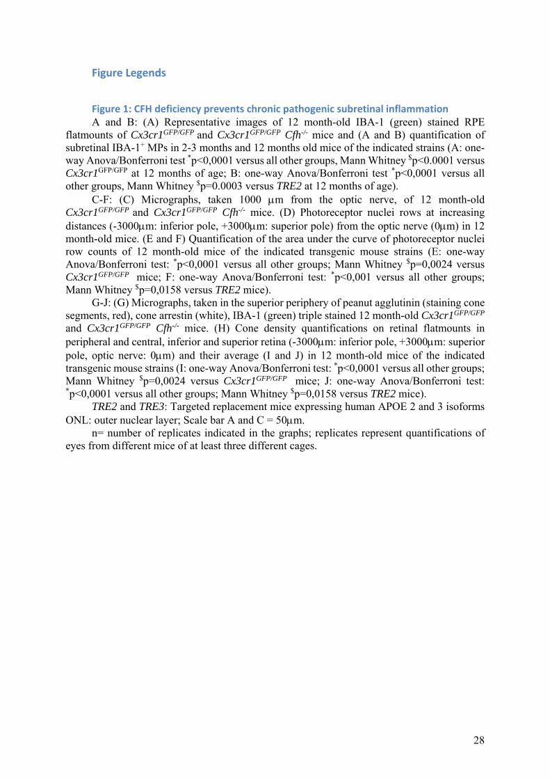
Figure 1: CFH deficiency prevents chronic pathogenic subretinal inflammation A and B: (A) Representative images of 12 month-old IBA-1 (green) stained RPE
flatmounts of Cx3cr1GFP/GFP and Cx3cr1GFP/GFP Cfh-/- mice and (A and B) quantification of subretinal IBA-1+ MPs in 2-3 months and 12 months old mice of the indicated strains (A: one-way Anova/Bonferroni test *p˂0,0001 versus all other groups, Mann Whitney $p<0.0001 versus Cx3cr1GFP/GFP at 12 months of age; B: one-way Anova/Bonferroni test *p˂0,0001 versus all other groups, Mann Whitney $p=0.0003 versus TRE2 at 12 months of age).
C-F: (C) Micrographs, taken 1000 m from the optic nerve, of 12 month-old Cx3cr1GFP/GFP and Cx3cr1GFP/GFP Cfh-/- mice. (D) Photoreceptor nuclei rows at increasing distances (-3000m: inferior pole, +3000m: superior pole) from the optic nerve (0m) in 12 month-old mice. (E and F) Quantification of the area under the curve of photoreceptor nuclei row counts of 12 month-old mice of the indicated transgenic mouse strains (E: one-way Anova/Bonferroni test: *p˂0,0001 versus all other groups; Mann Whitney $p=0,0024 versus Cx3cr1GFP/GFP mice; F: one-way Anova/Bonferroni test: *p˂0,001 versus all other groups; Mann Whitney $p=0,0158 versus TRE2 mice).
G-J: (G) Micrographs, taken in the superior periphery of peanut agglutinin (staining cone segments, red), cone arrestin (white), IBA-1 (green) triple stained 12 month-old Cx3cr1GFP/GFP
and Cx3cr1GFP/GFP Cfh-/- mice. (H) Cone density quantifications on retinal flatmounts in peripheral and central, inferior and superior retina (-3000m: inferior pole, +3000m: superior pole, optic nerve: 0m) and their average (I and J) in 12 month-old mice of the indicated transgenic mouse strains (I: one-way Anova/Bonferroni test: *p˂0,0001 versus all other groups; Mann Whitney $p=0,0024 versus Cx3cr1GFP/GFP mice; J: one-way Anova/Bonferroni test: *p˂0,0001 versus all other groups; Mann Whitney $p=0,0158 versus TRE2 mice).
TRE2 and TRE3: Targeted replacement mice expressing human APOE 2 and 3 isoforms ONL: outer nuclear layer; Scale bar A and C = 50m.
n= number of replicates indicated in the graphs; replicates represent quantifications of eyes from different mice of at least three different cages.
29
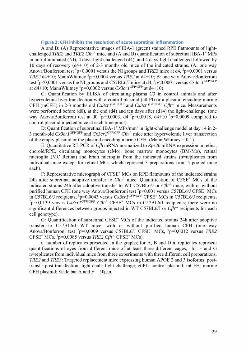
Figure 2: CFH inhibits the resolution of acute subretinal inflammation A and B: (A) Representative images of IBA-1 (green) stained RPE flatmounts of light-
challenged TRE2 and TRE2 Cfh-/- mice and (A and B) quantification of subretinal IBA-1+ MPs in non-illuminated (NI), 4 days-light challenged (d4), and 4 days-light challenged followed by 10 days of recovery (d4+10) of 2-3 months old mice of the indicated strains. (A: one way Anova/Bonferroni test *p˂0,0001 versus the NI groups and TRE3 mice at d4, $p<0.0001 versus TRE2 d4+10; MannWhitney $p=0,0004 versus TRE2 at d4+10; B: one way Anova/Bonferroni test *p˂0,0001 versus the NI groups and C57BL6/J mice at d4, $p<0.0001 versus Cx3cr1GFP/GFP at d4+10; MannWhitney $p=0,0002 versus Cx3cr1GFP/GFP at d4+10).
C: Quantification by ELISA of circulating plasma C3 in control animals and after hypervolemic liver transfection with a control plasmid (ctl Pl) or a plasmid encoding murine CFH (mCFH) in 2-3 months old Cx3cr1GFP/GFP and Cx3cr1GFP/GFP Cfh-/- mice. Measurements were performed before (d0), at the end (d4) and ten days after (d14) the light-challenge. (one way Anova/Bonferroni test at d0 *p=0,0003, d4 *p=0,0018, d4+10 *p=0,0009 compared to control plasmid injected mice at each time point).
D: Quantification of subretinal IBA-1+ MPs/mm2 in light-challenge model at day 14 in 2-3 month-old Cx3cr1GFP/GFP and Cx3cr1GFP/GFP Cfh-/- mice after hypervolemic liver transfection of the empty plasmid or the plasmid encoding murine CFH. (Mann Whitney = 0,1).
E: Quantitative RT-PCR of Cfh mRNA normalized to Rps26 mRNA expression in retina, choroid/RPE, circulating monocytes (cMo), bone marrow monocytes (BM-Mo), retinal microglia (MC Retina) and brain microglia from the indicated strains (n=replicates from individual mice except for retinal MCs which represent 3 preparations from 5 pooled mice each).
F: Representative micrograph of CFSE+ MCs on RPE flatmounts of the indicated strains 24h after subretinal adoptive transfer to Cfh-/- mice. Quantification of CFSE+ MCs of the indicated strains 24h after adoptive transfer to WT C57BL6/J or Cfh-/- mice, with or without purified human CFH (one way Anova/Bonferroni test *p˂0,001 versus C57BL6/J CFSE+ MCs in C57BL6/J recipients, $p=0,0043 versus Cx3cr1GFP/GFP CFSE+ MCs in C57BL6/J recipients, †p=0,0139 versus Cx3cr1GFP/GFP Cfh-/- CFSE+ MCs in C57BL6/J recipients; there were no significant differences between groups injected in WT C57BL6/J or Cfh-/- recipients for each cell genotype).
G: Quantification of subretinal CFSE+ MCs of the indicated strains 24h after adoptive transfer to C57BL6/J WT mice, with or without purified human CFH (one way Anova/Bonferroni test *p=0,0009 versus C57BL6/J CFSE+ MCs, $p=0,0012 versus TRE2 CFSE+ MCs, †p=0,0085 versus TRE2 Cfh-/- CFSE+ MCs).
n=number of replicates presented in the graphs; for A, B and D n=replicates represent quantifications of eyes from different mice of at least three different cages; for F and G n=replicates from individual mice from three experiments with three different cell preparations. TRE2 and TRE3: Targeted replacement mice expressing human APOE 2 and 3 isoforms; post-transf.: post-transfection; light-chall: light-challenge; ctlPL: control plasmid; mCFH: murine CFH plasmid; Scale bar A and F = 50m.
30
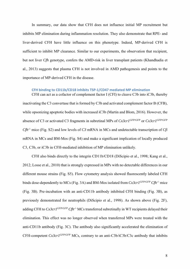
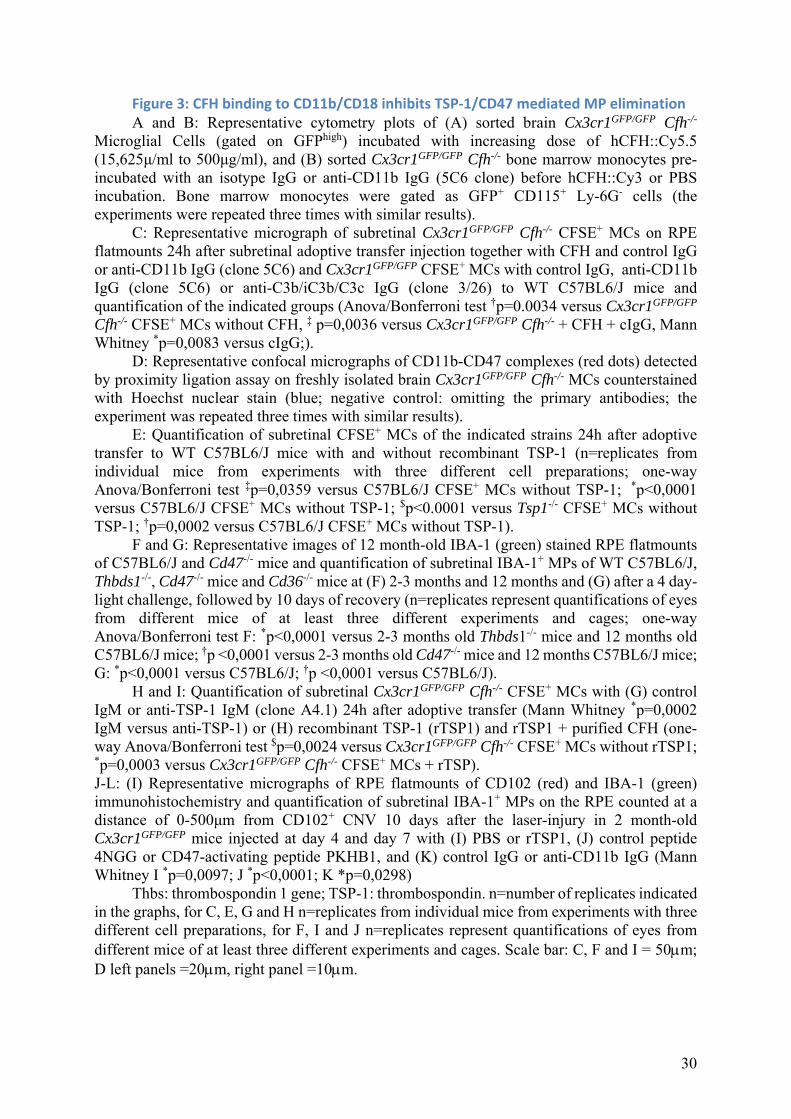
Figure 3: CFH binding to CD11b/CD18 inhibits TSP‐1/CD47 mediated MP elimination A and B: Representative cytometry plots of (A) sorted brain Cx3cr1GFP/GFP Cfh-/-
Microglial Cells (gated on GFPhigh) incubated with increasing dose of hCFH::Cy5.5 (15,625μ/ml to 500μg/ml), and (B) sorted Cx3cr1GFP/GFP Cfh-/- bone marrow monocytes pre-incubated with an isotype IgG or anti-CD11b IgG (5C6 clone) before hCFH::Cy3 or PBS incubation. Bone marrow monocytes were gated as GFP+ CD115+ Ly-6G- cells (the experiments were repeated three times with similar results).
C: Representative micrograph of subretinal Cx3cr1GFP/GFP Cfh-/- CFSE+ MCs on RPE flatmounts 24h after subretinal adoptive transfer injection together with CFH and control IgG or anti-CD11b IgG (clone 5C6) and Cx3cr1GFP/GFP CFSE+ MCs with control IgG, anti-CD11b IgG (clone 5C6) or anti-C3b/iC3b/C3c IgG (clone 3/26) to WT C57BL6/J mice and quantification of the indicated groups (Anova/Bonferroni test †p=0.0034 versus Cx3cr1GFP/GFP Cfh-/- CFSE+ MCs without CFH, ‡ p=0,0036 versus Cx3cr1GFP/GFP Cfh-/- + CFH + cIgG, Mann Whitney *p=0,0083 versus cIgG;).
D: Representative confocal micrographs of CD11b-CD47 complexes (red dots) detected by proximity ligation assay on freshly isolated brain Cx3cr1GFP/GFP Cfh-/- MCs counterstained with Hoechst nuclear stain (blue; negative control: omitting the primary antibodies; the experiment was repeated three times with similar results).
E: Quantification of subretinal CFSE+ MCs of the indicated strains 24h after adoptive transfer to WT C57BL6/J mice with and without recombinant TSP-1 (n=replicates from individual mice from experiments with three different cell preparations; one-way Anova/Bonferroni test ‡p=0,0359 versus C57BL6/J CFSE+ MCs without TSP-1; *p˂0,0001 versus C57BL6/J CFSE+ MCs without TSP-1; $p<0.0001 versus Tsp1-/- CFSE+ MCs without TSP-1; †p=0,0002 versus C57BL6/J CFSE+ MCs without TSP-1).
F and G: Representative images of 12 month-old IBA-1 (green) stained RPE flatmounts of C57BL6/J and Cd47-/- mice and quantification of subretinal IBA-1+ MPs of WT C57BL6/J, Thbds1-/-, Cd47-/- mice and Cd36-/- mice at (F) 2-3 months and 12 months and (G) after a 4 day-light challenge, followed by 10 days of recovery (n=replicates represent quantifications of eyes from different mice of at least three different experiments and cages; one-way Anova/Bonferroni test F: *p˂0,0001 versus 2-3 months old Thbds1-/- mice and 12 months old C57BL6/J mice; †p ˂ 0,0001 versus 2-3 months old Cd47-/- mice and 12 months C57BL6/J mice; G: *p˂0,0001 versus C57BL6/J; †p ˂0,0001 versus C57BL6/J).
H and I: Quantification of subretinal Cx3cr1GFP/GFP Cfh-/- CFSE+ MCs with (G) control IgM or anti-TSP-1 IgM (clone A4.1) 24h after adoptive transfer (Mann Whitney *p=0,0002 IgM versus anti-TSP-1) or (H) recombinant TSP-1 (rTSP1) and rTSP1 + purified CFH (one-way Anova/Bonferroni test $p=0,0024 versus Cx3cr1GFP/GFP Cfh-/- CFSE+ MCs without rTSP1; *p=0,0003 versus Cx3cr1GFP/GFP Cfh-/- CFSE+ MCs + rTSP). J-L: (I) Representative micrographs of RPE flatmounts of CD102 (red) and IBA-1 (green) immunohistochemistry and quantification of subretinal IBA-1+ MPs on the RPE counted at a distance of 0-500μm from CD102+ CNV 10 days after the laser-injury in 2 month-old Cx3cr1GFP/GFP mice injected at day 4 and day 7 with (I) PBS or rTSP1, (J) control peptide 4NGG or CD47-activating peptide PKHB1, and (K) control IgG or anti-CD11b IgG (Mann Whitney I *p=0,0097; J *p<0,0001; K *p=0,0298)
Thbs: thrombospondin 1 gene; TSP-1: thrombospondin. n=number of replicates indicated in the graphs, for C, E, G and H n=replicates from individual mice from experiments with three different cell preparations, for F, I and J n=replicates represent quantifications of eyes from different mice of at least three different experiments and cages. Scale bar: C, F and I = 50m; D left panels =20m, right panel =10m.
31
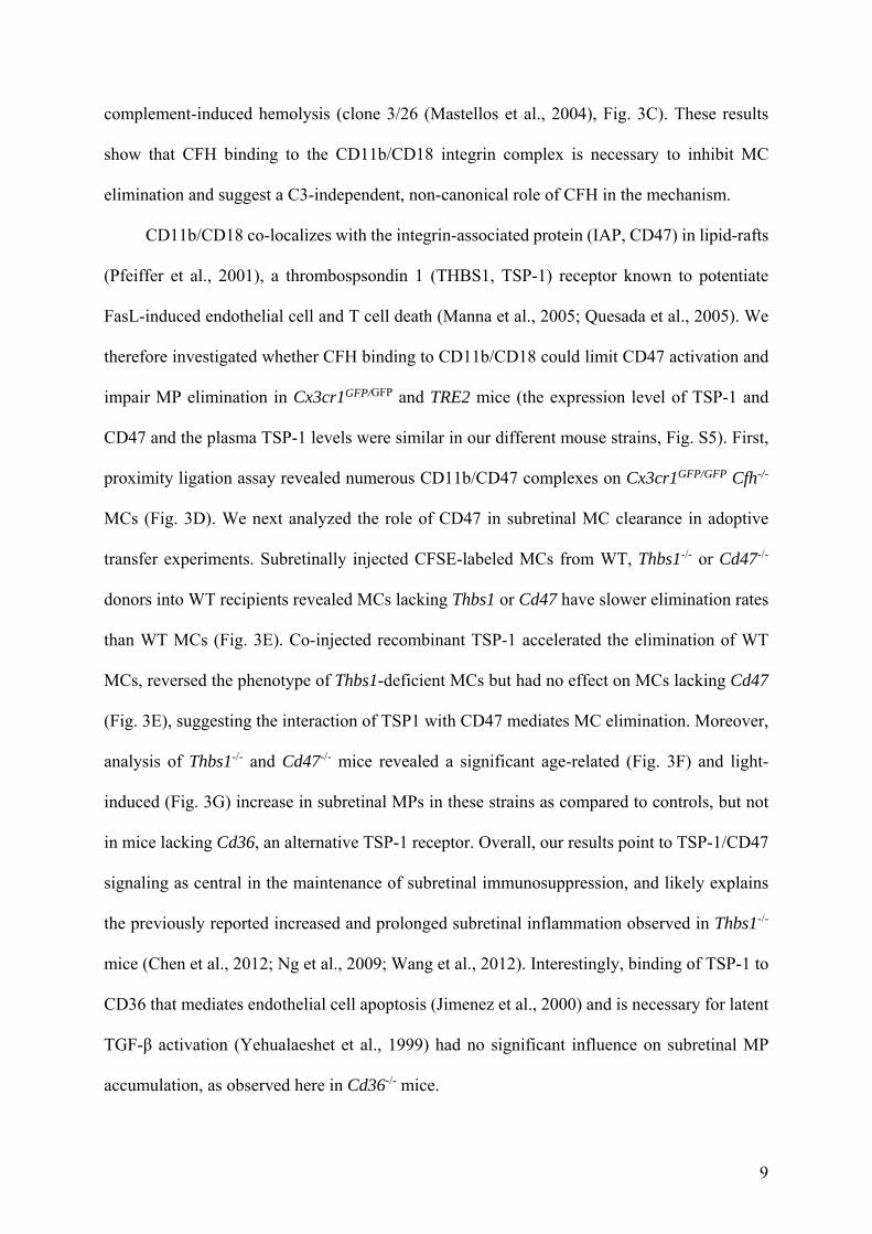
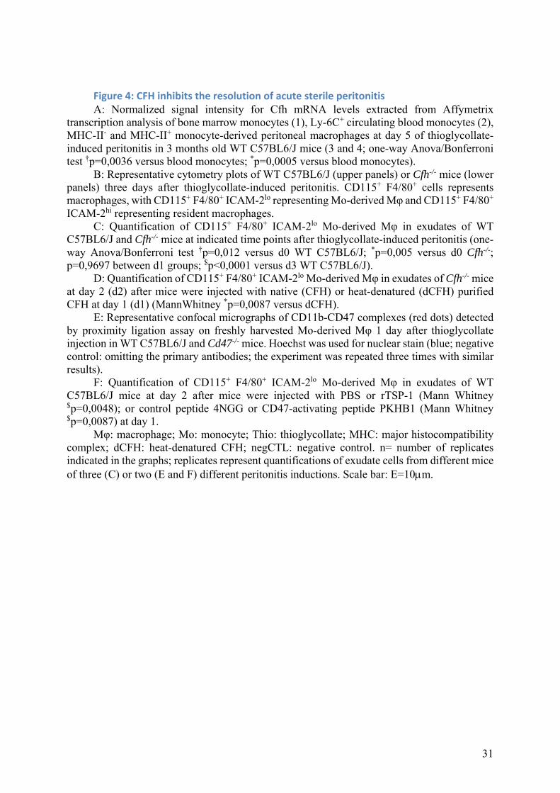
Figure 4: CFH inhibits the resolution of acute sterile peritonitis A: Normalized signal intensity for Cfh mRNA levels extracted from Affymetrix
transcription analysis of bone marrow monocytes (1), Ly-6C+ circulating blood monocytes (2), MHC-II- and MHC-II+ monocyte-derived peritoneal macrophages at day 5 of thioglycollate-induced peritonitis in 3 months old WT C57BL6/J mice (3 and 4; one-way Anova/Bonferroni test †p=0,0036 versus blood monocytes; *p=0,0005 versus blood monocytes).
B: Representative cytometry plots of WT C57BL6/J (upper panels) or Cfh-/- mice (lower panels) three days after thioglycollate-induced peritonitis. CD115+ F4/80+ cells represents macrophages, with CD115+ F4/80+ ICAM-2lo representing Mo-derived Mφ and CD115+ F4/80+ ICAM-2hi representing resident macrophages.
C: Quantification of CD115+ F4/80+ ICAM-2lo Mo-derived Mφ in exudates of WT C57BL6/J and Cfh-/- mice at indicated time points after thioglycollate-induced peritonitis (one-way Anova/Bonferroni test †p=0,012 versus d0 WT C57BL6/J; *p=0,005 versus d0 Cfh-/-; p=0,9697 between d1 groups; $p<0,0001 versus d3 WT C57BL6/J).
D: Quantification of CD115+ F4/80+ ICAM-2lo Mo-derived Mφ in exudates of Cfh-/- mice at day 2 (d2) after mice were injected with native (CFH) or heat-denatured (dCFH) purified CFH at day 1 (d1) (MannWhitney *p=0,0087 versus dCFH).
E: Representative confocal micrographs of CD11b-CD47 complexes (red dots) detected by proximity ligation assay on freshly harvested Mo-derived Mφ 1 day after thioglycollate injection in WT C57BL6/J and Cd47-/- mice. Hoechst was used for nuclear stain (blue; negative control: omitting the primary antibodies; the experiment was repeated three times with similar results).
F: Quantification of CD115+ F4/80+ ICAM-2lo Mo-derived Mφ in exudates of WT C57BL6/J mice at day 2 after mice were injected with PBS or rTSP-1 (Mann Whitney $p=0,0048); or control peptide 4NGG or CD47-activating peptide PKHB1 (Mann Whitney $p=0,0087) at day 1.
Mφ: macrophage; Mo: monocyte; Thio: thioglycollate; MHC: major histocompatibility complex; dCFH: heat-denatured CFH; negCTL: negative control. n= number of replicates indicated in the graphs; replicates represent quantifications of exudate cells from different mice of three (C) or two (E and F) different peritonitis inductions. Scale bar: E=10m.
32
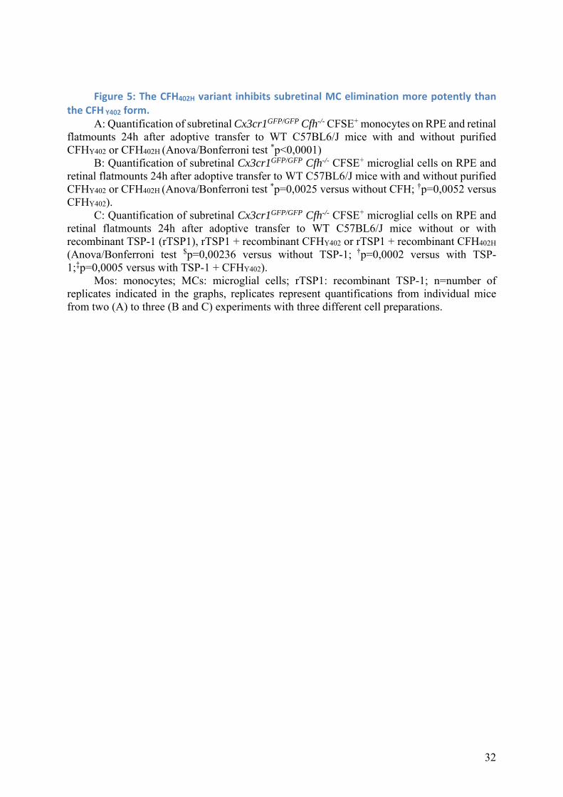
Figure 5: The CFH402H variant inhibits subretinal MC elimination more potently than the CFH Y402 form.
A: Quantification of subretinal Cx3cr1GFP/GFP Cfh-/- CFSE+ monocytes on RPE and retinal flatmounts 24h after adoptive transfer to WT C57BL6/J mice with and without purified CFHY402 or CFH402H (Anova/Bonferroni test *p˂0,0001)
B: Quantification of subretinal Cx3cr1GFP/GFP Cfh-/- CFSE+ microglial cells on RPE and retinal flatmounts 24h after adoptive transfer to WT C57BL6/J mice with and without purified CFHY402 or CFH402H (Anova/Bonferroni test *p=0,0025 versus without CFH; †p=0,0052 versus CFHY402).
C: Quantification of subretinal Cx3cr1GFP/GFP Cfh-/- CFSE+ microglial cells on RPE and retinal flatmounts 24h after adoptive transfer to WT C57BL6/J mice without or with recombinant TSP-1 (rTSP1), rTSP1 + recombinant CFHY402 or rTSP1 + recombinant CFH402H (Anova/Bonferroni test $p=0,00236 versus without TSP-1; †p=0,0002 versus with TSP-1;‡p=0,0005 versus with TSP-1 + CFHY402).
Mos: monocytes; MCs: microglial cells; rTSP1: recombinant TSP-1; n=number of replicates indicated in the graphs, replicates represent quantifications from individual mice from two (A) to three (B and C) experiments with three different cell preparations.
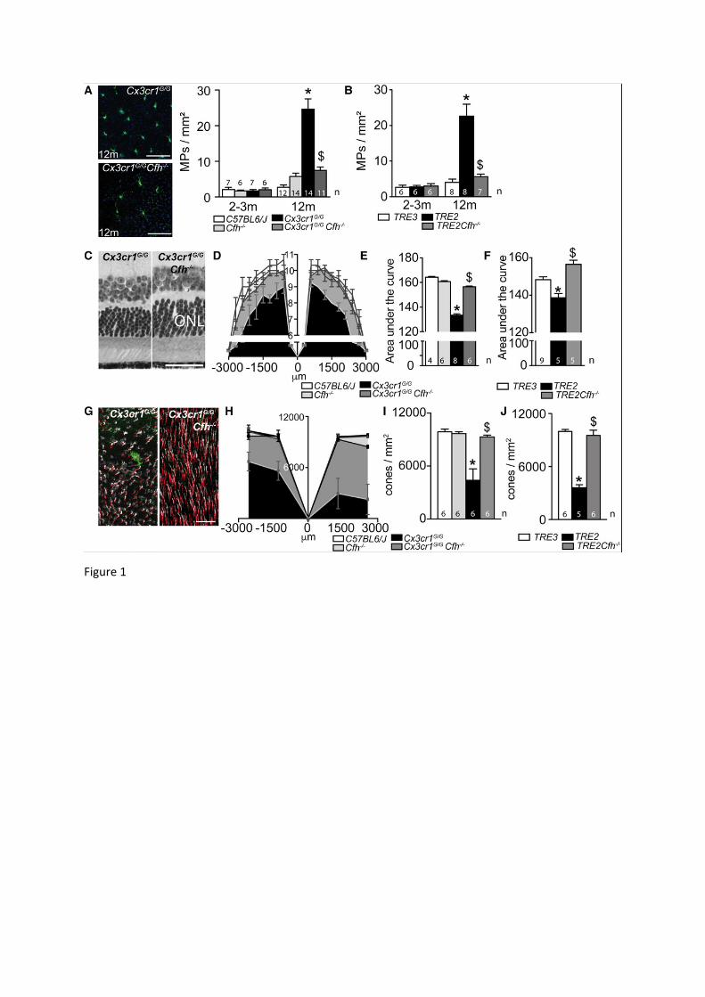
Figure 1
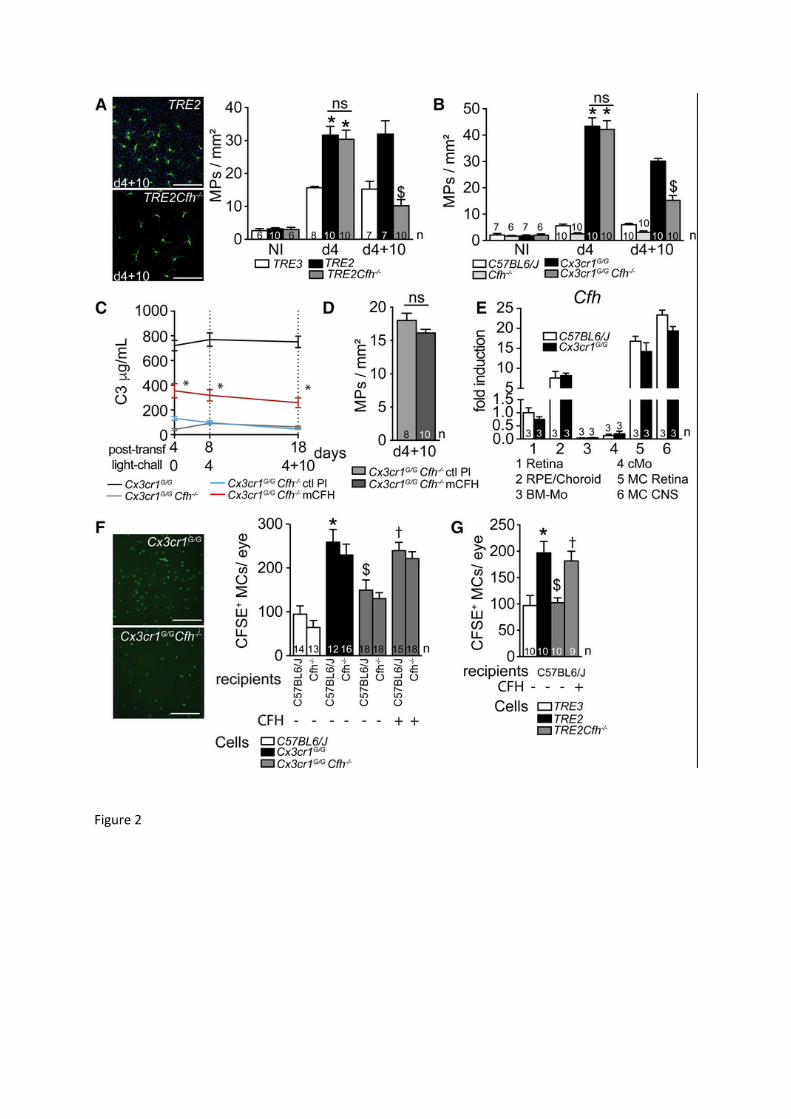
Figure 2
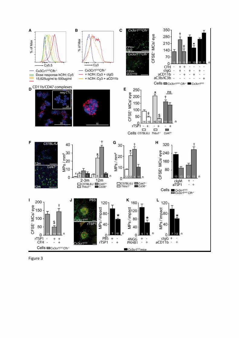
Figure 3
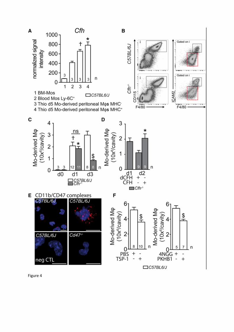
Figure 4
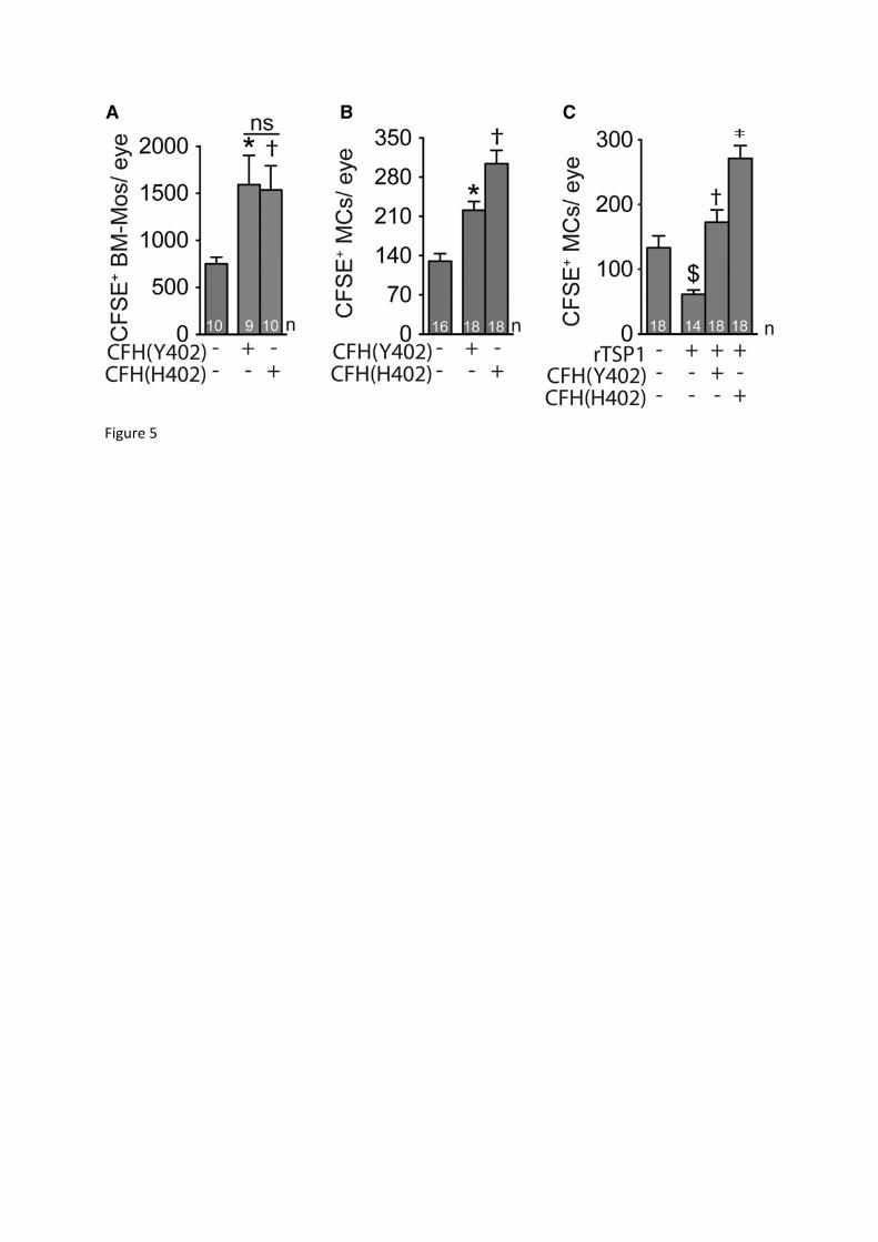
Figure 5
33
Figure S1: CFH‐deficiency does not alter in vitro pathogenic cytokine secretion of microglial cells and monocytes.
We previously showed that MP derived IL-1β, IL-6, and TNFα induce photoreceptor degeneration and deregulate RPE-cell functions (Eandi et al., 2016; Hu et al., 2015; Levy et al., 2015a; Mathis et al., 2016). Cytokine multiplex analysis of supernatants from cultured primary bone marrow monocytes (BM-Mos, 100 000 cells/well) and brain microglial cells (MCs, 200 000 cells/well), incubated for 24 h in serum free DMEM medium or stimulated with APOE3 (5
μg/ml as previously described (Levy et al., 2015b)) of Cx3cr1GFP/GFP and Cx3cr1GFP/GFP Cfh-/- mice revealed no significant CFH-dependent differences in the secretion of these pathogenic cytokines in basal or stimulated conditions, suggesting that CFH did not significantly affect the secretion of these cytokines.
Figure S2: Plasma C3 concentrations and C3, and C3 fragment, and CFH immunohistochemistry in the transgenic mice.
A: Complement factor C3 (C3)-ELISA measurements of plasma C3 concentrations from the indicated mouse strains indicate that C3 plasma levels in Cx3cr1GFP/GFP mice do not differ from WT C57BL6/J mice and TRE2 mice from TRE3 mice and Cfh-deficiency induces low circulating levels of C3 in both strains, likely due to un-inhibited plasma complement activation and exhaustion (Pickering et al., 2002).
B-D: Immunohistochemistry (red) for C3 (B, clone 11H9 Hycult biotech), C3b/iC3b/C3c (C, clone 3/26 Hycult biotech), and CFH (A, ab8842 Abcam) in 4d light-challenged Cx3cr1GFP/GFP and Cx3cr1GFP/GFP Cfh-/- mice (GFP in green, nuclear stain in blue). (B) C3 was strongly detected in the choriocapillaries (arrow) of Cx3cr1GFP/GFPmice, but not in and around subretinal MPs. In Cx3cr1GFP/GFP Cfh-/- mice that are characterized by low plasma C3 levels the signal in the choriocapillaries (adjacent to the RPE) was reduced. (C) Immunohistochemistry using the anti-mouse-C3b/iC3b/C3c antibody (clone 26/3 (Mastellos et al., 2004)) that specifically recognizes C3b and its fragments revealed a faint staining in the choriocapillaries (arrows), but no staining in the subretinal space of both mouse strains. (D) CFH was detected surrounding subretinal MPs (arrows), RPE and in the choriocapillaries (arrow head) in Cx3cr1GFP/GFP, but not in Cx3cr1GFP/GFP Cfh-/- mice.
ONL: outer nuclear layer; RPE retinal pigment epithelium. negative control: omitting the primary antibodies revealed no staining (not shown); the experiment was repeated three times with similar results. Scale bar = 50m.
Figure S3: Cfh induction in monocytes in vitro and monocyte elimination after subretinal adoptive transfer.
A: Quantitative RT-PCR of Cfh mRNA normalized with Rps26 mRNA in WT C57BL6/J or Cx3cr1GFP/GFP monocytes cultivated for 18 h with or without photoreceptor outer segments to simulate the subretinal environment (POSs, one way Anova/Bonferroni test *p=0,0082 versus C57BL6/J Monocytes without POS, †p<0,0001 versus C57BL6/J Monocytes with and without POS). Cfh mRNA is robustly induced in BM-Mos from WT C57BL6/J-, and even more so from Cx3cr1GFP/GFP-mice in contact with POS.
B: Representative micrograph of CFSE+ Monocytes on RPE flatmounts of the indicated strains 24h after subretinal adoptive transfer to Cfh-/- mice. Quantification of CFSE+ Mos of the indicated strains 24h after adoptive transfer to Cfh-/- mice (one way Anova/Bonferroni test *p˂0,001 versus C57BL6/J CFSE+ Mos, $p=0,0043 versus Cx3cr1GFP/GFP CFSE+ Mos, †p=0,0178 versus Cx3cr1GFP/GFP Cfh-/- CFSE+ Mos).
34
POS: photoreceptor outer segments, Mos: monocytes; n=number of replicates indicated in the graphs, for A n=replicates represent different culture wells of the indicated conditions, the experiment was repeated twice with similar results; for B n=replicates from individual mice from two experiments with two different cell preparations. Scale bar B = 50m.
Figure S4: Complement component expression in retina, liver, and MPs. Quantitative RT-PCR of Cfh (A-C), C3 (D-E), Cfb (G-I), and Cfi (J-K) mRNA,
normalized with Rps26 mRNA, in retina (1), retina without MCs (2, after sorting of MCs), liver (3), bone marrow monocytes (BM-Mo), retinal microglia (5, MC Retina) and brain microglia (6, CNS MC) from WT C57BL6/J mice (left column), and of BM-Mo, and MCs from retina and CNS of the indicated mouse strains (middle and right column). (A) MCs robustly expressed Cfh mRNA (~1/4 of liver expression level) significantly (~20-40 fold) more than BM-Mo (B and C). In contrast, C3 mRNA was found in BM-Mos (B, ~1/60 of liver expression level), significantly (~50 fold) more than in MCs (E and F). Cfb mRNA transcription in MPs was detectable (~1/1000 of liver expression level, G) in all MPs with a relatively stronger expression in MCs (H and I). Interestingly, Cfi mRNA, robustly expressed in the liver (J), but was not detectable in MPs (K and L). Retinal and CNS MCs expressed the transcripts similarly and except for Cfh mRNA in Cfh-/- strains, we did not detect strain-dependent differences in the MPs.
BM-Mo: bone marrow monocytes; MC: microglial cell. CNS: central nervous system. A, D, G, and J n= replicates from individual mice; B, C, E, F, H, I, K, and L n= replicates from mRNA pooled from two individual mice.
Supplementary Figure 5: Cd11b, Cd18, Cd47 and Thbs1 transcription in MPs and TSP‐1 plasma levels of the transgenic mice.
Quantitative RT-PCR of Cd11b (A-C), Cd18 (D-E), Cd47 (G-I), and Thbs1 (J-L) mRNA, normalized with Rps26 mRNA, in retina (1), retina without MCs (2, after sorting of MCs), liver (3), bone marrow monocytes (BM-Mo), retinal microglia (5, MC Retina) and brain microglia (6, CNS MC) from WT C57BL6/J mice (left column), and of BM-Mo, and MCs from retina and CNS of the indicated mouse strains (middle and right column). Both, Cd11b and Cd18 mRNA were expressed specifically in MPs and not detected in mRNA from retina without the MC population (A and D). While Cd11b mRNA was more strongly expressed in MCs than in BM-Mo, no significant difference was detected in Cd18 transcription (B, C, E, and F). (G-I): Cd47 mRNA was expressed ubiquitously with a tendentiously higher expression in MCs compared to BM-Mos (C57BL6/J-, Cx3cr1GFP/GFP, Cx3cr1GFP/GFP Cfh-/- mice). (J-L) Thbs1 mRNA expression was more strongly expressed in MCs than in BM-Mo of C57BL6/J-, Cx3cr1GFP/GFP, Cx3cr1GFP/GFP Cfh-/- mice, with differences less marked in TRE3- versus TRE2-mice. Retinal and CNS MCs expressed the transcripts similarly and we did not detect strain-dependent differences in the MPs.
(M and L) TSP-1 plasma concentrations of the indicated mouse strains measured by Elisa. BM-Mo: bone marrow monocytes; MC: microglial cell. CNS: central nervous system. A,
D, G, J, M, and N n= replicates from individual mice; B, C, E, F, H, I, K, and L n= replicates from mRNA pooled from two individual mice.


