Compartment Syndromes T. Toan Le, MD and Sameh Arebi, MD Original Author: Robert M. Harris, MD;...
74
Compartment Syndromes T. Toan Le, MD and Sameh Arebi, MD Original Author: Robert M. Harris, MD; Created March 2004 New Authors: T. Toan Le, MD and Sameh Arebi, MD; Revised December 2005; Revised May 2011 – Michael Sirkin
-
Upload
jayce-chenery -
Category
Documents
-
view
216 -
download
1
Transcript of Compartment Syndromes T. Toan Le, MD and Sameh Arebi, MD Original Author: Robert M. Harris, MD;...
Compartment Syndromes
T. Toan Le, MD and Sameh Arebi, MD
Original Author: Robert M. Harris, MD; Created March 2004New Authors: T. Toan Le, MD and Sameh Arebi, MD; Revised December 2005; Revised May 2011 – Michael Sirkin
Today
• What is it
• Pathophysiology
• Diagnosis
• Treatment
Increase in hydrostatic pressure in closed osteofascial space resulting in decreased perfusion of muscle and nerves within compartment
• RAISED PRESSURE RAISED PRESSURE WITHIN A CLOSED WITHIN A CLOSED SPACESPACE with a potential to cause irreversible damageirreversible damage to the contents of the closed space
Richard Von Volkmann, 1881
• “For many years I have noted on occasion, following the use of bandages too tightly appliedbandages too tightly applied, the occurrence of paralysis and contraction of the limb, NOT … due to paralysis and contraction of the limb, NOT … due to thethe paralysis of the nerve by pressureparalysis of the nerve by pressure, but as a quick and massive disintegration of the contractile substance and the effect of the ensuing reaction and degeneration.”

Definition
• Symptoms resulting from increased pressure within a limited space– compromising
• circulation
• function

Pathophysiology
• Local Blood Flow is reduced as a consequence:
LBF=Pa-Pv / R (A-V Gradient)
Pathophysiology
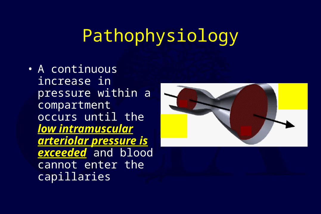
• A continuous increase in pressure within a compartment occurs until the low intramuscular low intramuscular arteriolar pressure is arteriolar pressure is exceededexceeded and blood cannot enter the capillaries
Pathophysiology
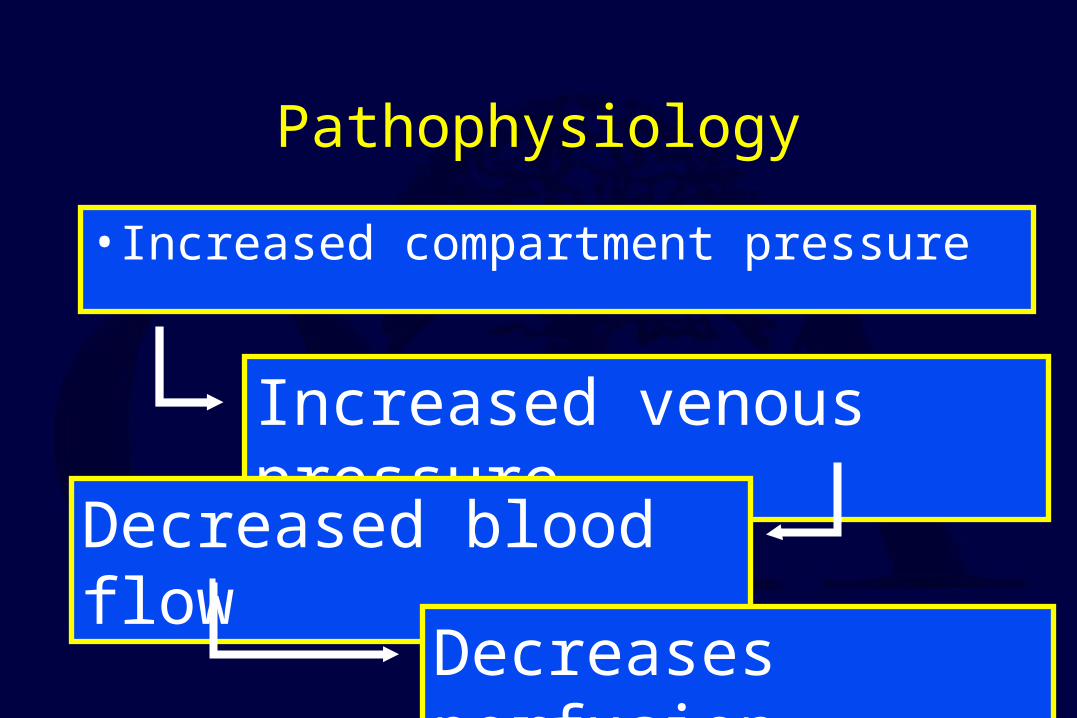
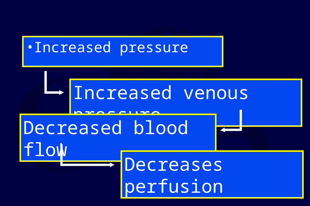
• Increased compartment pressure
Increased venous pressure
Decreased blood flow
Decreases perfusion
Pathophysiology
• Autoregulatory mechanisms may compensate:– Decrease in peripheral vascular resistance– Increased extraction of oxygen
• As system becomes overwhelmed: – Critical closing pressure is reached– Oxygen perfusion of muscles and nerves decreases
Muscle Ischemia
• 4 hours - reversible damage
• 8 hours - irreversible changes
• 4-8 hours - variable
Hargens JBJS 1981
Muscle Ischemia
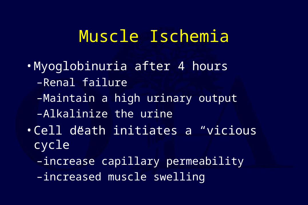
• Myoglobinuria after 4 hours– Renal failure
– Maintain a high urinary output
– Alkalinize the urine
• Cell death initiates a “vicious cycle”– increase capillary permeability
– increased muscle swelling
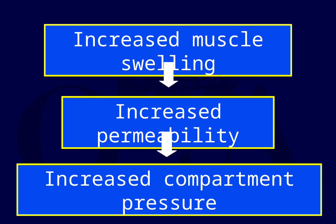
Increased muscle swelling
Increased permeability
Increased compartment pressure
• Increased pressure
Increased venous pressure
Decreased blood flow
Decreases perfusion
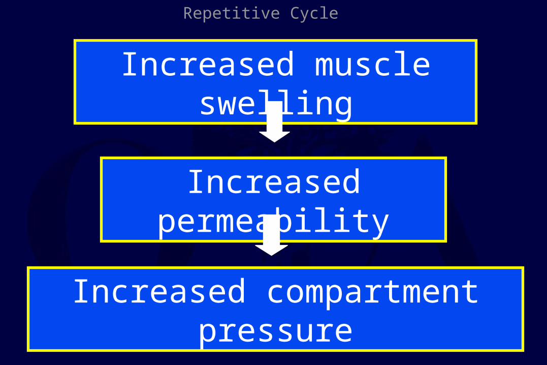
Increased muscle swelling
Increased permeability
Increased compartment pressure
Repetitive Cycle
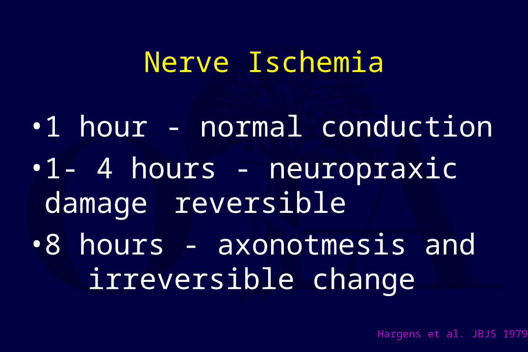
Nerve Ischemia
• 1 hour - normal conduction
• 1- 4 hours - neuropraxic damage reversible
• 8 hours - axonotmesis and irreversible change
Hargens et al. JBJS 1979
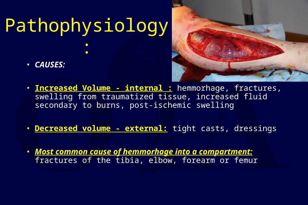
Pathophysiology:
• CAUSES:
• Increased Volume - internal :Increased Volume - internal : hemmorhage, fractures, swelling from traumatized tissue, increased fluid secondary to burns, post-ischemic swelling
• Decreased volume - external:Decreased volume - external: tight casts, dressings
• Most common cause of hemmorhage into a compartment:Most common cause of hemmorhage into a compartment: fractures of the tibia, elbow, forearm or femur
Etiology
• Fractures
• Soft Tissue Injury (Crush)
• Arterial Injury– Post-ischemic swelling
– Reperfusion injury
• Drug Overdose (limb compression)
• Burns
Pathophysiology:
Most common causeMost common cause of compartment syndrome is muscle injurymuscle injury that leads to edema
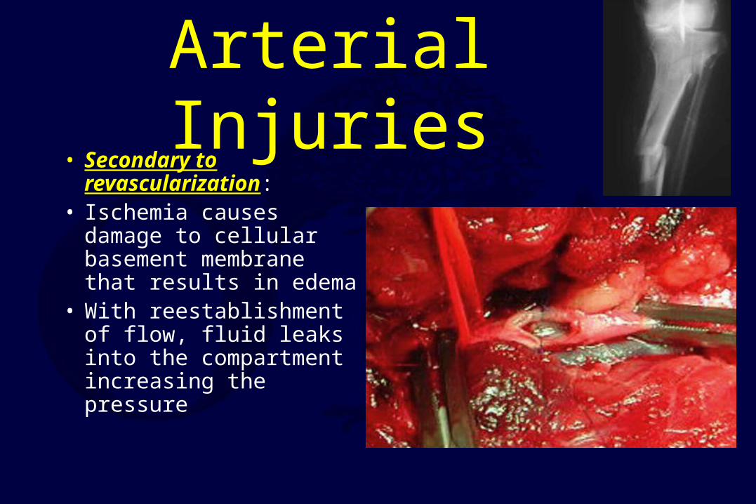
Arterial Injuries• Secondary to Secondary to
revascularizationrevascularization:• Ischemia causes damage
to cellular basement membrane that results in edema
• With reestablishment of flow, fluid leaks into the compartment increasing the pressure
Diagnosis
• Clinical diagnosis– High index of suspicion
• Syndrome– History– Physical Exam
Difficult Diagnosis• Classic signs of the 5 P’s - ARE NOT RELIABLE:
– pain– pallor – paralysis– pulselessness – paresthesias
• These are signs of an ESTABLISHED compartment syndrome where ischemic injury has already taken place
• These signs may be present in the absence of compartment syndrome.
Diagnosis
• Pain
• Compartment pressure– Confirmatory test– Don’t just measure
Diagnosis
• Palpable pulses are usually present in acute compartment syndromes unless an arterial injury occurs
• Sensory changes and paralysisSensory changes and paralysis do not occur until ischemia has been present for about 1 1 hour or morehour or more
Diagnosis
• The most important most important symptomsymptom of an impending compartment syndrome is PAIN PAIN DISPROPORTIONATE DISPROPORTIONATE TO THAT EXPECTED TO THAT EXPECTED FOR THE INJURYFOR THE INJURY
Signs & Symptoms
• Pain –Passive muscle stretching
–Out of proportion
–Progressive–Not relieved by immobilization
Signs & Symptoms
• Pain –May be worse with elevation
–Patient will not initiate motion on own
• Be careful with coexisting nerve injury
Signs & Symptoms
• Parasthesia–Secondary to nerve ischemia
• Must be differentiated from nerve injury
Signs & Symptoms
• Paralysis (Weakness)– Ischemic muscles lose function
Signs & Symptoms
• Tense compartment on palpation
• Elevated compartment pressure
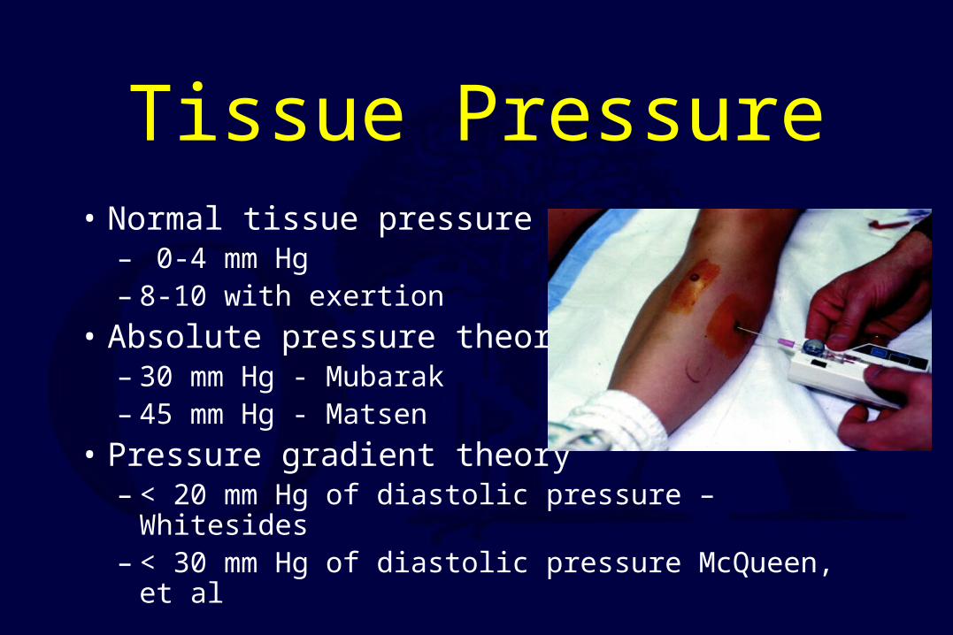
Tissue Pressure• Normal tissue pressure
– 0-4 mm Hg – 8-10 with exertion
• Absolute pressure theory– 30 mm Hg - Mubarak– 45 mm Hg - Matsen
• Pressure gradient theory– < 20 mm Hg of diastolic pressure – Whitesides– < 30 mm Hg of diastolic pressure McQueen, et al
Tissue-Pressure: Principles
• Originally, fasciotomies for tissue-pressures greater-than 30mmHg
• Whitesides et al in 1975Whitesides et al in 1975 was the first to suggest that the significance of tissue pressures was in their relation to diastolic relation to diastolic blood pressureblood pressure.
• McQueen et al: absolute compartment pressure is an UNRELIABLE absolute compartment pressure is an UNRELIABLE indication for the need for fasciotomies. indication for the need for fasciotomies. BUT, pressures within 30mmHg of DP indicate compartment syndrome
Tissue-Pressure: Principles
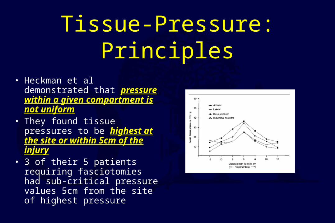
• Heckman et al demonstrated that pressure within a given pressure within a given compartment is not uniformcompartment is not uniform
• They found tissue pressures to be highest at the site or within 5cm highest at the site or within 5cm of the injuryof the injury
• 3 of their 5 patients requiring fasciotomies had sub-critical pressure values 5cm from the site of highest pressure

Who is at high risk?
High energy fractures• Severe
comminution
• Joint extension
• Segmental injuries
• Widely displaced
• Bilateral
• Floating knee
• Open fractures
Impaired Sensorium
• Alcohol
• Drug
• Decreased GCS
• Unconscious
• Chemically unconscious
• Neurologic deficit
• Cognitively challenged
Diagnosis
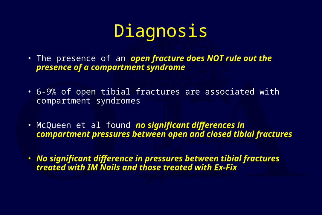
• The presence of an open fracture does NOT rule out the presence open fracture does NOT rule out the presence of a compartment syndromeof a compartment syndrome
• 6-9% of open tibial fractures are associated with compartment syndromes
• McQueen et al found no significant differences in compartment no significant differences in compartment pressures between open and closed tibial fracturespressures between open and closed tibial fractures
• No significant difference in pressures between tibial fractures No significant difference in pressures between tibial fractures treated with IM Nails and those treated with Ex-Fixtreated with IM Nails and those treated with Ex-Fix
Criteria-Compartment Pressure• Accurately examine
– Difference < 30mm Hg
• Impaired– Absolute > than 30mm Hg
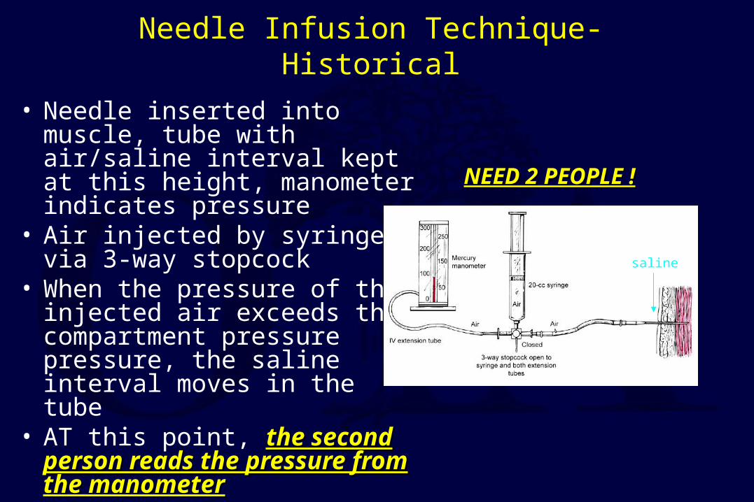
Needle Infusion Technique-Historical
• Needle inserted into muscle, tube with air/saline interval kept at this height, manometer indicates pressure
• Air injected by syringe via 3-way stopcock
• When the pressure of the injected air exceeds the compartment pressure pressure, the saline interval moves in the tube
• AT this point, the second person the second person reads the pressure from the reads the pressure from the manometermanometer
NEED 2 PEOPLE !NEED 2 PEOPLE !
saline
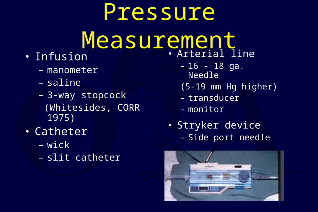
Pressure Measurement• Infusion
– manometer– saline– 3-way stopcock (Whitesides, CORR 1975)
• Catheter– wick– slit catheter
• Arterial line– 16 - 18 ga. Needle (5-19 mm Hg higher)– transducer– monitor
• Stryker device– Side port needle
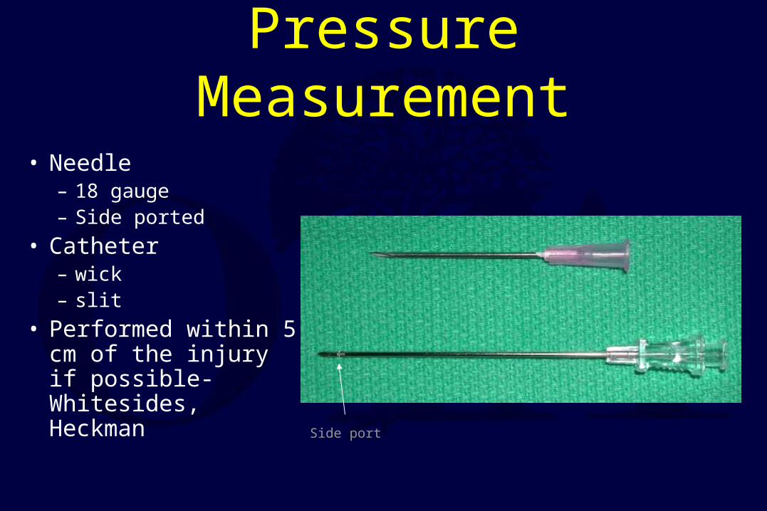
• Needle– 18 gauge– Side ported
• Catheter– wick– slit
• Performed within 5 cm of the injury if possible-Whitesides, Heckman Side port
Pressure Measurement

• Unit and needle set
• Assemble unit and prime
• Hold at angle to measure
• Zero machine
• Test each of 4 compartments– Keep calf off of bed

Most Common Locations
• Leg: deep posterior and the deep posterior and the anterioranterior compartmentscompartments
• Forearm: volar compartmentvolar compartment, especially in the deep flexor area
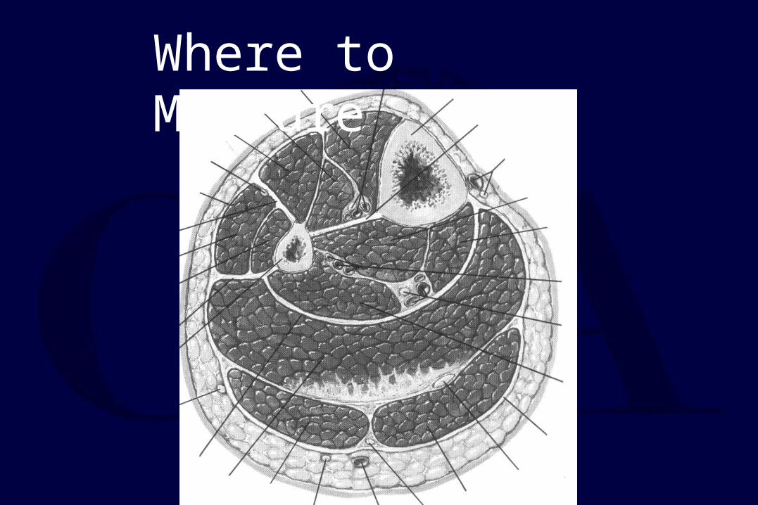
Where to Measure
Pressure
• Deeper muscles are initially involved
• Distance from fracture affects pressure
Heckmen et al. JBJS 1994
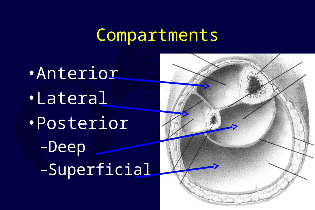
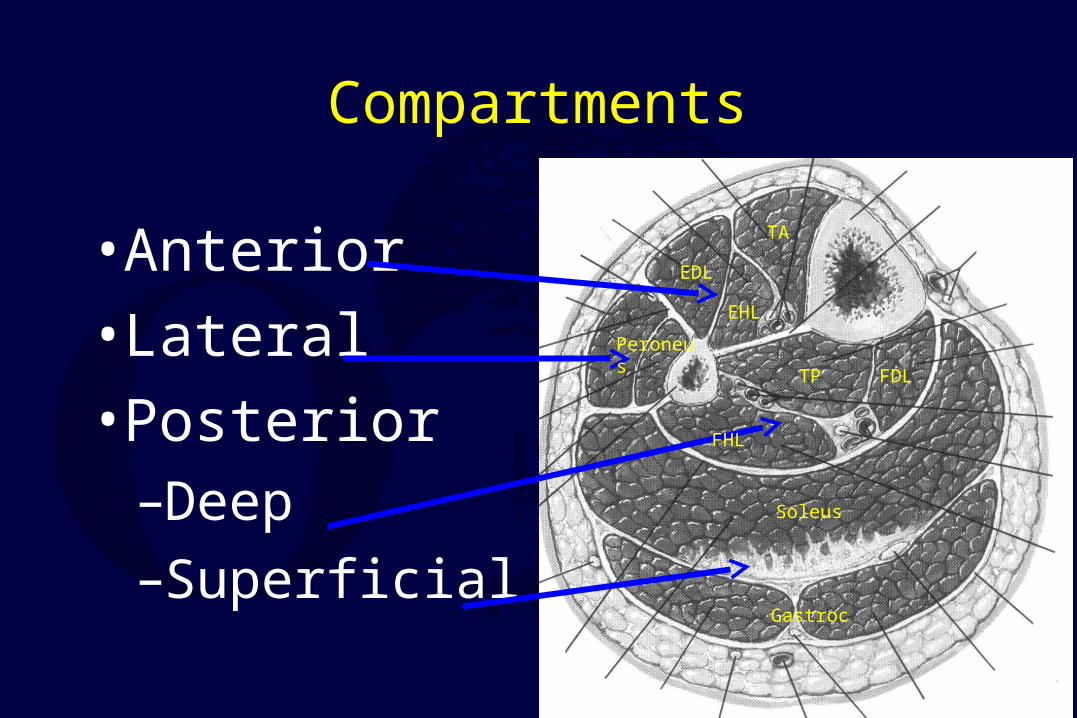
Compartments
• Anterior
• Lateral
• Posterior–Deep
–Superficial
Compartments
• Anterior
• Lateral
• Posterior–Deep
–Superficial
EDL
FDLTP
Gastroc
Soleus
TA
EHL
FHL
Peroneus

Treatment
• Remove restricting bandages
• Serial exams
• When diagnosis made– Immediate surgery
• 4 compartment fasciotomy
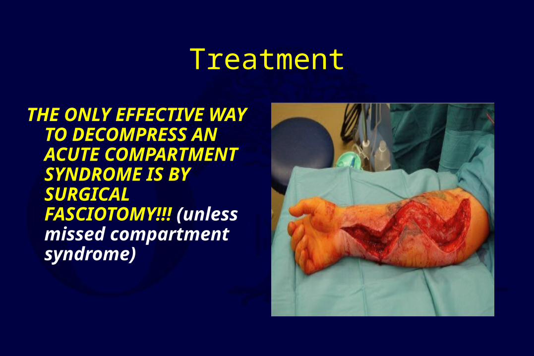
Treatment
THE ONLY EFFECTIVE WAY TO DECOMPRESS AN ACUTE COMPARTMENT SYNDROME IS BY SURGICAL FASCIOTOMY!!! (unless missed compartment syndrome)
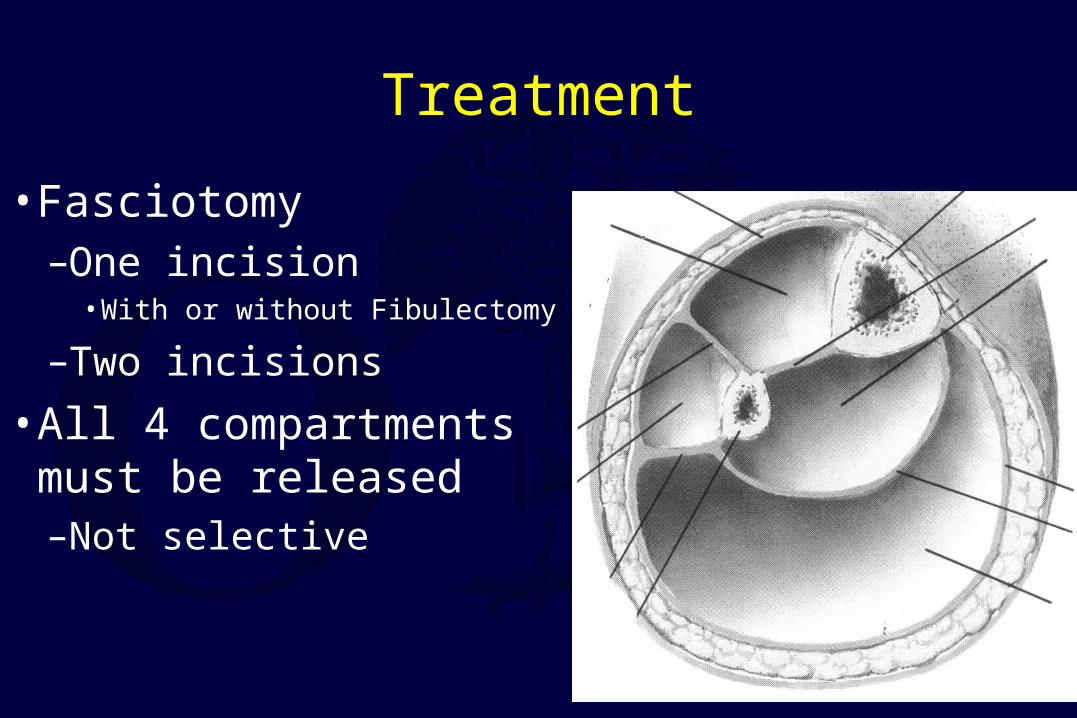
Treatment
• Fasciotomy–One incision
• With or without Fibulectomy
–Two incisions
• All 4 compartments must be released–Not selective
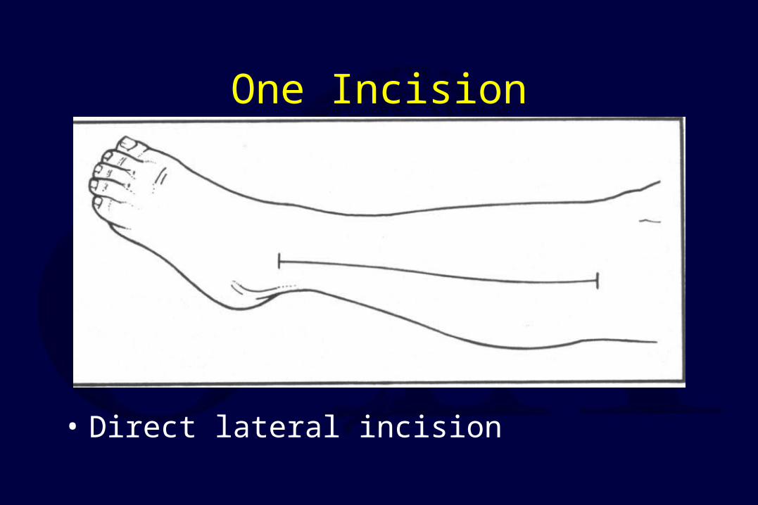
One Incision
• Direct lateral incision
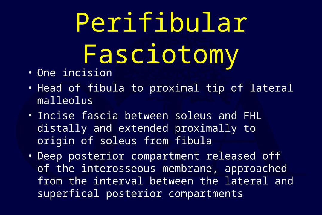
Perifibular Fasciotomy• One incision• Head of fibula to proximal tip of lateral malleolus• Incise fascia between soleus and FHL distally and
extended proximally to origin of soleus from fibula• Deep posterior compartment released off of the
interosseous membrane, approached from the interval between the lateral and superfical posterior compartments
• Lateral compartment
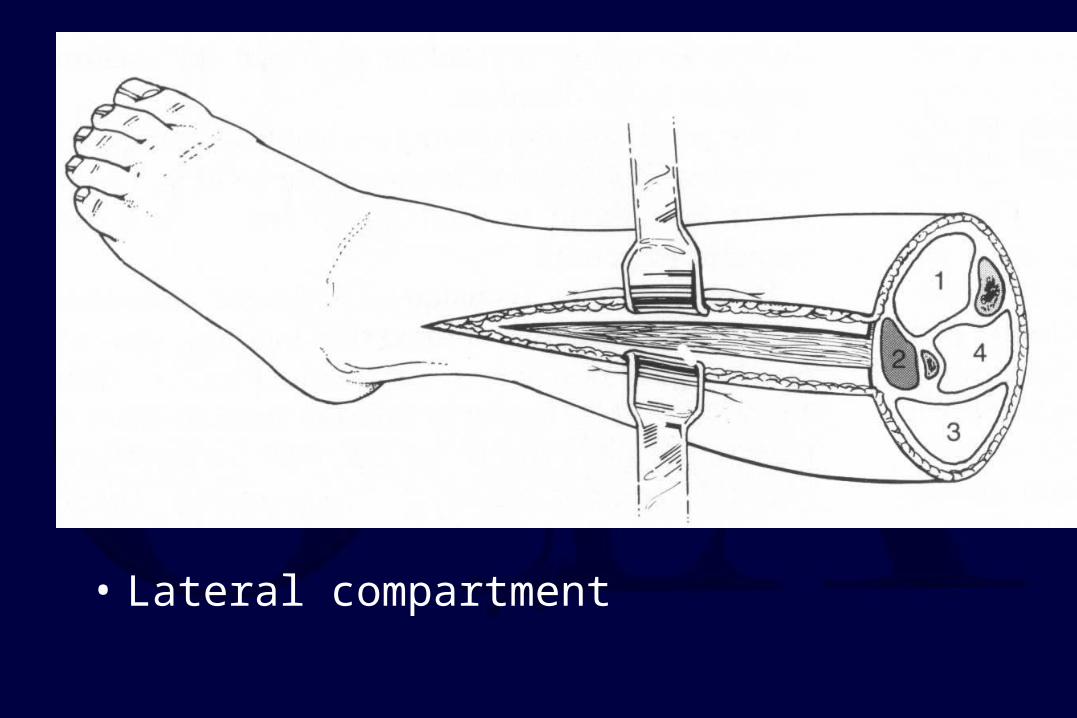
• Anterior compartment
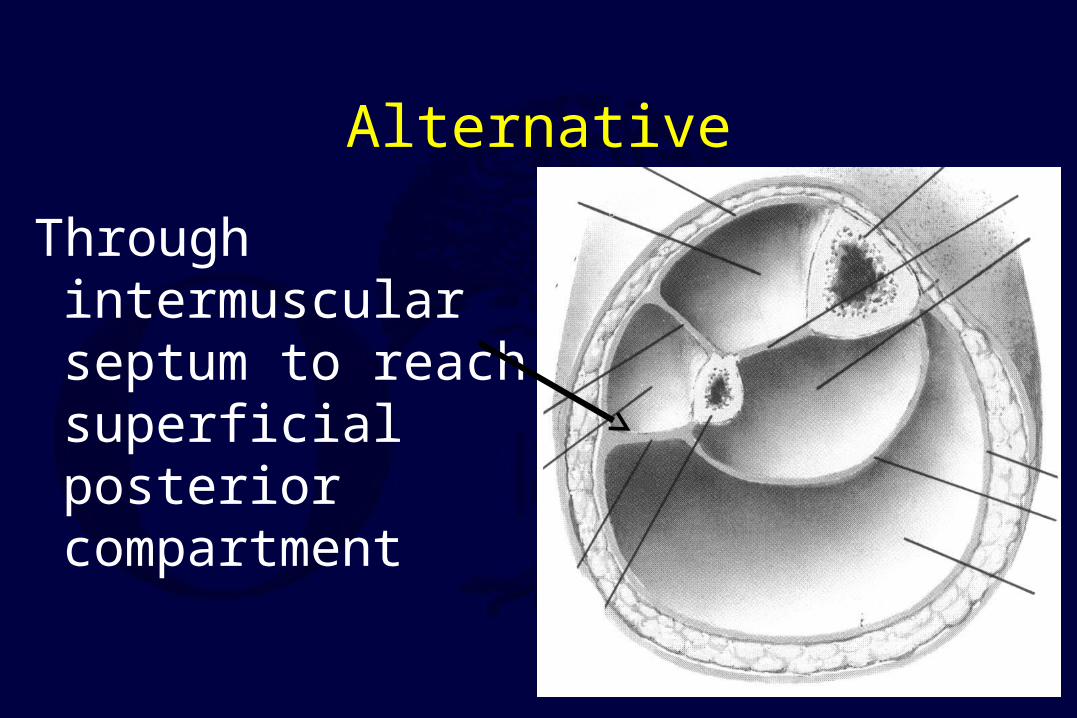
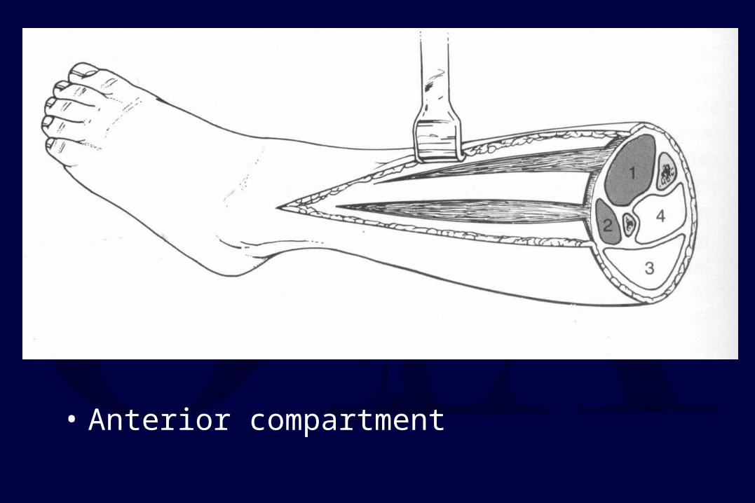
Alternative
Through intermuscular septum to reach superficial posterior compartment
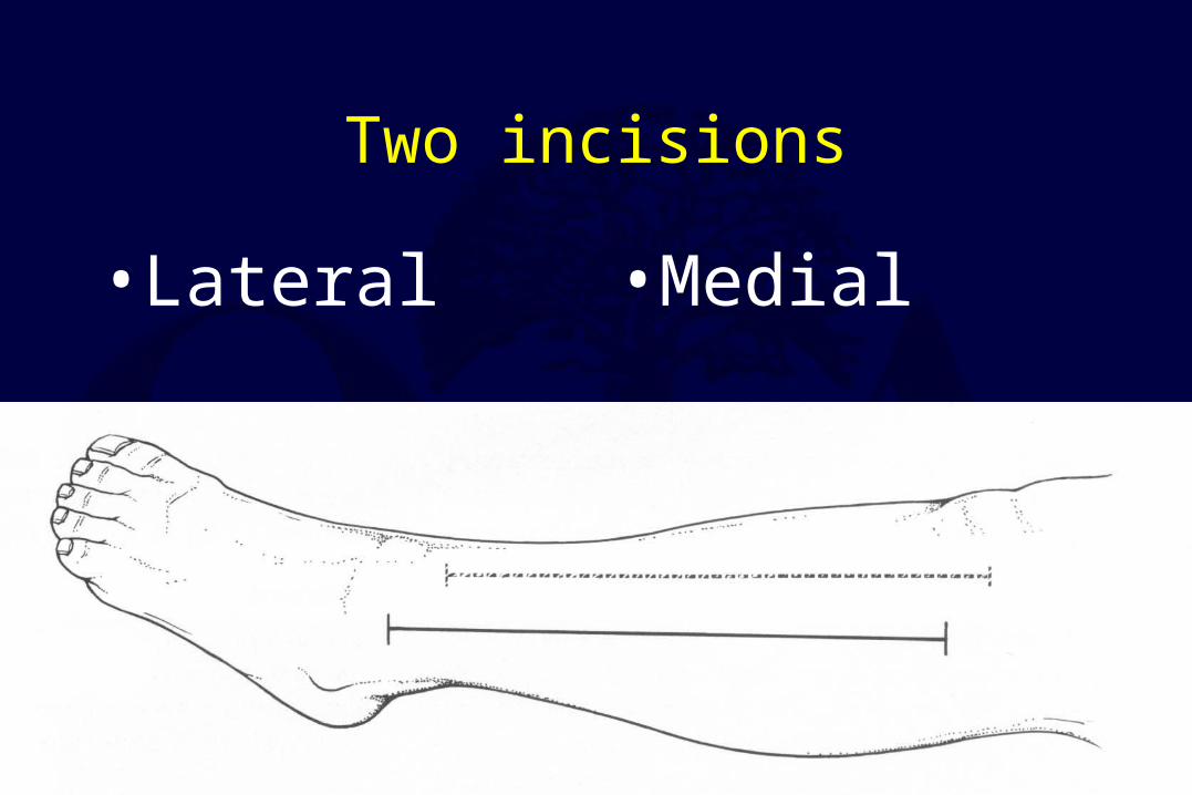
Two incisions
• Lateral • Medial
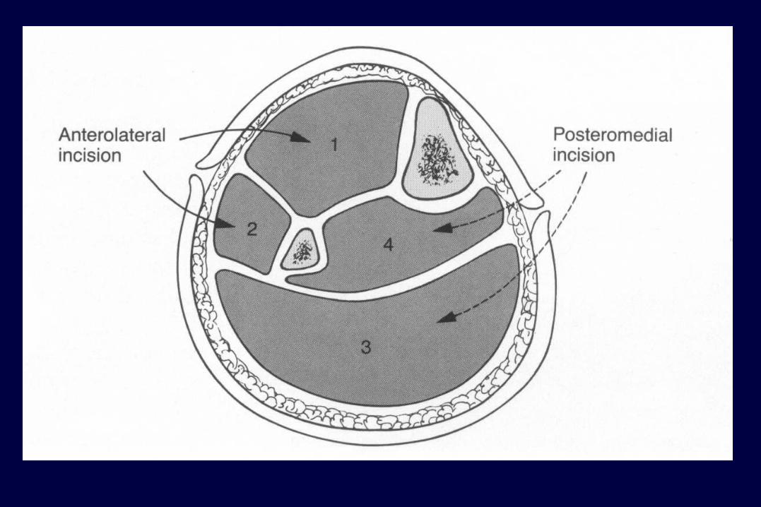
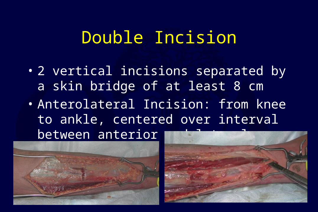
Double Incision
• 2 vertical incisions separated by a skin bridge of at least 8 cm
• Anterolateral Incision: from knee to ankle, centered over interval between anterior and lateral compartments
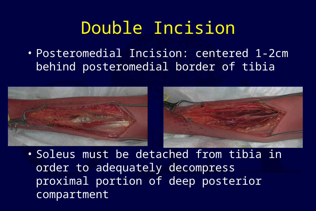
Double Incision
• Posteromedial Incision: centered 1-2cm behind posteromedial border of tibia
• Soleus must be detached from tibia in order to adequately decompress proximal portion of deep posterior compartment
Thigh
• Rare
• Crush injury with femur fracture
• Over distraction– relative under distraction
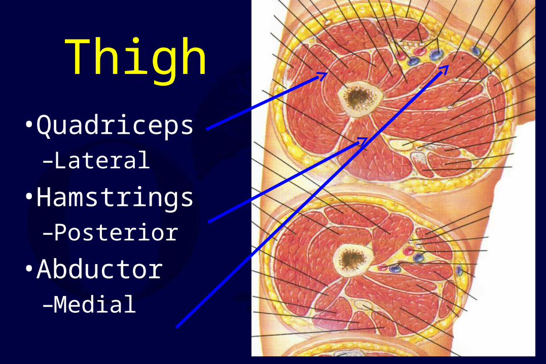
Thigh• Quadriceps
–Lateral
• Hamstrings–Posterior
• Abductor–Medial
Treatment
• Based upon involvement
• Usually Quadriceps and Hamstrings
• Usually, a single lateral incision will suffice
Compartments of the Forearm
• Forearm can be divided into 3 compartments: Dorsal, Volar and “Mobile Wad”
• Mobile Wad: Brachioradialis, ECRL, ECRB
• Dorsal: EPB, EPL, ECU, EDC
• Volar: FPL, FCR, FCU, FDS, FDP, PQ
Henry Approach
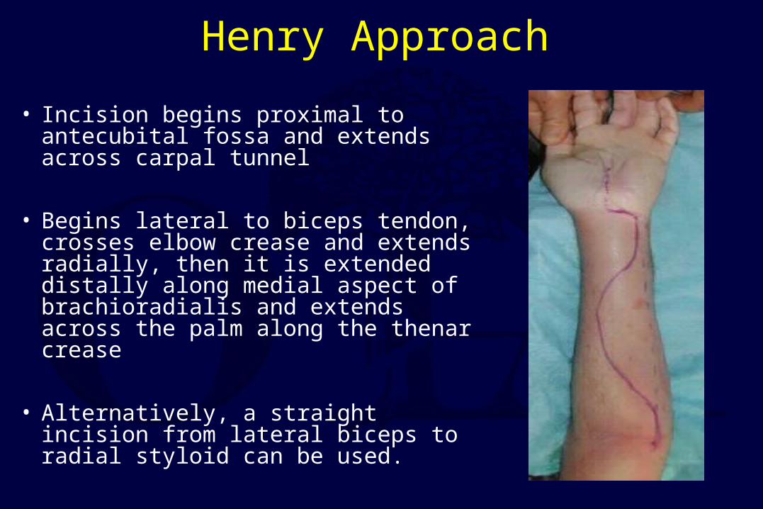
• Incision begins proximal to antecubital fossa and extends across carpal tunnel
• Begins lateral to biceps tendon, crosses elbow crease and extends radially, then it is extended distally along medial aspect of brachioradialis and extends across the palm along the thenar crease
• Alternatively, a straight incision from lateral biceps to radial styloid can be used.
Henry Approach
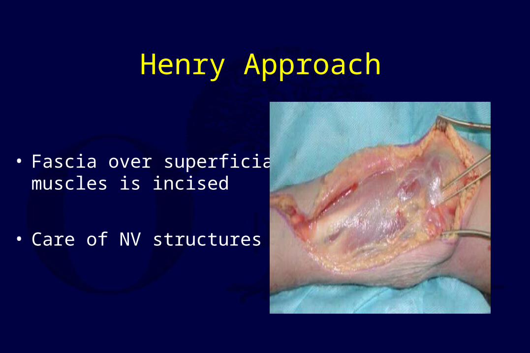
• Fascia over superficial muscles is incised
• Care of NV structures
Henry Approach
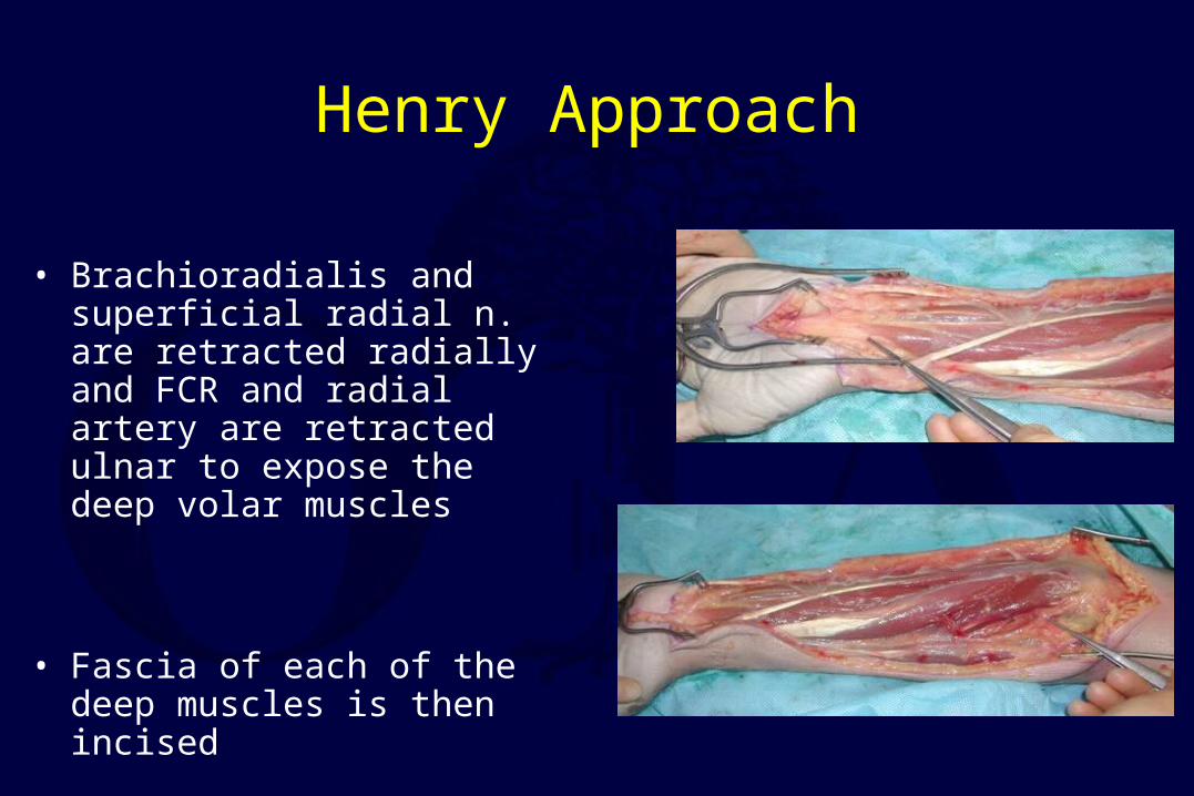
• Brachioradialis and superficial radial n. are retracted radially and FCR and radial artery are retracted ulnar to expose the deep volar muscles
• Fascia of each of the deep muscles is then incised
Post Fasciotomy…
• Must get bone stability– IMN
– exfix
• ~48hrs after procedure patient should be brought back to OR for further debridement
• Delayed skin closure or skin-grafting 3-7 days after the fasciotomies
Aftercare
• Xeroform
• VAC dressings
• Elevation of limb
• Delayed wound closure– Split thickness skin graft

Remember…
• Fasciotomies are not benign
• Complications are real >25%– Chronic swelling– Chronic pain– Muscle weakness– Iatrogenic NV injury– Cosmetic concerns
*** BUT if they are needed do not come up with *** BUT if they are needed do not come up with excuses to not do them !!!excuses to not do them !!!
Chronic (Exertional) Compartment Syndrome
• Transient rise in compartmental pressure following activity
• Symptoms –Pain
–Weakness
–Neurologic deficits
Chronic Compartment Syndrome
• Stress Test–Serial Compartment
Pressure• Resting >15mm Hg• 5 min post-ex. >25mm
Hg» Rydholm et al CORR 1983
–Volumetrics
–Nerve conduction Velocities
» Pedowitz et al. JHS 1988
Chronic Compartment Syndrome
• Treatment– Modification of activity– Splinting– Elective Fasciotomy
Conclusion
• Very important to make diagnosis
• Missed compartment is devastating
• Physical exam
• Re-examine patient!

Return to General/Principles
Index
E-mail OTA about
Questions/Comments
If you would like to volunteer as an author for the Resident Slide Project or recommend updates to any of the following slides, please send an e-mail to [email protected]