
Comparison of wild and cultivated extracts of Cordyceps ...
24
Union College Union | Digital Works Honors eses Student Work 6-2013 Comparison of wild and cultivated extracts of Cordyceps sinensis apoptotic potential Katelyn Staring Union College - Schenectady, NY Follow this and additional works at: hps://digitalworks.union.edu/theses Part of the Fungi Commons , and the Neuroscience and Neurobiology Commons is Open Access is brought to you for free and open access by the Student Work at Union | Digital Works. It has been accepted for inclusion in Honors eses by an authorized administrator of Union | Digital Works. For more information, please contact [email protected]. Recommended Citation Staring, Katelyn, "Comparison of wild and cultivated extracts of Cordyceps sinensis apoptotic potential" (2013). Honors eses. 736. hps://digitalworks.union.edu/theses/736
Transcript of Comparison of wild and cultivated extracts of Cordyceps ...

Union CollegeUnion | Digital Works
Honors Theses Student Work
6-2013
Comparison of wild and cultivated extracts ofCordyceps sinensis apoptotic potentialKatelyn StaringUnion College - Schenectady, NY
Follow this and additional works at: https://digitalworks.union.edu/theses
Part of the Fungi Commons, and the Neuroscience and Neurobiology Commons
This Open Access is brought to you for free and open access by the Student Work at Union | Digital Works. It has been accepted for inclusion in HonorsTheses by an authorized administrator of Union | Digital Works. For more information, please contact [email protected].
Recommended CitationStaring, Katelyn, "Comparison of wild and cultivated extracts of Cordyceps sinensis apoptotic potential" (2013). Honors Theses. 736.https://digitalworks.union.edu/theses/736
1
Running Title: Cordyceps sinensis Apoptotic Potential
Comparison of wild and cultivated extracts of Cordyceps sinensis apoptotic potential
By
Katelyn E. Staring
* * * * * * * * *
Submitted in partial fulfillment
of the requirements for
Honors in the Department of Neuroscience
UNION COLLEGE
June, 2013

2
ABSTRACT
STARING, KATELYN Comparison of wild and cultivated extracts of Cordyceps sinensis apoptotic potential. Department of Neuroscience, June 2013. ADVISOR: Brian D. Cohen
Cordyceps sinensis is a mushroom which contains the compound cordycepin (3’-
deoxyadenosine), an analogue of adenosine. In Traditional Chinese Medicine (TCM),
cordycepin has multipurpose pharmacological uses including purported anti-tumor
effects. In the present study, cordycepin was extracted from the wild mushroom as well
as from various commercially available cultivated extracts. Previous research in this lab
has demonstrated that cultivated extracts contain less cordycepin than the wild
mushroom. However, it is unclear if the decrease in cordycepin correlates with decreased
activity. To measure anti-tumor activity, extracts were used to treat human breast cancer
cells (MCF-7 cells). In other labs, cordycepin has been shown to induce apoptosis, or
programmed cell death in MCF-7 cells. Activity was evaluated using light microscopy to
observe cell morphology and DNA electrophoresis to discern DNA laddering, both of
which should be hallmarks of apoptosis. Using these methods the anticipated outcome is
to determine if there is a dose and time dependent manner to the cordycepin-induced cell
death as well as differential effects across the various cordycepin supplements which may
contain other active compounds. These hypotheses were supported by our data with a
dose-dependent cell death and marked differential in apoptotic potential among the
various types of cordycepin sources.

3
Introduction
For over 2,500 years Traditional Chinese Medicine (TCM) has been practiced as a study
of human physiology and pathology that is based on Chinese philosophy (Wang & Li,
2005). Originally based on Taoism and the idea of holism (the study of the whole
person—mind, body, and spirit), its practices include: herbal remedies, acupuncture,
moxibustion, cupping, and massage (Wang & Li, 2005; Russell & Patterson, 2008).
While it is often considered as part of complementary and alternative medicine (CAM) in
the United States, the popularity of TCM has been on the rise despite the limited
scientific evidence of its effectiveness (Russell & Patterson, 2008). It is important to
note, however, that this rise in use is as an adjunct to Western medicine and not as the
sole treatment.
TCM’s holistic approach gives rise to the difficulty found when trying to assess the
scientific validity. As it is based on complex principles that exist beyond strict natural
science, a unique approach must be taken when analyzing the data. However, in light of
new research, TCM treatments can no longer be dismissed as unscientific without
thorough experimentation.
Herbal supplements are probably the most popular of all the TCM treatments. They
abound in many stores and are not just limited to herbal shops and co-ops. Plus, they lend
themselves well to laboratory research. Of the over 13,000 herbal supplements employed
in TCM, one of the most sought after herbs comes from the mushroom species
Cordyceps. This herbal remedy uses cordycepin (3’-deoxyadenosine), an extract from
the rare mushroom Cordyceps sinensis (CS) found in parts of Asia including Nepal,
China, Japan, Korea, Vietnam, and Thailand. CS is a type of ascomycete fungi which

4
acts in a parasitic manner to insects. It paralyzes the insect victims, grows within their
host bodies, and eventually turns into a mature, fruiting fungus. In TCM, the fungus
body is boiled and used in chicken soup, duck soup, or herbal tea (Xie & Xie, 2010).
Likewise, in experimentation the biochemical cordycepin is extracted from the mycelium
of the fungi through a boiling water extraction.
Cordycepin is often considered a “wonder drug” due to its multipurpose pharmacological
uses (Russell & Paterson, 2008). It is prescribed for everything from anti-aging, to
hypertension, to cancer (He, et al., 2010). As the main active ingredient, cordycepin has
been found to have reno-protective activity (Li, He, Yang, & Wang, 2011), anti-
inflammation properties (Kim et al., 2011), and anti-tumor activity (He et al., 2010; Kim
et al., 2011; Jen et al., 2008; Pan, Lin, & Huang, 2011; Pao, Pan, Leu, Huang, 2012).
One of cordycepin’s most eye-catching claims is its anti-tumor effect, and previous
research has shown that this adenosine analogue can induce steroidogenesis in many cell
lines including: human colorectal cancer cells (SW480 & SW620), human breast cancer
cells (MDA-MB-231 & MCF-7), human neuroblastoma cells (SK-N-BE(2)-C),
melanoma cells (SK-MEL-2), human prostate carcinoma cells (PC-3), thyroid carcinoma
cells (CGTH W-2), human leukemia cells (U937 & THP-1), oral cancer cells (OEC-M1),
and mouse Leydig cells (MA-10). This anti-tumor effect is most likely due to the fact
that cordycepin is an analogue of the nucleotide adenosine. The biochemical differs at
the 3’ of the ribose moiety where it lacks a hydroxyl group found on adenosine (Fig. 1).
This hydroxyl group is necessary for 5’—3’ nucleotide elongation and interfering with
this process might be part of the anti-tumor effect (Chen, Stellrecht, & Gandhi, 2008).
Also due to the similarity with adenosine, cordycepin has been found to induce

5
steroidogenesis (steroid hormone synthesis) in many cell lines of the adrenal glands,
brain, placenta, testes, and ovaries (Ghayee & Auchus, 2007).
Figure 1. Adenosine and Cordycepin Structures. Adenosine is the purine nucleoside of adenine
and the attached ribose sugar molecule. As an analogue of adenosine, cordycepin lacks the 3’
hydroxyl on the attached sugar molecule.
Research has found that the anti-tumor role of cordycepin is due in part to its interaction
with the adenosine-3 receptor which subsequently activates ERK1/2 (Figure 2). ERK1/2
then interacts with other factors and causes proliferation of the cell and steroidogenesis or
apoptosis of the cell and cell death (Pao et al., 2012).

6
Figure 2. Adenosine-3 Receptor Pathway. A3 is one type of adenosine receptor that interacts
with alpha subunits of the G protein heterotrimer to instigate downstream effects that ultimately
activate the ERK1/2 protein and result in proliferation or apoptosis depending on other factors
within the cell.
The focus of this thesis is on cordycepin’s effects on the MCF-7 cell line, a clonal strain
of human breast cancer cells (Choi et al., 2011). Breast cancer is the most common
cancer among women in the world. The prevalence of breast cancer has been increasing
approximately 2% each year (Jemal et al., 2008). It is most often treated with surgical
interventions, chemotherapy, radiotherapy, and endocrine therapy. However, many
women also make use of CAM therapies both to lessen the symptoms and as an attempt
to cure their cancer (Yong et al., 2004). Therefore, research into cordycepin-induced cell
death could provide an alternative to the surgical route and the harsh chemotherapy while
also evolving with the widespread integration of CAM into the anti-cancer field.

7
Given cordycepin’s previously described wide range of uses there is a great demand for
this extract. However, CS is a rare mushroom that only exists in its wild form in high-
altitude areas of the Tibetan Plateau and is quite expensive outside of China. These
factors have led to an increase in various cultivation methods globally and in the United
States. As this production is generally unregulated the quality and purity of the cultivated
species of CS is questionable. It is often listed as having the same bioactive ingredients
and properties as wild CS but is sold at a much reduced price. In previous research done
by this lab by Lucas First, the cultivated species of CS that is commercially available was
found to contain less cordycepin.
This experiment will investigate the differential effects of various types of extracts from
cultivated CS, wild CS extracts, and Sigma-purchased pure cordycepin on the viability of
MCF-7 cells. At high enough concentrations, these treatments should induce apoptosis in
this cell line. Furthermore, even when the amount of cordycepin from each sample type
is normalized there might still be differential activity in the cells due to other compounds
located within the natural supplements used. In this way the anti-tumor claim of
cordycepin can be tested as it is available to the public in its supplemental form as well as
in its wild imported form. It will also provide valuable insight into further avenues of
cancer treatment and alternatives to chemotherapy.
In order to measure cell apoptosis light microscopy will be employed to determine the
cell morphology. DNA electrophoresis will also allow apoptosis to be monitored by the
presence or absence of DNA laddering. Using these methods the anticipated outcome is
to find a dose and time dependent manner to the cordycepin-induced cell death as well as
differential effects across the various cordycepin supplements.

8
9
Methods
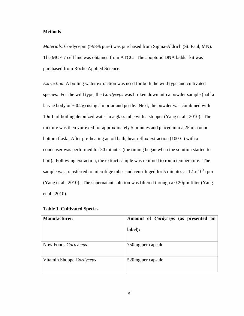
Materials. Cordycepin (>98% pure) was purchased from Sigma-Aldrich (St. Paul, MN).
The MCF-7 cell line was obtained from ATCC. The apoptotic DNA ladder kit was
purchased from Roche Applied Science.
Extraction. A boiling water extraction was used for both the wild type and cultivated
species. For the wild type, the Cordyceps was broken down into a powder sample (half a
larvae body or ~ 0.2g) using a mortar and pestle. Next, the powder was combined with
10mL of boiling deionized water in a glass tube with a stopper (Yang et al., 2010). The
mixture was then vortexed for approximately 5 minutes and placed into a 25mL round
bottom flask. After pre-heating an oil bath, heat reflux extraction (100ºC) with a
condenser was performed for 30 minutes (the timing began when the solution started to
boil). Following extraction, the extract sample was returned to room temperature. The
sample was transferred to microfuge tubes and centrifuged for 5 minutes at 12 x 103 rpm
(Yang et al., 2010). The supernatant solution was filtered through a 0.20µm filter (Yang
et al., 2010).
Table 1. Cultivated Species
Manufacturer: Amount of Cordyceps (as presented on
label):
Now Foods Cordyceps 750mg per capsule
Vitamin Shoppe Cordyceps 520mg per capsule

10
Jorrow Formulations Cordyceps 500mg per capsule
Holistic Herbal Solutions Cordyceps
Mushroom Powder
1lb bag of pure powder
For the cultivated species (listed in Table 1), the same water extraction method described
above was used for all four cultivated species. The amount of Cordyceps used in each
extraction was controlled by referencing the manufacturer’s published amount contained
in each capsule/tablet.
Cell Culture & Treatment. MCF-7 cells were maintained in DMEM medium in an
atmosphere of 5% CO2 at 37˚C. The cells were plated in 2 sets of 6-well dishes and
allowed to grow for 72 hours. Each well contained approximately 2.5x10⁵ cells and
2.5ml medium. They were treated with 3 doses (100, 150, and 200µM) of four different
samples (Sigma-purchased pure cordycepin, wild-type Cordyceps, Holistic Herbal
Solutions powder Cordyceps, and Vitamin Shoppe capsule Cordyceps) and incubated at
37˚C for 72 hours.
Cell Analysis. The cell morphology was observed and recorded using inverted light
microscopy. DNA gel electrophoresis (2% Agarose) was then performed to observe the
presence or absence of DNA laddering, a hallmark of apoptosis.

11
Results
To investigate possible cell growth inhibition, the effect of cordycepin on the MCF-7
human breast cancer cell line was visualized by light microscopy. Through this
examination many morphological changes were observed. The first set of wells were
treated with pure sigma-purchased cordycepin. In the control well (Figure 3a) the cells
appeared normal and attached to the well. In the well treated with 100µM (Figure 3b)
many cells appeared rounded-up, an indication of cell death. There was quite a bit of cell
debris from already dead cells and the remaining viable cells were irregularly shaped. In
the well treated with 200µM (Figure 3c) there was mostly cell debris and the few cells
that remained were rounded up.
Figure 3. Light microscopy of breast cancer cells treated with pure cordycepin. (a) The control well
contained normally-shaped cells. (b) The cells treated with 100µM pure cordycepin showed some
rounding of cells, cell debris, and irregularly shaped cells. (c) The cells treated with 200µM pure
cordycepin show mostly rounded up cells.
The second set of wells was treated with the wildtype Cordyceps mushroom extract. In
the well treated with 100µM (Figure 4a) there were once again some viable cells that
were irregularly shaped and a few rounded up cells. In the 200µM treatment (Figure 4b),
there were more rounded up cells and quite a bit of cell debris.

12
Figure 4. Light microscopy of breast cancer cells treated with wildtype Cordyceps extract. (a) The
100µM treatment has many irregularly shaped viable cells and some rounded up cells. (b) The 200µM
treatment has many rounded up cells, a few irregularly shaped viable cells, and visible cell debris.
The third set of wells was treated with the American cultivated Holistic Herbal Solutions
Cordyceps powder extract. The well treated with 100µM (Figure 5a) contains mostly
normal cells that are viable and still attached to the well bottom. There are only a few
rounded up cells at this treatment concentration and very little cell debris. At 200µM
(Figure 5b) there is signficantly more rounded up cells and very few normal viable cells.
There is also a marked increase in cell debris at this concentration.

13
Figure 5. Light microscopy of breast cancer cells treated with cultivated Holistic Herbal Solutions
Cordyceps powder extract. (a) The 100µM treatment resulted in many normal viable cells and a few
rounded up cells. (b) The 200µM treatment contains many rounded up cells and cell debris.
The last treatment utilized the Vitamin Shoppe Capsule Cordyceps extract. In the well
treated with 100µM (Figure 6a), most cells were normal and attached to the well bottom
with only a few rounded up cells. In the 200µM treatment (Figure 6b), there were still
many normal cells and few rounded up cells.
Figure 6. Light microscopy of breast cancer cells treated with Vitamin Shoppe Cordyceps extract. (a)
The 100µM treatment resulted in many normal cells and a few rounded up cells. (b) The 200µM treatment
also resulted in many normal cells and a few rounded up cells.
The results of the DNA electrophoresis (Figure 7) did not present DNA laddering which
would indicate apoptosis. Instead, there was non-specific DNA degradation that was
again consistent with the findings of Choi et al., (2011) and Lee et al., (2012) which also
worked with this cell line (MCF-7).

14
Figure 7. DNA electrophoresis of MCF-7 cells treated with cordycepin. The cells were treated with 4
sources of cordycepin at 3 different concentrations and then analyzed via agarose gel electrophoresis.

15
Discussion
There were two hypotheses for this study: (1) all forms of Cordyceps used would reduce
cell survival in the MCF-7 cell line and (2) there will be a differential apoptotic potential
across the various types of Cordyceps used—specifically that the cultivated form would
be less potent than the wildtype which would be less potent than the pure Sigma-
purchased cordycepin. To test these hypotheses MCF-7 cells were treated with cultivated
CS extracts, wild CS extracts, and their viability was first inferred by observing the cell
morphology via light microscopy.
The observations of rounding up of cells, large amounts of cell debris, and irregularly
shaped remaining cells in the treatment wells all indicate stages of cell death and support
the hypothesis that cell survival in the MCF-7 cell line was reduced due to the treatment
of Cordyceps. These findings are all consistent with the findings of Choi et al., (2011)
and Lee et al., (2012), both of which also did research using this cell line (MCF-7).
Furthermore, while the amount of cordycepin from each sample type was normalized
(using previous analysis values found in this lab using HPLC on an Agilent 6400 Series
Triple Quad LC/MS) there was still differential activity in the cells and a differential
apoptotic potential across the various types used. The pure Sigma-purchased cordycepin
treatment and the wildtype CS extract treatment both resulted in large amounts of cell
debris, many rounded up cells, and a large percentage of any remaining cells were
irregularly-shaped in both the 100µM and 200µM wells. In the American cultivated
Holistic Herbal Solutions Cordyceps extract treatment, however, there were only a few
rounded up cells at the 100µM dosage and there was not significant rounding-up of cells

16
and cell debris until the 200µM treatment. The Vitamin Shoppe capsule Cordyceps
extract also did not show many rounded-up cells at the 100µM dosage nor was there a
significant amount at the 200µM treatment. These results display the hypothesized
differential apoptotic potential across the various Cordyceps types as predicted. This
effect may have then been due to inherent differences in the wildtype versus cultivated
forms of Cordyceps such as other active compounds (Table 2) and not based on the actual
amount of cordycepin. The method of cultivation is not listed on the packaging for any
of the samples used in this study but previous studies have found that different methods
produce Cordyceps with varying amounts of the bioactive compounds generally found in
the wildtype. For example, in the submerged fermentation method there is a loss of
extra-cellular compounds which leads to less secondary bio-metabolites (Tuli, Sanhu, &
Sharma, 2013). While many studies have been done concerning cordycepin, the
remaining bioactive compounds have still not been fully researched to determine their
structure-function relationships and effects on the cell.
Table 1. Bioactive compounds extracted from Cordyceps (Tuli, Sanhu, & Sharma, 2013).
Number Bioactive Compound
1 Cordycepin
2 Cordycepic acid
3 N-acetylgalactosamine
4 Adenosine
5 Ergosterol and ergosteryl esters
6 Bioxanthracenes

17
7 Hypoxanthine
8 Acid deoxyribonuclease
9 Polysacchardie and exopolysaccharide
10 Chitinase
11 Macrolides
12 Cicadapeptins and myriocin
13 Superoxide dismutase
14 Protease
15 Naphthaquinone
16 Cordyheptapeptide
17 Dipicolinic acid
18 Fibrynolytical enzyme
19 Lectin
20 Cordymin
Apoptosis or programmed cell death is normally characterized by chromatin
condensation and then endonuclease cleavage of DNA (Cohen, Sun, Snowden, Dinsdale,
& Killeter, 1992). This cleavage results in in fragments of varying lengths that form a
DNA “ladder” when submitted to gel electrophoresis with bands in a ladder pattern
corresponding to the increasing fragment weights. This hallmark of apoptosis was,
however, not evident and indicated that the cordycepin activated a different pathway or
different branch of the normal apoptotic route.

18
This alternate pathway should not be used as evidence that cordycepin would not be an
effective treatment of breast cancer, but instead quite the opposite. In the MDA-MB-231
cell line (another human breast cancer cell line), MA-10 cell line (mouse Leydig tumor
cells), and many others, cordycepin treatment did in fact result in DNA laddering and
normal apoptosis. However, in the cell line used in this study a different pathway was
instead activated but the end goal of cell death was still accomplished. In a study by Choi
et al., (2011), a series of tests were ran to determine the exact differences between the
apoptosis in the MDA-MB-231 breast cancer cells and the cell death in the MCF-7 breast
cancer cells following treatment with cordycepin. This study found that the MCF-7 cells
underwent a different type of cell death that mainly consisted of autophagy. There were
autophagosome-like structures observed with transmission electron microscopy and many
molecules found that indicate autophagy (LC3-II, AVOs, and MDC-positive vacuoles).
Breast cancer cell lines are often referred to as ER-positive, meaning that estrogen
promotes tumor growth, or ER-negative, meaning that estrogen decreases tumor growth.
However, most breast cancer cell lines actually contain a combination of both. Treatment
with cordycepin, which acts irrespective of this ER-response could suppress both types of
tumor growth (Choi et al., 2011).
By activating multiple cell-death pathways this form of treatment could be very effective
in aggressive multi-drug resistant cancers. Likewise, due to its anti-cancer effects in
multiple cell lines across different body parts (testicular, oral, colorectal, neuroblastoma,
melanoma, prostate, thyroid, leukemia, etc.) it would also be a good candidate for
treatment in metastasized breast cancer.

19
All of this data concerning cordycepin indicates that it could be used in future therapeutic
applications with more research. In fact, much research is being done at this very
moment concerning the link between adenosine and cancer. In a review article by
Merighi et al., (2003), adenosine was proposed as a potent regulator of tumor cell growth.
The study reviewed many others where numerous adenosine receptors were found to play
a role in cancer including A1, A2A, A2B, and A3 (Figure 8). Current research is also been
doing on other adenosine analogues similar to cordycepin such as: 3”-ethynyladenosine,
pentostatin, and cladribine as well as adenosine antagonists such as caffeine.
Figure 8. Signaling pathways of A1, A2B, A2A, and A3 adenosine receptors. AC: adenylate cyclase; CHO: Chinese hamster ovary cells; ERK1/2: extracellular signal-regulated kinases 1 and 2; HEK-293: human embryonic kidney cells; JNK: c-Jun N-terminal kinase; MEK-1/2: MAP kinase kinases 1 and 2; PC12: pheochromocytoma cell line; PI3K: phosphoinositide 3-kinase; PKA: protein kinase A; PKB: protein kinase B; PKC: protein kinase C; PLC: phospholipase C; PLD: phospholipase D; p21: small G protein, p21(ras); rap 1: small G protein, rap 1; U937: monocytic lymphoma cell line.
Cordyceps has been used as a treatment for thousands of years safely and even with
present research that have only been a few cases where the treatment caused a negative
effect. The most common negative report involves dry mouth, nausea, and diarrhea with
very few patients reporting an allergic response. While there are recommended dosages

20
based on the condition that the Cordyceps is supposed to be triggering, there is not a
lethal dosage. Studies using mice and rabbits found no fatality even when the dosage was
increased dramatically for a long period of time (Tuli, Sandhu, & Sharma, 2013).
As cordycepin is similar to adenosine it is able to cause a wide variety of
polypharmacological effects based on the inhibition of RNA synthesis and
polyadenylation and should be considered for further research and eventually clinical
trials once more of the mechanism is further elucidated. Besides the missing gaps in the
mechanism, the effects of all the other bioactive compounds should be clarified and
proper cultivation methods should be followed for accurate production of the capsules
sold in many vitamin shops and websites. This research would provide a valuable insight
into further avenues of cancer treatment and alternatives to chemotherapy.

21
Reference
Chen, L.S., Stellrecht, C.M., Gandhi, V. (2008). RNA-directed agent, cordycepin,
induces cell death in multiple myeloma cells. British Journal of Haematology,
140, 682-691.
Choi, S., Lim, M.H., Kim, K.M., Jeon, B.H., Song, W.O., & Kim, T.W. (2011).
Cordycepin-induced apoptosis and autophagy in breast cancer cells are
independent of the estrogen receptor. Toxicology and Applied Pharmacology,
257(2), 165-173.
Cohen, G.M., Sun, X.M., Snowden, R.T., Dinsdale, D., & Skilleter, D.N. (1992). Key
morphological features of apoptosis may occur in the absence of internucleosomal
DNA fragmentation. Biochemical Journal, 286(Pt 2), 331.
Ghayee, H.K., & Auchus, R., J. 2007. Basic concepts and recent developments in human
steroid hormone biosynthesis. Reviews in Endocrine and Metabolic Disorders,
8(4), 289-300.
He, W., Zhang, M., Ye, J., Jiang, T., Fang, X., & Song, Y. (2010). Cordycepin induces
apoptosis by enhancing JNK and p38 kinase activity and increasing the protein
expression of Bcl-2 pro-apoptotic molecules. Biomedicine and Biotechnology,
11(9), 654-660.
Jemal, A., Siegel, R., Ward, E., Hao, Y., Xu, J., Murray, T., Thun, M.J. (2008). Cancer
statistics, CA: A Cancer Journal for Clinicians, 58, 71-96.
22
Jen, C., Lin, C., Huang, B., & Leu, S. (2008). Cordycepin induced MA-10 mouse leydig
tumor cell apoptosis through caspase-9 pathway. Evidence-Based Complementary
and Alternative medicine, 2011, 1-11.
Kim, H., Naura, A. S., Errami, Y., Ju, J., & Boulares, A. H. (2011). Cordycepin blocks
lung injury-associated inflammation and promotes BRCA1-deficient breast cancer
cell killing by effectively inhibiting PARP. Molecular Medicine, 17(9-10), 893-
900.
Li, L., He, D., Yang, J., & Wang, X. (2011). Cordycepin inhibits renal interstitial
myofibroblast activation by inducing hepatocyte growth factor expression.
Journal of Pharmacological Sciences, 117, 286-294.
Merighi, S., Mirandola, P., Varani, K., Gessi, S., Leung, E., Baraldi, P.G., & Borea, P.A.
(2003). A glance at adenosine receptors: novel target for antitumor therapy.
Pharmacology & Therapeutics, 100(1), 31-48.
Pan, B., Lin, C., & Huang, B. (2011). The effect of cordycepin on steroidogenesis and
apoptosis in MA-10 mouse leydig tumor cells. Evidence-Based Complementary
and Alternative Medicine, 2011.
Pao, H., Pan, B., Leu, S., & Huang, B. (2012). Cordycepin stimulated steroidogenesis in
MA-10 Mouse tumor cells through the protein kinase c pathway. Journal of
Agricultural and food chemistry, 60, 3905-4913.
Russell, R., & Paterson, M. (2008). Cordyceps—A traditional Chinese medicine and
another fungal therapeutic biofactory. Phytochemisty, 69, 1469-1495.
23
Tuli, H. S., Sandhu, S. S., & Sharma, A.K. (2013). Pharmacological and therapeutic
potential of cordyceps with special reference to cordycepin. 3 Biotech, 1-12.
Wang, S., & Li, Y. (2005). Traditional Chinese Medicine. In O. Devinsky, S. Schacter,
& S. Pacia (Eds.), Complementary and alternative therapies for epilepsy. New
York, NY: Demos Medical Publishing.
Xie, Z., & Xie, F. (2010). Contemporary Introduction to Chinese Medicine: Comparison
with Western Medicine. Beijing, China: Foreign Languages Press, 2010.
Yang, F., Li, D., Feng, K., Hu, D., & Li S. (2010). Determination of nucleotides,
nucleosides and their transformation products in cordyceps by ion-pairing
reversed-phase liquid chromatography-mass spectrometry. Journal of
Chromatography A, 1217, 5501-5510.
Yong, C., Xiao-Qu, S., Yutang, G., Wanging, W., et al. (2004). Use of complementary
and alternative medicine by Chinese women with breast cancer. Breast Cancer
Research and Treatment, 85(3), 263-270.


![Wild versus Cultivated Olive Leaves Extracts: Antioxidant ...jcbnet.info/journals/jcb/Vol_4_No_2_December_2016/6.pdf · of malaria and associated fever [9]. Olive leaf offered a capacity](https://static.fdocuments.net/doc/165x107/5d53343b88c993f4288b776a/wild-versus-cultivated-olive-leaves-extracts-antioxidant-of-malaria-and.jpg)


