Comparison of marginal bone loss and patient satisfaction ... · Comparison of marginal bone loss...
8
The Journal of Advanced Prosthodontics 191 Comparison of marginal bone loss and patient satisfaction in single and double-implant assisted mandibular overdenture by immediate loading Sara Tavakolizadeh 1 *, Fariborz Vafaee 2 *, Masume Khoshhal 2 , Zahra Ebrahimzadeh 3 1 TMD Center, INI Hospital, Hannover, Germany 2 Hamadan University of Dental Science, Hamadan, Iran 3 Periodontist, Tehran, Iran PURPOSE. The purpose of this study was to compare the coronal bone level and patient satisfaction in 1-implant and 2-implant assisted mandibular overdentures. MATERIALS AND METHODS. Twenty patients who had maladaptive mandibular dentures were treated in this study. Patients were randomly divided into two groups. The first group received 1 implant (Simple line II, Implantium, South Korea) in their mandibular midline and the second group received 2 implants in their B and D regions (according to Misch’s category). If the primary stability of each implant was at least 60 ISQ, ball attachment was placed and denture relined with soft liner. After 6 weeks, retentive cap incorporated with hard acrylic resin. In the 6 and 12 months recalls, periapical digital radiograph were made and visual analogue scale questionnaires were used to record patient satisfaction. The Friedman test was done for comparing the presurgical and postsurgical parameters in each group and the U-Mann Whitney test (P<.05) was done for comparison of post-treatment results between the two groups. RESULTS. All implants achieved sufficient primary stability to be immediately loaded. Patient satisfaction was high, and there were no significant differences between two groups (P>.05). In addition, mean marginal bone loss was 0.6 ± 0.67 mm in the first group and 0.6 ± 0.51 mm in the second group, after 12 month. Mean marginal bone loss showed no significant differences between two groups. CONCLUSION. This preliminary one- year result indicated that mandibular overdentures anchored to a single implant can be a safe and cost-effective method as a starting step for implant-overdenture treatment. [ J Adv Prosthodont 2015;7:191-8] KEY WORDS: Implant; Overdenture; Bone loss; Patients satisfaction http://dx.doi.org/10.4047/jap.2015.7.3.191 http://jap.or.kr J Adv Prosthodont 2015;7:191-8 INTRODUCTION Half of all conventional mandibular dentures demonstrate problems with prosthesis stability and retention, with reten- tion being the single most important deficiency reported. 1 Introduction of osseointegrated dental implant has improved the quality of life of many edentulous patients. Implant assisted mandibular overdentures can provide a highly suc- cessful restoration of both function and esthetics. 2-4 In the last 30 years, use of unloaded and submerged implant according to Brånemark protocol for achieving osseointegration was necessary. Current studies have dem- onstrated increasing success with immediately loaded dental implants. Immediate loading of implants in anterior mandible was reported to shorten the treatment time and show compa- rable clinical success with the delay loading approach. 5-7 Corresponding author: 1 Sara Tavakolizadeh, 2 Fariborz Vafaee 1 TMD center, INI hospital, Rudolf-Pichlmayr street, 30625, Hannover, Germany Tel: 004917646171743: e-mail, [email protected] 2 Hamadan University of Dental Science, Shahid Fahmide street, Hamadan, Iran e-mail, [email protected] Received October 12, 2014 / Last Revision March 16, 2015 / Accepted March 18, 2015 © 2015 The Korean Academy of Prosthodontics This is an Open Access article distributed under the terms of the Creative Commons Attribution Non-Commercial License (http://creativecommons. org/licenses/by-nc/3.0) which permits unrestricted non-commercial use, distribution, and reproduction in any medium, provided the original work is properly cited. pISSN 2005-7806, eISSN 2005-7814 This study was supported by a grant of the Hamadan Dental University, Iran.
Transcript of Comparison of marginal bone loss and patient satisfaction ... · Comparison of marginal bone loss...
The Journal of Advanced Prosthodontics 191
Comparison of marginal bone loss and patient satisfaction in single and double-implant assisted mandibular overdenture by immediate loading
Sara Tavakolizadeh1*, Fariborz Vafaee2*, Masume Khoshhal2, Zahra Ebrahimzadeh3
1TMD Center, INI Hospital, Hannover, Germany2Hamadan University of Dental Science, Hamadan, Iran3Periodontist, Tehran, Iran
PURPOSE. The purpose of this study was to compare the coronal bone level and patient satisfaction in 1-implant and 2-implant assisted mandibular overdentures. MATERIALS AND METHODS. Twenty patients who had maladaptive mandibular dentures were treated in this study. Patients were randomly divided into two groups. The first group received 1 implant (Simple line II, Implantium, South Korea) in their mandibular midline and the second group received 2 implants in their B and D regions (according to Misch’s category). If the primary stability of each implant was at least 60 ISQ, ball attachment was placed and denture relined with soft liner. After 6 weeks, retentive cap incorporated with hard acrylic resin. In the 6 and 12 months recalls, periapical digital radiograph were made and visual analogue scale questionnaires were used to record patient satisfaction. The Friedman test was done for comparing the presurgical and postsurgical parameters in each group and the U-Mann Whitney test (P<.05) was done for comparison of post-treatment results between the two groups. RESULTS. All implants achieved sufficient primary stability to be immediately loaded. Patient satisfaction was high, and there were no significant differences between two groups (P>.05). In addition, mean marginal bone loss was 0.6 ± 0.67 mm in the first group and 0.6 ± 0.51 mm in the second group, after 12 month. Mean marginal bone loss showed no significant differences between two groups. CONCLUSION. This preliminary one-year result indicated that mandibular overdentures anchored to a single implant can be a safe and cost-effective method as a starting step for implant-overdenture treatment. [ J Adv Prosthodont 2015;7:191-8]
KEY WORDS: Implant; Overdenture; Bone loss; Patients satisfaction
http://dx.doi.org/10.4047/jap.2015.7.3.191http://jap.or.kr J Adv Prosthodont 2015;7:191-8
INTRODUCTION
Half of all conventional mandibular dentures demonstrate problems with prosthesis stability and retention, with reten-tion being the single most important deficiency reported.1 Introduction of osseointegrated dental implant has improved the quality of life of many edentulous patients. Implant assisted mandibular overdentures can provide a highly suc-cessful restoration of both function and esthetics.2-4
In the last 30 years, use of unloaded and submerged implant according to Brånemark protocol for achieving osseointegration was necessary. Current studies have dem-onstrated increasing success with immediately loaded dental implants. Immediate loading of implants in anterior mandible was reported to shorten the treatment time and show compa-rable clinical success with the delay loading approach.5-7
Corresponding author: 1Sara Tavakolizadeh, 2Fariborz Vafaee1TMD center, INI hospital, Rudolf-Pichlmayr street, 30625, Hannover, GermanyTel: 004917646171743: e-mail, [email protected] University of Dental Science, Shahid Fahmide street, Hamadan, Irane-mail, [email protected] October 12, 2014 / Last Revision March 16, 2015 / Accepted March 18, 2015
© 2015 The Korean Academy of ProsthodonticsThis is an Open Access article distributed under the terms of the Creative Commons Attribution Non-Commercial License (http://creativecommons.org/licenses/by-nc/3.0) which permits unrestricted non-commercial use, distribution, and reproduction in any medium, provided the original work is properly cited.
pISSN 2005-7806, eISSN 2005-7814
This study was supported by a grant of the Hamadan Dental University, Iran.
192
Traditionally, an overdenture has based the use of two or more mandibular implants. Although, it seems that increasing the number of implants may improve the treat-ment outcome, but many studies showed that two implants, splinted or unsplinted, are sufficient for clinical success.8-12 McGill consensus statement suggested that two-implant overdenture should be the first choice of treatment for the edentulous mandible.13
Unfortunately, two-implant mandibular overdenture is outside the financial scope of many compromised edentu-lous patients.14 With the increased demand for a better den-ture, a more cost-effective alternative treatment with a sin-gle implant can be recommended. Improving patient’s satis-faction and predictable clinical success of single implant overdenture with a delay and immediate implant loading protocol was demonstrated in some studies.14-18
The aim of this study is to compare the coronal bone loss and patient satisfaction between 1 and 2 implant anchored mandibular overdenture by immediate loading protocol.
MATERIALS AND METHODS
Twenty completely edentulous patients (9 male and 11 female), with the mean age of 59 years, were included in this study. The main complaint among the patients was poor retention and instability of mandibular denture.
Inclusion criteria dictated that the patients have been completely edentulous for at least one year, be maladaptive to the mandibular denture and have enough bone for an implant length of at least 10 mm and diameter of 3.8 mm in the anterior and inverted U shape ridge in the posterior region of the mandible.5,14,19 Exclusion criteria included substance abuse, disorders to the implant sites that disturb bone regeneration or any physical or psychological problem that could affect the follow up. Smokers were encouraged and followed to quit for at least 2 months, but were not excluded from the study.14 Before initiation of treatment, an informed consent was signed by all patients. Approval of the Ethic Committee for Human Clinical Research and the IRB approval were produced.
All existing dentures were evaluated with the California Dental Association (CDA) quality evaluation system.20 Fabrication of new upper and lower dentures was done in any patients that dentures did not match with these criteria in terms of aesthetic, coordination of centric occlusion with centric relation, correct vertical dimension or absence of tissue irritation.
Preoperative cone beam computed tomography (CBCT) (Promax 3D, Planmeca, Helsinki, Finland) was taken with a radiopaque surgical stent. A transparent stent was duplicat-ed from the final patient’s mandibular denture, then drilled in B, C and D locations (according to Misch’s category).21 C location is the mandibular midline, B and D locations are the proximal contacts between the right and left lateral inci-sor-canine teeth, respectively. These holes were filled with gutta-percha cone (Guttapercha, Ariadent, Tehran, Iran) and polished surfaces painted with Barium sulfate (barium
sulfate was mixed with dentin bonding in ratio of 1/3 and painted by 2 layers) as radiopaque materials. The bone qual-ity and quantity of anterior mandibular ridge was analyzed with CBCT and the best implant angulations according to available bone and avoidance of anatomic structures was determined by electronic surgery on computer (Fig. 1).
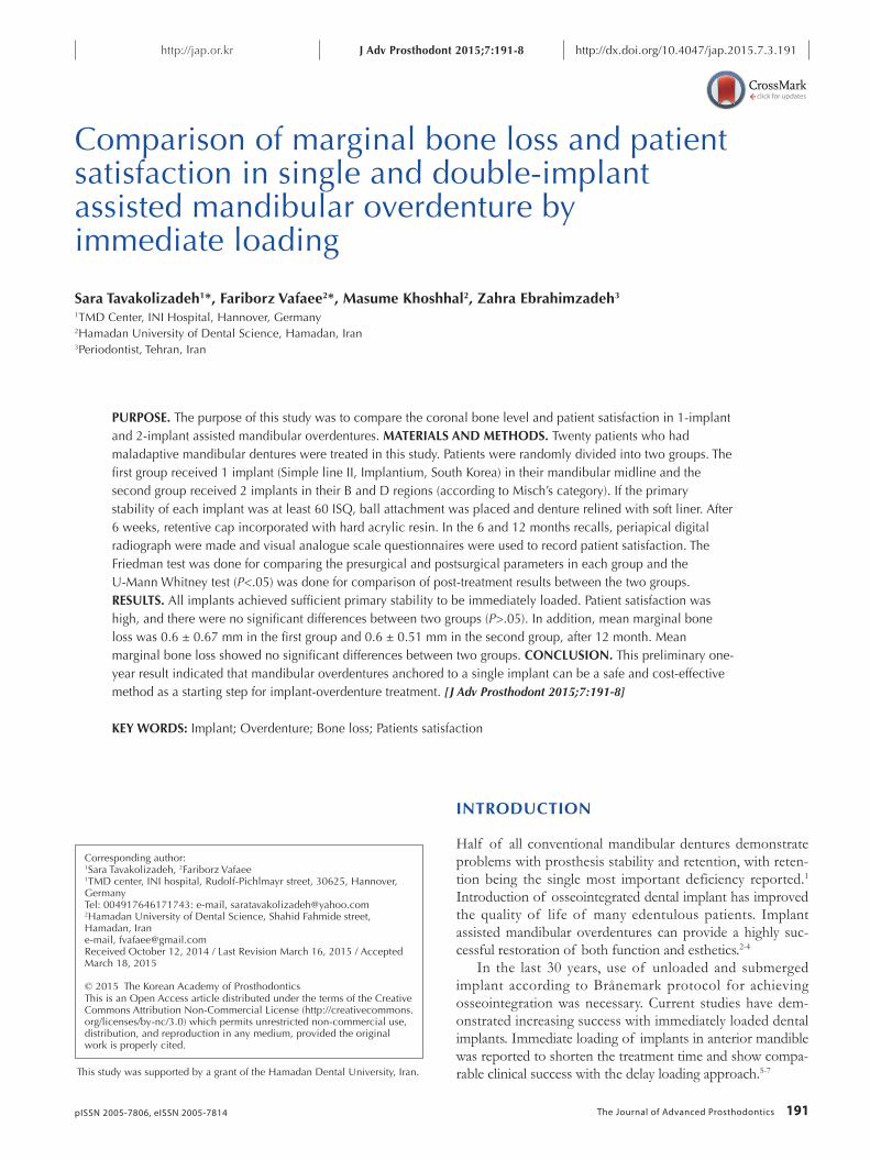
After removal of the radiopaque markers, pilot holes for the best implants positioning (according to the CBCT evalu-ation) were drilled. These holes diameter were matched with the initial 2 mm twist drill (Fig. 2).
For comparison of patients comfort and function, before and after implant treatment, self-administered questionnaire that followed the Visual Analog Scale (VAS) method was filled out by the patients preoperatively and then at each scheduled recall.22
Each VAS questionnaire consisted of a 100 mm line anchored at the beginning and end by opposing statements such as “not at all satisfied” to “extremely satisfied”. The participants marked a vertical line on the horizontal VAS line to indicate feelings. Scores determined by measuring the distance (in mm) from the right starting point of the line to the intersection of the response line. Questions were
Fig. 1. CBCT evaluation and electronic surgery.
Fig. 2. Pilot hole in surgical stent as a guide for initial twist drill. (A) tissue surface, (B) polished surface.
A B
J Adv Prosthodont 2015;7:191-8
The Journal of Advanced Prosthodontics 193
Comparison of marginal bone loss and patient satisfaction in single and double-implant assisted mandibular overdenture by immediate loading
in 5 categories: general satisfaction, social life, mastication of hard and soft foods and fit.14
Patients were randomly divided into 2 groups; in the first group a single regular platform implant of 3.8 mm diameter (Simple line II, SOFX483810R, Implantium, Seoul, South Korea) installed in the mandibular midline and in the second group, two implants were installed in the B and D locations (according to Misch’s category).
One hour before the surgery, all patients received sin-gle-dose prophylactic antibiotic (2 g of Amoxicillin or 600 mg of Clindamycin) orally.23 A mouth rinse (0.2% chlorhex-idineNajo, Tehran, Iran) was utilized for 60 seconds just prior to the local anesthetic: bilateral mental nerve blocks and local infiltration in the buccal and lingual sulcus with 2% HCL lidocaine with epinephrine 1:80,000 (Lidocaine, Darupakhsh, Tehran, Iran).
A minimal crestal incision was made to enable adequate visualization of the lingual aspect of the mandible and to evenly divide the available keratinized tissue. All implant installation sites were prepared using a standard dense bone drilling protocol following the manufacturer’s guidelines and all sites were tapped with a 3.8 mm diameter screw tap to the full implant length.21 The pre tap for D1 bones was done with hand ratchet (Simple line II, Implantium, Seoul, South Korea) and for D2 was done with hand piece (NSK, Tokyo, Japan).
The drilling (Surgical XT, NSK, Japan) speed was 2500 rpm and the torque was 75 Ncm. The insertion speed was 30 rpm and the torque was 75 Ncm.21
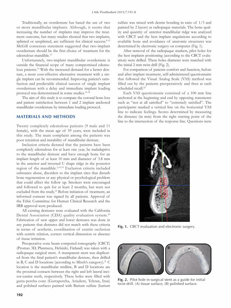
An insertion torque of at least 45 Ncm and Resonance Frequency Analysis (RFA) (Osstell, Integration Diagnostics, Gamlestads, Sweden) of at least 60 ISQ indicated primary implant stability (Fig. 3).4,6 This primary stability is neces-sary for immediate implant loading protocol. So, the ball attachment (Simple line II, SOBA4800, Implantium, Seoul, South Korea) was connected and tightened to 25 Ncm with special torque wrench (that is connected to the ball previ-ously). Then, the mucosa was sutured.
The 2-stage (delayed loading) protocol was considered for implants that did not have sufficient primary stability.
After the surgery, patients received antibiotic (20 cap-sule Amoxicillin (Amoxicillin, Darupakhsh, Tehran, Iran) 500 mg 3 times a day) and Gelofen (20 pearls, 400 mg 4
times a day) for pain control if needed. Immediate after surgery, the tissue surface of patient’s
denture was relieved around the ball attachments and implants sites. Then, denture was relined with soft liner (GC Corporation, Tokyo, Japan) and delivered to the patient. This protocol is in contrast to relining with hard acrylic Resin resulted in greater post-operative comfort and uneventful healing.
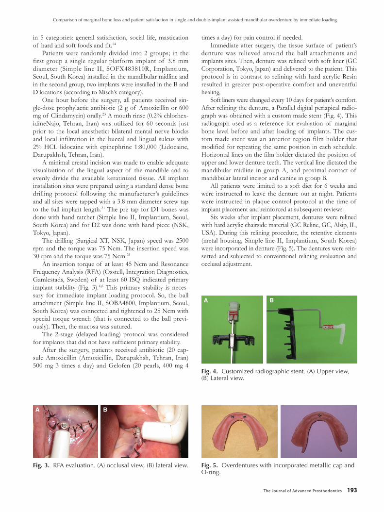
Soft liners were changed every 10 days for patient’s comfort. After relining the denture, a Parallel digital periapical radio-graph was obtained with a custom made stent (Fig. 4). This radiograph used as a reference for evaluation of marginal bone level before and after loading of implants. The cus-tom made stent was an anterior region film holder that modified for repeating the same position in each schedule. Horizontal lines on the film holder dictated the position of upper and lower denture teeth. The vertical line dictated the mandibular midline in group A, and proximal contact of mandibular lateral incisor and canine in group B.
All patients were limited to a soft diet for 6 weeks and were instructed to leave the denture out at night. Patients were instructed in plaque control protocol at the time of implant placement and reinforced at subsequent reviews.
Six weeks after implant placement, dentures were relined with hard acrylic chairside material (GC Reline, GC, Alsip, IL, USA). During this relining procedure, the retentive elements (metal housing, Simple line II, Implantium, South Korea) were incorporated in denture (Fig. 5). The dentures were rein-serted and subjected to conventional relining evaluation and occlusal adjustment.
Fig. 3. RFA evaluation. (A) occlusal view, (B) lateral view.
A B
Fig. 4. Customized radiographic stent. (A) Upper view, (B) Lateral view.
A B
Fig. 5. Overdentures with incorporated metallic cap and O-ring.
194
Patients were asked to refer if any unexpected problem happens. Patients recall was arranged for 6 and 12 months after surgery. In each recall schedule, self-administered questionnaire was filled by patients and parallel digital peri-apical radiograph was obtained.
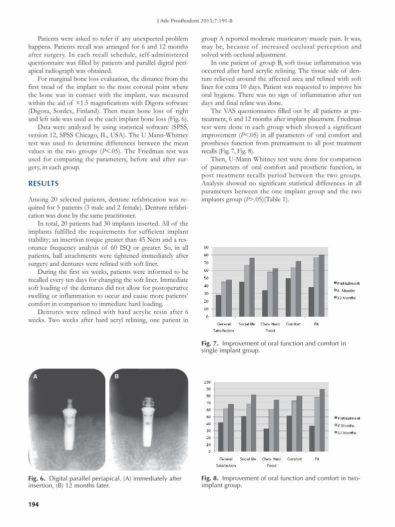
For marginal bone loss evaluation, the distance from the first tread of the implant to the most coronal point where the bone was in contact with the implant, was measured within the aid of ×1.5 magnifications with Digora software (Digora, Sordex, Finland). Then mean bone loss of right and left side was used as the each implant bone loss (Fig. 6).
Data were analyzed by using statistical software (SPSS, version 12, SPSS Chicago, IL, USA). The U Mann-Whitney test was used to determine differences between the mean values in the two groups (P<.05). The Friedman test was used for comparing the parameters, before and after sur-gery, in each group.
RESULTS
Among 20 selected patients, denture refabrication was re-quired for 5 patients (3 male and 2 female). Denture refabri-cation was done by the same practitioner.
In total, 20 patients had 30 implants inserted. All of the implants fulfilled the requirements for sufficient implant stability; an insertion torque greater than 45 Ncm and a res-onance frequency analysis of 60 ISQ or greater. So, in all patients, ball attachments were tightened immediately after surgery and dentures were relined with soft liner.
During the first six weeks, patients were informed to be recalled every ten days for changing the soft liner. Immediate soft loading of the dentures did not allow for postoperative swelling or inflammation to occur and cause more patients’ comfort in comparison to immediate hard loading.
Dentures were relined with hard acrylic resin after 6 weeks. Two weeks after hard acryl relining, one patient in
group A reported moderate masticatory muscle pain. It was, may be, because of increased occlusal perception and solved with occlusal adjustment.
In one patient of group B, soft tissue inflammation was occurred after hard acrylic relining. The tissue side of den-ture relieved around the affected area and relined with soft liner for extra 10 days. Patient was requested to improve his oral hygiene. There was no sign of inflammation after ten days and final reline was done.
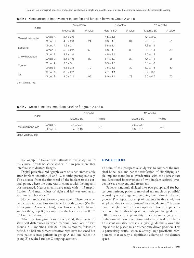
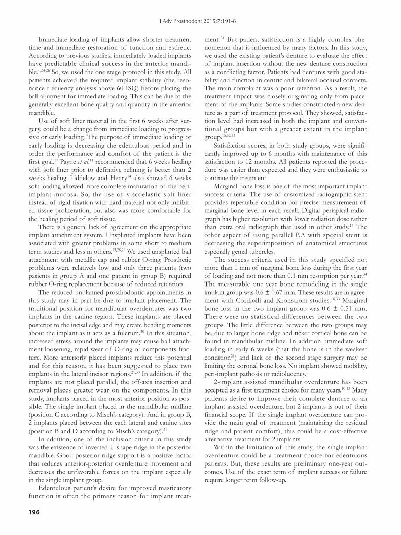
The VAS questionnaires filled out by all patients at pre-treatment, 6 and 12 months after implant placement. Friedman test were done in each group which showed a significant improvement (P<.05) in all parameters of oral comfort and prostheses function from pretreatment to all post treatment recalls (Fig. 7, Fig. 8).
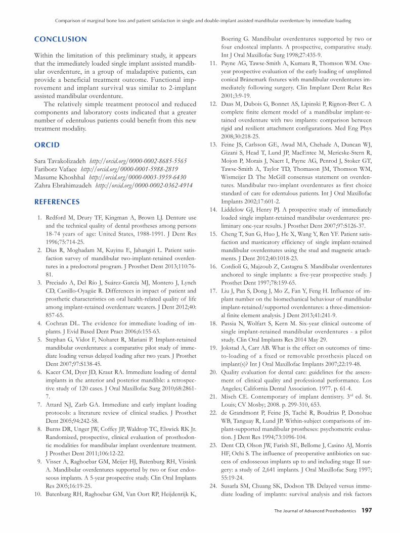
Then, U-Mann Whitney test were done for comparison of parameters of oral comfort and prosthetic function, in post treatment recalls period between the two groups. Analysis showed no significant statistical differences in all parameters between the one implant group and the two implants group (P>.05)(Table 1).
Fig. 6. Digital parallel periapical. (A) immediately after insertion, (B) 12 months later.
A B
Fig. 7. Improvement of oral function and comfort in single implant group.
Fig. 8. Improvement of oral function and comfort in two-implant group.
J Adv Prosthodont 2015;7:191-8
The Journal of Advanced Prosthodontics 195
Comparison of marginal bone loss and patient satisfaction in single and double-implant assisted mandibular overdenture by immediate loading
Radiograph follow-up was difficult in this study due to the clinical problems associated with film placement that interfere with denture flanges.
Digital periapical radiograph were obtained immediately after implant insertion, 6 and 12 months postoperatively. The distance from the first tread of the implant to the cor-onal point, where the bone was in contact with the implant, was measured. Measurements were made with ×1.5 magni-fication. And mean values of right and left was used as an each implant bone loss.14
No peri-implant radiolucency was noted. There was a lit-tle increase in bone loss over time for both groups (P=.16). In the group A (one implant), bone loss was 0.6 ± 0.67 mm and for the group B (two implants), the bone loss was 0.6 ± 0.51 mm in 12 months.
When the two groups were compared, there were no statistical differences between marginal bone loss of two groups in 12 months (Table 2). In the 12 months follow-up period, no ball attachment retentive caps have loosened but three patients (two patients in group A and one patient in group B) required rubber O-ring replacement.
DISCUSSION
The aim of this prospective study was to compare the mar-ginal bone level and patient satisfaction of simplifying sin-gle-implant mandibular overdenture with the success rate and functional improvement of two-implant assisted over-denture as a conventional treatment.
Patients randomly divided into two groups and for bet-ter comparison, patients matched (as much as possible) according to sex, age and smoking condition in the two groups. Presurgical work-up of patients in this study was simplified due to use of patient’s existing denture.14 A trans-parent acrylic template was duplicated from the patient’s denture. Use of this template as a radiographic guide with CBCT provided the possibility of electronic surgery with evaluation of bone condition and anatomical structures. This stent was also used as a surgical guide that allowed the implant to be placed in a prosthetically-driven position. This is particularly critical when relatively large prosthetic com-ponents that occupy a significant volume of the denture space.
Table 1. Comparison of improvement in comfort and function between Group A and B
IndexPretreatment 6 months 12 months
Mean ± SD P value Mean ± SD P value Mean ± SD P value
General satisfactionGroup A 2.7 ± 3.0 4.5 ± 1.6 7.1 ± 2.03
Group B 4.3 ± 2.3 .24 6.3 ± 1.5 .04 7.0 ± 1.5 .51
Social lifeGroup A 4.5 ± 2.1 5.8 ± 1.4 7.8 ± 1.1
Group B 5.2 ± 2.2 .55 6.8 ± 1.5 .36 8.3 ± 1.4 .83
Chew hardfoodsGroup A 3.4 ± 1.4 4.8 ± 2.1 7.5 ± 1.2
Group B 3.5 ± 1.9 .82 6.1 ± 1.9 .20 7.4 ± 1.4 .55
ComfortGroup A 5.0 ± 3.1 6.5 ± 1.3 8.1 ± 1.8
Group B 5.3 ± 2.8 .70 7.5 ± 1.6 .30 8.0 ± 1.3 .39
FitGroup A 3.6 ± 2.2 7.7 ± 1.1 8.2 ± 0.8
Group B 3.6 ± 2.2 .99 8.0 ± 1.1 .78 9.0 ± 0.7 .70
Mann-Whitney Test
Table 2. Mean bone loss (mm) from baseline for group A and B
Index6 months 12 months
Mean ± SD P value Mean ± SD P value
Marginal bone lossGroup A 0.4 ± 0.24
.910.6 ± 0.67
.74Group B 0.4 ± 0.16 0.6 ± 0.51
Mann-Whitney Test
196
Immediate loading of implants allow shorter treatment time and immediate restoration of function and esthetic. According to previous studies, immediately loaded implants have predictable clinical success in the anterior mandi-ble.6,24-26 So, we used the one stage protocol in this study. All patients achieved the required implant stability (the reso-nance frequency analysis above 60 ISQ) before placing the ball abutment for immediate loading. This can be due to the generally excellent bone quality and quantity in the anterior mandible.
Use of soft liner material in the first 6 weeks after sur-gery, could be a change from immediate loading to progres-sive or early loading. The purpose of immediate loading or early loading is decreasing the edentulous period and in order the performance and comfort of the patient is the first goal.27 Payne et al.11 recommended that 6 weeks healing with soft liner prior to definitive relining is better than 2 weeks healing. Liddelow and Henry14 also showed 6 weeks soft loading allowed more complete maturation of the peri-implant mucosa. So, the use of viscoelastic soft liner instead of rigid fixation with hard material not only inhibit-ed tissue proliferation, but also was more comfortable for the healing period of soft tissue.
There is a general lack of agreement on the appropriate implant attachment system. Unsplinted implants have been associated with greater problems in some short to medium term studies and less in others.13,28,29 We used unsplinted ball attachment with metallic cap and rubber O-ring. Prosthetic problems were relatively low and only three patients (two patients in group A and one patient in group B) required rubber O-ring replacement because of reduced retention.
The reduced unplanned prosthodontic appointments in this study may in part be due to implant placement. The traditional position for mandibular overdentures was two implants in the canine region. These implants are placed posterior to the incisal edge and may create bending moments about the implant as it acts as a fulcrum.30 In this situation, increased stress around the implants may cause ball attach-ment loosening, rapid wear of O-ring or components frac-ture. More anteriorly placed implants reduce this potential and for this reason, it has been suggested to place two implants in the lateral incisor regions.21,30 In addition, if the implants are not placed parallel, the off-axis insertion and removal places greater wear on the components. In this study, implants placed in the most anterior position as pos-sible. The single implant placed in the mandibular midline (position C according to Misch’s category). And in group B, 2 implants placed between the each lateral and canine sites (position B and D according to Misch’s category).21
In addition, one of the inclusion criteria in this study was the existence of inverted U shape ridge in the posterior mandible. Good posterior ridge support is a positive factor that reduces anterior-posterior overdenture movement and decreases the unfavorable forces on the implant especially in the single implant group.
Edentulous patient’s desire for improved masticatory function is often the primary reason for implant treat-
ment.31 But patient satisfaction is a highly complex phe-nomenon that is influenced by many factors. In this study, we used the existing patient’s denture to evaluate the effect of implant insertion without the new denture construction as a conflicting factor. Patients had dentures with good sta-bility and function in centric and bilateral occlusal contacts. The main complaint was a poor retention. As a result, the treatment impact was closely originating only from place-ment of the implants. Some studies constructed a new den-ture as a part of treatment protocol. They showed, satisfac-tion level had increased in both the implant and conven-tional groups but with a greater extent in the implant group.15,32,33
Satisfaction scores, in both study groups, were signifi-cantly improved up to 6 months with maintenance of this satisfaction to 12 months. All patients reported the proce-dure was easier than expected and they were enthusiastic to continue the treatment.
Marginal bone loss is one of the most important implant success criteria. The use of customized radiographic stent provides repeatable condition for precise measurement of marginal bone level in each recall. Digital periapical radio-graph has higher resolution with lower radiation dose rather than extra oral radiograph that used in other study.14 The other aspect of using parallel P.A with special stent is decreasing the superimposition of anatomical structures especially genial tubercles.
The success criteria used in this study specified not more than 1 mm of marginal bone loss during the first year of loading and not more than 0.1 mm resorption per year.34
The measurable one year bone remodeling in the single implant group was 0.6 ± 0.67 mm. These results are in agree-ment with Cordiolli and Kronstrom studies.16,33 Marginal bone loss in the two implant group was 0.6 ± 0.51 mm. There were no statistical differences between the two groups. The little difference between the two groups may be, due to larger bone ridge and ticker cortical bone can be found in mandibular midline. In addition, immediate soft loading in early 6 weeks (that the bone is in the weakest condition21) and lack of the second stage surgery may be limiting the coronal bone loss. No implant showed mobility, peri-implant pathosis or radiolucency.
2-implant assisted mandibular overdenture has been accepted as a first treatment choice for many years.10-13 Many patients desire to improve their complete denture to an implant assisted overdenture, but 2 implants is out of their financial scope. If the single implant overdenture can pro-vide the main goal of treatment (maintaining the residual ridge and patient comfort), this could be a cost-effective alternative treatment for 2 implants.
Within the limitation of this study, the single implant overdenture could be a treatment choice for edentulous patients. But, these results are preliminary one-year out-comes. Use of the exact term of implant success or failure require longer term follow-up.
J Adv Prosthodont 2015;7:191-8
The Journal of Advanced Prosthodontics 197
Comparison of marginal bone loss and patient satisfaction in single and double-implant assisted mandibular overdenture by immediate loading
CONCLUSION
Within the limitation of this preliminary study, it appears that the immediately loaded single implant assisted mandib-ular overdenture, in a group of maladaptive patients, can provide a beneficial treatment outcome. Functional imp-rovement and implant survival was similar to 2-implant assisted mandibular overdenture.
The relatively simple treatment protocol and reduced components and laboratory costs indicated that a greater number of edentulous patients could benefit from this new treatment modality.
ORCID
Sara Tavakolizadeh http://orcid.org/0000-0002-8685-5565Fariborz Vafaee http://orcid.org/0000-0001-5988-2819Masume Khoshhal http://orcid.org/0000-0003-3959-6430Zahra Ebrahimzadeh http://orcid.org/0000-0002-0362-4914
REFERENCES
1. Redford M, Drury TF, Kingman A, Brown LJ. Denture use and the technical quality of dental prostheses among persons 18-74 years of age: United States, 1988-1991. J Dent Res 1996;75:714-25.
2. Dias R, Moghadam M, Kuyinu E, Jahangiri L. Patient satis-faction survey of mandibular two-implant-retained overden-tures in a predoctoral program. J Prosthet Dent 2013;110:76-81.
3. Preciado A, Del Río J, Suárez-García MJ, Montero J, Lynch CD, Castillo-Oyagüe R. Differences in impact of patient and prosthetic characteristics on oral health-related quality of life among implant-retained overdenture wearers. J Dent 2012;40: 857-65.
4. Cochran DL. The evidence for immediate loading of im-plants. J Evid Based Dent Pract 2006;6:155-63.
5. Stephan G, Vidot F, Noharet R, Mariani P. Implant-retained mandibular overdentures: a comparative pilot study of imme-diate loading versus delayed loading after two years. J Prosthet Dent 2007;97:S138-45.
6. Kacer CM, Dyer JD, Kraut RA. Immediate loading of dental implants in the anterior and posterior mandible: a retrospec-tive study of 120 cases. J Oral Maxillofac Surg 2010;68:2861-7.
7. Attard NJ, Zarb GA. Immediate and early implant loading protocols: a literature review of clinical studies. J Prosthet Dent 2005;94:242-58.
8. Burns DR, Unger JW, Coffey JP, Waldrop TC, Elswick RK Jr. Randomized, prospective, clinical evaluation of prosthodon-tic modalities for mandibular implant overdenture treatment. J Prosthet Dent 2011;106:12-22.
9. Visser A, Raghoebar GM, Meijer HJ, Batenburg RH, Vissink A. Mandibular overdentures supported by two or four endos-seous implants. A 5-year prospective study. Clin Oral Implants Res 2005;16:19-25.
10. Batenburg RH, Raghoebar GM, Van Oort RP, Heijdenrijk K,
Boering G. Mandibular overdentures supported by two or four endosteal implants. A prospective, comparative study. Int J Oral Maxillofac Surg 1998;27:435-9.
11. Payne AG, Tawse-Smith A, Kumara R, Thomson WM. One-year prospective evaluation of the early loading of unsplinted conical Brånemark fixtures with mandibular overdentures im-mediately following surgery. Clin Implant Dent Relat Res 2001;3:9-19.
12. Daas M, Dubois G, Bonnet AS, Lipinski P, Rignon-Bret C. A complete finite element model of a mandibular implant-re-tained overdenture with two implants: comparison between rigid and resilient attachment configurations. Med Eng Phys 2008;30:218-25.
13. Feine JS, Carlsson GE, Awad MA, Chehade A, Duncan WJ, Gizani S, Head T, Lund JP, MacEntee M, Mericske-Stern R, Mojon P, Morais J, Naert I, Payne AG, Penrod J, Stoker GT, Tawse-Smith A, Taylor TD, Thomason JM, Thomson WM, Wismeijer D. The McGill consensus statement on overden-tures. Mandibular two-implant overdentures as first choice standard of care for edentulous patients. Int J Oral Maxillofac Implants 2002;17:601-2.
14. Liddelow GJ, Henry PJ. A prospective study of immediately loaded single implant-retained mandibular overdentures: pre-liminary one-year results. J Prosthet Dent 2007;97:S126-37.
15. Cheng T, Sun G, Huo J, He X, Wang Y, Ren YF. Patient satis-faction and masticatory efficiency of single implant-retained mandibular overdentures using the stud and magnetic attach-ments. J Dent 2012;40:1018-23.
16. Cordioli G, Majzoub Z, Castagna S. Mandibular overdentures anchored to single implants: a five-year prospective study. J Prosthet Dent 1997;78:159-65.
17. Liu J, Pan S, Dong J, Mo Z, Fan Y, Feng H. Influence of im-plant number on the biomechanical behaviour of mandibular implant-retained/supported overdentures: a three-dimension-al finite element analysis. J Dent 2013;41:241-9.
18. Passia N, Wolfart S, Kern M. Six-year clinical outcome of single implant-retained mandibular overdentures - a pilot study. Clin Oral Implants Res 2014 May 29.
19. Jokstad A, Carr AB. What is the effect on outcomes of time-to-loading of a fixed or removable prosthesis placed on implant(s)? Int J Oral Maxillofac Implants 2007;22:19-48.
20. Quality evaluation for dental care: guidelines for the assess-ment of clinical quality and professional performance. Los Angeles; California Dental Association. 1977. p. 61-4.
21. Misch CE. Contemporary of implant dentistry. 3rd ed. St. Louis; CV Mosby; 2008. p. 299-310, 653.
22. de Grandmont P, Feine JS, Taché R, Boudrias P, Donohue WB, Tanguay R, Lund JP. Within-subject comparisons of im-plant-supported mandibular prostheses: psychometric evalua-tion. J Dent Res 1994;73:1096-104.
23. Dent CD, Olson JW, Farish SE, Bellome J, Casino AJ, Morris HF, Ochi S. The influence of preoperative antibiotics on suc-cess of endosseous implants up to and including stage II sur-gery: a study of 2,641 implants. J Oral Maxillofac Surg 1997; 55:19-24.
24. Susarla SM, Chuang SK, Dodson TB. Delayed versus imme-diate loading of implants: survival analysis and risk factors

198
for dental implant failure. J Oral Maxillofac Surg 2008;66: 251-5.
25. Stricker A, Gutwald R, Schmelzeisen R, Gellrich NG. Immediate loading of 2 interforaminal dental implants supporting an overdenture: clinical and radiographic results after 24 months. Int J Oral Maxillofac Implants 2004;19:868-72.
26. Komiyama A, Klinge B, Hultin M. Treatment outcome of immediately loaded implants installed in edentulous jaws fol-lowing computer-assisted virtual treatment planning and flap-less surgery. Clin Oral Implants Res 2008;19:677-85.
27. Szmukler-Moncler S, Piattelli A, Favero GA, Dubruille JH. Considerations preliminary to the application of early and immediate loading protocols in dental implantology. Clin Oral Implants Res 2000;11:12-25.
28. Naert I, Gizani S, Vuylsteke M, Van Steenberghe D. A 5-year prospective randomized clinical trial on the influence of splint-ed and unsplinted oral implants retaining a mandibular over-denture: prosthetic aspects and patient satisfaction. J Oral Rehabil 1999;26:195-202.
29. Tokuhisa M, Matsushita Y, Koyano K. In vitro study of a mandibular implant overdenture retained with ball, magnet, or bar attachments: comparison of load transfer and denture stability. Int J Prosthodont 2003;16:128-34.
30. Taylor TD. Indications and treatment planning for mandibular implant overdntures. In: Fiene Js, carlsson GE eds. Implant overdentures as the standard of care for edentulous patients. Quintessence; Chicago; 2003. p. 71-81.
31. Fueki K, Kimoto K, Ogawa T, Garrett NR. Effect of im-plant-supported or retained dentures on masticatory perfor-mance: a systematic review. J Prosthet Dent 2007;98:470-7.
32. Geertman ME, Boerrigter EM, Van’t Hof MA, Van Waas MA, van Oort RP, Boering G, Kalk W. Two-center clinical trial of implant-retained mandibular overdentures versus complete dentures-chewing ability. Community Dent Oral Epidemiol 1996;24:79-84.
33. Kronstrom M, Davis B. Immediate functional loading of 1 or 2 dental implants supporting a ball retained mandibular overdenture: Interim results of a randomized prospective study. J Oral Maxillofac Surg 2010;68:e15.
34. Roos J, Sennerby L, Lekholm U, Jemt T, Gröndahl K, Albrektsson T. A qualitative and quantitative method for evaluating im-plant success: a 5-year retrospective analysis of the Brånemark implant. Int J Oral Maxillofac Implants 1997; 12:504-14.
J Adv Prosthodont 2015;7:191-8