COMPARISON OF GLYCEROL TREATMENT IN FROG SKELETAL MUSCLE AND
12
COMPARISONOFGLYCEROLTREATMENTIN FROGSKELETALMUSCLEANDMAMMALIANHEART AnElectrophysiologicalandMorphologicalStudy G . NIEMEYER andW .G . FORSSMANN FromtheInstituteofPhysiology,Universityof Berne, andtheInstitute ofHistology,Medical School,UniversityofGeneva,Switzerland .Dr .Forssmann'spresentaddress istheDepartmentof Anatomy,Universityof Heidelberg,Heidelberg, Germany .Dr .Niemeyer's presentaddressisthe NationalEyeInstitute,NationalInstitutesofHealth,Bethesda,Maryland20014 ABSTRACT INTRODUCTION Sincediscussionaboutthefunctionalandmorpho- logicaldifferencesbetweenskeletalandheart muscleisstillopen,itseemedtobeofinterestto comparethebehaviorofbothtissuesafterexposure tohypertonicglycerolsolutions .Itiswellknown thatthetransversetubularsystem(Tsystem)asa partofthesarcoplasmicreticulum(theterm sarcoplasmicreticulumisusedaccordingto PorterandPalade [32], seealsoForssmannand Girardier [9]) isanessentialpartoftheexcitation- contractioncouplingmechanisminskeletal muscle (20) . Itcanbedamagedinfrogskeletal musclebymeansofasuddenchangefromhyper- tonicglycerol-Ringer'stonormalRinger's (19) . Thisprocedureiscalled"glyceroltreatment ." Thismethodhasbeenappliedtomammalian heartmuscle,andinthepresentstudycontraction wasobservedandtransmembranepotentialswere Frogskeletalmuscleandmammalianheartmusclewerestudiedinvitrobeforeandafter glyceroltreatment .Lossofcontractility,changesintheactionpotentialanddisruption oftheTsystemwereobservedinskeletalmusclecells .InmammalianheartmuscletheT systemwasnotdisruptedwithhypertonicglyceroltreatment,andnosignificantelectro- physiologicalchangeswereobserved .ThecontinuitybetweentheTsystemandtheextra- cellularspacewasinvestigatedbydiffusiontracermethods .Decreaseofcontractilityduring thehypertonicphaseintheglyceroltreatmentwasfoundtodependontonicity .There- sultsofthisstudyclearlyshowthatnotonlyaretheredifferencesinmorphologybetween skeletalandcardiacmuscle,buttherearealsodifferencesintheresistancetoosmotic changes . measuredbeforeandafterglyceroltreatment .The structuralstatusoftheTsystemwasfollowed throughoutthewholeexperimentbyadiffusion tracermethod .Toindicatewhethertheconnec- tionbetweentheTsystemandtheextracellular spacewasintact,asoaking-outprocedureforthe tracersubstancewasused . MATERIALANDMETHODS Electrophysiology Muscletrabeculae(averagediameter0 .7mm, averagelength8mm)fromtherightventricleof sheepandcalfheartswerecutoutwithin40min afterslaughtering .Allexperimentsweredonewithin 7hrafterthepreparation .Equilibrationinthe differentsolutionsat23 ° or35 ° Cwasperformedina perfusedPerspexchamber,inwhichstimulating 28 8 Tua JOURNAL OF CELL BIOLOGY • VOLUME 50, 1971 . pages 288-299 on November 27, 2018 jcb.rupress.org Downloaded from http://doi.org/10.1083/jcb.50.2.288 Published Online: 1 August, 1971 | Supp Info:
Transcript of COMPARISON OF GLYCEROL TREATMENT IN FROG SKELETAL MUSCLE AND
COMPARISON OF GLYCEROL TREATMENT IN
FROG SKELETAL MUSCLE AND MAMMALIAN HEART
An Electrophysiological and Morphological Study
G . NIEMEYER and W . G . FORSSMANN
From the Institute of Physiology, University of Berne, and the Institute of Histology, MedicalSchool, University of Geneva, Switzerland . Dr . Forssmann's present address is the Department ofAnatomy, University of Heidelberg, Heidelberg, Germany. Dr . Niemeyer's present address is theNational Eye Institute, National Institutes of Health, Bethesda, Maryland 20014
ABSTRACT
INTRODUCTION
Since discussion about the functional and morpho-logical differences between skeletal and heartmuscle is still open, it seemed to be of interest tocompare the behavior of both tissues after exposureto hypertonic glycerol solutions . It is well knownthat the transverse tubular system (T system) as apart of the sarcoplasmic reticulum (the termsarcoplasmic reticulum is used according toPorter and Palade [32], see also Forssmann andGirardier [9]) is an essential part of the excitation-contraction coupling mechanism in skeletalmuscle (20) . It can be damaged in frog skeletalmuscle by means of a sudden change from hyper-tonic glycerol-Ringer's to normal Ringer's (19) .This procedure is called "glycerol treatment ."This method has been applied to mammalianheart muscle, and in the present study contractionwas observed and transmembrane potentials were
Frog skeletal muscle and mammalian heart muscle were studied in vitro before and afterglycerol treatment . Loss of contractility, changes in the action potential and disruptionof the T system were observed in skeletal muscle cells . In mammalian heart muscle the Tsystem was not disrupted with hypertonic glycerol treatment, and no significant electro-physiological changes were observed . The continuity between the T system and the extra-cellular space was investigated by diffusion tracer methods . Decrease of contractility duringthe hypertonic phase in the glycerol treatment was found to depend on tonicity . The re-sults of this study clearly show that not only are there differences in morphology betweenskeletal and cardiac muscle, but there are also differences in the resistance to osmoticchanges .
measured before and after glycerol treatment . Thestructural status of the T system was followedthroughout the whole experiment by a diffusiontracer method. To indicate whether the connec-tion between the T system and the extracellularspace was intact, a soaking-out procedure for thetracer substance was used .
MATERIAL AND METHODS
Electrophysiology
Muscle trabeculae (average diameter 0.7 mm,average length 8 mm) from the right ventricle ofsheep and calf hearts were cut out within 40 minafter slaughtering . All experiments were done within7 hr after the preparation . Equilibration in thedifferent solutions at 23 ° or 35 ° C was performed in aperfused Perspex chamber, in which stimulating
288
Tua JOURNAL OF CELL BIOLOGY • VOLUME 50, 1971 . pages 288-299
on November 27, 2018jcb.rupress.org Downloaded from http://doi.org/10.1083/jcb.50.2.288Published Online: 1 August, 1971 | Supp Info:

and recording electrodes were placed (26) . Themuscle strips were driven at a frequency of 0 .5-1cycle/sec by extracellular electrodes from a Grassstimulator (Grass Instrument Co ., Quincy, Mass .),through a stimulus isolation unit . The transmem-brane potentials were recorded differentially withglass microelectrodes filled with 3 M KCI, by meansof a cathode follower and a Tektronix 502 oscilloscope(Tektronix, Inc., Beaverton, Ore .) . The electroderesistance was between 12 and 30 MS2 . The contrac-tion was continuously observed with a binocularmicroscope (magnification 40) . After equilibrationin normal Tyrode's solution (solution I) for at least30 min, the perfusion solution was abruptly changedeither to 400 rum glycerol-Tyrode's or to 1000 mmglycerol-Tyrode's solution (solution II or III) .The preparation was perfused with the glycerol-containing solution for 60 min and then the perfusionwas abruptly changed back to normal Tyrode'ssolution . In order to hyperpolarize the preparationof heart muscle the sucrose gap method of Wood et al .(36) was employed . In this technique a three-com-partment chamber was used, the middle chamberhaving a width of 2 mm. This middle chamber wasperfused with isotonic sucrose solution, providing ahigh extracellular resistance . The other two chamberswere perfused with Tyrode's solution . The length ofthe fiber extending into the front chamber was Imm. Constant current pulses were applied through a50-100 kI2 series resistor to the posterior chamber,and the changes in the membrane potential weremeasured in the anterior chamber. When this methodwas used, it was also possible to apply glycerol treat-ment. In order to do this, all three chambers wereperfused with the appropriate glycerol solution. Inthe sucrose gap technique, hyperpolarizations ofless than 10 my were used in order to have fairlyuniform voltage distribution along the fiber (calcula-tion from cable equations, McGuigan, unpublished) .In all experiments the electrode position remainedunchanged in the middle of the front segment ofthe fiber. To calculate the time constant, a value of696]0 of the final value was taken . This was calculatedby Weidmann by means of the superimpositionmethod from Hodgkin and Rushton, Table I (17) .
To check the behavior of frog twitch muscle,bundles containing 5-30 fibers were prepared fromm. semitendinosus of Rana temporaria. Perfusion withRinger's solution (V) and glycerol solution (VI)was performed at room temperature in the sameway as in heart muscle .
SolutionsI . Tyrode's, bubbled with 95% 02/5% CO2,
containing (in millimoles per liter) : NaCI136.9, KCI 5.4, NaHCO3 11.9, NaH2PO40.4, CaCl2 1 .8, MgC1s 0 .5, and glucose 1 g.
II . The same as I, with the addition of 400 mmolesglycerol/liter.
III. The same as I, with the addition of 1000mmoles glycerol/liter .
IV. Sucrose with ions, oxygenated as I, containing(in millimoles per liter) : KCI 2.1, CaCl2 1 .1,glucose 1 g, sucrose 94 .5 g/liter.
V. Ringer's, containing (in millimoles per liter)NaCl 115.5, KCI 2.5, CaCl2 1 .8, NaH 2PO40.9, Na2HPO4 2 .2 .
VI. The same as V, containing 400 mmolesglycerol/liter.
Electron Microscopy
The solutions used for the physiological experi-ments were also used during the preparation forelectron microscopy, and in some cases the samepiece of tissue was utilized for both studies . Atdifferent stages during the experiment (see Table I)the physiological solutions were replaced by a 2%glutaraldehyde phosphate-buffered fixative (34) .After fixation for 1 hr the tissue was cut into smallblocks which were then washed three times in phos-phate buffer solution . The blocks were then post-fixed with phosphate-buffered osmium tetroxide (29) .If a tracer experiment had been done, the blockswere first treated for the histochemical demonstrationof horseradish peroxidase (see below) . After theseprocedures the blocks were dehydrated and em-bedded in Epon (28), sectioned on an LKB ultra-microtome (LKB Produkter, Stockholm, Sweden),and contrasted after the method of Karnovsky (23) .The sections were examined with a Zeiss EM 9 or aPhilips EM 300 electron microscope . Semithin sec-tions were observed and photographed in a Zeissphotomicroscope with a phase-contrast device .The diffusion tracer (horseradish peroxidase,
HRP) technique of Karnovsky (24) and Grahamand Karnovsky (15) was applied with some slightmodifications (Forssmann [8]) . The histochemicalcontrols were performed as indicated by Karnovsky(25) . In all experiments the quantity of HRP addedto the solutions was between 2 and 5 mg HRP/ml .
RESULTS
Electrophysiology
FROG SKELETAL MUSCLE : The results asobtained by others (3, 5, 11, 18, 19) could be fullyconfirmed . After glycerol treatment (using solu-tions V and VI), the mechanical response toelectrical stimulation disappeared completelywithin 40 min . Single fibers twitched spontaneouslyduring the first minutes after the removal of thehypertonic glycerol-Ringer's . The action potential
G. NIEMEYER AND W. G. FORSSMANN Glycerol Treatment in Muscle and Heart
289

290
TABLE I
Frog Skeletal Muscle and Mammalian Heart _Muscle : Electrophysiological Results with Glycerol Treatment
Frog Skeletal Muscle
Mammalian Heart Muscle
was still present, but the early afterpotential wasmarkedly decreased and the late afterpotential,following a tetanic stimulation (50-100 cycle/sec),disappeared completely . After this change (seeFig. I, Table I), the resting membrane potentialwas in general slightly reduced, occasionally to avalue above the mechanical threshold . Thesechanges were irreversible .MAMMALIAN HEART MUSCLE : Similar re-
THE JOURNAL OF CELL BIOLOGY ' VOLUME 50, 1971
suits were obtained in 17 experiments with sheepor calf trabeculae. In the hypertonic glycerolsolutions, a marked decrease of contraction (seeTable I) and a decrease in fiber diameter of theorder of 10 0/0 was observed . Both effects weremore pronounced in the solution with the highertonicity (solution III) . With solution III, thecontractility sometimes disappeared completelyfor several minutes in the hypertonic phase of the
Tyrode'ssolution (I)
400 mm glycerol-Tyrode's(II) Tyrode's (I) after glycerol
Contractility normal slightly reduced normalResting mcm- normal normal normalbrane poten-tial
Action potential normal normal normalUpstroke veloc- normal normal normal
ityTime constant 4.9 f 0.99 5 .2 f 0.4 msec
of hyperpo- mseclarization
Ringer'ssolution (V)
400 mm glycerol-Ringer's(VI) After glycerol (V)
Contractility normal normal absentResting mem- normal normal normal, reducedbrane poten-tial
Action potential normal normal present with highlydecreased earlyafterpotential
Late afterpo- normal normal absenttential
Tyrode'ssolution (I)
1000 ram glycerol-Tyrode's(III)
Tyrode's (I) afterglycerol
Contractility normal greatly reduced, normalsometimes initi-ally absent
Resting mem- normal normal normalbrane poten-tial
Action potential normal normal normalUpstroke veloc- normal normal normal

OA
C
A
C
100msec
treatment, but always recovered in normalTyrode's .
The resting membrane potential showed minorvariation after every change of solution . Since, inseveral experiments, a microelectrode remainedin the same cell for as long as 90 min, it waspossible to follow these changes in detail . Theaction potentials did not show any remarkablechange in shape, amplitude, upstroke velocity orduration during and after application of either400 or 1000 mm glycerol solution . The presentmethod would have allowed detection of changes
B
D
1 msec
1OmV
1Omsec
50mV
10msec
50mV
200 msec
FIGURE 1 Frog skeletal muscle . Action potential before (A) and after (B) glycerol treatment, C, lateafterpotential, following a train of pulses before glycerol, and D, lack of the late afterpotential afterglycerol treatment are shown .
FIGURE 2 Sheep heart muscle, right ventricle . A, Action potential before (continuous line), during(dotted line), and after (dashed line) glycerol, are graphed . B Shows corresponding tracings with a muchfaster sweep speed ; there is an indication of the upstroke . Hyperpolarizing pulses in an equivalent prepa-ration, recorded with the sucrose gap method, before (C) and after (D) glycerol treatment (base line =resting potential, 83 mv), are recorded .
of -f- 7 my in over-shoot, +10% in upstrokevelocity, and ±50 msec in duration. No furtherchanges were observed when the time of observa-tion, after glycerol removal, was increased to 4 hr .Even when glycerol perfusion time was increasedfrom 60 min to 4 hr, the results were similar . Areduction in temperature to 23 °C did not in-fluence these results .
Hyperpolarizing current pulses produced atransmembrane voltage displacement of a similaramplitude before and after glycerol treatment (seeFig. 2, C and D), the time constant was 4 .9 f 0.99
G. NIEMEYER AND W. G. FORSSMANN Glycerol Treatment in Muscle and Heart
291

FIGURE 3 Frog skeletal muscle after incubation for 30 min in 400 mm glycerol-Ringer's and 30 min innormal Ringer's . Fig . 3 a shows a localized swelling of a T tubule (arrow) ; Fig. 3 b shows disrupted Ttubules (T) showing no clear membrane limit with the L system . X 30,000.
FIGURE 4 Frog skeletal muscle, incubated for 30 min in Ringer's, followed by 30 min in 400 .mm glyc-erol-Ringer's containing 3 mg/ml horseradish peroxidase, and then fixed . Note the normal structureof the T system and the tracer substance filling all T tubules . X 30,000.
292

FIGURE 5 Frog skeletal muscle incubated for 30 min in Ringer's, 30 min in 400 mM glycerol-Ringer's,30 min in Ringer's, and finally 30 min in Ringer's containing peroxidase . T tubules are not filled withtracer substance . Tracer is found only in the interstitial space. X 32,000 .
FIGURE 6 Frog skeletal muscle incubated throughout the glycerol treatment with solutions containingthe tracer, then washed for 30 min in normal Ringer's . The tracer is washed out from the interstitialspace but not from the T system. X 32,000 .
G. NIEMEYER AND W. G. FGRSSMANN Glycerol Treatment in Muscle and Heart
293
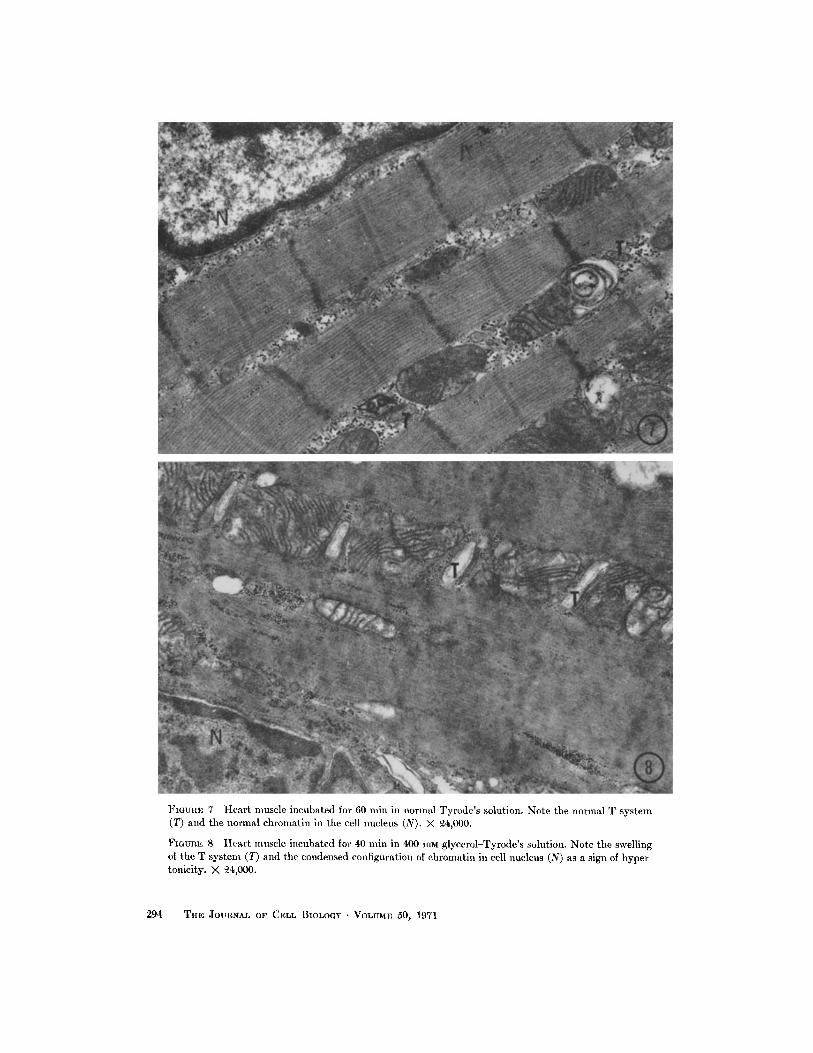
FIGUE: 7 Heart muscle incubated for 60 min in normal Tyrode's solution . Note the normal T system(T) and the normal chromatin in the cell nucleus (N) . X 94,000 .
FIGURE 8 Heart muscle incubated for 40 min in 400 mm glycerol-Tyrode's solution . Note the swellingof the T system (T) and the condensed configuration of chromatin in cell nucleus (N) as a sign of hyper-tonicity. X 24,000 .
2 94
THE: JOURNAL OF CELL BIOLOGY • VOLUME 50, 1971

FIGURE 9 Heart muscle incubated for 30 min in Tyrode's solution, and for 30 min in 400 mm glycerol-Tyrode's with peroxidase . All T tubules are labeled by the tracer substance, and there is no markeddilatation of T tubules (T) . X 18,000 .
FIGURE 10 Heart muscle incubated in Tyrode's with peroxidase after glycerol treatment with 1000mm glycerol-Tyrode solution . A slight swelling of T tubules (T) persists, but no disruption is observed ;all T tubules are labeled. X 22,000 .
G. NIEMEYER AND W. G. FORSSMANN Glycerol Treatment in Muscle and Heart
295

msec before and 5,2 f 0 .49 msec after (meanvalues and sD, one experiment, 10 impalements ;the difference is not statistically significant) .
Morphology
FROG SKELETAL MUSCLE : Examination offrog skeletal muscle in the different glycerolexperiments showed that the results of Howell andJenden (19) could be reproduced (Figs. 3 a and3 b) . As is seen in Table II and Fig . 4, there areno ultrastructural alterations in isolated fibersduring the whole period of incubation in eitherRinger's solution or Ringer's solution containing400 mm glycerol and HRP, even if the muscleswere fixed some hours after incubation . Themaximal incubation time in disruption experi-ments was 3 hr.
If the HRP was added to the solutions as atracer, it penetrated both the interstitial space andthe T system even if the muscle fibers were alreadyincubated in glycerol-Ringer's (Fig . 4) . In all ofthese stages the tracer could be washed out byusing the same solution without HRP, demon-strating that the T system was intact throughoutthe incubation with Ringer's or glycerol-Ringer's .
If the muscle fibers were prepared for electron-microscopy 30 min after the removal of glycerol,the T tubules were locally swollen or disrupted,forming a large clear space from the Z band some-times reaching as far as the next sarcomere (Fig .3 b). Frequently the T membrane was observed
TABLE II
Results of Tracer Experiments in Glycerol-Treated Frog Skeletal Muscle and Mammalian Heart Muscle
Tracer Tracer inin T
intersti-Trestrnent
system
ti-
Control electrolyte solutionElectrolyte solution containing
+
+HRP
Glycerol electrolyte solution
+
+containing HRP
Electrolyte containing HRP
-
+after glycerol treatment*
Glycerol treatment with solu-
+tions all containing HRP,then washing in HRP-freeelectrolyte solution
Strietal muscle
* For explanation of glycerol treatment, see Introduction .
296
THE JOURNAL OF CELL BIOLOGY . VOLUME 50, 1971
to be disrupted, and continuity between T andL cisternae appeared to result .The disruption of the T system was demon-
strated with tracer experiments . It was observedthat the tracer did not penetrate into the T tubulesafter glycerol treatment (Fig . 5), and that thetracer substances when present in glycerol-Ringer's,as well as in normal Ringer's during the periodof disruption, could not subsequently be washedout from the T tubules (Fig . 6) . However, thetracer was washed out completely from the inter-stitial space after several changes of the Ringer'swithout HRP (Fig. 6) .SHEEP AND CALF VENTRICULAR FIBERS :
The investigation of mammalian heart muscleshowed that the T system was not disrupted if thesame treatment, or even treatment with 1000 mmglycerol, was carried out . In the controls the heartmuscles did not change in their morphologicalfeatures during several hours of incubation innormal, oxygenated Tyrode's at 35 °C (Fig. 7) . Ifglycerol was added, a swelling of the T system wasobserved (Fig . 8) .
Horseradish peroxidase could be added at anystage of the glycerol treatment or in controlincubations in normal Tyrode's (Figs . 9 and 10),and it was found to diffuse freely to all of the Ttubules . If the fibers were washed with tracer-freeTyrode's before fixation, a complete removal ofHRP was possible in all stages of the experiment(Table II). This indicates that T tubules are onlyslightly swollen after glycerol treatment, anddisruption never occurred in our experiments .
Heart muscle
Tracer Tracer inin T
intersti-Morphology
system
tium
Morphology
normal
-
normalnormal
+
+
normal
nor.nal
+
+
swelling of Tsystem
disrupted T
+
+
swelling of Tsystem
systemdisrupted T
-
-
swelling of Tsystem
system

DISCUSSION
demonstrated that the diffusion of tracers isabolished in both directions.
The function of the T system in skeletal musclewith respect to excitation-contraction coupling isquite well defined by the work of Huxley et al .(20-22) and the more recent work dealing withglycerol treatment . However, little is known aboutthe role of the T system in mammalian heart (7,9), even though its structure has been well studied .In contrast to skeletal muscle, heart muscle cellsare smaller and the action potential is so muchlonger that an implication of a T system in ex-citation-contraction coupling might not be re-quired. Thus, the function of the T system inmammalian heart still remains obscure . In addi-tion to the recently reported findings concerningthe response to local stimulation in sheep heart byMueller (30) and the detailed morphologicaldescription of the T system in rat heart (forliterature, see Fawcett and McNutt [7]), theresults presented here again emphasize the dif-ferent characteristics of this intracellular mem-brane system in heart. Since the experimentscarried out in 400 mm glycerol solution were noteffective in heart tissue, we increased the tonicityby adding 1000 mmoles glycerol/liter Tyrode's .Even with this increase of tonicity, the onlymorphological change caused by glycerol treat-ment was a slight swelling of the T system .Swelling of the T system could be observed as aneffect of osmotic or ionic changes (1, 27), andincrease of osmolarity of the bathing solution alsoproduces swelling of the T system in skeletalmuscles (2, 10) .
During perfusion with 400 mm glycerol solutionand for a short time after the change back tonormal Tyrode's, a decrease in contraction wasobserved . In the presence of 1000 mss glycerol-Tyrode's, the contractions sometimes disappearedfor periods of 5-15 min.
Since this decrease in contraction may not berelated to the T system, other possibilities have tobe considered. A loss of contraction may occur asa simple consequence of removal of cell water,explained by changes in filament lattice spacing .In fact, a decrease in filament lattice volume byX-ray diffraction studies has been shown to occurin hypertonic solutions (33) . In addition to thispossibility, Gordon and Godt (14) have recentlydiscussed in detail the possible direct effects ofhypertonic solutions on the contractile proteins,such as an influence on actomyosin and on ATPase
In frog skeletal muscle a change from electrolytesolution to a hypertonic, glycerol-containingelectrolyte solution does not produce physiologicalor morphological alterations . However, if thepreparation is brought back to normal Ringer'ssolution, drastic changes in both physiological andmorphological characteristics are observed .
In 1967, Howell and Jenden (19) first demon-strated electron microscopically that the failure ofexcitation-contraction coupling was associatedwith a disruption of the T system . Mechanicalresponse of glycerol-treated fibers could only beobtained with caffeine. In the same year, Eisen-berg and Gage (5, 11) examined the electricalproperties of frog muscle after disruption of theT system and were able (6) to differentiate be-tween the surface and tubular fraction of themembrane capacity and specific ionic conduct-ances for K+ and Cl-. For further details con-cerning physiological and morphological prop-erties of the T system, see references 3, 4, 6, 12, 13,18 and 31 .
On the other hand, Howell (18) has shown thatT-tubular disruption is observed only with anabrupt, but not with a stepwise, removal ofglycerol-Ringer's . The same effect could also beobtained with urea, but not with sucrose or withfast-penetrating substances such as ethyleneglycol . He also investigated slow muscle fibers ofof the frog and found that they are not sensitiveto glycerol treatment .
Our own results on frog skeletal muscle are inagreement with those discussed above. Themorphological investigations carried out on frogskeletal muscle were especially related to theproblem of finding when the disruption of thetubular system occurs . It was previously shown byEisenberg and Eisenberg (3, 4) using peroxidaseand by Nakajima et al . (31) using ferritin asdiffusion tracers, that the T-tubule compartmentis separated from the interstitial space by glyceroltreatment. Our present study reveals beyond anydoubt that the lumen of the T tubules mustbecome discontinuous during the removal ofglycerol, since we have demonstrated that peroxi-dase can not only penetrate to the T system beforethe disruption but can also be removed by soakingin HRP-free solution . If the T system is disrupted,peroxidase is removed only from the interstitialspace, but never from the T system. This clearly
G. NIEMEYER AND W . G. FORSSMANN Glycerol Treatment in Muscle and Heart
297

activity. Such a mechanism was first reported byHasselbach (16), and more recently Portzehl(unpublished, personal communication) has found,in extracted rabbit skeletal muscle, a significantdecrease in ATPase activity in hypertonic glycerol-Tyrode's (500-1000 HIM) .
Certain problems concerning the differencesbetween skeletal and cardiac muscle stressed in arecent paper by one of us (9) now become moreprominent. Two main morphological propertiesmay be responsible for the insensitivity of the heartmuscle T system to glycerol treatment . First, theT system membrane in heart has been shown to becoated with a 200 A thick basement membrane,which is absent in skeletal muscle . Second, thesize of the lumen of the T tubules is much largerin heart muscle than it is in skeletal muscle . Since,after the change to normal Tyrode's solution,water will flow through the surface and tubularmembrane into the fiber, glycerol will slowlydiffuse to the bathing solution through the surfacemembrane and via the T system . The differentdiffusion rates of water and glycerol as well as aglycerol concentration gradient between the Tsystem and the extracellular fluid immediatelyafter removal of the hypertonic solution may causeextreme swelling and disruption of the tubules inskeletal muscle . In heart muscle the T tubules aremuch shorter and wider, so an equilibrium is morelikely to be reached between the T system and thebathing solution before extreme swelling anddisruption . This may be the reason that the Tsystem in heart muscle is mechanically less stressedduring osmotic changes than that in skeletalmuscle . Not only are there morphological dif-ferences between the T systems in skeletal muscleand in heart, but there also exist differences in thephysiological response to glycerol treatment as hasbeen shown in this paper .
In the experiments with glycerol in heartmuscle, only slight reversible changes of the actionpotential characteristics were found . These smallchanges are in the range of normal variability(Weidmann [35], and personal communication) .Furthermore, in heart muscle the slope and timeconstant of hyperpolarizing constant currentpulses did not change with the glycerol treatment,and the amount of applied current for a smallvoltage displacement remained constant .
The fact that no changes in the shape of ahyperpolarizing step were observed is in agree-ment with the lack of disruption of the T system,
298
THE JOURNAL OF CELL BIOLOGY . VOLUME 50, 1971
since marked changes were found by Gage andEisenberg (13) in skeletal muscle where disruptionof the T system occurs .The agreement between the negligible changes
in heart muscle with respect to electrophysiologicaland electron microscope findings after the glyceroltreatment is thus confirmed in the present study .
We wish to thank Prof. Silvio Weidmann for stimu-lating ideas, criticisms and help during the wholework, Prof. Charles Rouiller for facilities of workingin his laboratory, and Dr . John A. S. McGuiganand Dr . Harry A . Fozzard for help in preparing themanuscript. We thank as well Mrs. U. Westphal,Mrs. J . Boucherat, Mrs. M. Sidler-Ansermet, Mrs .E. Sturzenegger, and Mr. C. Cigada for technicalassistance.
This work was supported by grants of the SwissNational Science Foundation .
Received for publication 23 September 1970, and in revisedform 30 November 1970 .
REFERENCES
1 . DREIFUSS, J. J ., L . GIRARDIER, and W. G .FORSSMANN . 1966. Etude de la propagation del'excitation dans le ventricle de rat moyen desolutions hypertoniques . Pflügers Arch . GesamtePhysiol. Menschen Tiere . 292:13 .
2. DYDYNSKA, M ., and D . R. WILKIE . 1963. Theosmotic properties of striated muscle fibers inhypertonie solutions. J. Physiol . (London) . 169 :312 .
3 . EISENBERG, B., and R. S . EISENBERG . 1968 .Selective disruption of the sarcotubular systemin frog sartorius muscle. A quantative studywith exogenous peroxidase as a marker. J.Cell Biol. 39:451 .
4 . EISENBERG, B., and R. S . EISENBERG . 1968 .Transverse tubular system in glycerol-treatedskeletal muscle. Science (Washington) . 160:1243 .
5 . EISENBERG, R. S., and P. W . GAGE . 1967. Frogskeletal muscle fibers : changes in electricalproperties after disruption of transversetubular system. Science (Washington) . 158:1700 .
6 . EISENBERG, R. S ., and P. W. GAGE . 1969. Ionicconductances of frog sartorius fibers . J. Gen .Physiol . 53 :279 .
7 . FAWCETT, D. W., and N. S. MCNUTT. 1969. Theultrastructure of the cat myocardium . I .Ventricular papillary muscle . J. Cell Biol. 41 :1 .
8 . FORSSMANN, W. G. 1969 . A method for in vivodiffusion tracer studies combining perfusionfixation with intravenous tracer injection .Histochemie . 20:277 .

9 . FORSSMANN, W . G., and L . GIRARDIER . 1970. Astudy of the T system in rat heart . J. Cell Biol .44 :1 .
10. FOULKS, J . G., J . A . PACEY, and F. A . PERRY .1965. Contractures and swelling of the trans-verse tubules during chloride withdrawal infrog skeletal muscle. J. Physiol. (London) . 180 :96 .
11 . GAGE, P . W., and R. S . EISENBERG . 1967 . Actionpotentials without contraction in frog skeletalmuscle fibers with disrupted transverse tubules .Science (Washington) . 158:1702 .
12 . GAGE, P . W., and R. S . EISENBERG . 1969 .Capacitance of the surface and transversetubular membrane of frog sartorius musclefibers. J. Gen. Physiol. 53 :265 .
13 . GAGE, P . W., and R. S . EISENBERG . 1969 . Actionpotentials, afterpotentials and excitation-con-traction coupling in frog . J. Gen. Physiol . 53 :298 .
14. GORDON, A. M., and R. E . GODT . 1970. Someeffects of hypertonic solutions on contractionand excitation-contraction coupling in frogskeletal muscles . J. Gen . Physiol . 55:254 .
15. GRAHAM, R . C., and M. J. KARNOVSKY . 1966 .The early stages of absorption of injectedhorseradish peroxidase in the proximal tubulesof mouse kidney : ultrastructural cytochemistryby a new technique . J. Histochem . Cytochem . 14 :291 .
16. HASSELBACH, W. 1952. Die Umwandlung vonAktomyosin-ATPase in L-Myosin-ATPasedurch Aktivatoren and die resultierendenAktivierungseffekte. Z. Naturforsch. 7b :163 .
17. HODGKIN, A. L., and W. A. H. RUSHTON . 1946 .The electrical constants of crustacean nervefibre . Proc. Roy . Soc. Ser. B. Biol . Sci . 133 :444 .
18. HOWELL, J. N. 1969 . A lesion of the transversetubules of skeletal muscle . J. Physiol . (London) .201:515 .
19. HOWELL, J. N., and D . J. JENDEN . 1967. Ttubules of skeletal muscle : morphologicalalterations which interrupt excitation-con-traction coupling . Fed. Proc . 26:553 .
20 . HUXLEY, A. F. 1959. Local activation in muscle .Ann. N. Y. Acad. Sci. 81:446 .
21 . HUXLEY, A. F., and R. W. STRAUB . 1958 . Localactivation and interfibrillar structures instriated muscle. J. Physiol. (London) . 143:40 P .
22 . HUXLEY, A. F., and R. E. TAYLOR . 1958 . Local
activation of striated muscle fibres . J. Physiol .(London) . 144:426 .
23 . KARNOVSKY, M. J . 1961. A simple method forstaining with lead at high pH in electronmicroscopy . J. Biophys . Biochem . Cytol. 11 :729 .
24. KARNOVSKY, M. J . 1965 . Vesicular transport ofexogenous peroxidase across capillary endo-thelium into the T system of muscle . J. CellBiol . 27(2) :49 A. (Abstr .) .
25 . KARNOVSKY, M. J. 1967. The ultrastructuralbasis of capillary permeability studied withperoxidase as a tracer . J. Cell Biol. 35 :213 .
26. KAVALER, F. 1959 . Membrane depolarization asa cause of tension development in mammalianventricular muscle . Amer . J. Physiol. 197 :968 .
27. LEGATO, M. J., D. SPIRO, and G. A. LANGER .1968. Ultrastructural alterations produced inmammalian myocardium by variation inperfusate ionic composition . J. Cell Biol. 37 :1 .
28 . LUFT, J . H. 1961 . Improvements in epoxy resinembedding methods . J. Biophys. Biochem . Cy-tol. 9:409 .
29 . MILLONIG, G. 1961 . Advantages of a phosphatebuffer for OS04 solutions in fixation. J. Appl.Physiol . 32:1637.
30. MUELLER, P. 1966 . Lokale Kontraktionsauslö-sung am Herzmuskel. Hely . Physiol. Pharmacol.Acta . 24:C 106.
31 . NAKAJIMA, S ., Y. NAKAJIMA, and L. D. PEACHEY .1969. Speed of repolarization and morphologyof glycerol-treated muscle fibers . J. Physiol .(London) . 200:115 P.
32 . PORTER, K. R., and G . E . PALADE . 1957. Studieson the endoplasmic reticulum . III . Its formand distribution in striated muscle cells . J.Biophys. Biochem. Cytol . 3:269 .
33 . ROME, E. 1968. X-ray diffraction studies of thefilament lattice of striated muscle in variousbathing media . J. Mol. Biol. 37 :331 .
34 . SABATINI, D . D., K . BENSCH, and R . J. BARRNETT .1963. Cytochemistry and electron microscopy.The preservation of cellular ultrastructure andenzymatic activity by aldehyde fixation . J.Cell Biol . 17:19.
35 . WEIDMANN, S . 1956 . Elektrophysiologie derHerzmuskelfaser. Hans Huber Gmbh., Stutt-gart .
36. WOOD, E. H., R . L . HEPPNER, and S . WEIDMANN .1969. Inotropic effects of electrical currents .Circ . Res . 24:409.
G. NIEMEYER AND W . G. FoRSSMANN Glycerol Treatment in Muscle and Heart 299