Comparison of arabidopsis stomatal density mutants ...Comparison of Arabidopsis Stomatal Density...
12
J. Plant Biol. (2014) 57:162-173 DOI 10.1007/s12374-014-0017-1 Comparison of Arabidopsis Stomatal Density Mutants Indicates Variation in Water Stress Responses and Potential Epistatic Effects Shaneka S. Lawson 1,2 * , Paula M. Pijut 1,2 and Charles H. Michler 1,2 1 USDA Forest Service, Northern Research Station, Hardwood Tree Improvement and Regeneration Center (HTIRC), 195 Marstellar Street, West Lafayette, Indiana USA 47907 2 Purdue University, Department of Forestry and Natural Resources, HTIRC, 715 West State Street, West Lafayette, Indiana USA 47907 Received: January 10, 2014 / Accepted: March 18, 2014 © Korean Society of Plant Biologists 2014 Abstract Recent physiological analysis of Arabidopsis stomatal density (SD) mutants indicated that SD was not the major factor controlling aboveground biomass accumulation. Despite the general theory that plants with fewer stomata have limited biomass acquisition capabilities, epf1 and several other Arabidopsis mutants varied significantly in leaf fresh weight despite having similar stomatal numbers. The in- depth mechanisms controlling increased or decreased leaf area biomass remain undetermined. This work used calculations of SD, overnight water-loss, and LI6400XT measurements to reject the premise that SD is a primary factor controlling leaf biomass accumulation in Arabidopsis. With respect to our data, SD is not the primary factor influencing biomass accumulation in Arabidopsis epf1 mutants as it did not positively correlate to any of the physiological parameters examined. Further observation of morphological differences between the mutants hinted that additional pathways were interrupted when these mutants were generated. Each mutant examined showed a variation in physiological measurements despite SD. Many SD mutants also showed morphological abnormalities in addition to altered stomatal numbers. These phenotypes may indicate epistatic effects related to the mutation of SD genes in the studied mutants. Keywords: Morphology, Stomatal conductance, Stomatal density, Transpiration Introduction Stomatal density (SD) is known to vary with respect to a number of physiological and environmental factors. This study of Arabidopsis mutants included positive and negative SD regulators. The EPIDERMAL PATTERNING FACTOR (EPF)-LIKE (EPFL) family of secreted cysteine-rich peptides is composed of three homologous negative SD regulators, where overexpression leads to reduced stomatal number and increased pavement cell formation [EPF1/2 and EPFL6 or CHALLAH (CHAL)], and one positive regulator [(EPFL9 or STOMAGEN (STOM)] (Sugano et al. 2010). Mutations in TOO MANY MOUTHS ( TMM) or EPF1 result in increased SD and a stomatal bunching phenotype (Hoover 1986; Hara et al. 2009) rather than adequate one-cell spacing (Yang and Sack 1995). Overexpression of EPF1 (extremely high levels led to total ablation of stomata and infertility) resulted in a decreased stomatal index (SI), and overexpression of TMM led to increased stomatal numbers. Dependent upon TMM, overexpression of the STOMATAL DENSITY AND DISTRIBUTION1 (SDD1) gene also led to decreased stomatal numbers and formation of arrested stomata (von Groll et al. 2002; Yoo et al. 2010). Since sdd1 mutants cannot alleviate EPFL6 overexpression phenotypes, SDD1 seemed to be divergent from the ER and TMM pathways. However, mutations in sdd1, epf1, and tmm led to increased SI and occasional bunching (Lampard et al. 2008; Hara et al. 2009; Abrash and Bergmann 2010). Mutations in ER family members er , erl1, and erl2, and yda (YODA) resulted in almost exclusive production of guard cells and few pavement cells (Bergmann et al. 2004; Shpak et al. 2004). Required for stomatal pore formation, CYCLIN-DEPENDENT KINASE B1;1 (CDKB1;1), a cyclin dependent kinase responsible for arresting cell cycle proliferation (Porter 2008; Boudolf et al. ORIGINAL ARTICLE *Corresponding author; Shaneka S. Lawson Tel : +1-765-412-6119 E-mail : [email protected]
Transcript of Comparison of arabidopsis stomatal density mutants ...Comparison of Arabidopsis Stomatal Density...

J. Plant Biol. (2014) 57:162-173
DOI 10.1007/s12374-014-0017-1
Comparison of Arabidopsis Stomatal Density Mutants Indicates Variation
in Water Stress Responses and Potential Epistatic Effects
Shaneka S. Lawson1,2*, Paula M. Pijut1,2 and Charles H. Michler1,2
1USDA Forest Service, Northern Research Station, Hardwood Tree Improvement and Regeneration Center (HTIRC), 195
Marstellar Street, West Lafayette, Indiana USA 479072Purdue University, Department of Forestry and Natural Resources, HTIRC, 715 West State Street, West Lafayette, Indiana
USA 47907
Received: January 10, 2014 / Accepted: March 18, 2014
© Korean Society of Plant Biologists 2014
Abstract Recent physiological analysis of Arabidopsis
stomatal density (SD) mutants indicated that SD was not the
major factor controlling aboveground biomass accumulation.
Despite the general theory that plants with fewer stomata
have limited biomass acquisition capabilities, epf1 and several
other Arabidopsis mutants varied significantly in leaf fresh
weight despite having similar stomatal numbers. The in-
depth mechanisms controlling increased or decreased leaf
area biomass remain undetermined. This work used calculations
of SD, overnight water-loss, and LI6400XT measurements
to reject the premise that SD is a primary factor controlling
leaf biomass accumulation in Arabidopsis. With respect to
our data, SD is not the primary factor influencing biomass
accumulation in Arabidopsis epf1 mutants as it did not
positively correlate to any of the physiological parameters
examined. Further observation of morphological differences
between the mutants hinted that additional pathways were
interrupted when these mutants were generated. Each mutant
examined showed a variation in physiological measurements
despite SD. Many SD mutants also showed morphological
abnormalities in addition to altered stomatal numbers. These
phenotypes may indicate epistatic effects related to the
mutation of SD genes in the studied mutants.
Keywords: Morphology, Stomatal conductance, Stomatal
density, Transpiration
Introduction
Stomatal density (SD) is known to vary with respect to a
number of physiological and environmental factors. This
study of Arabidopsis mutants included positive and negative
SD regulators. The EPIDERMAL PATTERNING FACTOR
(EPF)-LIKE (EPFL) family of secreted cysteine-rich peptides
is composed of three homologous negative SD regulators,
where overexpression leads to reduced stomatal number and
increased pavement cell formation [EPF1/2 and EPFL6 or
CHALLAH (CHAL)], and one positive regulator [(EPFL9
or STOMAGEN (STOM)] (Sugano et al. 2010). Mutations
in TOO MANY MOUTHS (TMM) or EPF1 result in increased
SD and a stomatal bunching phenotype (Hoover 1986; Hara
et al. 2009) rather than adequate one-cell spacing (Yang and
Sack 1995). Overexpression of EPF1 (extremely high levels
led to total ablation of stomata and infertility) resulted in
a decreased stomatal index (SI), and overexpression of
TMM led to increased stomatal numbers. Dependent upon
TMM, overexpression of the STOMATAL DENSITY
AND DISTRIBUTION1 (SDD1) gene also led to decreased
stomatal numbers and formation of arrested stomata (von
Groll et al. 2002; Yoo et al. 2010). Since sdd1 mutants
cannot alleviate EPFL6 overexpression phenotypes, SDD1
seemed to be divergent from the ER and TMM pathways.
However, mutations in sdd1, epf1, and tmm led to increased
SI and occasional bunching (Lampard et al. 2008; Hara et al.
2009; Abrash and Bergmann 2010). Mutations in ER family
members er, erl1, and erl2, and yda (YODA) resulted in
almost exclusive production of guard cells and few pavement
cells (Bergmann et al. 2004; Shpak et al. 2004). Required for
stomatal pore formation, CYCLIN-DEPENDENT KINASE
B1;1 (CDKB1;1), a cyclin dependent kinase responsible for
arresting cell cycle proliferation (Porter 2008; Boudolf et al.
ORIGINAL ARTICLE
*Corresponding author; Shaneka S. Lawson
Tel : +1-765-412-6119
E-mail : [email protected]
J. Plant Biol. (2014) 57:162-173 163
2009), is involved in symmetric cell division. This last cellular
division leads to guard cell formation and is required for the
proper functioning of FOUR LIPS (FLP), a developmental
regulator that targets CDKB1;1 and coordinates cell cycle
exit before and after asymmetric division (Xie et al. 2010a).
The FLP (MYB124) gene and its paralog MYB88, two
proteins which share an amino acid substitution specific to
members of the MYB family involved in development of
stomata, were not required for stomatal fate (Lai et al. 2005).
These proteins functioned to limit GMCs to one symmetric
division, and thus prevented additional “daughter-cell” divisions
as well as coerced guard cell formation.
During periods of water stress it was expected that
stomatal numbers and pore apertures would be decreased,
a common occurrence in Arabidopsis and grass species
(Nawazish et al. 2006; Liu et al. 2009) however in some
plant species research data showed that decreased water
availability resulted in increased SD (Fraser et al. 2009).
It was suggested that fluctuations in temperature also
influenced SD (Xu and Zhou 2008). In Arabidopsis, a
general decrease in SD has been shown in response to
elevated temperatures, light, and CO2 levels (Berger and
Altmann 2000; Pospisilova 2003). Arabidopsis SD varies
with genotype and species. However, analysis of the
variations in three primary physiological factors such as
E, A, and gs between a subset of mutants involved in
stomatal development has not been conducted. Stomata
are found on nearly all aerial plant parts, but this study
focused only on the leaf. Pore aperture controls the
amount of water leaving the leaf during E. In response to
environmental influences, stomatal densities have been
known to increase or decrease over long periods of time (Chen
et al. 2001) however genetic mutations can also influence
density and distribution of stomata (Alonso et al. 2003).
Evidence is presented here to indicate relative differences in A,
E, and gs between a subset of Arabidopsis SD mutants.
Stomatal development and density in Arabidopsis has been
described and characterized and the reference data concerning
the various mutant phenotypes has increased. By examining
the developmental cues for stomatal patterning, a greater
degree of comprehension can be achieved when evaluating
developmental signaling pathways. Studies that address
stomatal size, density, and aperture have been described and
a number of physiological variations as a result of changes in
water availability have been recorded amongst various
species including Arabidopsis (Serna 2009; Peterson et al.
2010). A number of SD mutants have been characterized,
however this study incorporated some of those most widely
described in the literature and provided background and
expression data for several others (see Supplemental Table 1).
It is hypothesized that analysis of SD in addition to several
physiological parameters would result in significant
correlations between CO2 assimilation rate (A) and SD.
Decreased SD could be presumed to coincide with
decreased leaf transpiration (E) or provide data to support
assumptions that increased SD does not greatly influence
growth and development. Despite our knowledge that a
variety of endogenous signals could be at work to override
SD effects, we are using these data to demonstrate variation in
physiological parameters of mutants with varied SD. The
objective was to examine the density of leaf stomata in a
selection of Arabidopsis SD mutants and examine the
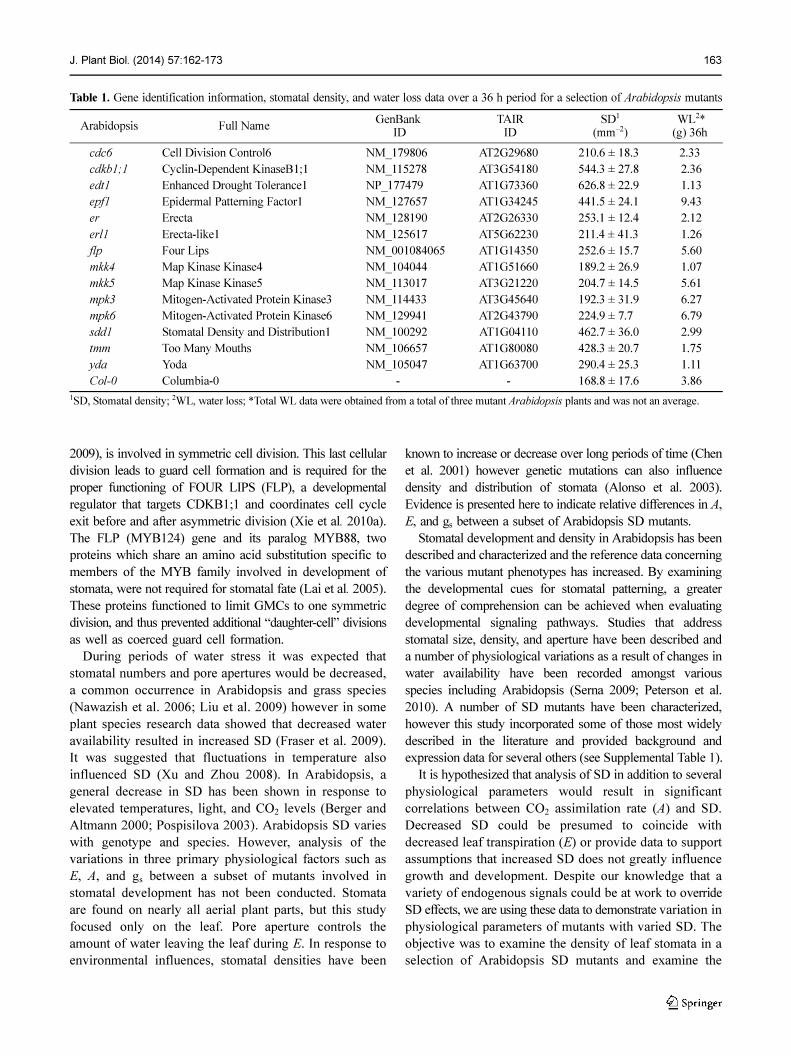
Table 1. Gene identification information, stomatal density, and water loss data over a 36 h period for a selection of Arabidopsis mutants
Arabidopsis Full NameGenBank
ID TAIR
ID SD1
(mm−2) WL2*(g) 36h
cdc6 Cell Division Control6 NM_179806 AT2G29680 210.6 ± 18.3 2.33
cdkb1;1 Cyclin-Dependent KinaseB1;1 NM_115278 AT3G54180 544.3 ± 27.8 2.36
edt1 Enhanced Drought Tolerance1 NP_177479 AT1G73360 626.8 ± 22.9 1.13
epf1 Epidermal Patterning Factor1 NM_127657 AT1G34245 441.5 ± 24.1 9.43
er Erecta NM_128190 AT2G26330 253.1 ± 12.4 2.12
erl1 Erecta-like1 NM_125617 AT5G62230 211.4 ± 41.3 1.26
flp Four Lips NM_001084065 AT1G14350 252.6 ± 15.7 5.60
mkk4 Map Kinase Kinase4 NM_104044 AT1G51660 189.2 ± 26.9 1.07
mkk5 Map Kinase Kinase5 NM_113017 AT3G21220 204.7 ± 14.5 5.61
mpk3 Mitogen-Activated Protein Kinase3 NM_114433 AT3G45640 192.3 ± 31.9 6.27
mpk6 Mitogen-Activated Protein Kinase6 NM_129941 AT2G43790 224.9 ± 7.70 6.79
sdd1 Stomatal Density and Distribution1 NM_100292 AT1G04110 462.7 ± 36.0 2.99
tmm Too Many Mouths NM_106657 AT1G80080 428.3 ± 20.7 1.75
yda Yoda NM_105047 AT1G63700 290.4 ± 25.3 1.11
Col-0 Columbia-0 - - 168.8 ± 17.6 3.86
1SD, Stomatal density; 2WL, water loss; *Total WL data were obtained from a total of three mutant Arabidopsis plants and was not an average.
164 J. Plant Biol. (2014) 57:162-173
physiological parameters involved in growth under identical
experimental conditions.
Results
Arabidopsis SD Mutants Varied in Water Loss
Stomatal pore apertures decreased as guard cell lengths
increased in Arabidopsis control plants exposed to water-
stress (Fig. 1). Stomatal apertures of well-watered plants were
2.55±0.29 µm however, apertures of mildly water-stressed
and severely water-stressed plants were significantly decreased
(1.75±0.16 µm) and (1.23±0.21 µm) respectively (Fig. 1A).
The observed increase in guard cell length was also
statistically significant (Fig. 1B). These data indicated that
care needed to be taken when examining SD results to
ensure that the mutant Arabidopsis plants being used for
analysis had not suffered from water stress. Measurements of
water loss in Arabidopsis SD mutants indicated that mutants
with the greatest SD were not necessarily associated with the
greatest water loss. The data collected were the result of a
Fig. 1. Changes in stomatal pore aperture and guard cell length in water-stressed Arabidopsis. (A) Bar graph of differences in guard celllength clearly indicated a decreased stomatal aperture in response to water stress. (B) Bar graph of stomatal apertures indicated theexpected corresponding variation in pore width. (C) Illustration of a mildly-stressed plant stoma with demarcations as to wheremeasurements were taken. Well-watered (WW), Mildly-stressed (MS) and severely stressed (SS) plants were indicated. Meanssuperscripted with the same letter were not significantly different at p < 0.05. Bar = 2.5 µm Error bars (±SEM)
Fig. 2. Arabidopsis mutant gravimetric water loss data. (A) Transpiration data of mutants randomly selected for additional physiologicalanalysis and (B) those mutants not further analyzed. (C) Compilation of data from all mutants in the study indicated epf1 displayed thegreatest water loss. Dark and light periods were clearly marked.
J. Plant Biol. (2014) 57:162-173 165
single measurement of three plants with no replication (Table
1). Gravimetric analysis of individual mutants over the
course of 36 h showed that SD mutants varied widely in
degree of water loss under continued water-withdrawal
conditions (Fig. 2, Table 1). The epf1 mutant line, over the
course of 36 h, showed the greatest degree of water loss.
Excessive water loss from these lines was initially attributed
to the increased stomatal numbers found in the mutant
plants. The SD variation between the mutants in this study
ranged from 168.8 mm−2 to 626.8 mm−2 (Table 1). An in-
depth analysis of published Arabidopsis SD mutant data was
used to help formulate theories to explain this phenomenon.
Suppositions to explain this occurrence were: incomplete
stomatal closure at night, premature stomatal opening, or
abscisic acid (ABA) insensitivity. Theories of incomplete
stomatal closure were illustrated by examination of overnight
gravimetric water loss for each mutant when compared to
wild-type plants while ABA insensitivity was not tested in
this study. Although Arabidopsis plants typically exhibit a
small degree of nighttime respiration, epf1 mutants represented
the greatest volume of overall water loss over 36 h when
compared to controls and the other mutant lines (Fig. 2).
Closer inspection of the gravimetric water loss data showed
that the mutant lines with the highest water loss over 36 h
were epf1, mpk3, and mpk6 while the tmm, mkk4, and yoda
mutants had lower amounts of overnight water loss (Fig. 2).
Transpiration analysis data also indicated epf1 mutant stomata
did not open or close prematurely to bolster water loss rates.
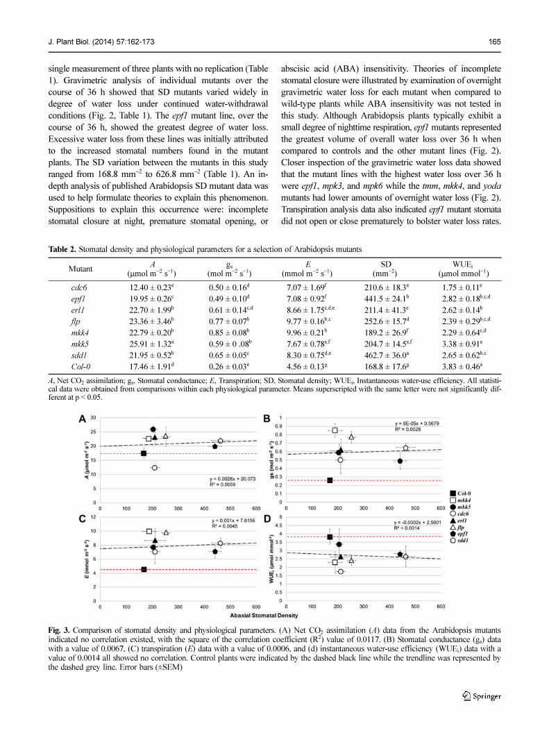
Fig. 3. Comparison of stomatal density and physiological parameters. (A) Net CO2 assimilation (A) data from the Arabidopsis mutantsindicated no correlation existed, with the square of the correlation coefficient (R2) value of 0.0117. (B) Stomatal conductance (gs) datawith a value of 0.0067, (C) transpiration (E) data with a value of 0.0006, and (d) instantaneous water-use efficiency (WUEi) data with avalue of 0.0014 all showed no correlation. Control plants were indicated by the dashed black line while the trendline was represented bythe dashed grey line. Error bars (±SEM)
Table 2. Stomatal density and physiological parameters for a selection of Arabidopsis mutants
Mutant A
(µmol m−2 s−1)gs
(mol m−2 s−1) E
(mmol m−2 s−1) SD
(mm−2)WUEi
(µmol mmol−1)
cdc6 12.40 ± 0.23e 0.50 ± 0.16d 7.07 ± 1.69f 210.6 ± 18.3e 1.75 ± 0.11e
epf1 19.95 ± 0.26c 0.49 ± 0.10d 7.08 ± 0.92f 441.5 ± 24.1b 2.82 ± 0.18b,c,d
erl1 22.70 ± 1.99b 0.61 ± 0.14c,d 8.66 ± 1.75c,d,e 211.4 ± 41.3e 2.62 ± 0.14b
flp 23.36 ± 3.46b 0.77 ± 0.07b 9.77 ± 0.16b,c 252.6 ± 15.7d 2.39 ± 0.29b,c,d
mkk4 22.79 ± 0.20b 0.85 ± 0.08b 9.96 ± 0.21b 189.2 ± 26.9f 2.29 ± 0.64c,d
mkk5 25.91 ± 1.32a 0.59 ± 0 .08b 7.67 ± 0.78e,f 204.7 ± 14.5e,f 3.38 ± 0.91a
sdd1 21.95 ± 0.52b 0.65 ± 0.05c 8.30 ± 0.75d,e 462.7 ± 36.0a 2.65 ± 0.62b,c
Col-0 17.46 ± 1.91d 0.26 ± 0.03e 4.56 ± 0.13g 168.8 ± 17.6g 3.83 ± 0.46a
A, Net CO2 assimilation; gs, Stomatal conductance; E, Transpiration; SD, Stomatal density; WUEi, Instantaneous water-use efficiency. All statisti-cal data were obtained from comparisons within each physiological parameter. Means superscripted with the same letter were not significantly dif-ferent at p < 0.05.
166 J. Plant Biol. (2014) 57:162-173
Arabidopsis Mutant SD and Physiological Parameters
Individual Arabidopsis SD mutant lines grown under identical
conditions demonstrated varied rates of gs, net A, and E
(Table 2). Graphical analysis of A and average SD indicated
that a number of mutant lines demonstrated greater rates of
A than Col-0 plants (Fig. 3A, Table 2). These data indicated
that previous findings where decreased SD led to decreased
A were likely true in one situation, but not another. The
variation in water loss among lines led to the theory that
stomatal aperture and not density may be a contributing
factor to irregularities in assimilation. The mkk5, flp, mkk4,
and erl1 mutant lines displayed the higher A and the lower
assimilation rates were recorded for cdc6 and the wild-type
Col-0 (Fig. 3A, Table 2). These high and low assimilation rates
varied across a wide range (from 12.4 to 25.9 µmol m−2 s−1)
and could have contributed to the slower initial growth for
those mutants with lower assimilation rates when observed
in the greenhouse (Table 2, data not shown). Although cdc6
plants generated the lowest assimilation values, total leaf
fresh weight data indicated that this line did not have the
lowest fresh weight recorded (Table 3). Upon bolting, these
mutants were indistinguishable from the other lines with the
exception of yda. Several mutants with lower stomatal
densities had greater A rates than those Arabidopsis mutants
with higher SD. No observable patterns were found for A
among the selected mutants and SD in this study.
It has been speculated that reduced SD leads to reduced gs,
because with fewer available stomata a more limited amount
of CO2 would be obtained and in response, less water can be
transpired. There was no correlation observed between gs
data and SD for the mutant lines examined in this study (R2=
0.0028) (Fig. 3B). A common pattern emerged between gs
and A in cdc6 and several other mutant lines. In addition to
having the lowest overall A rates (12.40±0.23 µmol m−2 s−1),
cdc6 lines also displayed one of the lower gs rates (0.50±
0.16 mol m−2 s−1) and E rates (7.07±1.69 mmol m−2 s−1) (Fig.
3A, B). The greatest conductance rates were seen in mkk4
(0.85±0.08 mol m−2 s−1) and flp (0.77±0.07 mol m−2 s−1) mutant
plants. Lines that displayed the lower conductance readings
were Col-0, epf1, and cdc6. With the exception of Col-0, gs
data from the remaining mutants were all within 0.36 mol m−2
s−1 from highest to the lowest.
Analysis of the Arabidopsis SD mutant transpiration rates
indicated that E varied independently of SD (Fig. 3C). Yoo
et al. (2010) reported that decreased SD led to decreased E
in gtl1, a SD mutant with a close association with the sdd1
mutant examined in this study, and increased WUEi. No
correlation existed (R2=0.0045) among mutants in this study
despite SD levels for some mutants being greater than control
plants. Those lines with greater SD than wild-type did not
also exhibit higher rates of E (Fig. 3C). The greater transpiration
rates were seen in mkk4 plants (9.96±0.21 mmol m−2 s−1) and
flp (9.77±0.16 mmol m−2 s−1) while the lower transpiration
rates were seen in wild-type (9.96±0.21 mmol m−2 s−1), cdc6
(7.07±0.69 mmol m−2 s−1), and epf1 (7.08±0.92 mmol m−2 s−1)
lines (Table 2). The WUEi data from the individual mutant
lines demonstrated no correlation (R2=0.0014) to SD in the
Arabidopsis mutant lines examined (Fig. 3D). Calculation of
WUEi among all of the mutants showed a two-fold difference
between the lowest and highest efficiency lines (Fig. 3D).
The lowest WUEi was observed in cdc6 (1.75±0.11 µmol
mmol−1) while the highest efficiencies were recorded, as
expected, in Col-0 (Table 2). The Arabidopsis mkk5 mutants
had a WUEi that was closer to Col-0 than any of the other
mutant lines. When all physiological data were compared to
SD, no clear patterns emerged (Fig. 3A-E, 4, Table 2). There
existed a number of differences between SD, gs, A, and E
between each mutant line (Table 3). None of the mutants
studied were similar to Col-0 based on any observed
physiological parameter with the exception of A and WUEi.
Col-0 and mkk5 had similar WUE values, however the majority
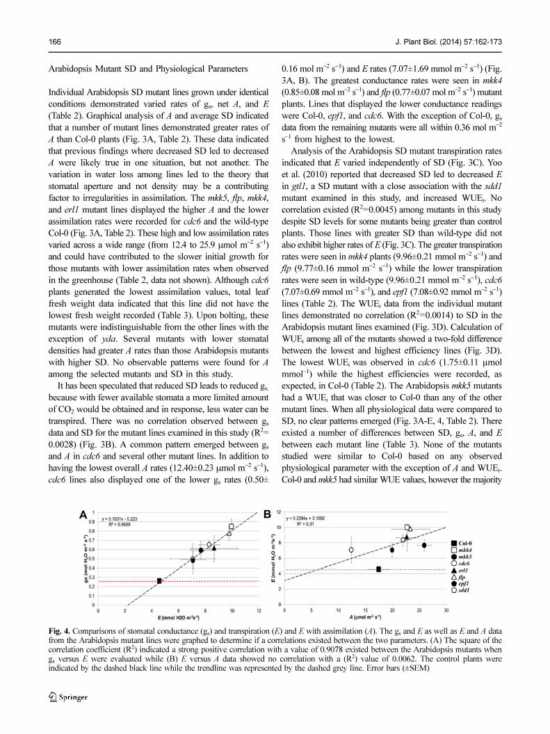
Fig. 4. Comparisons of stomatal conductance (gs) and transpiration (E) and E with assimilation (A). The gs and E as well as E and A datafrom the Arabidopsis mutant lines were graphed to determine if a correlations existed between the two parameters. (A) The square of thecorrelation coefficient (R2) indicated a strong positive correlation with a value of 0.9078 existed between the Arabidopsis mutants whengs versus E were evaluated while (B) E versus A data showed no correlation with a (R2) value of 0.0062. The control plants wereindicated by the dashed black line while the trendline was represented by the dashed grey line. Error bars (±SEM)
J. Plant Biol. (2014) 57:162-173 167
all other parameters for the two lines were significantly
different (Table 2). Comparisons of SD with the physiological
parameters resulted in no correlations being seen between
SD and A (R2=0.0059), gs (R2=0.0028), E (R2=0.0045), or
WUEi (R2=0.0014). A strong positive correlation (R2=0.9689)
was seen between gs and E as expected (Fig. 4A). There was
a slight correlation between E and A (R2=0.31) (Fig. 4B).
Arabidopsis Leaf Fresh Weight (LFW) and Physiological
Parameters
Visual observation of above-ground biomass at the start of
the study indicated no difference in the size of these mutant
plants with the exception of the yda mutants, however
statistical analysis of leaf fresh weight (LFW) indicated
significant differences that may help to explain why more
correlations were not seen within the generated data (Fig. 5,
Table 3). Measurements of LFW were necessary to verify
that undulations in water loss were not attributed to differences
in initial plant size. A mutant that displays a dwarf phenotype,
it was not a surprise to note the resultant diminutive growth
of plants in the yda line. Attempts to find relationships
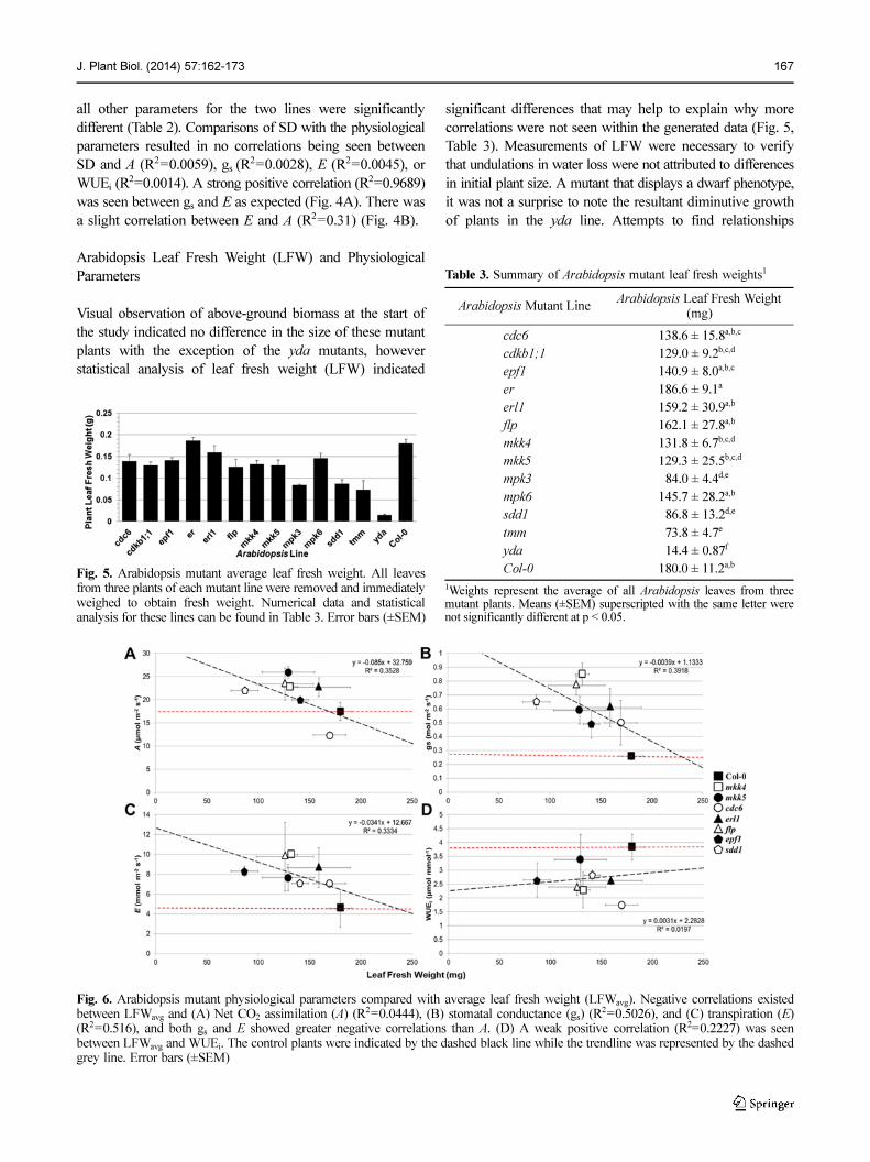
Fig. 5. Arabidopsis mutant average leaf fresh weight. All leavesfrom three plants of each mutant line were removed and immediatelyweighed to obtain fresh weight. Numerical data and statisticalanalysis for these lines can be found in Table 3. Error bars (±SEM)
Table 3. Summary of Arabidopsis mutant leaf fresh weights1
Arabidopsis Mutant LineArabidopsis Leaf Fresh Weight
(mg)
cdc6 138.6 ± 15.8a,b,c
cdkb1;1 129.0 ± 9.2b,c,d
epf1 140.9 ± 8.0a,b,c
er 186.6 ± 9.1a
erl1 159.2 ± 30.9a,b
flp 162.1 ± 27.8a,b
mkk4 131.8 ± 6.7b,c,d
mkk5 129.3 ± 25.5b,c,d
mpk3 0 84.0 ± 4.4d,e
mpk6 145.7 ± 28.2a,b
sdd1 0 86.8 ± 13.2d,e
tmm 0 73.8 ± 4.7e
yda 0 14.4 ± 0.87f
Col-0 180.0 ± 11.2a,b
1Weights represent the average of all Arabidopsis leaves from threemutant plants. Means (±SEM) superscripted with the same letter werenot significantly different at p < 0.05.
Fig. 6. Arabidopsis mutant physiological parameters compared with average leaf fresh weight (LFWavg). Negative correlations existedbetween LFWavg and (A) Net CO2 assimilation (A) (R2=0.0444), (B) stomatal conductance (gs) (R
2=0.5026), and (C) transpiration (E)(R2=0.516), and both gs and E showed greater negative correlations than A. (D) A weak positive correlation (R2=0.2227) was seenbetween LFWavg and WUEi. The control plants were indicated by the dashed black line while the trendline was represented by the dashedgrey line. Error bars (±SEM)

168 J. Plant Biol. (2014) 57:162-173
between LFW and physiological parameters resulted in
negative correlation being drawn between gs, net A, and E.
No correlation was observed between leaf area and WUEi.
Associations between fresh weight and WUEi were the
poorest (R2=0.0197) followed by E (R2=0.3334), net A (R2=
0.3528), and gs (R2=0.3918) (Fig. 6).
Abnormal Mutant Arabidopsis Phenotypes
In addition to the bunching phenotype often found in some
of these SD mutants, a number of other morphological
alterations were observed in these plants. Exceedingly long
pavement cells in two of the mutated protein kinase lines
mpk3 and mpk6 were observed (Fig. 7A, B). These significantly
lengthened pavement cells were found on both leaf surfaces
and clearly indicated a break-down in cell division signaling
when compared to WT (Fig. 7C). Statistical analysis indicated
that pavement cells of mpk3 plants were longer while those
of mpk6 were wider than pavement cells in WT plants (Fig.
7C-E). The characteristic abnormalities observed were joined
and clustered stomata. Many lines demonstrated clustered
stomata, but consistent pairing phenotypes were observed in
epf1, erl1, mpk3, mkk5, and sdd1 (Fig. 8). All of the SD
mutants have different mechanisms of action, yet many of
these displayed the same or similar paired phenotypes. This
pairing tendency found in a number of the studied mutants
emphasized the ablation of the one-cell spacing rule that
EPF1 and other genes control.
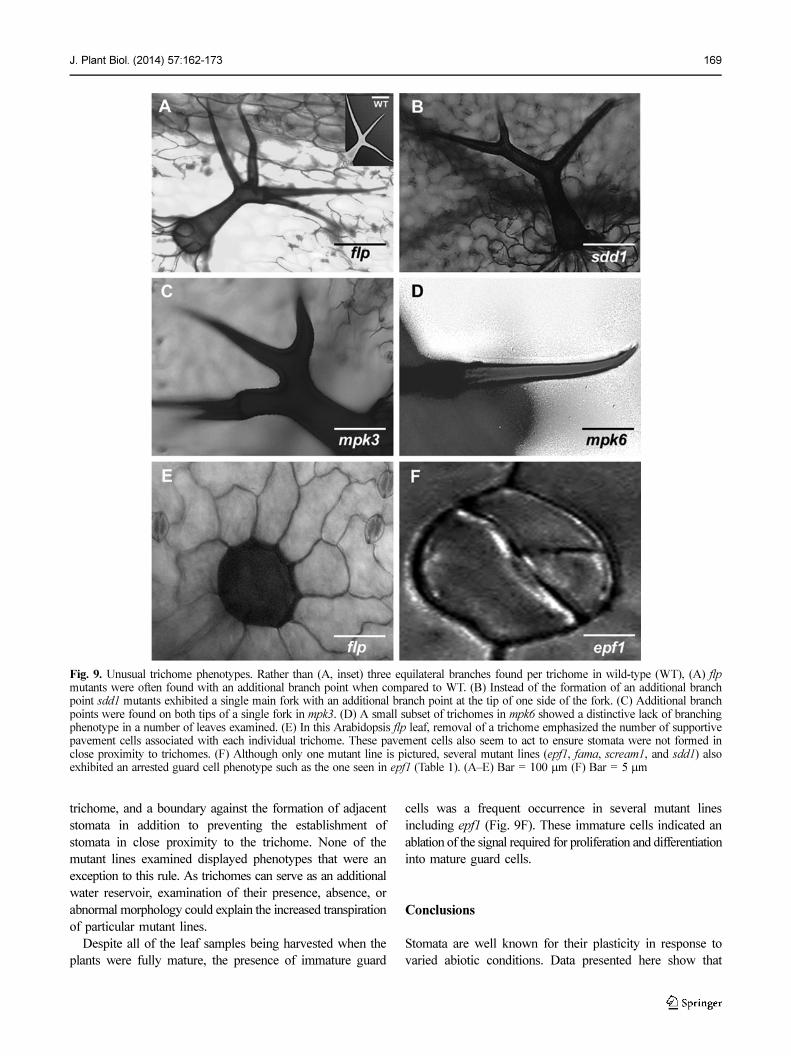
An unusual phenotype discovered that has not previously
been described was variation in trichome morphology between
mutant lines (Fig. 9A-D). Although abnormal trichomes
were not highly prevalent, these were present in noticeable
numbers. Rather than the typical trident-shaped trichomes,
flp, mpk3, mpk6, and sdd1 lines displayed abnormal formations
of trichomes in addition to the described stomatal mutations
and morphologies (Fig. 9A-D). When trichomes were removed,
the supportive pavement cells surrounding the base were
revealed (Fig. 9E).
Examination of these cells led to the hypothesis that these
particular pavement cells provided a foundation for the
Fig. 7. Abnormally shaped pavement cells. (A) The pavement cells in mpk3 were greater than four times longer than adjacent cells whilethe pavement cells in mpk6 (B) were elongated both lengthwise and widthwise when compared to (C) wild-type. Statistical analysisconfirmed observations for (D) length and (E) width. Means superscripted with the same letter were not significantly different at p < 0.05.Bar = 200 µm Error bars (±SEM)
Fig. 8. Paired stomata were found in a number of mutant lines. Anumber of joining morphologies were observed from (A) barelytouching parallel joins in epf1 to (B) barely touching perpendicularjoining in mkk5. (C) A greater degree of parallel joining was seenin erl1, (D) mpk3, and (E) sdd1. (A) Bar = 5 µm, (B–E) Bar = 10 µm
J. Plant Biol. (2014) 57:162-173 169
trichome, and a boundary against the formation of adjacent
stomata in addition to preventing the establishment of
stomata in close proximity to the trichome. None of the
mutant lines examined displayed phenotypes that were an
exception to this rule. As trichomes can serve as an additional
water reservoir, examination of their presence, absence, or
abnormal morphology could explain the increased transpiration
of particular mutant lines.
Despite all of the leaf samples being harvested when the
plants were fully mature, the presence of immature guard
cells was a frequent occurrence in several mutant lines
including epf1 (Fig. 9F). These immature cells indicated an
ablation of the signal required for proliferation and differentiation
into mature guard cells.
Conclusions
Stomata are well known for their plasticity in response to
varied abiotic conditions. Data presented here show that
Fig. 9. Unusual trichome phenotypes. Rather than (A, inset) three equilateral branches found per trichome in wild-type (WT), (A) flpmutants were often found with an additional branch point when compared to WT. (B) Instead of the formation of an additional branchpoint sdd1 mutants exhibited a single main fork with an additional branch point at the tip of one side of the fork. (C) Additional branchpoints were found on both tips of a single fork in mpk3. (D) A small subset of trichomes in mpk6 showed a distinctive lack of branchingphenotype in a number of leaves examined. (E) In this Arabidopsis flp leaf, removal of a trichome emphasized the number of supportivepavement cells associated with each individual trichome. These pavement cells also seem to act to ensure stomata were not formed inclose proximity to trichomes. (F) Although only one mutant line is pictured, several mutant lines (epf1, fama, scream1, and sdd1) alsoexhibited an arrested guard cell phenotype such as the one seen in epf1 (Table 1). (A–E) Bar = 100 µm (F) Bar = 5 µm
170 J. Plant Biol. (2014) 57:162-173
decreased leaf SD does not prevent uptake of additional
CO2 or hinder leaf biomass accumulation. Measurements
of total leaf area indicated that mutants with higher
stomatal densities than Col-0 plants did not exhibit the
expected increase in leaf area. Therefore, additional studies
with SD mutants or studies that involve the highest
biomass producing Arabidopsis mutant plants are needed to
more convincingly state whether or not leaf area would be
significantly affected in plants with decreased stomatal
densities. Destructive sampling of each mutant line
indicated significant differences in fresh weights for many
of the examined mutant lines with the greatest difference
being seen in yda, a mutant with a dwarf phenotype and
delayed growth. Further studies that characterize mutants
with progressively fewer stomata may uncover a threshold
where normal growth and development begins to decrease,
however this study did not reach that physiological limit.
The results obtained here indicated that present levels of
CO2 were sufficient to satisfy growth requirements for
these Arabidopsis mutants even in the absence of uniform
stomatal numbers. The fact that the majority of the plants
measured in this study were mutant lines could be a factor
that contributed to the variations in physiological parameters.
It was also possible that additional epistatic or downstream
effects may have influenced resultant observations. The
effect that each mutation has on growth has not been
overlooked. Thorough examination of previously published
research indicated an absence of data regarding large
numbers of Arabidopsis SD mutants examined for these
parameters. Future studies should focus either on two or
three mutant lines that are closely related in SD to observe
growth and biomass variation as well as morphological
abnormalities throughout the lifetime of the plant, or a
single mutant line and its growth in response to a host of
environmental conditions. These milieus may be responsible
for variation seen both here and in the literature regarding
correlations between A, gs, E, and WUEi. It was not the
objective of this work to thoroughly exhaust all possible
causes for biomass variation in these mutants. These data
uncover and highlight variation in SD mutant biomass,
morphology, and physiological characteristics and provide
plausible justifications worthy of further study.
Materials and Methods
Plant Materials and Growth Conditions
Arabidopsis plants were grown in the greenhouse under controlledconditions. Average greenhouse temperatures ranged between 23.2°−23.7°C each day with the highest and lowest recorded temperaturesbeing 18.4° and 30.7°C on one day of the growth period. Relativehumidity remained steady at 60% under a long-day photoperiod (16/8-h)and light levels ranged from 200−400 µmol m−2s−1 based on natural
light. All plants were grown in individual 10 cm pots. Pots were placedin watering trays (28 cm × 43 cm) that were filled to the brim with waterevery 3 d. All pots were removed from the trays after 1.5 h and placed ongreenhouse benches. The water regime was only altered at the beginningof the water withdrawal experiment. No additional water was providedonce the experiment started. The soil mix was a 3:1 mixture ofsuperfine germinating mix (Farfand) composed of 55% Canadiansphagnum peat, perlite and vermiculite to turface (MVP).
Arabidopsis Seed Disinfestation
Seeds of Arabidopsis (Col-0) were rinsed with distilled water andsurface-disinfested with 15% commercial bleach solution (5.25%sodium hypochlorite) and 0.1% sodium dodecyl sulfate (SDS) for 10min. Seeds were then rinsed three times with sterile, distilled water.Aseptic seeds were stratified at 4°C in a 0.1% agar solution for 4 daysbefore being germinated on half-strength Murashige and Skoog (MS;[Sigma M519]) medium (Murashige and Skoog 1962).
Arabidopsis Mutant Material and Seed Screening
Mutant lines used for this experiment were: cdc6 (SALK_093678),cdkB1;1 (SALK_073457), epf1 (SALK_137549), er (CS20), erl1(SALK_019571), flp (SALK_033970C), mkk4 (SALK_058307),mkk5 (SALK_050700), mpk3 (SALK_100651), mpk6 (SALK_073907), sdd1 (SALK_035560), tmm (SALK_017816), and yoda(CS85662; Torii et al. 1996; Till et al. 2003). Lines were grown to theT3 generation after continuous screening for kanamycin resistancebased on the protocol by Harrison et al. 2006. PCR confirmed thepresence of kanamycin (data not shown). All mutant seeds wereobtained from the Arabidopsis Biological Research Center (ABRC)found at The Arabidopsis Resource Center (TAIR) website (www.arabidopsis.org). Each mutant was generated and reported by Alonsoet al. (2003) unless otherwise noted.
Gravimetric Analysis
Gravimetric transpiration data were obtained from 5-week-oldArabidopsis mutants grown in inverted 50 ml conical tubes in agrowth chamber. The solid end cap was replaced by a wire meshand a punctured end cap during pre-analysis growth to allow foradequate watering. At the start of the analysis period the mesh andpunctured end caps on the 50 ml conical tubes were replaced with asolid end cap for all mutants. Soil evaporation was eliminated bysealing the bottom and top of the tube. No additional water wasprovided once the experiment began and only the aerial parts of theplant protruded. Whole plant transpiration data (mmol H2O
−1) wasobtained by placing a total of three individually grown plants onto abalance that recorded weight measurements in 5 min intervals overthe course of 36 h. The graph depicts data from every 30 min forimproved clarity. Only one plant was placed on each balance in thisstudy. Data represent transpirational water loss from each mutant.Each “icon” represents the average of 3 trials performed within thegrowth chamber. Statistical error bars (± SEM) are not visualizedfor clarity.
Leaf Fresh Weight
Arabidopsis mutant average leaf total fresh weight data were obtainedby carefully removing all of the leaves from three plants from eachmutant line. All of the individual leaves were immediately weighed toobtain fresh weight data. No stems or petioles were included.
Stomatal Imaging by Microscopy
Three fully-expanded rosette leaves were collected from healthy

J. Plant Biol. (2014) 57:162-173 171
mature Arabidopsis plants several weeks after bolting and siliquegrowth, cleared with 70% ethanol, and stained with Safranin-O (1 µgmL−1). Differential Interference Microscopy (DIC) images of theabaxial surface were captured with a Zeiss LSM710 microscope (CarlZeiss, Inc.). Images of four mid-leaf sections (0.5 mm2) from each ofthree leaf rosettes were obtained for each mutant. Counts of stomatalnumber were performed by outlining an image to include a single 1mm2 area. Photos were then printed and stomata were counted byhand and average densities were calculated through the examinationof three photos per rosette leaf per Arabidopsis mutant.
Guard Cell Length and Pore Aperture Measurements
Two fully-expanded rosette leaves were collected from healthy matureArabidopsis plants several weeks after bolting and silique growth,immediately coated with a thin layer of clear nail polish, and placedon a cover slip. After 30 seconds, polish was peeled from the leavesand placed on a labeled cover slip. Images of each leaf were capturedusing DIC with a Zeiss LSM710 microscope (Carl Zeiss, Inc.). Twohundred fifty stomata were measured per leaf. This study wascompleted in triplicate with 3 blocks of plants (well-watered, mildlywater-stressed, severely water-stressed).
Physiological Measurement Collection
A Li-Cor LI6400XT (LICOR Biosciences) was used to obtaintranspiration, photosynthesis, and stomatal conductance data. All datawere collected from plants distributed in a complete random blockdesign. Measurements were taken in triplicate on the same day for 3days in a row. Final calculations were based on three consecutivedays of data collection from three leaves obtained from threeindividual plants. Instantaneous water-use efficiency [WUEi; (µmolmmol−1)] was calculated from A divided by E.
Statistical Analysis
Calculation of standard error of the mean (SEM) and balancedanalysis of variance were performed on SD data. Results of p < 0.001were deemed significant. Analysis was conducted on data gatheredfrom abaxial leaf surfaces only. All analyses were performed usingSAS software programs (SAS Institute Inc. 2008).
Acknowledgements
The authors thank Drs. Yiwei Jiang and Jaemo Yang for their thoughtfulcomments on a previous version of this manuscript. This work wassupported by the Fred M. van Eck Foundation [grant numberFVE51020058] and the Alliance for Graduate Education andProfessoriate (AGEP) [grant number NSF0450373] at Purdue University.Mention of a trademark, proprietary product, or vendor does notconstitute a guarantee or warranty of the product by the USDepartment of Agriculture and does not imply its approval to theexclusion of other products or vendors that also may be suitable.
Author’s Contributions
SSL designed the experimental plans, acquired and screened theArabidopsis seeds, grew and maintained the plants, collected andanalyzed the data, and wrote the manuscript. PMP and CHMreviewed a first draft of the manuscript. All the authors agreed on thecontents of the paper and post no conflicting interest.
Supporting Information
Table S1. Summary of some of the most studied Arabidopsisstomatal density and development genes.
References
Abrash E, Bergmann DC (2010) Regional specification of stomatalproduction by the putative ligand CHALLAH. Development137:447–455. DOI: 10.1242/dev.040931
Abrash E, Lampard GR (2010) A view from the top: new ligandscontrolling stomatal development in Arabidopsis. New Phytol186:561–564. DOI: 10.1111/j.1469-8137.2010.03265.x
Alonso JM, Stepanova AN, Leisse TJ, Kim CJ, Chen H, Shinn P,Stevenson DK, Zimmerman J, Barajas P, Cheuk R, Gadrinab C,Heller C, Jeske A, Koesema E, Meyers CC, Parker H, Prednis L,Ansari Y, Choy N, Deen H, Geralt M, Hazari N, Hom E, KarnesM, Mulholland C, Ndubaku R, Schmidt I, Guzman P, Aguilar-Henonin L, Schmid M, Wrigel D, Carter DE, Marchand T,Risseeuw E, Brogden D, Zeko A, Crosby WL, Berry CC, EckerJR (2003) Genome-wide insertional mutagenesis of Arabidopsisthaliana. Science 301:653–657. DOI: 10.1126/science.1086391
Alwerdt JL, Gibson DJ, Ebbs SD, Wood AJ (2006) Intraspecificinteractions in Arabidopsis thaliana and the stomatal mutantstmm1-1 and sdd1-2. Biol Plantarun 50:205–209. DOI: 10.1007/s10535-006-0008-2
Berger D, Altmann T (2000) A subtilisin-like serine protease involved inthe regulation of stomatal density and distribution in Arabidopsisthaliana. Gene Dev 14:1119–1131. DOI: 10.1101/gad.14.9.1119
Bergmann DC, Lukowitz W, Somerville CR (2004) Stomataldevelopment and pattern controlled by a MAPKK kinase. Science304:1494–1497. DOI: 10.1126/science.1096014
Bertoni G (2009) Integration of signaling pathways in stomataldevelopment. Plant Cell 21:2542. DOI: 10.1105/tpc.109.210910
Bhave NS, Veley KM, Nadeau JA, Lucas JR, Bhave SL, Sack FD(2009) TOO MANY MOUTHS promotes cell fate progressionin stomatal development of Arabidopsis stems. Planta 229:357–367. DOI: 10.1007/s00425-008-0835-9
Boudolf V, Lammens T, Boruc J, van Leene J, van Den Daele H,Maes S, van Isterdael G, Russinova E, Kondorski E, Witters E,De Jeager G (2009) CDKB1;1 forms a functional complex withCYCA2;3 to suppress endocycle onset. Plant Physiol 150:1482–1493. DOI: 10.1104/pp.109.140269
Brownlee C (2001) The long and the short of stomatal density signals.Trends Plant Sci 6:441442. DOI: 10.1016/S1360-1385(01)02095-7
Büssis D, von Groll U, Fisahn J, Altmann T (2006) Stomatal aperturecan compensate altered stomatal density in Arabidopsis thalianaat growth light conditions. Funct Plant Biol 33:1037–1043. DOI:10.1071/FP06078
Chen LQ, Cheng-Sen L, Chaloner WG, Beerling DJ, Sun Q-G,Collinson ME, Mitchell PL (2001) Assessing the potential forthe stomatal characters of extant and fossil Ginkgo leaves tosignal atmospheric CO2 change. Am J Bot 88:1309–1315. DOI:10.2307/3558342
Chinnusamy V, Ohta M, Kanrar S, Lee BH, Hong X, Agrawal M,Zhu JK (2003) ICE1: a regulator of cold-induced transcriptomeand freezing tolerance in Arabidopsis. Gene Dev 17:1043–1054.DOI: 10.1101/gad.1077503
Dong J, MacAlister CA, Bergmann DC (2009) BASL controlsasymmetric cell division in Arabidopsis. Cell 137:1320–1330.DOI: 10.1016/j.cell.2009.04.018
Fraser LH, Carlyle C, Turkington R, Friedman CR (2009) Adaptivephenotypic plasticity of Pseudoroegneria spicata: response of
172 J. Plant Biol. (2014) 57:162-173
stomatal density, leaf area and biomass to changes in watersupply and increased temperature. Ann Bot- Lon 103:769–775.DOI: 10.1093/aob/mcn252
Gao T, Wu Y, Zhang Y, Liu L, Ning Y, Wang D, Tong H, Chen S, ChuC, Xie Q (2011) OsSDIR1 overexpression greatly improvesdrought tolerance in transgenic rice. Plant Mol Biol 76:145–156.DOI: 10.1007/s11103-011-9775-z
Geisler M, Nadeau J, Sack FD (2000) Oriented asymmetric divisionsthat generate the stomatal spacing pattern in Arabidopsis aredisrupted by the too many mouths mutation. Plant Cell 12:2075–2086. DOI: 10.2307/3871106
Guseman JM, Lee JS, Bogenschutz NL, Peterson KM, Virata RE, XieB, Kanaoka MM, Hong Z, Torii KU (2010) Dysregulation ofcell-to-cell connectivity and stomatal patterning by loss-of-functionmutation in Arabidopsis CHORUS (GLUCAN SYNTHASE-LIKE 8). Development 137:1731–1741. DOI: 10.1242/dev.049197
Hara K, Kajita R, Torii KU, Bergmann DC Kakimoto T (2007) Thesecretory peptide gene EPF1 enforces the stomatal one-cell-spacingrule. Gene Dev 21:1720–1725. DOI: 10.1101/gad.1550707
Hara K, Yokoo T, Kajita R, Onishi T, Yahata S, Peterson KM, ToriiKU, Kakimoto T (2009) Epidermal cell density is autoregulatedvia a secretory peptide, EPIDERMAL PATTERNING FACTOR2in Arabidopsis leaves. Plant Cell Physiol 50:1019–1031. DOI:10.1093/pcp/pcp068
Harrison SJ, Mott EK, Parsley K, Aspinall S, Gray JC, Cottage A(2006) A rapid and robust method of identifying transformedArabidopsis thaliana seedlings following floral dip transformation.Plant Methods 2:19. DOI: 10.1186/1746-4811-2-19
Hoover WS (1986) Stomata and stomatal clusters in Begonia: Ecologicalresponse in 2 Mexican species. Biotropica 18:16–21. DOI:10.2307/2388356
Hord CLH, Sun Y-J, Pillitteri LJ, Wang H, Zhang S, Ma H (2008)Regulation of Arabidopsis early anther development by themitogen-activated protein kinases, MPK3and MPK6, and theERECTA and related receptor-like kinases. Mol Plant 1:645–658. DOI: 10.1093/mp/ssn029
Hunt L, Gray JE (2009) The signaling peptide EPF2 controls asymmetriccell divisions during stomatal development. Curr Biol 19:864–869. DOI: 10.1016/j.cub.2009.03.069
Hunt L, Gray JE (2010) BASL and EPF2 act independently to regulateasymmetric divisions during stomatal development. Plant SignalBehav 5:278–280. PMCID: PMC2881277
Kanaoka MM, Pillitteri LJ, Fujii H, Yoshida Y, Bogenschutz NL,Takabayashi J, Zhu J-K, Torii KU (2008) SCREAM/ICE1 andSCREAM2 specify three cell-state transitional steps leading toArabidopsis stomatal differentiation. Plant Cell 20:1775–1785.DOI: 10.1105/tpc.108.060848
Kondo T, Kajita R, Miyazaki A, Hokoyama M, Nakamura-Miura T,Mizuno S, Masuda Y, Irie K, Tanaka Y, Takada S, Kakimoto T,Sakagami Y (2010) Stomatal density is controlled by a mesophyll-derived signaling molecule. Plant Cell Physiol 51:1–8. DOI:10.1093/pcp/pcp180
Korn RW (2009) A new hypothesis for stomatal placement inArabidopsis. J Theor Biol 260:172–174. DOI: 10.1016/j.jtbi.2009.05.012
Lai LB, Nadeau JA, Lucas J, Lee E-K, Nakagawa T, Zhao L, GeislerM, Sack FD (2005) The Arabidopsis R2R3 MYB proteinsFOUR LIPS and MYB88 restrict divisions late in the stomatallineage. Plant Cell 17:2754–2767. DOI: 10.1105/tpc.105.034116
Lammertsma EI, de Boer HJ, Dekker SC, Dilcherc DL, Lotter AF,Wagner-Cremer F (2011) Global CO2 rise leads to reducedmaximum stomatal conductance in Florida vegetation. Proc NatlAcad Sci USA 108:4035–4040. DOI: 10.1073/pnas.1100371108
Lampard GR, Lukowitz W, Ellis BE, Bergmann DC (2009) Noveland expanded roles for MAPK signaling in Arabidopsis stomatalcell fate revealed by cell type–specific manipulations. Plant Cell
21:3506–3517. DOI: 10.1105/tpc.109.070110Lampard GR, MacAlister CA, Bergmann DC (2008) Arabidopsis
stomatal initiation is controlled by MAPK-mediated regulationof the bHLH SPEECHLESS. Science 322:1113–1116. DOI:10.1126/science.1162263
Liu T, Ohashi-Ito K, Bergmann DC (2009) Orthologs of Arabidopsisthaliana stomatal bHLH genes and regulation of stomataldevelopment in grasses. Development 136:2265–2276. DOI:10.1242/dev.032938
Lukowitz W, Roeder A, Parmenter D, Somerville C (2004) A MAPKKkinase gene regulates extra-embryonic cell fate in Arabidopsis.Cell 116:109–119. DOI: 10.1016/S0092-8674(03)01067-5
Lumbreras V, Vilela B, Irar S, Sole M, Capellades M, Valls M, CocaM, Pages M (2010) MAPK phosphatase MKP2 mediates diseaseresponses in Arabidopsis and functionally interacts with MPK3and MPK6. Plant J 63:1017–1030. DOI: 10.1111/j.1365-313X.2010.04297.x
MacAlister CA, Ohashi-Ito K, Bergmann DC (2007) Transcriptionfactor control of asymmetric cell divisions that establish thestomatal lineage. Nature 445:537–540. DOI: 10.1038/nature05491
Metzinger CA, Bergmann DC. (2010) Plant asymmetric cell divisionregulators: pinch-hitting for PARs? F1000 Biol Reports. 2:25.DOI:10.3410/B2-25
Murashige T, Skoog F (1962) A revised medium for rapid growth andbioassays with tobacco tissue cultures. Physiol Plantarum15:473–497. DOI: 10.1111/j.1399-3054.1962.tb08052.x
Nawazish S, Hameed M, Naurin S (2006) Leaf anatomical adaptationsof Cenchrus cilliaris L., from the salt range, Pakistan againstdrought stress. Pakistan J Bot 38:1723–1730. http://www.pakbs.org/pjbot/PDFs/38(5)/PJB38(5)1723.pdf
Nadeau JA, Sack FD (2002) Control of stomatal distribution on theArabidopsis leaf surface. Science 296:1697–1700. DOI: 10.1126/science.1069596
Ohashi-Ito K, Bergmann DC (2006) Arabidopsis FAMA controlsthe final proliferation/differentiation switch during stomataldevelopment. Plant Cell 18:2493–2505. DOI: 10.1105/tpc.106.046136
Peterson KM, Rychel AL, Torii KU (2010) Out of the mouths ofplants: The molecular basis of the evolution and diversity ofstomatal development. Plant Cell 22:296–306. DOI: 10.1105/tpc.109.072777
Pillitteri LJ, Bogenschutz NL, Torii KU (2008) The bHLH protein,MUTE, controls differentiation of stomata and the hydathodepore in Arabidopsis. Plant Cell Physiol 49:934–943. DOI:10.1093/pcp/pcn067
Pillitteri LJ, Sloan DB, Bogenschutz NL, Torii KU (2007) Terminationof asymmetric cell division and differentiation of stomata.Nature 445:501–505. DOI: 10.1038/nature05467
Porter ACG (2008) Preventing DNA over-replication: a Cdkperspective. Cell Division 3:3. DOI:10.1186/1747–1028-3-3.
Pospíšilová J (2003) Participation of phytohormones in the stomatalregulation of gas exchange during water stress. Biol Plantarum46:491–506. DOI: 10.1023/A:1024894923865
Rodriguez MCS, Peterson M, Mundy J (2010) Mitogen-activatedprotein kinase signaling in plants. Annu Rev Plant Biol 61:621–649. DOI: 10.1146/annurev-arplant-042809-112252
Rychel AL, Peterson KM, Torii KU (2010) Plant twitter: ligandsunder 140 amino acids enforcing stomatal patterning. J Plant Res123:275–280. DOI: 10.1007/s10265-010-0330-9
SAS Institute Inc (2008) Available at www.sas.com/, AccessedJanuary 5, 2014.
Serna L (2009) Cell fate transitions during stomatal development.BioEssays 31:865–873. DOI: 10.1002/bies.200800231
Shpak ED, Berthiaume CT, Hill EJ, Torii KU (2004) Synergisticinteraction of three ERECTA-family receptor-like kinasescontrols Arabidopsis organ growth and flower development by
J. Plant Biol. (2014) 57:162-173 173
promoting cell proliferation. Development 131:1491–1501.DOI: 10.1242/dev.01028
Shpak ED, McAbee JM, Pillitteri LJ, Torii KU (2005) Stomatalpatterning and differentiation by synergistic interactions ofreceptor kinases. Science 309:290–293. DOI: 10.1126/science.1109710
Sugano SS, Shimada T, Imai Y, Okawa K, Tamai A, Mori M, Hara-Nishimura I (2010) Stomagen positively regulates stomataldensity in Arabidopsis. Nature 463:241–244. DOI: 10.1038/nature08682
The Arabidopsis Information Resource (TAIR) (2008) Available athttp://www.arabidopsis.org, Accessed January 1, 2014.
Till BJ, Reynolds SH, Greene EA, Codomo CA, Enns LC, JohnsonJE, Burtner C, Odden AR, Young K, Taylor NE, Henikoff JG(2003) Large-scale discovery of induced point mutations withhigh-throughput tilling. Genome Res 13:524–530. DOI:10.1101/gr.977903
Torii KU, Mitsukawa N, Oosumi T, Matsuura Y, Yokoyama R,Whittier RF, Komeda Y (1996) The Arabidopsis ERECTA geneencodes a putative receptor protein kinase with extracellularleucine-rich repeats. Plant Cell 8:735–746. DOI: http://dx.doi.org/10.1105/tpc.8.4.735
Umbrasaite J, Schweighofer A, Kazanaviciute V, Magyar Z, Ayatollahi Z,Unterwurzacher V, Choopayak C, Boniecka J, Murray JAH,Bogre L, Meskiene I (2010) MAPK phosphatase AP2C3 inducesectopic proliferation of epidermal cells leading to stomatadevelopment in Arabidopsis. PLOS One 5:e15357. DOI:10.1371/journal.pone.0015357
von Groll U, Berger D, Altmann T (2002) The subtilisin-like serineprotease SDD1 mediates cell-to-cell signaling during Arabidopsisstomatal development. Plant Cell 14:1527–1539. DOI: 10.1105/
tpc.001016Wang H, Liu Y, Bruffett K, Lee J, Hause G, Walker JC, Zhang S
(2008) Haplo-insufficiency of MPK3 in MPK6 mutant backgrounduncovers a novel function of these two MAPKs in Arabidopsisovule development. Plant Cell 20:602–613. DOI: 10.1105/tpc.108.058032
Wang H, Ngwenyama N, Liu Y, Walker JC, Zhang S (2007) Stomataldevelopment and patterning are regulated by environmentallyresponsive mitogen-activated protein kinases in Arabidopsis.Plant Cell 19:63–73. DOI: 10.1105/tpc.106.048298
Xie Z, Lee E-K, Lucas JR, Morohashi K, Li D, Murray JAH, SackFD, Grotewold E (2010b) Regulation of cell proliferation in thestomatal lineage by the Arabidopsis MYB FOUR LIPS viadirect targeting of core cell cycle genes. Plant Cell 22:2306–2321. DOI: 10.1105/tpc.110.074609
Xie Z, Li D, Wang L, Sack FD, Grotewold E (2010a) Role of thestomatal development regulators FLP/MYB88 in abiotic stressresponses. Plant J 64:731–739. DOI: 10.1111/j.1365-313X.2010.04364.x
Xu Z, and Zhou G (2008) Responses of leaf stomatal density to waterstatus and its relationship with photosynthesis in a grass. J ExpBot 59:3317–3325. DOI: 10.1093/jxb/ern185
Yang M, Sack FD (1995) The too many mouths and four lipsmutations affect stomatal production in Arabidopsis. Plant Cell7:2227–2239. DOI: 10.2307/3870164
Yoo CY, Pence HE, Jin JB, Miura K, Gosney MJ, Hasegawa PM,Michelbart MV (2010) The Arabidopsis GTL1 transcriptionfactor regulates water use efficiency and drought tolerance bymodulating stomatal density via transrepression of SDD1. PlantCell 22:4128–4141. DOI: 10.1105/tpc.110.078691
![Generation of Targeted Knockout Mutants in Arabidopsis ... · Keywords: CRISPR/Cas9, Genome editing, Arabidopsis thaliana, Plants, Knockout [Background] The CRISPR/Cas9 system (Cas9)](https://static.fdocuments.net/doc/165x107/5fcbdfb69ddbe939ee10f004/generation-of-targeted-knockout-mutants-in-arabidopsis-keywords-crisprcas9.jpg)