Comparative study of treatment of the dry eye syndrome … · Comparative study of treatment of the...
12
Klin Monatsbl Augenheilkd 2006; 223: 974-983 Comparative study of treatment of the dry eye syndrome due to disturbances of the tear film lipid layer with lipid-containing tear substitutes Efficacy of lipid-containing tear substitutes Authors Dausch Dieter 1, 2, Lee Suwan 1, 2, Dausch Sabine 1, Kim Jae Chan 2, Schwert Gregor 3, Michelson Wanda 4 Instituts 1 Abteilung Augenheilkunde, Klinikum St. Marien, Amberg 2 Department of Ophthalmology, Chung-Ang University Hospital, Seoul, Korea 3 augenärztliche Praxis, Beckum 4 augenärztliche Praxis, Kronshagen Key words Dry eye – keratoconjunctivitis sicca – lipid layer – phospholipids – polar lipids – triglyceride – nonpolare lipids – eye spray – eye gel – carbomer Abstract Backround: A deficiency in the tear film lipid layer is aetiological in about 80 % of the patients suffering from dry eye, which results in excessive evaporation (so-called hyperevaporative dry eye). The treatment with conventional artificial tears did not prove to be successful here. In this study the treatment with two tear substitutes containing lipids were set in contrast with each other. Material and Methods: The randomised, controlled, multicenter cross-over study included 74 patients suffering from dry eye caused by a deficiency of the tear film lipid layer, which were organised into two groups. Group A (n = 38) was treated for the first 6 weeks with a liposomal eye spray (Tears Again ® ), while the patients of the group of B (n = 36) were treated with an eye gel containing triglycerides (Liposic ® ) in the same period. After 6 weeks the crossover was performed. The patients were treated in the following 6 weeks with the product which was not used before. Control examinations by masked examiners took place at the beginning of the study as well as after 6 and 12 weeks, considering following parameters: eyelidedge-parallel conjunctival folds (LIPCOF), BREAK UP time (BUT), Schimer-I Test, measurement of the tear meniscus, investigation of the edges of eyelid and visual acuity. In addition the subjective feelings of the patients were also determined by means of questionnaires. Results: At the beginning of the study both groups did not differ significantly with respect to the initial values. After the first treatment period the improvement of the examined parameters LIPCOF, BUT, Schirmer, visual acuity and inflammation of the lid margin in group A (eye spray) proved to be significant superior in comparison to group B (eye gel). The results of the second treatment period after the crossover were similar and showed an analogical supremacy of the liposomal eye spray. The interview of the patients resulted that the subjective evaluation concerning efficacy and compatibility of the eye spray turned out to be more favourable explicitly than that concerning the eye gel. 74,6 % of the patients favoured the application as an eye spray onto the closed eyelids over the eye gel into the conjunctival sac. 62,5 % of the patients rated the liposomal eye spray to be better all in all, 12,5 % rated that both preparations are equal and 25 % favoured the eye gel. Conclusions: The treatment with phospholipid-liposomes shows statistically significant clinical advantages and proves to be favourably and explicitly superior compared to the conventional standard treatment all in all.
Transcript of Comparative study of treatment of the dry eye syndrome … · Comparative study of treatment of the...
Klin Monatsbl Augenheilkd 2006; 223: 974-983
Comparative study of treatment of the dry eye syndrome due to
disturbances of the tear film lipid layer with lipid-containing tear
substitutes
Efficacy of lipid-containing tear substitutes
Authors Dausch Dieter 1, 2, Lee Suwan 1, 2, Dausch Sabine 1, Kim Jae Chan 2, Schwert Gregor 3, Michelson Wanda 4
Instituts 1 Abteilung Augenheilkunde, Klinikum St. Marien, Amberg 2 Department of Ophthalmology, Chung-Ang University Hospital, Seoul, Korea
3 augenärztliche Praxis, Beckum 4 augenärztliche Praxis, Kronshagen
Key words
Dry eye – keratoconjunctivitis sicca – lipid layer –
phospholipids – polar lipids – triglyceride – nonpolare lipids
– eye spray – eye gel – carbomer
Abstract �
Backround: A deficiency in the tear film lipid layer is
aetiological in about 80 % of the patients suffering from dry
eye, which results in excessive evaporation (so-called
hyperevaporative dry eye). The treatment with conventional
artificial tears did not prove to be successful here. In this
study the treatment with two tear substitutes containing
lipids were set in contrast with each other.
Material and Methods: The randomised, controlled,
multicenter cross-over study included 74 patients suffering
from dry eye caused by a deficiency of the tear film lipid
layer, which were organised into two groups. Group A (n =
38) was treated for the first 6 weeks with a liposomal eye
spray (Tears Again®), while the patients of the group of B (n
= 36) were treated with an eye gel containing triglycerides
(Liposic®) in the same period. After 6 weeks the crossover
was performed. The patients were treated in the following 6
weeks with the product which was not used before. Control
examinations by masked examiners took place at the
beginning of the study as well as after 6 and 12 weeks,
considering following parameters: eyelidedge-parallel
conjunctival folds (LIPCOF), BREAK UP time (BUT),
Schimer-I Test, measurement of the tear meniscus,
investigation of the edges of eyelid and visual acuity. In
addition the subjective feelings of the patients were also
determined by means of questionnaires.
Results: At the beginning of the study both groups did not
differ significantly with respect to the initial values. After
the first treatment period the improvement of the examined
parameters LIPCOF, BUT, Schirmer, visual acuity and
inflammation of the lid margin in group A (eye spray)
proved to be significant superior in comparison to group B
(eye gel). The results of the second treatment period after
the crossover were similar and showed an analogical
supremacy of the liposomal eye spray. The interview of the
patients resulted that the subjective evaluation concerning
efficacy and compatibility of the eye spray turned out to be
more favourable explicitly than that concerning the eye gel.
74,6 % of the patients favoured the application as an eye
spray onto the closed eyelids over the eye gel into the
conjunctival sac. 62,5 % of the patients rated the liposomal
eye spray to be better all in all, 12,5 % rated that both
preparations are equal and 25 % favoured the eye gel.
Conclusions: The treatment with phospholipid-liposomes
shows statistically significant clinical advantages and proves
to be favourably and explicitly superior compared to the
conventional standard treatment all in all.
Introduction �
The tear film is a very complex structure made up of
numerous different components, such as lipids, proteins,
salts, mucin and water, whose interactions are essential to its
stability.
The lipid layer forms the outer layer of the tear film located
at the air interface and plays a key role in the composition
and functionality of the tear film.
An intact lipid layer can reduce the evaporation rate of tear
fluid by 90-95% (36).
Further, it ensures a smooth surface of the tear film and is
therefore critical for the quality of vision. In addition, the
spreading of the lipid layer reduces free energy at the tear
film surface and reduces surface tension by 25%, which is
crucial for the structure of the entire tear film [63].
Disorders of this exterior lipid layer are responsible for
almost 80% of tear film disorders embraced by the term "dry
eye" [25, 26] and are its major etiology [33,54].
Correspondingly, patients suffering from dry eye exhibit an
increased evaporation rate at the eye surface [19, 40,50,51]
and a higher surface tension of the tear liquid [47,71].
A lipid layer disturbance leads to an excessive evaporation
rate and damage to the eye surface due to the resulting
increased osmolarity [18, 42, 39].
The use of conventional artificial tear products does not
appear to be an appropriate therapy for hyperevaporative
dry eye, since it has been shown to additionally increase
evaporation rates [38, 66, 67].
After application of the artificial tear product, it takes
approximately 40 minutes for the evaporation rate to return
to the original value [68]. The application process itself
creates a significant disturbance in the lipid layer [24].
Additional preservatives, especially benzalconium chloride,
do lasting damage to the lipid layer [28] by dissolving it due
to their detergent action [20, 21, 26]. There is a reason why
56% of patients report applying their ophthalmic agents
more than 8 times daily [25].
Successful treatment of lipid phase disturbances would
instead require a targeted addition of the lipids that are
actually lacking.
The lipid phase itself is made up of two layers; the inner
layer borders the aqueous phase, consists of polar lipids,
and is surface-active. The thick outer layer is composed of
neutral lipids with anti-evaporative properties [44].
Phospholipid-liposome therapy has already proven useful in
several previous studies [3, 32, 52, 60].
The authors are not aware of any specific study results that
would justify a recommendation of therapy with the
triglyceride-containing eye gel.
Therefore, this study compared both therapy options
regarding their suitability for successful treatment of lipid
layer disturbances.
Methods �
A randomized, controlled, multi-center cross-over study had
been carried out in the participating centers beginning in
December 2004, with blinded examiners and including 74
patients.
Participants suffered from dry eye due to disturbance of the
lipid phase; they were assigned to treatment groups A (n=
38) or B (n= 36) and treated with the corresponding
preparations.
Lipid layer disturbances were diagnosed using reduced tear
break-up time and the findings of a slit-lamp examination of
the lids, with particular regard to signs of chronic
blepharitis [25].
Patients in both groups were treated with a lipid-containing
preparation applied 3 times daily in accordance with
manufacturer recommendations.
Tears Again® liposomal eye spray (Optima Pharmazeutische
GmbH) contains phospholipids in liposomal form (polar
lipids). Liposic® eye gel (Dr. Mann Pharma) is an artificial
tear preparation based on carbomer (polyacrylic acid)
containing triglycerides (neutral lipids).
Lipid layer disturbances were diagnosed using reduced tear
break-up time and the findings of a slit-lamp examination of
the lids, with particular regard to signs of chronic
blepharitis [25].
Patients in group A were initially treated for 6 weeks with
the liposomal eye spray, while patients in group B were
treated with the triglyceride-containing eye gel for the same
time period.
The crossover occurred after this first treatment period, with
patients of group A being treated with the triglyceride-
containing eye gel for the following 6 weeks, while patients
in group B received treatment with the liposomal eye spray.
A washout phase prior to switching preparations at
crossover was deemed unnecessary since both preparations
do not contain pharmacologically active ingredients;
therefore, long-term effects were not expected.
Examinations were carried out at the beginning of the study,
at six and at twelve weeks. The following parameters were
examined and documented by classification into categories:
Lid parallel conjunctival folds (LIPCOF), break-up time
(BUT), Schirmer-I test, tear meniscus measurement, slit-
lamp examination of the lid margins, and visual acuity.
The established data were documented by assigning
categories or severity levels derived from the recommended
classification of stages and severities of dry eye (4, 6). The
Schirmer-I test values were classified into four categories:
>10mm/5 min, <10mm/5min, > 5mm/5 min, < 5 mm/5 min.
The values of the tear break-up time were also classified into
four categories: > 15 s, 10–15 s, 5–10 s, < 5 s. LIPCOF results
were classified into degrees from 0 to 4.
In a slit-lamp exam, the lid margin was evaluated for
edema, inflammation, hyperaemia, keratinization and
abnormalities of the Meibomian gland orifices
(centralization, blockage, scarring), the lashes (abnormal
position, loss), and the lash base.
In addition, subjective patient evaluations were recorded
using questionnaires administered during control
examinations.
Statistical analysis was performed using the statistics
software program SPSS v. 12.0.
To illustrate how rapidly the liposomal eye spray reaches
the tear film after application onto closed lids, the
application of liposomal eye spray mixed with 10%
fluorescein solution in a 1:1000 ratio was observed and
photographed under blue light conditions in a volunteer.
The study was designed and carried out in accordance with
the regulations of the declaration of Helsinki.
Results �
Study population
The study population (N = 75) consisted of 28 male and 47
female patients. One patient was less than 25 years old, 9
patients were between 25 and 45 years of age, 16 patients
were between 46 and 60 years old, and the remaining 49
patients were over 60 years old. A correlation between age
and gender or a non-random representation was not found
(χ²-test: χ²-value = 2.134; df = 3; p> .100; n.s.).
Regarding the comparability of the two patient groups, it
was shown that the distribution of gender (χ²-test: χ²-value =
0.175; df = 1; p> .100; n.s.) and age categories (χ²-test: χ²-
value = 1.815; df = 3; p> .100; n.s.) was homogenous between
the two groups. The study was completed according to
protocol by 74 patients. One patient chose to discontinue
participation, not due to side effects or medical reasons.
Tear Break-up Time
The tear break-up time measured with the BUT test does not
significantly differ between groups A and B at the beginning
of therapy (Mann-Whitney U-test:
.s.n;.p;.z 10426 >−= ).
Over the course of the study, both groups exhibited a
significant improvement of break-up time (GLM with
repeated measures: Factor time:
001.;409.16)122,2( <= pF ). However, the increase in
tear break-up time differs between the two groups (time by
treatment interaction: 055.;962.2)122,2( == pF ). The
group that first received the liposomal eye spray exhibited a
significantly greater improvement during the first six weeks
and showed less improvement in the second part of the
study (after crossover), when using Liposic (test of the
square of the contrast of time by treatment group
interaction: F(1,61) = 6.713; p<.05).
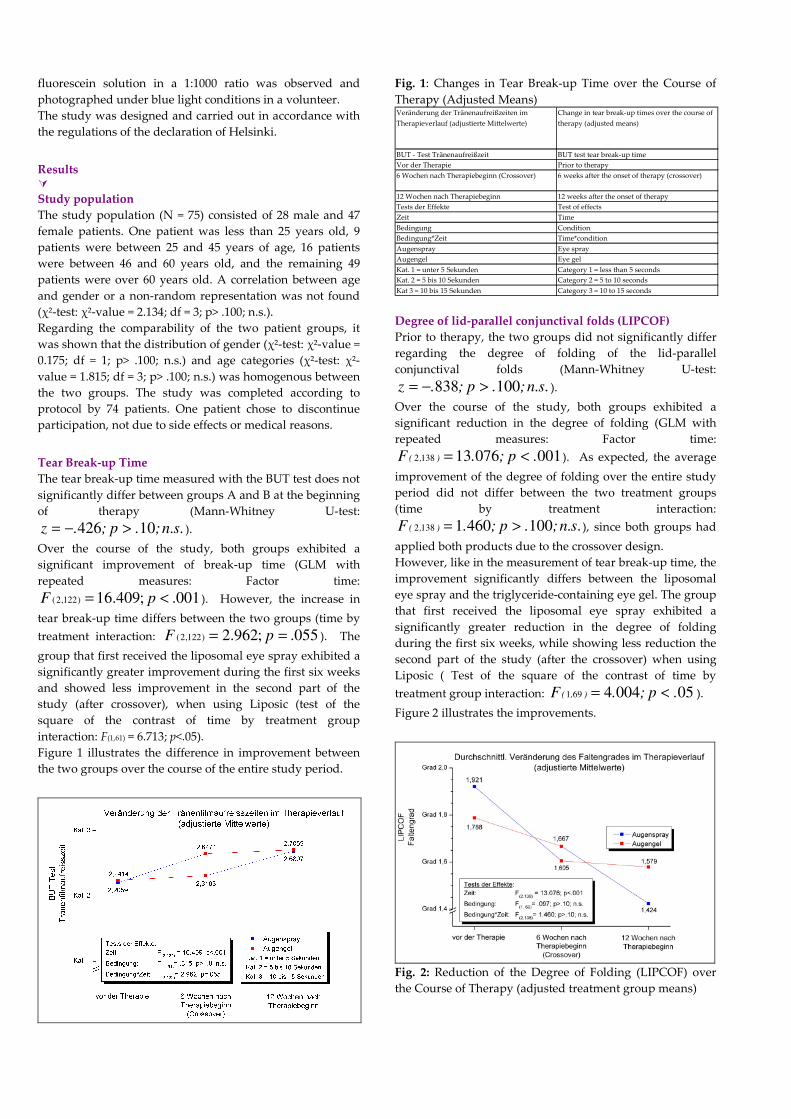
Figure 1 illustrates the difference in improvement between
the two groups over the course of the entire study period.
Fig. 1: Changes in Tear Break-up Time over the Course of
Therapy (Adjusted Means) Veränderung der Tränenaufreißzeiten im
Therapieverlauf (adjustierte Mittelwerte)
Change in tear break-up times over the course of
therapy (adjusted means)
BUT - Test Tränenaufreißzeit BUT test tear break-up time
Vor der Therapie Prior to therapy
6 Wochen nach Therapiebeginn (Crossover) 6 weeks after the onset of therapy (crossover)
12 Wochen nach Therapiebeginn 12 weeks after the onset of therapy
Tests der Effekte Test of effects
Zeit Time
Bedingung Condition
Bedingung*Zeit Time*condition
Augenspray Eye spray
Augengel Eye gel
Kat. 1 = unter 5 Sekunden Category 1 = less than 5 seconds
Kat. 2 = 5 bis 10 Sekunden Category 2 = 5 to 10 seconds
Kat 3 = 10 bis 15 Sekunden Category 3 = 10 to 15 seconds
Degree of lid-parallel conjunctival folds (LIPCOF)
Prior to therapy, the two groups did not significantly differ
regarding the degree of folding of the lid-parallel
conjunctival folds (Mann-Whitney U-test:
.s.n;.p;.z 100838 >−= ).
Over the course of the study, both groups exhibited a
significant reduction in the degree of folding (GLM with
repeated measures: Factor time:
001076131382 .p;.F ),( <= ). As expected, the average
improvement of the degree of folding over the entire study
period did not differ between the two treatment groups
(time by treatment interaction:
.s.n;.p;.F ),( 10046011382 >= ), since both groups had
applied both products due to the crossover design.
However, like in the measurement of tear break-up time, the
improvement significantly differs between the liposomal
eye spray and the triglyceride-containing eye gel. The group
that first received the liposomal eye spray exhibited a
significantly greater reduction in the degree of folding
during the first six weeks, while showing less reduction the
second part of the study (after the crossover) when using
Liposic ( Test of the square of the contrast of time by
treatment group interaction: 050044691 .p;.F ),( <= ).
Figure 2 illustrates the improvements.
Fig. 2: Reduction of the Degree of Folding (LIPCOF) over
the Course of Therapy (adjusted treatment group means)
Durchschnittliche Veränderung des Faltengrades
im Therapieverlauf (adjustierte Mittelwerte)
Average Change in degree of folding over the
course of therapy (adjusted means)
LIPCOF Faltengrad LIPCOF degree
Grad 1,4 Degree 1.4
Grad 1,6 Degree 1.6
Grad 1,8 Degree 1.8
Vor der Therapie Prior to therapy
6 Wochen nach Therapiebeginn (Crossover) 6 weeks after the onset of therapy (crossover)
12 Wochen nach Therapiebeginn 12 weeks after the onset of therapy
Tests der Effekte Test of effects
Zeit Time
Bedingung Condition
Bedingung*Zeit Time*condition
Augenspray Eye spray
Augengel Eye gel
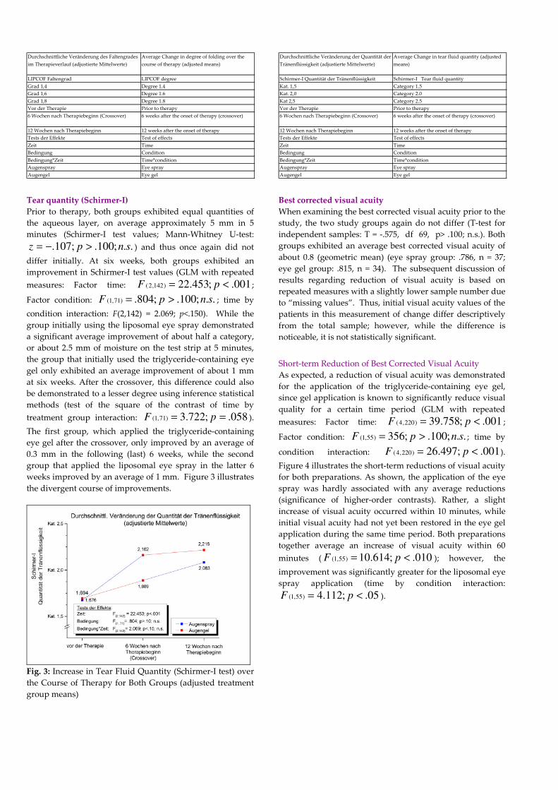
Tear quantity (Schirmer-I)
Prior to therapy, both groups exhibited equal quantities of
the aqueous layer, on average approximately 5 mm in 5
minutes (Schirmer-I test values; Mann-Whitney U-test:
..;100.;107. snpz >−= ) and thus once again did not
differ initially. At six weeks, both groups exhibited an
improvement in Schirmer-I test values (GLM with repeated
measures: Factor time: 001.;453.22)142,2( <= pF ;
Factor condition: ..;100.;804.)71,1( snpF >= ; time by
condition interaction: F(2,142) = 2.069; p<.150). While the
group initially using the liposomal eye spray demonstrated
a significant average improvement of about half a category,
or about 2.5 mm of moisture on the test strip at 5 minutes,
the group that initially used the triglyceride-containing eye
gel only exhibited an average improvement of about 1 mm
at six weeks. After the crossover, this difference could also
be demonstrated to a lesser degree using inference statistical
methods (test of the square of the contrast of time by
treatment group interaction: 058.;722.3)71,1( == pF ).
The first group, which applied the triglyceride-containing
eye gel after the crossover, only improved by an average of
0.3 mm in the following (last) 6 weeks, while the second
group that applied the liposomal eye spray in the latter 6
weeks improved by an average of 1 mm. Figure 3 illustrates
the divergent course of improvements.
Fig. 3: Increase in Tear Fluid Quantity (Schirmer-I test) over
the Course of Therapy for Both Groups (adjusted treatment
group means)
Durchschnittliche Veränderung der Quantität der
Tränenflüssigkeit (adjustierte Mittelwerte)
Average Change in tear fluid quantity (adjusted
means)
Schirmer-I Quantität der Tränenflüssigkeit Schirmer-I Tear fluid quantity
Kat. 1,5 Category 1.5
Kat. 2,0 Category 2.0
Kat 2,5 Category 2.5
Vor der Therapie Prior to therapy
6 Wochen nach Therapiebeginn (Crossover) 6 weeks after the onset of therapy (crossover)
12 Wochen nach Therapiebeginn 12 weeks after the onset of therapy
Tests der Effekte Test of effects
Zeit Time
Bedingung Condition
Bedingung*Zeit Time*condition
Augenspray Eye spray
Augengel Eye gel
Best corrected visual acuity
When examining the best corrected visual acuity prior to the
study, the two study groups again do not differ (T-test for
independent samples: T = -.575, df 69, p> .100; n.s.). Both
groups exhibited an average best corrected visual acuity of
about 0.8 (geometric mean) (eye spray group: .786, n = 37;
eye gel group: .815, n = 34). The subsequent discussion of
results regarding reduction of visual acuity is based on
repeated measures with a slightly lower sample number due
to “missing values”. Thus, initial visual acuity values of the
patients in this measurement of change differ descriptively
from the total sample; however, while the difference is
noticeable, it is not statistically significant.
Short-term Reduction of Best Corrected Visual Acuity
As expected, a reduction of visual acuity was demonstrated
for the application of the triglyceride-containing eye gel,
since gel application is known to significantly reduce visual
quality for a certain time period (GLM with repeated
measures: Factor time: 001.;758.39)220,4( <= pF ;
Factor condition: ..;100.;356)55,1( snpF >= ; time by
condition interaction: 001.;497.26)220,4( <= pF ).
Figure 4 illustrates the short-term reductions of visual acuity
for both preparations. As shown, the application of the eye
spray was hardly associated with any average reductions
(significance of higher-order contrasts). Rather, a slight
increase of visual acuity occurred within 10 minutes, while
initial visual acuity had not yet been restored in the eye gel
application during the same time period. Both preparations
together average an increase of visual acuity within 60
minutes ( 010.;614.10)55,1( <= pF ); however, the
improvement was significantly greater for the liposomal eye
spray application (time by condition interaction:
05.;112.4)55,1( <= pF ).
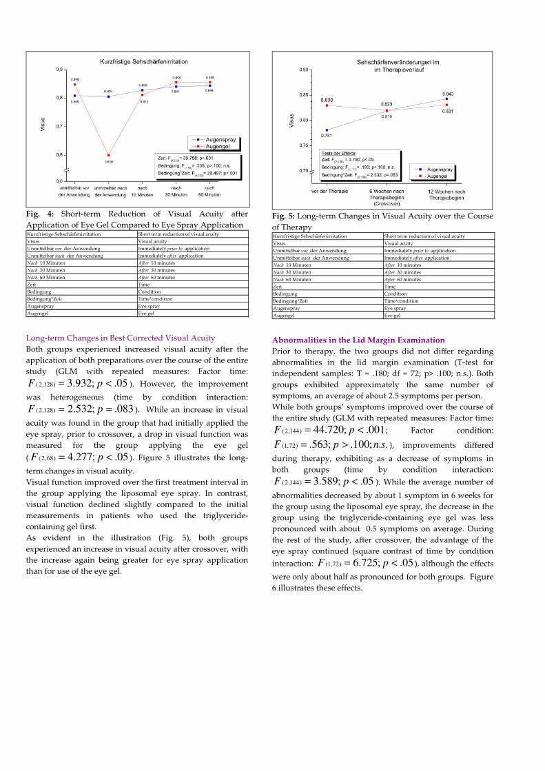
Fig. 4: Short-term Reduction of Visual Acuity after
Application of Eye Gel Compared to Eye Spray Application Kurzfristige Sehschärfenirritation Short term reduction of visual acuity
Visus Visual acuity
Unmittelbar vor der Anwendung Immediately prior to application
Unmittelbar nach der Anwendung Immediately after application
Nach 10 Minuten After 10 minutes
Nach 30 Minuten After 30 minutes
Nach 60 Minuten After 60 minutes
Zeit Time
Bedingung Condition
Bedingung*Zeit Time*condition
Augenspray Eye spray
Augengel Eye gel
Long-term Changes in Best Corrected Visual Acuity
Both groups experienced increased visual acuity after the
application of both preparations over the course of the entire
study (GLM with repeated measures: Factor time:
05.;932.3)128,2( <= pF ). However, the improvement
was heterogeneous (time by condition interaction:
083.;532.2)128,2( == pF ). While an increase in visual
acuity was found in the group that had initially applied the
eye spray, prior to crossover, a drop in visual function was
measured for the group applying the eye gel
( 05.;277.4)68,2( <= pF ). Figure 5 illustrates the long-
term changes in visual acuity.
Visual function improved over the first treatment interval in
the group applying the liposomal eye spray. In contrast,
visual function declined slightly compared to the initial
measurements in patients who used the triglyceride-
containing gel first.
As evident in the illustration (Fig. 5), both groups
experienced an increase in visual acuity after crossover, with
the increase again being greater for eye spray application
than for use of the eye gel.
Fig. 5: Long-term Changes in Visual Acuity over the Course
of Therapy Kurzfristige Sehschärfenirritation Short term reduction of visual acuity
Visus Visual acuity
Unmittelbar vor der Anwendung Immediately prior to application
Unmittelbar nach der Anwendung Immediately after application
Nach 10 Minuten After 10 minutes
Nach 30 Minuten After 30 minutes
Nach 60 Minuten After 60 minutes
Zeit Time
Bedingung Condition
Bedingung*Zeit Time*condition
Augenspray Eye spray
Augengel Eye gel
Abnormalities in the Lid Margin Examination
Prior to therapy, the two groups did not differ regarding
abnormalities in the lid margin examination (T-test for
independent samples: T = .180; df = 72; p> .100; n.s.). Both
groups exhibited approximately the same number of
symptoms, an average of about 2.5 symptoms per person.
While both groups’ symptoms improved over the course of
the entire study (GLM with repeated measures: Factor time:
001.;720.44)144,2( <= pF ; Factor condition:
..;100.;563.)72,1( snpF >= ), improvements differed
during therapy, exhibiting as a decrease of symptoms in
both groups (time by condition interaction:
05.;589.3)144,2( <= pF ). While the average number of
abnormalities decreased by about 1 symptom in 6 weeks for
the group using the liposomal eye spray, the decrease in the
group using the triglyceride-containing eye gel was less
pronounced with about 0.5 symptoms on average. During
the rest of the study, after crossover, the advantage of the
eye spray continued (square contrast of time by condition
interaction: 05.;725.6)72,1( <= pF ), although the effects
were only about half as pronounced for both groups. Figure
6 illustrates these effects.
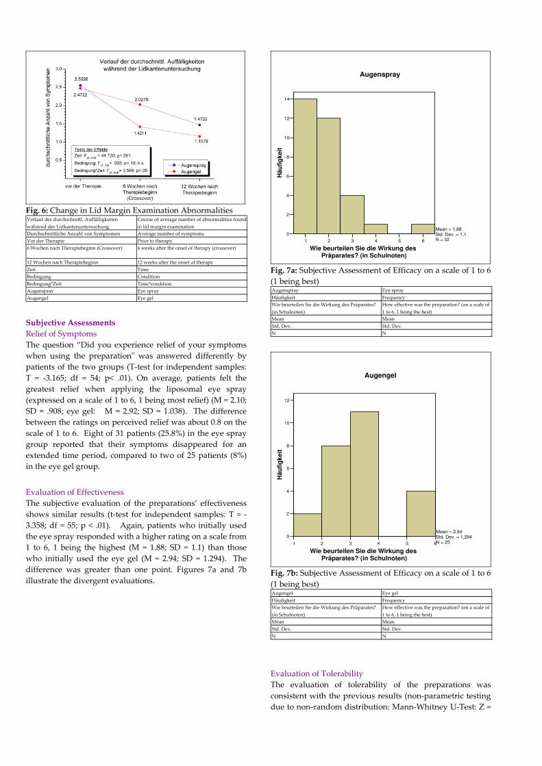
Fig. 6: Change in Lid Margin Examination Abnormalities Verlauf der durchschnittl. Auffälligkeiten
während der Lidkantenuntersuchung
Course of average number of abnormalities found
in lid margin examination
Durchschnittliche Anzahl von Symptomen Average number of symptoms
Vor der Therapie Prior to therapy
6 Wochen nach Therapiebeginn (Crossover) 6 weeks after the onset of therapy (crossover)
12 Wochen nach Therapiebeginn 12 weeks after the onset of therapy
Zeit Time
Bedingung Condition
Bedingung*Zeit Time*condition
Augenspray Eye spray
Augengel Eye gel
Subjective Assessments
Relief of Symptoms
The question “Did you experience relief of your symptoms
when using the preparation" was answered differently by
patients of the two groups (T-test for independent samples:
T = -3.165; df = 54; p< .01). On average, patients felt the
greatest relief when applying the liposomal eye spray
(expressed on a scale of 1 to 6, 1 being most relief) (M = 2.10;
SD = .908; eye gel: M = 2.92; SD = 1.038). The difference
between the ratings on perceived relief was about 0.8 on the
scale of 1 to 6. Eight of 31 patients (25.8%) in the eye spray
group reported that their symptoms disappeared for an
extended time period, compared to two of 25 patients (8%)
in the eye gel group.
Evaluation of Effectiveness
The subjective evaluation of the preparations’ effectiveness
shows similar results (t-test for independent samples: T = -
3.358; df = 55; p < .01). Again, patients who initially used
the eye spray responded with a higher rating on a scale from
1 to 6, 1 being the highest (M = 1.88; SD = 1.1) than those
who initially used the eye gel (M = 2.94; SD = 1.294). The
difference was greater than one point. Figures 7a and 7b
illustrate the divergent evaluations.
1 2 3 4 5 6
Wie beurteilen Sie die Wirkung des Präparates? (in Schulnoten)
0
2
4
6
8
10
12
14
Hä
ufi
gk
eit
Mean = 1,88Std. Dev. = 1,1N = 32
Augenspray
Fig. 7a: Subjective Assessment of Efficacy on a scale of 1 to 6
(1 being best) Augenspray Eye spray
Häufigkeit Frequency
Wie beurteilen Sie die Wirkung des Präparates?
(in Schulnoten)
How effective was the preparation? (on a scale of
1 to 6, 1 being the best)
Mean Mean
Std. Dev. Std. Dev.
N N
1 2 3 4 5 6
Wie beurteilen Sie die Wirkung des Präparates? (in Schulnoten)
0
2
4
6
8
10
12
Hä
ufi
gk
eit
Mean = 2,94Std. Dev. = 1,294N = 25
Augengel
Fig. 7b: Subjective Assessment of Efficacy on a scale of 1 to 6
(1 being best) Augengel Eye gel
Häufigkeit Frequency
Wie beurteilen Sie die Wirkung des Präparates?
(in Schulnoten)
How effective was the preparation? (on a scale of
1 to 6, 1 being the best)
Mean Mean
Std. Dev. Std. Dev.
N N
Evaluation of Tolerability
The evaluation of tolerability of the preparations was
consistent with the previous results (non-parametric testing
due to non-random distribution: Mann-Whitney U-Test: Z =
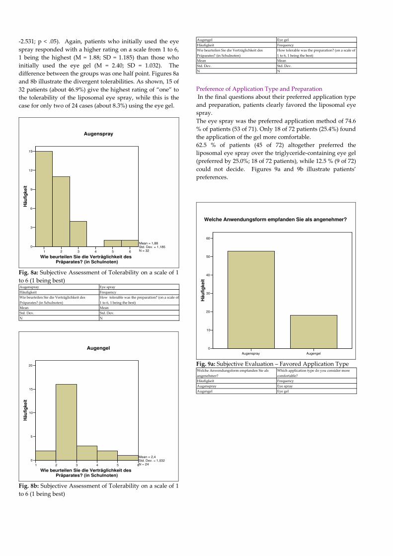
-2.531; p < .05). Again, patients who initially used the eye
spray responded with a higher rating on a scale from 1 to 6,
1 being the highest (M = 1.88; SD = 1.185) than those who
initially used the eye gel (M = 2.40; SD = 1.032). The
difference between the groups was one half point. Figures 8a
and 8b illustrate the divergent tolerabilities. As shown, 15 of
32 patients (about 46.9%) give the highest rating of “one” to
the tolerability of the liposomal eye spray, while this is the
case for only two of 24 cases (about 8.3%) using the eye gel.
1 2 3 4 5 6
Wie beurteilen Sie die Verträglichkeit des Präparates? (in Schulnoten)
0
3
6
9
12
15
Hä
ufi
gk
eit
Mean = 1,88Std. Dev. = 1,185N = 32
Augenspray
Fig. 8a: Subjective Assessment of Tolerability on a scale of 1
to 6 (1 being best) Augenspray Eye spray
Häufigkeit Frequency
Wie beurteilen Sie die Verträglichkeit des
Präparates? (in Schulnoten)
How tolerable was the preparation? (on a scale of
1 to 6, 1 being the best)
Mean Mean
Std. Dev. Std. Dev.
N N
1 2 3 4 5 6
Wie beurteilen Sie die Verträglichkeit des Präparates? (in Schulnoten)
0
5
10
15
20
Hä
ufi
gke
it
Mean = 2,4Std. Dev. = 1,032N = 24
Augengel
Fig. 8b: Subjective Assessment of Tolerability on a scale of 1
to 6 (1 being best)
Augengel Eye gel
Häufigkeit Frequency
Wie beurteilen Sie die Verträglichkeit des
Präparates? (in Schulnoten)
How tolerable was the preparation? (on a scale of
1 to 6, 1 being the best)
Mean Mean
Std. Dev. Std. Dev.
N N
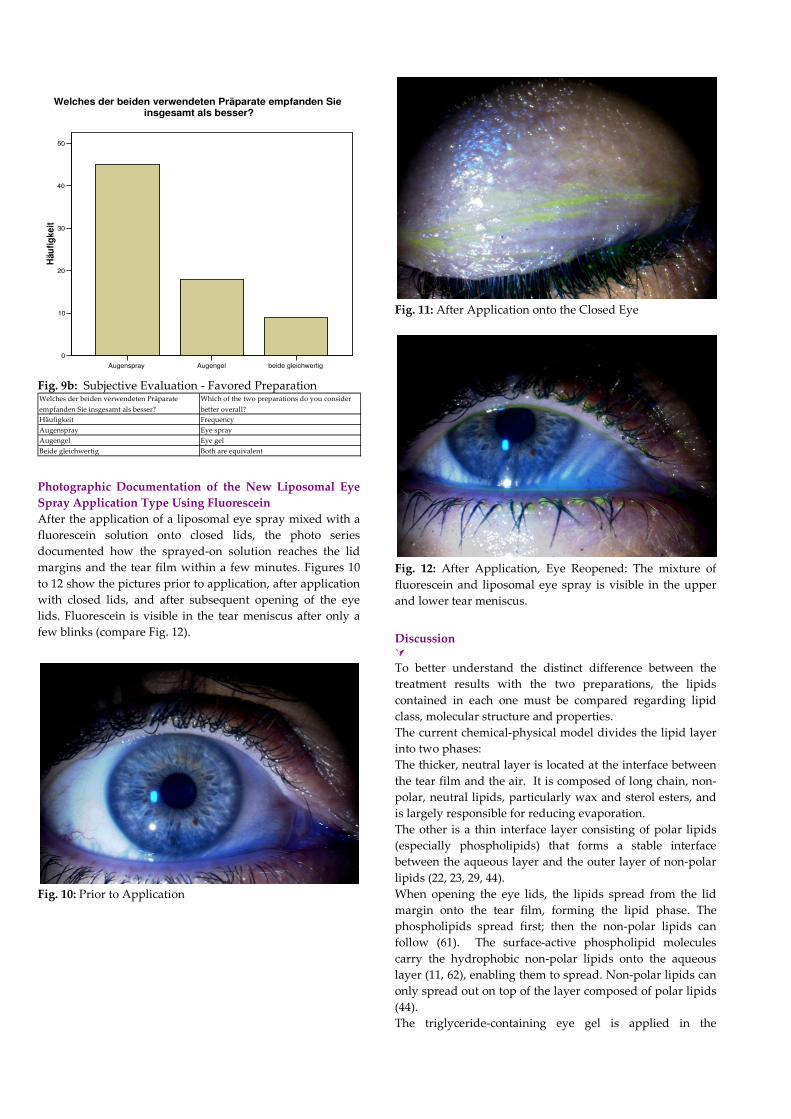
Preference of Application Type and Preparation
In the final questions about their preferred application type
and preparation, patients clearly favored the liposomal eye
spray.
The eye spray was the preferred application method of 74.6
% of patients (53 of 71). Only 18 of 72 patients (25.4%) found
the application of the gel more comfortable.
62.5 % of patients (45 of 72) altogether preferred the
liposomal eye spray over the triglyceride-containing eye gel
(preferred by 25.0%; 18 of 72 patients), while 12.5 % (9 of 72)
could not decide. Figures 9a and 9b illustrate patients’
preferences.
Augenspray Augengel
0
10
20
30
40
50
60
Häu
fig
keit
Welche Anwendungsform empfanden Sie als angenehmer?
Fig. 9a: Subjective Evaluation – Favored Application Type Welche Anwendungsform empfanden Sie als
angenehmer?
Which application type do you consider more
comfortable?
Häufigkeit Frequency
Augenspray Eye spray
Augengel Eye gel
Augenspray Augengel beide gleichwertig
0
10
20
30
40
50
Häu
fig
ke
it
Welches der beiden verwendeten Präparate empfanden Sie insgesamt als besser?
Fig. 9b: Subjective Evaluation - Favored Preparation Welches der beiden verwendeten Präparate
empfanden Sie insgesamt als besser?
Which of the two preparations do you consider
better overall?
Häufigkeit Frequency
Augenspray Eye spray
Augengel Eye gel
Beide gleichwertig Both are equivalent
Photographic Documentation of the New Liposomal Eye
Spray Application Type Using Fluorescein
After the application of a liposomal eye spray mixed with a
fluorescein solution onto closed lids, the photo series
documented how the sprayed-on solution reaches the lid
margins and the tear film within a few minutes. Figures 10
to 12 show the pictures prior to application, after application
with closed lids, and after subsequent opening of the eye
lids. Fluorescein is visible in the tear meniscus after only a
few blinks (compare Fig. 12).
Fig. 10: Prior to Application
Fig. 11: After Application onto the Closed Eye
Fig. 12: After Application, Eye Reopened: The mixture of
fluorescein and liposomal eye spray is visible in the upper
and lower tear meniscus.
Discussion �
To better understand the distinct difference between the
treatment results with the two preparations, the lipids
contained in each one must be compared regarding lipid
class, molecular structure and properties.
The current chemical-physical model divides the lipid layer
into two phases:
The thicker, neutral layer is located at the interface between
the tear film and the air. It is composed of long chain, non-
polar, neutral lipids, particularly wax and sterol esters, and
is largely responsible for reducing evaporation.
The other is a thin interface layer consisting of polar lipids
(especially phospholipids) that forms a stable interface
between the aqueous layer and the outer layer of non-polar
lipids (22, 23, 29, 44).
When opening the eye lids, the lipids spread from the lid
margin onto the tear film, forming the lipid phase. The
phospholipids spread first; then the non-polar lipids can
follow (61). The surface-active phospholipid molecules
carry the hydrophobic non-polar lipids onto the aqueous
layer (11, 62), enabling them to spread. Non-polar lipids can
only spread out on top of the layer composed of polar lipids
(44).
The triglyceride-containing eye gel is applied in the
traditional manner directly onto the eye’s conjunctival sac.
The application interferes with the tear film by significantly
damaging the already disturbed lipid phase, causing its
rupture (24, 68). Eye gels and salves in particular lead to
long-term disturbance of the entire tear film (24). Their
application causes a 7-fold increase in the liquid volume at
the eye surface when compared with the normal volume
(68). Further, it has been shown that the use of eye gels also
increases the evaporation rate (38).
The applied eye gel is based on the thickening agent
carbomer (polyacrylic acid).
Carbomer preparations, both with and without
preservatives, have a toxic effect on corneal cells in vitro,
causing severe damage after more than 30 minutes of
exposure (14).
It has been shown that carbomer gels remain on the cornea
for more than 35 minutes (70), raising concerns about in vivo
corneal damage due to these toxic effects, at least with long-
term use.
The lipid components of the eye gel are non-polar
triglycerides, which make up only 3.7 % of the lipid
secretions of the Meibomian glands (8) and thus are likely to
play a minor role in the stability of the lipid layer. There is
no conceivable triglyceride-deficiency in the lipid secretions
either, particularly not related to dry eye.
Even among non-polar lipids, triglycerides play a
subordinate role, whereas sterol and wax esters together
total almost 60% of Meibomian secretions (8).
While the use of lipid-containing artificial tear products has
been shown to thicken the lipid layer (30), one can not
necessarily deduce that patients are free of symptoms and
that treatment was successful (15).
In contrast to the gel, the liposomal eye spray is simply
applied to the closed eye. It has been established for a long
time that lipids applied to the outer skin of the lid near the
lid margins reach the lid margins and thus the tear film
during blinking (37, 48).
Intraocular penetration has even been demonstrated for
aqueous eye drops applied onto the inner canthus (1, 35, 58).
Although the application onto the closed eye lids may
transfer resident bacteria onto the eye surface, there is no
risk of infection since the bacterial florae on the eye lids and
on the eye surface are necessarily identical (34).
Endogenous lipid secretions of the Meibomian glands are
secreted onto the lid margin, forming a reservoir (8). Chew
et al. calculated that the lipid volume in this reservoir is
probably 40 times greater than the amount that actually
spreads onto the tear film to form the lipid layer (12).
In the closed eye, the lipid layer is compressed between the
lid margins. The lipids that were part of the tear film mix
once again with the lipid reservoir on the lid margin. During
the opening of the lids, some of the lipids from this reservoir
spread onto the tear film, forming a lipid layer (9).
The sprayed-on phospholipid liposomes reach the lid
margins via the same effect and mix with the endogenous
lipids located there.
The lipid phase is stabilized by the added phospholipids.
Polar lipids comprise 15% of the total Meibomian secretions
and play a key role in the stability of the lipid layer due to
their surface-active properties described earlier (29, 44).
Phospholipids make up 70% of the polar lipids (57).
The liposomes (vesicles made of a phospholipid bilayer)
contained in the eye spray are made from highly purified
soy lecithin, consisting of 94% phosphatidylcholine and a
small amount of other phospholipids (32) that have also
been shown to be present in the tear film (22, 23).
At 38%, phosphatidylcholine makes up the largest amount
of phospholipids in the tear film (57).
Lipid phase instability is not caused by a quantitative
deficiency of total lipid secretions (43) but by their
inadequate composition.
This is also supported by the fact that effective evaporation
protection is primarily determined by the stability, not the
thickness, of the lipid layer. (13, 41).
Lipid phase disturbances and the resulting
hyperevaporative dry eye are traced back to a phospholipid
deficiency (55, 56), particularly in chronic blepharitis (45, 56,
59).
Observations of an increased surface tension in dry eye
patients support an incorrect composition of lipid secretions
as well (47, 71).
Surface-active polar lipids are very important for the
maintenance of tear film stability:
The spreading of the tear film is thought to take place in a
two-step process. In the first step, the (upward) movement
of the upper lid draws a tear film layer over the cornea via
capillary action. In the second step, an upward flow of the
outer lipid layer increases the thickness of the tear film
significantly, because the spreading of the lipid layer draws
the tear fluid into the tear film (10).
The second step is based on a so-called boundary-layer
phenomenon:
Langmuir has demonstrated that the behavior of fluid films
of a thickness less than 100 micrometers is completely
controlled by surface or boundary forces (31), and is not
affected by gravity (27, 53).
The spreading of the lipid layer reduces the surface tension
of the tear film, causing a flow of tear fluid from the upper
and lower tear meniscus onto the tear film at the eye surface
(known as Marangoni effect or Marangoni flow) (2, 36).
The development of a surface film – such as the lipid phase
of the tear film – significantly alters the surface properties of
the liquid substrate (the tear fluid), such as surface tension,
viscosity, and elasticity, as well as light reflection.
The film formation of the lipids largely depends on the
chemical makeup of the molecules. Lipid molecules forming
a monolayer – a monomolecular film – must have a bipolar
molecular structure, consisting of a hydrophilic polar head
and a hydrophobic (fatty acid) chain.
During spreading onto the aqueous phase, hydrogen
bonding occurs between the polar hydrophilic heads and
the water molecules of the aqueous phase, “anchoring” the
monolayer on the aqueous phase (16). Phospholipids are
amphiphilic, their fatty acid chains interacting with the non-
polar part of the lipid phase, while their polar heads interact
with the molecules of the aqueous phase.
It is widely known that neutral lipids and water do not mix;
therefore, neutral lipids alone could only partially cover the
aqueous layer of the tear film, which would result in the
formation of a lens (16) similar to the fat droplets in a soup.
The formation of a monolayer at the surface of the aqueous
layer results in decreased surface tension (16). The biggest
decrease in surface tension is caused by the phospholipids,
especially by phosphatidylcholine (47).
However, in dry eye, a significantly increased surface
tension was demonstrated (65).
Therefore, these results support a phospholipid deficit as a
cause of dry eye.
It was shown that phosphatidylcholine results in greater tear
film stability and significantly increases tear break-up time
(49).
The significant improvement in break-up time during use of
the liposomal eye spray also supports the results of the
previous studies (3, 32, 52, 60).
The slight improvement of tear film break-up time observed
when using the eye gel is consistent with previous reports
(5).
Tear film stability is essential for visual function. The lipid
layer forms the outer layer of the tear film and is therefore
the first structure of the visual system that is hit by entering
light (64).
The lipid layer provides a smooth tear film surface. The
results of this study indicate that the polar lipids
(phospholipids) play a key role in the optical quality of the
tear film as well.
In agreement with other reports, the use of the eye gel in this
study resulted in a significant disturbance of visual acuity
(14, 17). Even 10 minutes after application, the original value
had not yet been restored. On the other hand, use of the
liposomal eye spray did not decrease, but slightly improved
visual acuity, also in agreement with prior results (32).
The significant improvement of Schirmer-I test values when
using the liposomal eye spray confirms the results of
previous studies (3, 32, 52), while the slight improvements
during eye gel use correspond to the observations of other
studies as well (7).
When evaluating the subjective assessments, it must be
noted that besides adding triglycerdies, the eye gel also
initially moistens the eye surface, which is perceived as a
relief. However, its use significantly disturbs the natural
lipid layer (24) and increases the tear fluid evaporation rate
(38), potentially resulting in a so-called rebound-effect (68).
Conclusion �
The liposomal eye spray shows statistically significant
clinical advantages compared to the triglyceride-containing
eye gel. The patients’ subjective direct comparisons of the
two preparations in the crossover study are particularly
enlightening and demonstrate a clear preference for the
liposomal eye spray regarding its application type onto the
closed eye as well as for its effectiveness and tolerability.
Direct comparison reveals that the phospholipid-liposome
therapy is altogether advantageous and distinctly superior
to conventional standard therapy.^
References 1 Alster Y, Herlin L, Lazar M, Loewenstein A.
Intraocular penetration of vancomycin eye drops
after application to the medial canthus with closed
lids. Br J Ophthalmol 2000; 84: 300-302 2 Berke A. Blinking frequency and the thickness of
the lipid layer.In: Sullivan D et al. (Hrsg.). Lacrimal
Gland, Tear Film, and Dry Eye Syndromes 3.
Kluwer Academic/ Plenum Publishers, 2002: 513-
516 3 Brandl H. Therapie des Trockenen Auges bei
Störungen der Lipidphase. Wehrpharmazie und
Wehrmedizin 2005; 29 (2): 55-56 4 Brewitt H, Höh H, Kaercher T, Stolze HH. Das
„Trockene Auge“ – Diagnostik und Therapie. Z
prakt Augenheilkd 1997; 18: 371-379 5 Brewitt H, Joost P. Klinische Studie zur
Wirksamkeit eines nicht konservierten
Tränenersatzmittels. Klin Monatsbl Augenheilkd
1991; 199: 160-164 6 Brewitt H, Rüfer F. Das trockene Auge. Klin
Monatsbl Augenheilkd 2004; 221: R51-R70 7 Bron AJ, Daubas P, Siou-Mermet R, Trinquand C.
Comparison of the efficacy and safety of two eye
gels in the treatment of dry eyes: Lacrinorm and
Viscotears. Eye 1998; 12
(Pt 5): 839-847 8 Bron AJ, Tiffany JM. The meibomian glands and
tear film lipids. Structure, function, and control. In:
Sullivan D et al (Hrsg). Lacrimal Gland, Tear Film,
and Dry Eye Syndromes 2. New York: Plenum
Press, 1998: 281-295 9 Bron AJ, Tiffany JM, Gouveia SM, Yokoi N, Voon
LW. Functional aspects of the tear film lipid layer.
Exp Eye Res. 2004; 78: 347-360 10 Brown SI, Dervichian DG. Hydrodynamics of
blinking. In vitro study of tue Interaktion of the
superficial oily layer and the tears. Arch
Ophthalmol. 1969; 82: 541-547 11 Brown SI, Dervichian DG. The oils of the
meibomian glands. Physical and surface
characteristics. Arch Ophthalmol. 1969; 82: 537-540 12 Chew CKS, Jansweijer C, Tiffany JM, Dikstein S,
Bron AJ. An instrument for quantifying meibomian
lipid on the lid margin: the Meibometer. Curr Eye
Res. 1993; 12: 247-254 13 Craig JP, Tomlinson A. Importance of the lipid
layer in human tear stability and evaporation.
Optom Vis Sci 1997; 74: 8-13 14 Diebold Y, Herreras JM, Callejo S, Argüeso P,
Calonge M. Carbomer- Versus Cellulose-Based
Artificial-Tear Formulations: Morphologic and
Toxicologic Effects on a Corneal Cell Line. Cornea
1998; 17: 433-440 15 Di Pascuale MA, Goto E, Tseng SCG. Sequential
Changes of Lipid Tear Film after tue Instillation of
a Single Drop of a New Emulsion Eye Drop in Dry
Eye Patients. Ophthalmology 2004; 111: 783-791 16 Dörfler HD. Grenzflächen und kolloid-disperse
Systeme. Springer Verlag, Berlin, Heidelberg 2002 17 Dudinski O, Finnin BC, Reed BL. Acceptability of
thickened eye drops to human subjects. Curr Ther
Res Clin Exp 1983; 33: 322- 327 18 Gilbard JP, Rossi SR, Heyda KG. Tear film and
ocular surface changes after closure of the
meibomian gland orifices in the rabbit.
Ophthalmology 1989; 96: 1180-1186 19 Goto E, Endo K, Suzuki A, Fujikura Y, Matsumoto
Y, Tsubota K. Tear evaporation dynamics in normal
subjects and subjects with obstructive meibomian
gland dysfunction. Invest Ophthalmol Vis Sci. 2003;
44: 533-539 20 Göbbels M, Spitznas M. Corneal epithelial
permeability of dry eyes before and after treatment
with artificial tears. Ophthalmology 1992; 99: 873-
878 21 Göbbels M, Spitznas M. Influence of artificial tears
on corneal epithelium in dry-eye syndrome.
Graefes Arch Clin Exp Ophthalmol 1989; 227: 139-
141 22 Greiner JV, Glonek T, Korb DR, Leahy CD.
Meibomian gland phospholipids. Curr Eye Res.
1996; 15: 371-375 23 Greiner JV, Glonek T, Korb DR, Booth R, Leahy CD.
Phospholipids in meibomian gland secretion.
Ophthalmic Res. 1996; 28: 44-49 24 Guillon JP. Abnormal Lipid Layer. Observation,
Differential Diagnosis, and Classification. In:
Sullivan D et al (Hrsg). Lacrimal Gland, Tear Film,
and Dry Eye Syndromes 2. New York: Plenum
Press, 1998: 309-313 25 Heiligenhaus A, Koch JM, Kruse FE, Schwarz C,
Waubke TN. Diagnostik und Differenzierung von
Benetzungsstörungen. Ophthalmologe 1995; 92: 6-
11 26 Heiligenhaus A, Koch JM, Kemper D, Kruse FE,
Waubke TN. Therapie von Benetzungsstörungen.
Klin Monatsbl Augenheilkd. 1994; 204: 162-168 27 Holly FJ. The preocular tear film; a small but highly
complex part of the eye.
Arch Soc Esp Oftalmol. 2005; 80: 65-68 28 Kaercher T, Honig D, Barth W. How the most
common preservative affects the Meibomian lipid
layer. Orbit 1999; 18: 89-97 29 Korb DR, Greiner JV, Glonek T. The effects of
anionic and zwitterionic phospholipids on the tear
film lipid layer. Adv Exp Med Biol. 2002;506(Pt
A):495-499 30 Korb DR, Scaffidi RC, Greiner JV, Kenyon KR,
Herman JP, Blackie CA, Glonek T, Case CL,
Finnemore VM, Douglass T. The effect of two novel
lubricant eye drops on tear film lipid layer
thickness in subjects with dry eye symptoms.
Optom Vis Sci. 2005; 82: 594-601 31 Langmuir I. Oil leises on water and tue nature of
monomolekular extended films. J Chem Phys 1933;
1: 756-776 32 Lee S, Dausch S, Maierhofer G, Dausch D. Ein
neues Therapiekonzept zur Behandlung des
Trockenen Auges – die Verwendung von
Phospholipid-Liposomen. Klin Monatsbl
Augenheilkd 2004; 221: 825-836 33 Lemp MA. Report of the National Eye
Institute/Industry workshop on Clinical Trials in
Dry Eyes. CLAO J. 1995; 21: 221-232 34 Locatcher-Khorazo D, Seegal BC. The bacterial flora
of the healthy eye. In: Locatcher-Khorazo D, Seegal
BC, eds. Microbiology of the eye. St. Louis: CV
Mosby, 1972: 13-23 35 Loewenstein A, Bolocinic S, Goldstein M, Lazar M.
Application of eye drops to the medial canthus.
Graefes Arch Clin Exp Ophthalmol. 1994; 232: 680-
682 36 Lozato PA, Pisella PJ, Baudouin C. Phase lipidique
du film lacrymal : physiologie et pathologie. J Fr
Ophtalmol 2001; 24: 643-658 37 MacKeen D, Roth H, Doane M. Ocular drug
delivery by the lid (lower lid delivery). Invest
Ophthalmol Vis Sci 1996; 37: 77 38 Mathers W. Evaporation from the ocular surface.
Exp Eye Res. 2004; 78: 389-394 39 Mathers WD. Ocular evaporation in meibomian
gland dysfunction and dry eye. Ophthalmology
1993; 100: 347-351 40 Mathers WD, Daley TE. Tear flow and evaporation
in patients with and without dry eye.
Ophthalmology 1996; 103: 664-669 41 Mathers WD, Lane JA. Meibominan gland lipids,
evaporation, and tear film stability. Adv Exp Med
Biol. 1998; 438: 349-360 42 Mathers WD, Shields WJ, Sachdev MS, Petroll WM,
Jester JV. Meibomian gland morphology and tear
osmolarity: changes with Accutane therapy.
Cornea. 1991; 10: 286-290 43 McCulley JP, Sciallis GF. Meibomian
keratoconjunctivitis. Am J Ophthalmol. 1977; 84:
788-793 44 McCulley JP, Shine W. A compositional based
model for the tear film lipid layer.
Trans Am Ophthalmol Soc. 1997; 95: 79-88 45 McCulley JP, Shine WE. Eyelid disorders: the
meibomian gland, blepharitis, and the contact
lenses. Eye Contact Lens. 2003; 29 (1 Suppl): 93-95 46 McCulley JP, Shine WE. The lipid layer: the outer
surface of the ocular surface tear film. Biosci Rep.
2001; 21: 407-418 47 Nagyova B, Tiffany JM. Components responsible
for the surface tension of human tears. Curr Eye
Res. 1999; 19: 4-11 48 Norn MS. Natural fat in external eye. Vital-stained
by Sudan III powder. Acta Ophthalmol (Copenh)
1980; 58: 331-336 49 Peters K, Millar TJ. The role of different
phospholipids on tear break-up time using a model
eye. Curr Eye Res. 2002; 25: 55-60 50 Rolando M, Refojo MF, Kenyon KR. Increased tear
evaporation in eyes with keratoconjunctivitis sicca.
Arch Ophthalmol. 1983; 101: 557-558 51 Rolando M, Refojo MF, Kenyon KR. Tear water
evaporation and eye surface diseases.
Ophthalmologica 1985; 190: 147-149 52 Roth HW, Koulen-Reitz G, Brunk G. Zur
Langzeittherapie des Trockenen Auges mit einem
Liposomenspray. Augenspiegel 2002; 48 (9): 54-58 53 Sharma A, Tiwari S, Khanna R, Tiffany JM.
Hydrodynamics of meniscus-induced thinning of
the tear film. In: Sullivan D et al (Hrsg). Lacrimal
Gland, Tear Film, and Dry Eye Syndromes 2. New
York: Plenum Press, 1998: 425-431 54 Shimazaki J, Sakata M, Tsubota K. Ocular surface
changes and discomfort in patients with meibomian
gland dysfunction. Arch Ophthalmol. 1995; 113:
1266-1270 55 Shine WE, McCulley JP. Keratoconjunctivitis sicca
associated with meibomian secretion polar lipid
abnormality. Arch Ophthalmol 1998; 116: 849-852 56 Shine WE, McCulley JP. Meibomianitis: polar lipid
abnormalities. Cornea 2004; 23: 781-783 57 Shine WE, McCulley JP. Polar lipids in human
meibomian gland secretions. Curr Eye Res. 2003; 26:
89-94 58 Smith SE. Eyedrop instillation for reluctant
children. Br J Ophthalmol. 1991; 75: 480-481 59 Song CH, Choi JS, Kim DK, Kim JC. Enhanced
secretory group II PLA2 activity in the tears of
chronic blepharitis patients. Invest Ophthalmol Vis
Sci. 1999; 40: 2744-2748 60 Strempel I, Roth HW. Zur Therapie des Trockenen
Auges mit einem Spray. Augenspiegel 1999; 45 (11):
16-21 61 Tiffany JM. Composition and biophysical
properties of the tear film: knowledge and
uncertainty. Adv Exp Med Biol. 1994; 350: 231-238 62 Tiffany JM. Lipid films in water conservation of
biological systems. Cell Biochem Funct. 1995;
13: 177-180 63 Tiffany JM. Physiological functions of the
meibomian glands. Prog. Retinal Res. 1995; 14: 47-
74 64 Tiffany JM. The Lipid Secretion of the Meibomian
Glands. Adv Lipid Res. 1987; 22: 1- 65 Tiffany JM, Winter N, Bliss G. Tear film stability
and tear surface tension. Curr Eye Res. 1989; 8: 507-
515 66 Toda I, Shinozaki N, Tsubota K. Hydroxypropyl
methylcellulose for the treatment of severe dry eye
associated with Sjogren's syndrome. Cornea 1996;
15: 120-128 67 Tomlinson A, Trees GR. Effect of preservatives in
artificial tear solutions on tear film evaporation.
Ophthalmic Physiol Opt. 1991; 11: 48-52 68 Trees GR, Tomlinson A. Effect of artificial tear
solutions and saline on tear film evaporation.
Optom Vis Sci. 1990 ; 67 : 886-890 69 Tsubota K, Monden Y, Yagi Y, Goto E, Shimmura S.
New treatment of dry eye: the effect of calcium
ointment through eyelid skin delivery. Br J
Ophthalmol. 1999; 83: 767-770 70 Wilson CG, Zhu YP, Frier M, Rao LS, Gilchrist P,
Perkins AC. Ocular contact time of a carbomer gel
(GelTears) in humans. Br J Ophthalmol 1998; 82:
1131-1134 71 Zhao J, Manthorpe R, Wollmer P. Surface activity
of tear fluid in patients with primary Sjogren's
syndrome. Clin Physiol Funct Imaging. 2002; 22:
24-27