Comparative proteomic analysis ... - BMC Plant Biology
13
RESEARCH ARTICLE Open Access Comparative proteomic analysis provides novel insights into the regulation mechanism underlying papaya (Carica papaya L.) exocarp during fruit ripening process Bian Jiang 1† , Siyan Ou 2† , Ling Xu 1† , Wanyi Mai 1† , Meijun Ye 1 , Haiping Gu 1 , Tao Zhang 1 , Changchun Yuan 1 , Chenjia Shen 3 , Jinxiang Wang 2* and Kaidong Liu 1* Abstract Background: Papaya (Carica papaya L.) is a popular climacteric fruit, undergoing various physico-chemical changes during ripening. Although papaya is widely cultivated and consumed, few studies on the changes in metabolism during its ripening process at the proteasome level have been performed. Using a newly developed TMT-LCMS analysis, proteomes of papaya fruit at different ripening stages were investigated. Results: In total, 3220 proteins were identified, of which 2818 proteins were quantified. The differential accumulated proteins (DAPs) exhibited various biological functions and diverse subcellular localizations. The KEGG enrichment analysis showed that various metabolic pathways were significantly altered, particularly in flavonoid and fatty acid metabolisms. The up-regulation of several flavonoid biosynthesis-related proteins may provide more raw materials for pigment biosynthesis, accelerating the color variation of papaya fruit. Variations in the fatty acid metabolism- and cell wall degradation-related proteins were investigated during the ripening process. Furthermore, the contents of several important fatty acids were determined, and increased unsaturated fatty acids may be associated with papaya fruit volatile formation. Conclusions: Our data may give an intrinsic explanation of the variations in metabolism during the ripening process of papaya fruit. Keywords: Differential expressed protein, Fatty acid metabolism, Fruit, Hormones, Papaya, Ripening Background Fruit is a mature ovary including carpel tissues, and sev- eral fleshy fruits are important components of human and animal diets [1]. Fruit ripening is a complex process defined by dramatic metabolisms and textural transformations, color changes, softening, nutritional in- gredient accumulation and flavor compound production [1, 2]. Although softening increases the nutritional and flavor properties of fruit, it also leads to biomass loss and postharvest deterioration [3]. Fruit softening is a deep-studied process that in- volves cell wall degradation [4]. The cell walls of fruit are composed of pectins, poly-galacturonans, cellu- loses and hemicelluloses, and degradation of these components is the major cause of tissue softening © The Author(s). 2019 Open Access This article is distributed under the terms of the Creative Commons Attribution 4.0 International License (http://creativecommons.org/licenses/by/4.0/), which permits unrestricted use, distribution, and reproduction in any medium, provided you give appropriate credit to the original author(s) and the source, provide a link to the Creative Commons license, and indicate if changes were made. The Creative Commons Public Domain Dedication waiver (http://creativecommons.org/publicdomain/zero/1.0/) applies to the data made available in this article, unless otherwise stated. * Correspondence: [email protected]; [email protected] † Bian Jiang, Siyan Ou, Ling Xu and Wanyi Mai are contributed equally to this work. 2 Root Biology Center, College of Natural Resources and Environment, South China Agricultural University, Guangzhou 510642, China 1 Life Science and Technology School, Lingnan Normal University, Zhanjiang 524048, China Full list of author information is available at the end of the article Jiang et al. BMC Plant Biology (2019) 19:238 https://doi.org/10.1186/s12870-019-1845-4
Transcript of Comparative proteomic analysis ... - BMC Plant Biology
RESEARCH ARTICLE Open Access
Comparative proteomic analysis providesnovel insights into the regulationmechanism underlying papaya (Caricapapaya L.) exocarp during fruit ripeningprocessBian Jiang1†, Siyan Ou2†, Ling Xu1†, Wanyi Mai1†, Meijun Ye1, Haiping Gu1, Tao Zhang1, Changchun Yuan1,Chenjia Shen3, Jinxiang Wang2* and Kaidong Liu1*
Abstract
Background: Papaya (Carica papaya L.) is a popular climacteric fruit, undergoing various physico-chemicalchanges during ripening. Although papaya is widely cultivated and consumed, few studies on the changesin metabolism during its ripening process at the proteasome level have been performed. Using a newlydeveloped TMT-LCMS analysis, proteomes of papaya fruit at different ripening stages were investigated.
Results: In total, 3220 proteins were identified, of which 2818 proteins were quantified. The differentialaccumulated proteins (DAPs) exhibited various biological functions and diverse subcellular localizations. TheKEGG enrichment analysis showed that various metabolic pathways were significantly altered, particularly inflavonoid and fatty acid metabolisms. The up-regulation of several flavonoid biosynthesis-related proteins mayprovide more raw materials for pigment biosynthesis, accelerating the color variation of papaya fruit.Variations in the fatty acid metabolism- and cell wall degradation-related proteins were investigated duringthe ripening process. Furthermore, the contents of several important fatty acids were determined, andincreased unsaturated fatty acids may be associated with papaya fruit volatile formation.
Conclusions: Our data may give an intrinsic explanation of the variations in metabolism during the ripeningprocess of papaya fruit.
Keywords: Differential expressed protein, Fatty acid metabolism, Fruit, Hormones, Papaya, Ripening
BackgroundFruit is a mature ovary including carpel tissues, and sev-eral fleshy fruits are important components of humanand animal diets [1]. Fruit ripening is a complex processdefined by dramatic metabolisms and textural
transformations, color changes, softening, nutritional in-gredient accumulation and flavor compound production[1, 2]. Although softening increases the nutritional andflavor properties of fruit, it also leads to biomass lossand postharvest deterioration [3].Fruit softening is a deep-studied process that in-
volves cell wall degradation [4]. The cell walls of fruitare composed of pectins, poly-galacturonans, cellu-loses and hemicelluloses, and degradation of thesecomponents is the major cause of tissue softening
© The Author(s). 2019 Open Access This article is distributed under the terms of the Creative Commons Attribution 4.0International License (http://creativecommons.org/licenses/by/4.0/), which permits unrestricted use, distribution, andreproduction in any medium, provided you give appropriate credit to the original author(s) and the source, provide a link tothe Creative Commons license, and indicate if changes were made. The Creative Commons Public Domain Dedication waiver(http://creativecommons.org/publicdomain/zero/1.0/) applies to the data made available in this article, unless otherwise stated.
* Correspondence: [email protected]; [email protected]†Bian Jiang, Siyan Ou, Ling Xu and Wanyi Mai are contributed equally to thiswork.2Root Biology Center, College of Natural Resources and Environment, SouthChina Agricultural University, Guangzhou 510642, China1Life Science and Technology School, Lingnan Normal University, Zhanjiang524048, ChinaFull list of author information is available at the end of the article
Jiang et al. BMC Plant Biology (2019) 19:238 https://doi.org/10.1186/s12870-019-1845-4
during ripening [5]. The pectin matrix, composed ofboth linear and branched polysaccharides, mainly ac-cumulates in the primary cell wall. Several specificenzymes, such as pectate lyase, polygalacturonase andpectin methylesterase, show essential roles in the hy-drolysis of polysaccharide chains of the pectin matrix[5, 6].For climacteric fruits, the softening phenomenon is
also controlled by a complex series of non-enzymaticevents [7, 8]. During fruit ripening, abundant flavorand nutritional metabolites significantly change,including sugars, organic acids, lipids and aminoacids, leading to modification of fruit texture [9]. Forinstance, sugars, the major flavor and aroma compo-nents, increase rapidly apricot fruit during develop-ment and ripening process [10]. In starch-rich fruit,starch and organic acids are converted to sugar by ametabolic degradation pathway [11]. Additionally,lipid components of fruits are assumed to enhancethe formation of aromas and flavors in most climac-teric fruits [12]. The variations in the composition offatty acids during the fruit ripening process have beenrevealed in different climacteric fruits [13]. For ex-ample, most fatty acids increased during the ripeningof mango fruit [14].Papaya (Carica papaya L.), a climacteric fruit widely
planted and consumed, displays quick softening and ashort shelf-life [15, 16]. Once the climacteric fruit ismature, its soft texture and susceptibility to patho-genic fungi make the storage life short [17]. To over-come the commercial trading problem and prolongthe shelf life, many studies on papaya have been car-ried out [18]. In papaya fruit, the physico-chemicalchanges during ripening are affected by the expres-sion of ripening-related genes. A previous transcrip-tome identified 414 ripening-related genes, includingthose of MADS-box, NAC and ERF families, provid-ing molecular information on papaya [19]. Severalyears ago, a fruit-specific expressed subtilase gene wasidentified in papaya by rapid amplification of cDNAends and PCR primer walking techniques [20]. Appli-cation of 1-MCP (1-methylcyclopropene) can blockethylene signaling and so improve the shelf life [21].Expression analysis of ethylene signaling pathway-related genes showed a relationship between chillinginjury and ethylene signaling in papaya [16]. Physio-logical degradation of pectin in papaya cell walls hasalso been studied. For example, up-regulated expres-sion levels of genes for PG, endoxylanase and β-galactosidase (β-GAL) were positively correlated withpostharvest papaya fruit ripening [22, 23]. However,molecular and enzymatic mechanisms underlying thesoftening of papaya fruit during ripening remainlargely unknown.
Many studies on the physiological and genomic var-iations during the ripening process of papaya fruithave been done [19, 23, 24]. To date, several prote-omic data sets of papaya have been published. Usingthe 2-DE approach, differential accumulated proteinsresponsive to 1-MCP treatment during ripening havebeen identified [25]. Another comparative proteomicanalysis showed that certain proteins, such as enolase,esterase and ADH3, show an essential role in matur-ation of somatic papaya embryo cells [26]. Recently, adifferential proteome of virus-infected pre-floweringC. papaya vs. uninfected plants was published [27].In our study, we employed an integration of the basicHPLC fractionation and LC–MS/MS approach toidentify a total of 2818 differential accumulated pro-teins (DAPs) of papaya fruit during ripening. Our re-sults offer important information about how toexpend the shelf life of postharvest papaya fruit.
MethodsPlant materials and samplingTwo-year-old papaya trees of cultivar ‘Sunrise’ wereplanted in a 4 m × 4m arrangement. The seedlings ofpapaya tree were purchased by the Yixin horticulturecompany (Zhanjiang, China), who provided permissionto use the seedlings for our scientific research. The ex-perimental station was at Lingnan Normal Universitycampus in Zhanjiang, China. Plant experiment was per-formed in the Life Science and Technology School, Ling-nan Normal University, according to a plant protocolapproved by the Research Ethics Committee of LingnanNormal University. All papaya trees were fertilized twicea week with a standard nutrient solution. Two fruitgroups at similar color break stage (5% ≤ peel color≤10% yellow) were harvested. The fruit coloring index
was defined and calculated as follows: coloring index
¼ P ðcoloring grade�number of fruitÞtotal number of fruit :
There are three papaya in one sample, and three sam-ples for one group. The selected fruit was cleaned withdeionized H2O, and then dipped into 0.2% of hypochlor-ide solution for 15 min to eliminate potential microbesin growth chamber of our lab. The conditions of thechamber were set as: temperature of 26 ± 1 °C, a light/dark cycle of 16/8 h and humidity of 65–70%.
Physiological parameter measurementsMeasurements of the physiological parameters of pa-paya fruit were performed according to the previousstudy [15]. Three biological replicate were used.Briefly, the firmness of fruit was determined using aGY-J mini fruit tester (Top Instrument Co Ltd.,Shanghai, China). For total soluble solid (TSS) meas-urement, 5.0 g of fruit pulp from three independent
Jiang et al. BMC Plant Biology (2019) 19:238 Page 2 of 13

fruit was crushed manually with a mortar and pestleto extract the juice. The TSS (%) was measures usinga J1-3A refractometer (Guangdong Scientific Instru-ments, Guangzhou, China). The same juice was ti-trated against 0.1 N NaOH until a faint pink colorwas obtained at the end point and titratable aciditywas expressed as percentage citric acid. For respir-ation rate determination, three independent papayafruit from each group were weighed and sealed in 2 Lcontainers at 25 °C. The concentration of CO2 in eachcontainer was measured using an infrared Li-6262CO2/H2O gas analyzer (LI-COR, USA).
Observation by light microscopy and scanning electronmicroscopyFragments (0.5 cm long) of surface tissues were collectedfrom whole fruit at different ripening stages. Sampleswere fixed in formalin acetic acid (FAA)50 for 48 h andtransferred into 70% ethanol. The tissue samples weredehydrated in several varied concentrations of ethanol,and then embedded in wax. Next, fruit samples weresliced into a number of 12-μm thick sections using aRM2255 rotary microtome (Leica, Wetzlar, Germany)and spreaded on glass slides. The wax sections wereeliminated in xylol:ethanol (1:1, v/v) solution for 5 minand rehydrated in several decreasing concentrations ofethanol, including 100, 95, 85 and 70%, for 1 min eachconcentration. The sample slides were stained in 3% saf-ranin for 24 h and washed with a series of increasingethanol concentrations of 70, 85 and 95%, for 5 s eachconcentration. Next, the sample slides were dipped into1% fast green dye solution for 3 s each, treated twicewith 100% of ethanol for 5 s each, and transferred intoxylol:ethanol (1:1, v/v) solution for 5 min. All slides weremounted with synthetic resin and observed under aLSM510 laser scanning system (Carl Zeiss, Wetzlar,Germany).Samples for scanning electron microscopy were pre-
pared as above. Sample segments were gently dippedinto xylol at 45 °C for 4 h to clean the wax. The sam-ples were treated with xylol:ethanol (1:1, v/v) solutionand 100% ethanol for 1 h and then put in tert-butylalcohol. Finally, dried segments were metalized withgold for scanning electron microscope observation.
Protein extractionProtein extraction was performed according to the previ-ous study [28]. Approximately 0.5-g exocarp sampleswere kept in N2 and pulverized. The fine powder wassuspended with pre-cooled lysis buffer, containing 8Murea, 2 mM EDTA, 10 mM DTT and 1% Protease Inhibi-tor Cocktail, on ice overnight. The precipitate was re-moved at 15,000 g by centrifugation at 4 °C for 15 min.The final supernatants were redissolved in buffer
containing 8M urea, and 100 mM triethylammonium bi-carbonate (TEAB) and quantified by a 2-D Quant kit(GE Healthcare, Beijing, China).
Digestion and tandem mass tag (TMT) labelingThe soluble samples were dissolved in 10 mM dithio-threitol buffer for 1 h at room temperature. The sam-ple solution was alkylated with 20 mM iodoacetamidefor 30 min at 25 °C in darkness. Then, the sampleswere redissolved in 100 μL of TEAB (100 mM). Forthe first digestion, trypsin (Sequencing GradeModified Trypsin, V5113, Promega) was added at 1:50(trypsin:protein) overnight and 1:100 (trypsin:protein)for a second digestion for 4 h. Approximately 200 μgof protein for each sample was treated for furtherexperiments. The digestion was carried out at 25 °C.Then, samples were desalted by a Strata X 33 μm
C18 SPE column (8B-S100-AAK, Phenomenex,Torrance, CA, USA) and reconstituted using a 6-plexTMT kit (Thermo-Scientific, Rockford, USA). The su-pernatants were collected by centrifugation at 10,000×g for 15 min. Finally, the supernatants were sub-sequently labeled randomly with different TMT agents(Thermo-Scientific). Specifically, the amount of TMT-label solution needed to label 100 μg protein samplewas reconstituted in 24 μL of acetonitrile. The mix-ture was incubated at room temperature for 2 h. Atlast, samples were pooled and dried b for furtheranalysis.
Fractionation and LC–MS/MS analysisThe peptide samples were separated into various frac-tions by reverse-phase HPLC using an Agilent 300Ex-tend C18 column (5 μm particles, 4.6 mm ID, 250 mmlength, Santa Clara, CA, USA). The samples were de-tected at wavelength 250 nm. Then, the peptides werecombined into 18 fractions and dried.Fractionated samples were dissolved in 0.1% formic
acid (FA) solution and directly loaded onto a AcclaimPepMap 100 reversed-phase pre-column (25 cmlength, 2 μm, Thermo, Shanghai, China). The gradientcomprised increases in solution B, containing 0.1% FAin 98% ACN, from 4 to 22% in 26 min, from 22 to35% in 8 min, climbing to 80% in 3 min and main-tained at 80% for 3 min, with a constant flow rate of320 nL/min using an EASY-nLC 1000 UPLC system(Thermo, Shanghai, China). The peptide samples weresubjected to an Nano-Spray Ionization sourcefollowed by MS/MS in a Q Exactive™ (Thermo,Shanghai, China) coupled to the online UPLC [29].The MS proteomics data have been set to the Proteo-meXchange Consortium by PRIDE partner repositoryprogram under identifier PXD008871.
Jiang et al. BMC Plant Biology (2019) 19:238 Page 3 of 13

Validation of DAPs by parallel reaction monitoring (PRM)The PRM method was used to confirm the changesin DAPs identified in the LC–MS–TMT analysis. Inthis experiment, three independent samples wereidentified by an acquired MS/MS spectrum. Threebiological replicates were used. The tryptic peptideswere dissolved in solution (0.1% FA), directly loadedonto a reversed-phase analytical column (150 mmlength, 75 μm, Thermo, Shanghai, China). The gradi-ent of solution B containing 0.1% FA and 98% CAN,which comprised an increasing contents of 6 to 25%during 38 min, from 25 to 38% in 14 min, climbing to80% in 4 min, and maintained at 80% for 4 min, witha constant flow rate of 400 nL/min.
Database searchThe MS/MS data were processed using MaxQuantwith integrated Andromeda search engine v.1.5.2.8.MS/MS data were searched against the papaya gen-ome data (http://phytozome.jgi.doe.gov/pz/portal.html)concatenated with a reverse decoy database. Trypsin/P was used as cleavage enzyme allowing up to twomissing cleavages. For precursor ions, the mass errorwas set to 10 ppm, and for fragment ions the masserror was set to 0.02 Da. For protein quantification,TMT 6-plex was selected in MaxQuant. For peptideand protein identification, the false discovery rate(FDR) threshold was adjusted to < 1% and the peptideion score was set to ≥20.
Annotation methodsGene ontology (GO) annotation of our proteome wasderived from the UniProt-GOA database (www.http://www.ebi.ac.uk/GOA/). Firstly, all identified pro-tein IDs were converted to UniProt IDs and mappedto GO IDs. Then, InterProScan soft was used to an-notate the proteins that were not annotated byUniProt-GOA database based on protein sequencealignment method.Kyoto Encyclopedia of Genes and Genomes (KEGG)
database (http://www.genome.jp/kegg/) was used toannotate protein pathways. Firstly, KEGG online ser-vice tools KAAS (http://www.genome.jp/tools/kaas/)was used to annotate the KEGG description of pro-teins. Then, the annotation results were mapped tothe KEGG pathway database using online tool KEGGMapper (http://www.kegg.jp/kegg/mapper.html).A subcellular localization predication software, WOLFP-
SORT, was used to predict subcellular localization(https://wolfpsort.hgc.jp/).
Functional enrichment analysisWithin the DAPs, a Fisher’s test was used to analyzethe GO and KEGG functional enrichments. Correction
hypothesis was carried out using the FDR controlmethod. GO and KEGG categories with a corrected P-value < 0.05 were considered significant. K-means clus-ter was analyzed using the MeV software. In order tomeet the requirements of the hierarchical clusteringmethod [30].
Fatty acid content determinationFatty acid methyl ester was prepared according to theprevious study [31] with several minor alterations. Threebiological replicates were used. A 40-mg fruit samplewas put into a 15-mL tube. Then, 2 ml of mixture ofethyl ether and n-hexane (1:1, v/v), 2 ml of 2-mol/l KOHin methanol solution and 2ml of methanol were addedto the glass tube. The mixture solution was kept in awater bath at 50 °C for 30 min with shaking. When thesolution was cool, 2 mL of deionized water was added tothe tube. The up layer was collected and loaded to GC–MS. A constant flow rate of 0.8 mL/min was used as thecarrier gas. The temperature of oven was set initially at170 °C for 30 s, then raised to 230 °C at a rate of 5 °C/min and kept at 230 °C for 13 min. The MS was appliedin an electron mode at 70 eV with a scan range of 30–450 m/z.
ResultsMicromorphology and physiology changes of papayafruit during ripeningTwo groups of papaya fruit (five per group) at thetime points of 0 and 8 d after harvest were collectedand analyzed. Papaya peel during ripening exhibitedmorphological changes in the epidermal and sub-epidermal cells. Fruit at 0 d had a glossy surface andat 8 d had an irregular surface (Fig. 1a–d). On 0 day,peel was comprised of epidermal cells with bulliformshape (Fig. 1e). By day 8, the upper layers showedhorizontal elongation with an associated decrease incell height (Fig. 1f ). The firmness of fruit was rapidlydown-regulated during ripening. Respiration rate offruit was much higher in 8 d than that in 0 d. Thepapaya had a significantly higher TSS and lower TAduring fruit ripening (Fig. 1g–i).
Quality control and quantitative proteome analysisThe changes in the proteome of papaya fruit at twodifferent ripening stages were quantified. The correl-ation coefficients of two stages × three replicates indi-cated a good repeatability of MS data (Fig. 2a). Themass errors of the identified peptides were measured– most mass errors were < 0.02 Da, and their distribu-tion was near zero (Fig. 2b). The lengths of all identi-fied peptides were within 8–16 Aa (Fig. 2c). To getmore detailed information on the identified and quan-tified proteins, GO, KEGG, domain and subcellular
Jiang et al. BMC Plant Biology (2019) 19:238 Page 4 of 13

localization annotations of all proteins were showedin Additional file 1.
Identification of DAPs during fruit ripeningA total of 960 DAPs, including 425 up- and 535down-regulated proteins, were identified (Additionalfiles 2 and 3). Among the DAPs, the top five up-regulated proteins were a triphosphate hydrolasesuperfamily protein (9.3 fold), followed by an inhibitorof trypsin and hageman factor-like protein (8.4 fold),a groES-like zinc-binding dehydrogenase family pro-tein (7.7 fold), an 1-aminocyclopropane-1-carboxylateoxidase (7.6 fold) and a glutamine aminotransferase(7.6 fold). The top five down-regulated proteins werethree chlorophyll a–b binding proteins (34.5, 21.2 and14.7 fold, respectively), a PsaB protein (16.7 fold) anda Photosystem I 11 K family protein (15.6 fold)(Additional file 3). Subcellular locations of the DAPswere predicted (Additional file 4). For the inducedproteins, a total of 11 different groups were identified,such as chloroplast- (146 proteins), cytosol- (122), nu-cleus- (58), mitochondria- (29) and plasmamembrane-localized proteins (28 proteins) (Additional
file 2). For reduced proteins, 14 components wereidentified, including chloroplast- (193 proteins), cyto-sol- (167), nucleus- (58), extracellular- (25) andplasma membrane-localized protein (25) (Additionalfile 2).
Enrichment analysis of DAPs during fruit ripeningIn total, 332 DAPs were assigned to at least one GOterm. For up-regulated proteins, the highly enriched‘Molecular Function’ GO terms were ‘alpha-aminoacid biosynthetic process’, ‘cellular amino acid biosyn-thetic process’, ‘carboxylic acid biosynthetic process’,‘pigment metabolic process’, ‘cellular lipid metabolicprocess’, ‘isoprenoid metabolic process’, ‘organic acidbiosynthetic process’ and ‘small molecule biosyntheticprocess’; and the significantly enriched ‘BiologicalProcess’ GO terms were ‘Photosystem I’, ‘PhotosystemII’, ‘thylakoid membrane’, ‘extrinsic component ofmembrane’, ‘photosynthetic membrane’ and ‘thylakoidpart’ (Fig. 3a). For down-regulated proteins, the highlyenriched ‘Biological Process’ GO terms were relatedto ‘Photosystem I’, ‘Photosystem II’ and ‘thylakoidmembrane’; within the ‘Molecular Function’, the most
Fig. 1 Micromorphology and physiology changes of papaya fruit during ripening process. Pictures of papaya fruit under time points D0 (a) andD8 (b) after harvest. Light microscopy observation of surface tissues collected from the fruit under time points D0 (c) and D8 (d) after harvest.Scanning electron microcopy observation of surface tissues collected from the fruit under time points D0 (e) and D8 (f) after harvest. Thephysiological features, including firmness (g), respiration rate (h), total soluble solids (i) and titratable acidity (j), of papaya fruit under time pointsafter harvest
Jiang et al. BMC Plant Biology (2019) 19:238 Page 5 of 13

significantly enriched terms were ‘calcium ion binding’,‘endopeptidase inhibitor activity’ and ‘endopeptidaseregulator activity’; and the most enriched terms in the‘Cellular Component’ were ‘cell wall modification’,‘photosynthetic electron transport chain’ and ‘photo-synthesis, light reaction’ (Fig. 3b).The up-regulated proteins were mostly associated
with ‘Biosynthesis of secondary metabolites’ and ‘Fattyacid biosynthesis’; and the down-regulated proteinswere mostly involved in ‘Photosynthesis’ and ‘Glyoxy-late and dicarboxylate metabolism’ (Additional file 5).The up-regulated proteins mostly contained a ‘Isopeni-
cillin N synthase-like’ domain and down-regulated pro-teins mostly contained a ‘Chlorophyll a/b bindingprotein’ domain (Additional file 6).
Involvement of pathogen defense-related proteins in thefruit ripening processWe identified a number of pathogen defense-relatedproteins: five pathogenesis-related (PR) proteins, four
tubulin (TUB) family proteins, one suppressor of G2allele of SKP1 (SUGT) protein, one plant–pathogeninteracting (PTI) protein, four nucleolin (NCL) pro-teins, five heat shock proteins (HSPs), one glycerolkinase (glpK) protein, four calcium-Dependent proteinkinase (CPK) proteins, three cyclic nucleotide gatedchannel (CNGF) proteins, five calcium-binding pro-teins (CMLs), two calmodulin (CALM) proteins andone brassinosteroid-insensitive 1-associated receptorkinase 1 (BAK1) (Additional file 7). Then, the subcel-lular locations of these pathogen defense-related pro-teins were predicted, mostly in chloroplast, cytosoland nucleus. Additionally, two CNGFs and one CMLwere predicted as plasma membrane located, one PRprotein was extracellularly located and one HSP90protein was located in vacuolar membrane (Fig. 4a).Among these pathogen defense-related proteins, nine
were quantified as DAPs during papaya fruit ripeningand, interestingly, most were significantly down-regulated (Fig. 4b).
Fig. 2 Quantitative proteome analysis and QC validation of MS data. Protein were extracted in three biological replicates for each fruit group.Proteins were trypsin digested and analyzed by HPLC-MS/MS. (a) Pearson’s correlation of protein quantitation. (b) Mass delta of all identifiedpeptides. (c) Length distribution of all identified peptides
Jiang et al. BMC Plant Biology (2019) 19:238 Page 6 of 13

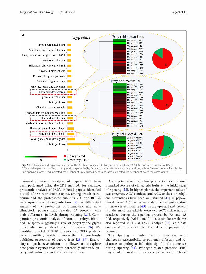
Involvement of fatty acid metabolism in the fruit ripeningprocessThree fatty acid related metabolism pathways were sig-nificantly changed during the ripening process of papayafruit: ‘Fatty acid biosynthesis’, ‘Fatty acid metabolism’ and‘Fatty acid degradation’ (Fig. 5a). For the ‘Fatty acid bio-synthesis’ pathway, two down-regulated and 11 up-regulated proteins were identified (Fig. 5b); for the ‘Fattyacid metabolism’ pathway, four down-regulated and 13up-regulated (Fig. 5c); and for the ‘Fatty acid degrad-ation’ pathway, four down-regulated and five up-regulated (Fig. 5d). Enzymes involved in fatty acid me-tabolism are listed in Additional file 8.Furthermore, the contents of several important fatty
acids were determined during the fruit ripening process.In total, 16 kinds of fatty acids were identified and re-spectively quantified at 0 and 8 d (Fig. 6). Notably, fourfatty acids – methyl-decanoate, methyl laurate, methylpalmitoleate and methyl α-octadecatrienoic acid – weresignificantly up-regulated at 8 d compared with 0 d; and
four fatty acids – methyl stearate, methyl oleate, methylerueate and methyl linoleate – were significantly down-regulated. Contents of the remaining eight fatty acidsdid not significantly change.
Involvement of cell wall-related proteins in the fruitripening processA number of enzymes, including PG, pectinesterase(PE), α-galactosidase (α-GAL), β-GAL, β-xylosidase, PL,expansin (EXP), xyloglucan-endotransglycosylase (XET)and endoglucanase (EG), are known to be involved incell wall metabolism [32]. In our study, three PGs, sixPEs, two α-GALs, one β-GAL, two EXPs, two XETs andtwo EGs were identified as DAPs. The expression pat-terns of the cell wall metabolism-related proteins werethus analyzed during the ripening process: all PGs, α-GALs and most PEs (except for Unigene0023139) weresignificantly down-regulated; and all β-GAL, EXPs, EGsand one EXT (Unigene0032975) were significantly up-regulated (Additional file 9).
Fig. 3 GO enrichment analysis of DAPs. (a) Distribution of the up-regulated proteins with GO annotation. Different color blocks representdifferent terms, including cellular component, molecular function, and biological process. Number of the up-regulated proteins in each secondlevel term was showed in a pie chart. (b) Distribution of the down-regulated proteins with GO annotation. Different color blocks representdifferent terms, including cellular component, molecular function, and biological process. Number of the down-regulated proteins in each secondlevel term was showed in a pie chart
Jiang et al. BMC Plant Biology (2019) 19:238 Page 7 of 13

Verification of the changes in fruit ripening-relatedproteins using PRMBecause fruit ripening is extremely complex, it is difficultto identify a single sensitive biomarker. Instead, theidentification of a panel of candidate enzymes that re-flect the process of fruit ripening would offer a betterunderstanding of this complex process. To validate thedifferential expression of several key enzymes involvedin papaya fruit ripening, PRM was applied. In total, 16key proteins, including five fatty acid metabolism-related, two pathogen defense-related, two ethylenebiosynthesis-related and seven cell wall biosynthesis-related proteins, were selected for PRM verification(Additional file 10). The relative abundances of severalkey proteins from different sample groups are presentedin Figs. 7 and 8. The trend of these DAPs determined by
PRM was consistent with our TMT-label quantificationresults.
DiscussionFruit ripening involves a process of several modifica-tions, including starch–sugar conversion, cell wall deg-radation, flavor substance and pigment biosynthesis, andmetabolic rearrangement [32–34]. As climacteric fruit,softening of papaya is an important feature that deter-mines its marketing value [35]. Increasing numbers ofstudies have focused on molecular and biochemicalmechanisms underlying the regulation of climactericfruit ripening. In our study, comprehensive analysis wascarried out to identify the DAPs involved in the ripeningprocess.
Fig. 4 Analysis of the pathogen defense-related proteins during the papaya fruit ripening process. (a) The subcellular locations of these pathogendefense-related proteins in papaya. (b) Relative expression levels of the proteins related to pathogen defense. Significant differences in expressionlevel were indicated by “*”
Jiang et al. BMC Plant Biology (2019) 19:238 Page 8 of 13
Several proteomic analyses of papaya fruit havebeen performed using the 2DE method. For example,proteomic analysis of PMeV-infected papaya identifieda total of 486 reproducible spots, among which calre-ticulin and the proteasome subunits 20S and RPT5awere upregulated during infection [36]. A differentialanalysis of the proteomes of climacteric and non-climacteric papaya fruit revealed 27 proteins withhigh differences in levels during ripening [37]. Com-parative proteomic analysis of somatic embryo identi-fied 76 spots, suggesting a role of polyethylene glycolin somatic embryo development in papaya [26]. Weidentified a total of 3220 proteins and 2818 proteinswere quantified, which is more than in previouslypublished proteomes of papaya fruit [25, 37]. Embra-cing comprehensive information allowed us to explorenew proteins/genes that were potentially involved, dir-ectly and indirectly, in the ripening process.
A sharp increase in ethylene production is considereda marked feature of climacteric fruits at the initial stageof ripening [38]. In higher plants, the important roles oftwo enzymes, ACC synthase and ACC oxidase, in ethyl-ene biosynthesis have been well-studied [39]. In papaya,two different ACO genes were identified as participatingin papaya fruit ripening [40]. In the up-regulated proteinlist, the most remarkable were two ACC oxidases, up-regulated during the ripening process by 7.6 and 1.8fold, respectively (Additional file 1). A similar result wasalso reported in a 2DE-DIGE analysis [37]. Our dataconfirmed the critical role of ethylene in papaya fruitripening.The ripening of fleshy fruit is associated with
changes in susceptibility to pathogen infection and re-sistance to pathogen infection significantly decreasesduring ripening [41]. Pathogen-related proteins (PRs)play a role in multiple functions, particular in defense
Fig. 5 Identification and expression analysis of the KEGG terms related to Fatty acid metabolism. (a) KEGG enrichment analysis of DAPs.Differential expression profiling of ‘fatty acid biosynthesis’-(b), ‘fatty acid metabolism’-(c), and ‘fatty acid degradation’-related genes (d) under thefruit ripening process. Red indicated the number of up-regulated genes and green indicated the number of down-regulated genes
Jiang et al. BMC Plant Biology (2019) 19:238 Page 9 of 13

against pathogens [42]. In papaya fruit, eightpathogen-related factors were significantly reducedand only one significantly induced (Fig. 4b), indicatingthat the disease resistance of papaya fruit might de-crease during ripening.The metabolic shifts throughout ripening cause al-
terations in primary and secondary metabolites, suchas amino acids, fatty acids, sugars, polyphenols andcinnamic acids [43]. Our KEGG enrichment analysisrevealed that various metabolic pathways were signifi-cantly changed during ripening, suggesting a close re-lationship between metabolism and fruit ripening ofpapaya. Flavonoids, the ubiquitous fruit secondarymetabolites, play important roles in coloring, abioticstress defense and other biological functions [44]. Indate palm, proteins involved in anthocyanin biosyn-thesis were up-regulated particularly from the onsetof ripening and during ripening [45]. Similarly, duringthe ripening of papaya, several flavonoid biosynthesis-related proteins were up-regulated: cinnamate-4-hydroxylase, coumaroyl 3-hydroxylase and flavanone-3-hydroxylase (Fig. 5 and Additional file 1). Increases
in flavonoids may provide raw materials for pigmentbiosynthesis, accelerating the color variation of papayafruit [46].Interestingly, three fatty acid metabolic pathways
were highlighted by our KEGG enrichment analysis,and a number of fatty acid metabolism-related en-zymes were also identified as DAPs (Fig. 5). Studieson the changes in fatty acid composition during fruitdevelopment and ripening have been performed forvarious fruit plants. For example, the levels of ω-6and ω-3 fatty acids significantly changed duringmango fruit development and ripening [47]. Ripeningraises the content of polyunsaturated fatty acids in to-mato fruit [48]. In our study, assessment and profilingassessed the variation of fatty acids during the ripen-ing process of papaya fruit (Fig. 6). Among 16 quanti-fied fatty acids, eight showed significant changesduring the ripening process (Fig. 6). In tomato fruit,ripening increases the contents of unsaturated fattyacids [48]. In papaya, two unsaturated fatty acids,methyl palmitoleate and methyl α-octadecatrienoicacid, were significantly up-regulated during ripening.
Fig. 6 The changes in the contents of various fatty acids during the papaya fruit ripening process. Significant differences in contents wereindicated by “*”
Jiang et al. BMC Plant Biology (2019) 19:238 Page 10 of 13

Fig. 7 Verification of the changes in fatty acid- and pathogen defense-related proteins using PRM. Five representative proteins, including threeproteins involved in fatty acid metabolism (a), two pathogen defense-related proteins (b), were randomly selected for PRM verification. For eachprotein, the abundances of two peptides were determined
Fig. 8 Verification of the changes in ethylene biosynthesis- and cell wall-related proteins using PRM. Nine representative proteins, including twoproteins involved in ethylene biosynthesis (a), and two cell wall-related proteins (b), were randomly selected for PRM verification. For eachprotein, the abundances of two peptides were determined
Jiang et al. BMC Plant Biology (2019) 19:238 Page 11 of 13

Increases in unsaturated fatty acids might be associ-ated with papaya fruit volatile formation [49].
ConclusionsIn summary, a total of 3220 proteins were detected,2828 of these were quantified. Among the DAPs, mostpathogen-related proteins were down-regulated duringthe ripening process. Moreover, flavonoid and fatty acidmetabolisms were greatly changed during ripening; andunsaturated fatty acids may be associated with papayafruit volatile formation. Our data may give an intrinsicexplanation of the variations in metabolism and offerimportant information about how to expend the shelflife of postharvest papaya fruit.
Additional files
Additional file 1: Table S1. The detail information of the identifiedproteins. (PDF 316 kb)
Additional file 2: Figure S1. Variations in protein abundances duringthe papaya fruit ripening. (XLSX 747 kb)
Additional file 3: Table S2. The detail information of the DAPs duringfruit ripening. (XLSX 118 kb)
Additional file 4: Table S3. The scores for the subcellular location ofeach protein. (XLSX 141 kb)
Additional file 5: Figure S2. KEGG enrichment analysis of the DAPsduring the ripening process of papaya fruits. (PDF 175 kb)
Additional file 6: Figure S3. Protein domain enrichment analysis of theDAPs during the ripening process of papaya fruits. (PDF 308 kb)
Additional file 7: Table S4. The detail information of the pathogendefense-related proteins. (XLSX 17 kb)
Additional file 8: Table S5. The information of the fatty acidmetabolism-related proteins. (XLSX 14 kb)
Additional file 9: Table S6. The information of the proteins involved inthe cell-related process. (XLSX 10 kb)
Additional file 10: Table S7. The PRM results of 16 key proteins. (XLSX 13 kb)
Abbreviations1-MCP: 1-methylcyclopropene; 2-DE: two-dimensional gel electrophoresis;ABA: abscisic acid; ACO: ACC oxidase; BAK1: brassinosteroid-insensitive 1-associated receptor kinase 1; CALM: calmodulin; CML: calcium-bindingproteins; CNGF: cyclic nucleotide gated channel; CPK: calcium-Dependentprotein kinase; DAP: differential accumulated protein; EG: endoglucanase;EXP: expansin; FA: formic acid; FAA: formalin acetic acid; glpK: glycerol kinase;GO: Gene ontology; HPLC: high-performance liquid chromatography;HSP: heat shock protein; JA: jasmonic acid; KEGG: Kyoto Encyclopedia ofGenes and Genomes; LC–MS/MS: liquid chromatography-mass spectrometer;NCL: nucleolin; PE: pectinesterase; PR: pathogenesis-related; PRM: ParallelReaction Monitoring; PTI: plant–pathogen interacting; SUGT: suppressor ofG2 allele of SKP1; TEAB: triethylammonium bicarbonate; TUB: tubulin;XET: xyloglucan-endotransglycosylase; α-GAL: α-galactosidase; β-GAL: β-galactosidase
AcknowledgementsWe are grateful to Dr. Xiaolin Zheng (Zhejiang Gongshang University), Dr.Zhiping Deng (Zhejiang Academy of Agricultural Sciences) and Dr. Jiang Tian(South China Agricultural University) for technical support. The authorsdeclare that they have no competing interests.
Authors’ contributionsAll authors have read and approved the final version. JW, LX and KLconceived and designed the study. BJ, LX, TZ, CY and JW collected and
taken care of the plant samples. BJ, LX, SO, WM, MY, and HG performed theexperiments. CY analyzed the data. CS and KL wrote the manuscript.
FundingThis work was funded by the Science and Technology Program ofGuangdong, China (2015A020208018 and 2017A040403069); Natural ScienceFoundation of Guangdong Province, China (2016A030307016 and2018A0303130161); Science and Technology Project of Zhanjiang City, China(2016A03016) and College Student Climbing Program of GuangdongProvince, China (pdjh2019b0299). There is on role of the funding bodies inthe design of the study and collection, analysis, and interpretation of dataand in writing the manuscript.
Availability of data and materialsThe datasets generated and analysed during the current study are availablein the ProteomeXchange Consortium by PRIDE partner repository programunder identifier PXD008871.
Ethics approval and consent to participateThis project uses plant materials and does not utilize transgenic technology.The seedlings of papaya tree were purchased by the Yixin horticulturecompany (Zhanjiang, China), who provided permission to use the seedlingsfor our scientific research. Collection of plant material complied with theConvention on the Trade in Endangered Species of Wild Fauna and Flora:https://www.cites.org/.
Consent for publicationNot applicable.
Competing interestsThe authors declare that they have no competing interests.
Author details1Life Science and Technology School, Lingnan Normal University, Zhanjiang524048, China. 2Root Biology Center, College of Natural Resources andEnvironment, South China Agricultural University, Guangzhou 510642, China.3College of Life and Environmental Sciences, Hangzhou Normal University,Hangzhou 310036, China.
Received: 18 October 2018 Accepted: 22 May 2019
References1. Giovannoni JJ. Genetic regulation of fruit development and ripening. The
Plant cell, vol. 16 Suppl; 2004. p. S170–80.2. Wang WQ, Møller IM, Song SQ: Proteomic analysis of embryonic axis of
Pisum sativum seeds during germination and identification of proteinsassociated with loss of desiccation tolerance. J Proteomics Journal ofproteomics 2012, 77(24):68–86.
3. Brummell DA. Cell wall disassembly in ripening fruit. Funct Plant Biol. 2006;33(2):103–19.
4. Carpita NC, Gibeaut DM. Structural models of primary cell walls in floweringplants: consistency of molecular structure with the physical properties ofthe walls during growth. The Plant journal : for cell and molecular biology,vol. 3; 1993. p. 1):1–30.
5. Brummell DA, Harpster MH. Cell wall metabolism in fruit softening andquality and its manipulation in transgenic plants. Plant Mol Biol. 2001;47(1–2):311–40.
6. Yong W, Link B, O'Malley R, Tewari J, Hunter CT, Lu CA, et al. Genomics ofplant cell wall biogenesis. Planta. 2005;221(6):747–51.
7. Dumville JC, Fry SC. Solubilisation of tomato fruit pectins by ascorbate:a possible non-enzymic mechanism of fruit softening. Planta. 2003;217(6):951–61.
8. Fry SC, Dumville JC, Miller JG. Fingerprinting of polysaccharides attacked byhydroxyl radicals in vitro and in the cell walls of ripening pear fruit. TheBiochemical journal. 2001;357(Pt 3:729–37.
9. Jha SN, Jaiswal P, Narsaiah K, Kaur PP, Singh AK, Kumar R. Texturalproperties of mango cultivars during ripening. J Food Sci Technol. 2013;50(6):1047–57.
Jiang et al. BMC Plant Biology (2019) 19:238 Page 12 of 13

10. Xi W, Zheng H, Zhang Q, Li W. Profiling taste and aroma compoundmetabolism during apricot fruit development and ripening. Int J Mol Sci.2016;17(7):pii: E998.
11. Yao BN, Tano K, Konan HK, Bedie GK, Oule MK, Koffi-Nevry R, et al. The roleof hydrolases in the loss of firmness and of the changes in sugar contentduring the post-harvest maturation of Carica papaya L. var solo 8. J FoodSci Technol. 2014;51(11):3309–16.
12. Baldwin EA, Scott JW, Shewmaker CK, Schuch W. Flavor trivia and tomatoaroma: biochemistry and possible mechanisms for control of importantaroma components. Hortscience. 2000;35(6):1013–22.
13. Villa-Rodríguez JA, Molina-Corral FJ, Ayala-Zavala JF, Olivas GI, González-Aguilar GA. Effect of maturity stage on the content of fatty acids andantioxidant activity of ‘Hass’ avocado. Food Res Int. 2011;44(5):1231–7.
14. Lalel HJD, Singh Z, Tan SC. Aroma volatiles production during fruit ripeningof ‘Kensington Pride’ mango. Postharvest Biology & Technology. 2003;27(3):323–36.
15. Liu K, Wang J, Li H, Zhong J, Feng S, Pan Y, et al. Identification, expression andIAA-amide synthetase activity analysis of Gretchen Hagen 3 in papaya fruit(Carica papaya L.) during postharvest process. Front Plant Sci. 2016;7:1555.
16. Zou Y, Zhang L, Rao S, Zhu X, Ye L, Chen W, et al. The relationship betweenthe expression of ethylene-related genes and papaya fruit ripening disordercaused by chilling injury. PLoS One. 2014;9(12):e116002.
17. Paull RE. Postharvest atemoya fruit splitting during ripening. PostharvestBiology & Technology. 1996;8(4):329–34.
18. Liu K, Yuan C, Li H, Lin W, Yang Y, Shen C, et al. Genome-wide identificationand characterization of auxin response factor (ARF) family genes related toflower and fruit development in papaya (Carica papaya L.). BMC Genomics.2015;16:901.
19. Fabi JP, Seymour GB, Graham NS, Broadley MR, May ST, Lajolo FM et al:Analysis of ripening-related gene expression in papaya using an Arabidopsis-based microarray. BMC Plant Biol 2012, 12:242–242.
20. Othman R, Nuraziyan A. Fruit-specific expression of papaya subtilase gene. JPlant Physiol. 2010;167(2):131–7.
21. Shiga TM, Fabi JP, do Nascimento JR, Petkowicz CL, Vriesmann LC, LajoloFM, et al. Changes in cell wall composition associated to the softening ofripening papaya: evidence of extensive solubilization of large molecularmass galactouronides. J Agric Food Chem. 2009;57(15):7064–71.
22. do Prado SBR, Melfi PR, Castro-Alves VC, Broetto SG, Araújo ES, doNascimento JRO, et al. Physiological degradation of pectin in papaya cellwalls: release of long chains galacturonans derived from insoluble fractionsduring postharvest fruit ripening. Front Plant Sci. 2016;7:1120.
23. Fabi JP, Broetto SG, da Silva SL, Zhong S, Lajolo FM, do Nascimento JR.Analysis of papaya cell wall-related genes during fruit ripening indicates acentral role of polygalacturonases during pulp softening. PLoS One. 2014;9(8):e105685.
24. Fu CC, Han YC, Qi XY, Shan W, Chen JY, Lu WJ, et al. Papaya CpERF9 acts asa transcriptional repressor of cell-wall-modifying genes CpPME1/2 andCpPG5 involved in fruit ripening. Plant Cell Rep. 2016;35(11):2341–52.
25. Huerta-Ocampo JA, Osuna-Castro JA, Lino-Lopez GJ, Barrera-Pacheco A,Mendoza-Hernandez G, De Leon-Rodriguez A, et al. Proteomic analysis ofdifferentially accumulated proteins during ripening and in response to 1-MCP in papaya fruit. J Proteome. 2012;75(7):2160–9.
26. Vale EdM, Heringer AS, Barroso T, Ferreira ATdS, da Costa MN, Perales JEA etal: Comparative proteomic analysis of somatic embryo maturation in Caricapapaya L. Proteome Sci 2014, 12:37–37.
27. Soares EA, Werth EG, Madronero LJ, Ventura JA, Rodrigues SP, Hicks LM, etal. Label-free quantitative proteomic analysis of pre-flowering PMeV-infectedCarica papaya L. J Proteome. 2017;151:275–83.
28. Hao J, Guo H, Shi X, Wang Y, Wan Q, Song YB, et al. Comparative proteomicanalyses of two Taxus species (Taxus × media and Taxus mairei) revealsvariations in the metabolisms associated with paclitaxel and othermetabolites. Plant & cell physiology. 2017;58(11):1878–90.
29. Xu D, Yuan H, Tong Y, Zhao L, Qiu L, Guo W, et al. Comparative proteomicanalysis of the graft unions in hickory (Carya cathayensis) provides insightsinto response mechanisms to grafting process. Front Plant Sci. 2017;8:676.
30. Yu C, Guo H, Zhang Y, Song Y, Pi E, Yu C, et al. Identification of potential genesthat contributed to the variation in the taxoid contents between two Taxusspecies (Taxus × media and Taxus mairei). Tree Physiol. 2017;37(12):1659–71.
31. He Z, Zhu H, Li W, Zeng M, Wu S, Chen S, et al. Chemical components ofcold pressed kernel oils from different Torreya grandis cultivars. Food Chem.2016;209:196–202.
32. Brownleader MD, Jackson P, Mobasheri A, Pantelides AT, Sumar S, Trevan M,et al. Molecular aspects of cell wall modifications during fruit ripening. CritRev Food Sci Nutr. 1999;39(2):149–64.
33. Xiao YY, Kuang JF, Qi XN, Ye YJ, Wu ZX, Chen JY, et al. A comprehensiveinvestigation of starch degradation process and identification of atranscriptional activator MabHLH6 during banana fruit ripening. PlantBiotechnol J. 2017.
34. Huo W, Li B, Kuang J, He P, Xu Z, Wang J. Functional characterization of thesteroid reductase genes GmDET2a and GmDET2b form Glycine max. Int JMol Sci. 2018;19(3).
35. Paull RE, Chen NJ. Postharvest variation in cell wall-degrading enzymes ofpapaya (Carica papaya L.) during fruit ripening. Plant Physiol. 1983;72(2):382–5.
36. Rodrigues SP, Ventura JA, Aguilar C, Nakayasu ES, Almeida IC, Fernandes PM,et al. Proteomic analysis of papaya (Carica papaya L.) displaying typicalsticky disease symptoms. Proteomics. 2011;11(13):2592–602.
37. Nogueira SB, Labate CA, Gozzo FC, Pilau EJ, Lajolo FM, Oliveira doNascimento JR. Proteomic analysis of papaya fruit ripening using 2DE-DIGE.J Proteome. 2012;75(4):1428–39.
38. Liu M, Pirrello J, Chervin C, Roustan JP, Bouzayen M. Ethylene control of fruitripening: revisiting the complex network of transcriptional regulation. PlantPhysiol. 2015;169(4):2380–90.
39. López-Gómez R, Cabrera-Ponce JL, Saucedo-Arias LJ, Carreto-Montoya L,Villanueva-Arce R, Díaz-Perez JC, et al. Ripening in papaya fruit is altered byACC oxidase cosuppression. Transgenic Res. 2009;18(1):89–97.
40. Chen YT, Lee YR, Yang CY, Wang YT, Yang SF, Shaw JF. A novel papaya ACCoxidase gene (CP-ACO2) associated with late stage fruit ripening and leafsenescence. Plant Sci. 2003;164(4):531–40.
41. Cantu D, Blanco-Ulate B, Yang L, Labavitch JM, Bennett AB, Powell AL.Ripening-regulated susceptibility of tomato fruit to Botrytis cinerea requiresNOR but not RIN or ethylene. Plant Physiol. 2009;150(3):1434–49.
42. Jia X, Zeng H, Wang W, Zhang F, Yin H. Chitosan oligosaccharide inducesresistance to Pseudomonas syringae pv. Tomato DC3000 in Arabidopsisthaliana by activating both salicylic acid- and jasmonic acid-mediatedpathways. Molecular plant-microbe interactions. MPMI. 2018;31(12):1271–9.
43. Liang Z, Sang M, Fan P, Wu B, Wang L, Duan W, et al. Changes ofpolyphenols, sugars, and organic acid in 5 Vitis genotypes during berryripening. J Food Sci. 2011;76(9):C1231–8.
44. Broun P. Transcriptional control of flavonoid biosynthesis: a complexnetwork of conserved regulators involved in multiple aspects ofdifferentiation in Arabidopsis. Curr Opin Plant Biol. 2005;8(3):272–9.
45. Marondedze C, Gehring C, Thomas L. Dynamic changes in the date palmfruit proteome during development and ripening. Horticulture research.2014;1:14039.
46. Mou W, Li D, Luo Z, Mao L, Ying T. Transcriptomic analysis reveals possibleinfluences of ABA on secondary metabolism of pigments, flavonoids andantioxidants in tomato fruit during ripening. PLoS One. 2015;10(6):e0129598.
47. Deshpande AB, Chidley HG, Oak PS, Pujari KH, Giri AP, Gupta VS. Data onchanges in the fatty acid composition during fruit development andripening of three mango cultivars (alphonso, Pairi and Kent) varying inlactone content. Data in Brief. 2016;9:480–91.
48. Saini RK, Zamany AJ, Keum YS: Ripening improves the content ofcarotenoid, alpha-tocopherol, and polyunsaturated fatty acids in tomato(Solanum lycopersicum L.) fruits. 3 Biotech 2017, 7(1):43.
49. Wang JJ, Liu HR, Gao J, Huang YJ, Zhang B, Chen KS. Two omega-3FADs are associated with peach fruit volatile formation. Int J Mol Sci.2016;17(4):464.
Publisher’s NoteSpringer Nature remains neutral with regard to jurisdictional claims inpublished maps and institutional affiliations.
Jiang et al. BMC Plant Biology (2019) 19:238 Page 13 of 13