comparative phosphorescence quenching of dna's of different ...
9
COMPARATIVE PHOSPHORESCENCE QUENCHING OF DNA'S OF DIFFERENT COMPOSITION I. ISENBERG, R. ROSENBLUTH, and S. L. BAIRD, JR. From the Science Research Institute, Oregon State University, Corvallis, Oregon 97331 ABsTRAcr Small amounts of paramagnetic cations quench the phosphorescence of DNA. Although the emission intensity is monotonic with increasing thymine con- tent, the quenching efficiency is not. The cations quench from phosphate sites. INTRODUCTION Recent studies have indicated that the low-temperature ultraviolet-induced phos- phorescence of DNA may be attributed to thymine-negative ions (1, 2). Guanine and adenine emit at neutral pH (3, 4). However, adenylic acid emission is quenched at acid pH (5, 6) while thymine, which does not emit at neutral pH, shows a strong emission at high pH (7). The electron spin resonance position of the DNA triplet is close to that of the thymine-negative ion, and no resonance has as yet been found corresponding to an adenine or guanine triplet (2). Both the DNA and the thymine-negative ion emissions have about the same decay constant and appear unstructured, in contrast to those of adenine and guanine which have struc- ture and much longer decay times. Furthermore, GMP emission is quenched by CMP or poly C (2). All of the evidence is thus consistent with a model in which the guanine emission is quenched by hydrogen bonding to cytosine and, in the excited state, the NJ proton of thymine is shifted to adenine. With this shift, the adenine no longer emits, and the emission is that due to the resulting thymine-negative ion. The DNA phosphorescence is quenched by relatively low concentrations of paramagnetic cations (2, 4, 8). The fluorescence is quenched very little, if at all. In order to explain the observed amount of DNA phosphorescence quenched per added paramagnetic cation, it has been postulated (4, 8) that the triplet energy wanders from one base to another, being quenched when the excitation is in the vicinity of a bound paramagnetic cation. This type of triplet migration has been shown in studies of the emission of crystals (9) and is now well recognized. However, as will be seen, any discussion of triplet migration in polynucleotides must include consideration of the possible occurrence of specific barriers to such migration. The work reported here involves a comparative study of the phosphorescence 365
Transcript of comparative phosphorescence quenching of dna's of different ...

COMPARATIVE PHOSPHORESCENCE QUENCHING
OF DNA'S OF DIFFERENT COMPOSITION
I. ISENBERG, R. ROSENBLUTH, and S. L. BAIRD, JR.
From the Science Research Institute, Oregon State University, Corvallis, Oregon 97331
ABsTRAcr Small amounts of paramagnetic cations quench the phosphorescence ofDNA. Although the emission intensity is monotonic with increasing thymine con-tent, the quenching efficiency is not. The cations quench from phosphate sites.
INTRODUCTION
Recent studies have indicated that the low-temperature ultraviolet-induced phos-phorescence of DNA may be attributed to thymine-negative ions (1, 2).Guanine and adenine emit at neutral pH (3, 4). However, adenylic acid emission
is quenched at acid pH (5, 6) while thymine, which does not emit at neutral pH,shows a strong emission at high pH (7). The electron spin resonance position ofthe DNA triplet is close to that of the thymine-negative ion, and no resonance hasas yet been found corresponding to an adenine or guanine triplet (2). Both the DNAand the thymine-negative ion emissions have about the same decay constant andappear unstructured, in contrast to those of adenine and guanine which have struc-ture and much longer decay times. Furthermore, GMP emission is quenched byCMP or poly C (2).
All of the evidence is thus consistent with a model in which the guanine emissionis quenched by hydrogen bonding to cytosine and, in the excited state, the NJproton of thymine is shifted to adenine. With this shift, the adenine no longer emits,and the emission is that due to the resulting thymine-negative ion.The DNA phosphorescence is quenched by relatively low concentrations of
paramagnetic cations (2, 4, 8). The fluorescence is quenched very little, if at all.In order to explain the observed amount of DNA phosphorescence quenched peradded paramagnetic cation, it has been postulated (4, 8) that the triplet energywanders from one base to another, being quenched when the excitation is in thevicinity of a bound paramagnetic cation.
This type of triplet migration has been shown in studies of the emission of crystals(9) and is now well recognized. However, as will be seen, any discussion of tripletmigration in polynucleotides must include consideration of the possible occurrenceof specific barriers to such migration.The work reported here involves a comparative study of the phosphorescence
365
and the quenching of phosphorescence of three double-stranded polynucleotidesdiffering in base composition. It also reports the inhibition of quenching in thepresence of large amounts of Mg++.Rahn, Shulman, and Longworth (2) found that, with minor variations, the phos-
phorescence of DNA's from varying sources appeared the same, except for thequantum yield which was larger for samples with more A-T content. Our dataverify theirs.
This paper presents evidence that the quenching efficiency, and thus presumablythe amount of triplet wandering, is not a simple function of A-T content but ap-parently depends on base sequence. It also shows that the site of a quenching ionis at the polynucleotide phosphate.
MATERIALS AND METHODS
All emissions were measured at 77°K, in 50% glycerol glasses, on an Aminco-Keirs spectro-phosphorimeter (American Instrument Co., Silver Spring, Md.) equipped with a Hruskaphotometer (Hruska Radio Co., Lutherville, Md.). Samples were in quartz capillary tubesof 0.2-0.4 mm diameter. Whenever comparative quantum yields were desired, the samecapillary tube and concentrations of DNA giving the same optical density were used. Wehave observed that samples of Micrococcus lysodeikticus DNA, permitted to stand in 50%glycerol for more than several hours at room temperature, showed increased quantum yieldsand gave irreproducible results. We, therefore, examined all samples in less than M hr aftermixing, during which time no detectable time variations could be seen.
Sources of the DNA were as follows.(a) salmon sperm DNA. This was obtained commercially and was further purified as
previously described (10).(b) Micrococcus lysodeikticus DNA. Commercially dried cells (Miles Chemicals Co.,
Elkhart, Ind.) were extracted, and DNA was purified according to Marmur's procedure (11).The final precipitate was redissolved in 0.0025 M NaCl. The solution (OD = 5.3) was thenput on a Sephadex G-25 fine column and eluted with 0.0025 M NaCl. It was then concen-trated by flash evaporation at 36°C.
(c) poly dAT. Synthetic poly dAT was a gift of Dr. Thomas M. Jovin, Stanford Uni-versity. It was dialysed extensively against 0.0025 M NaCl. In three batches, a total of 0.882 mlof concentration 4.96 X 10⁻⁴ M in phosphates was evaporated in a weighed 0.5 ml poly-ethylene centrifuge tube to a final weight of 40.9 mg dAT solution. Evaporation was per-formed below the dAT melting point. The concentrations of dAT were calculated using e =6650 liter - cm- - moles-1, based on phosphate, at 262 m,u (12).
Preparation of Samples
Samples used for emission contained 0.03 M NaCl in 50% glycerol-water mixtures. The di-valent cations were added generally as chloride salts, but controls run with perchlorate saltsyielded the same results. The pH of the samples was 5.5 at room temperature in 50% glycerol.The actual volume of sample required in the tubes used for emission studies was only
about 1 ,ul. However, samples of at least 0.1 ml had to be prepared in order to have confidencein the accuracy of their composition. For salmon sperm and M. lysodeikticus DNA separatesamples were mixed for each emission measurement. However, only small amounts of dAT
BIOPHYSICAL JOURNAL VOLUME 7 1967366

were available, and special micromixing techniques were employed to obtain quenchingcurves on single 0.1 ml samples. The technique was checked by obtaining identical quenchingcurves for salmon sperm DNA using both ordinary and micro methods. For the micro tech-nique, MnCl2 was added in increments to one sample, and the phosphorescence was measuredon an aliquot after each addition. Changes in the sample size and concentration due to addi-tion of MnCl2 and evaporation during mixing were monitored by weighing and were adjustedwhen necessary. At the end of the experiment the OD at 262 m, was measured and comparedwith the value expected from all monitoring weighings. The two values differed by only 10%.Two independent series of measurements yielded the same quenching curve. After running a
4.0
3.0
U~~~~~~~~~~
°2.0 _CL0
0
a.
1.0
350 400 450 500 550 600
myJFIGURE 1 Phosphorescence of native and denatured DNA: salmon sperm, 4 X 10-' MDNA-P. Curve A, native DNA; curve B, denatured DNA: the same sample as A afterheating for 30 min at 90°C followed by quick chilling; ordinate: phosphorescence, rela-tive units.
quenching curve, a melting point curve of the sample was run, still in 50% glycerol. It wasfound to melt at 37°C with a hypochromism of 42%. This, together with the absence of ade-nine emission, indicated that spectra had been run on hydrogen-bonded dAT.In general, an internal check on the nativeness of the DNA's used is obtained by observa-
tion of the phosphorescence (see results below).
RESULTSFig. 1, curve A, shows the phosphorescence spectrum for native salmon sperm DNA.Curve B is a typical spectrum obtained from heat-denatured DNA. Denaturationbrings with it a much more structured spectrum and an increase in signal. The in-crease in quantum yield upon denaturation is variable, ranging from about 2 to
ISENBERG, ROSENBLUTH, AND BAIRD Phosphorescence Quenching ofDNA 367

about 8 depending upon the concentration of DNA and the extent of heating. Simi-lar changes have been reported by Rahn et al. (2). The increased structure is pre-sumably due to the phosphorescence of purines which are now no longer hydrogenbonded.The quantum yield of DNA phosphorescence depends on the DNA examined.
Fig. 2 shows the relative emissions of M. lysodeikticus (28% A-T), salmon spermDNA (58% A-T), and poly dAT. It may be seen that the size of the emission in-creases with an increasing amount of A-T base pairs. The emissions are roughly in
my mPD
FiouRE 2, a-b. Phosphorescence of three different DNA's excited at 284 mp. All sampleshad same absorbance at 284 m,u. Curve A, Micrococcus lysodeikticus DNA; curve B, salmonsperm DNA; curve C, poly dAT; ordinate: phosphorescence, relative units.
the ratio of 1:1.6:10. Rahn et al. (2) have also reported that dAT has a largeremission than calf thymus DNA.Upon turning off the exciting light, the time decays for the emissions were found
to be the same, having a time constant of approximately 0.3 sec. This, together withthe similarity in shape, suggests that the source of the emission is the same in all
cases.
Let R = moles of cation added per mole of DNA phosphate. Then, for smallamounts of cation added, compared to nucleic acid phosphate present, the phospho-rescence intensity, S, is given by:
S = S0 (1- #R).
BIOPHYSICAL JOURNAL VoLUME 7 1967
0c0
a010
10IL
368

S.C
5.0
L
0~RS~=*'
40o
3.01
2.01
1.01-I I I I I I I
-- ...
0 1 3 4 5 6 766 10 1 2 3 4 5 7 10R* jQS0 Ra
FIGURE 3. Quenching of phosphorescence by MnCl2; comparison of three differentDNA's: salmon sperm DNA 000E, Micrococcus lysodeikticus DNA A-A, poly dAT0 0. R is the molar ratio of MnCl2 to DNA phosphates. Ordinate: amplitude of phos-phorescence, same relative units used.
0
If,CO)
'i| \\ FIGURE 4 Total emission, salmon
J | \ . . A sperm DNA. Curve A, no added Cu";e- | \ >.^ curve B, 0.1 moles of Cu++ per mole
| \ \ \ of DNA phosphate. Note that there is/>, \little or no change in fluorescence in
\ \,, the vicinity of 365 m,u, but phospho-rescence at 450 mp is severely quenched.
[Compare with Mn" quenching, Fig.f- Uf@ , _ 6 of Rahn et al. (2).1
350. 400 40 5606
is a measure of the quenching efficiency. It is the average region of DNA, as
measured in DNA phosphates, that is quenched by one added cation. Fig. 3 showsthe comparative quenching of M. lysodeikticus DNA, salmon sperm DNA, andpoly dAT by Mn §. These have 6 values of 5, 11, and 6, respectively.As previously reported (8), quenching also occurs upon the addition of Co++,
ISENBERo. ROseNLUTH, AND BAD Phosphorescence Quenching ofDNA 369_w -1 4_
__
;- (L,- f2.
LGfr
0.61
o 4
0
L OL2a.S.0 M
-.
n_n . . .s-. -WD----w--I a I a I I I I I a
1-k
Ni++, or Cu++, and #3 appears to show an increase, within a factor of 2, as oneprogresses along the series Mn++, Co++, Ni++, Cu 1. This quenching occurs withlittle or no change in the fluorescence (Fig. 4).
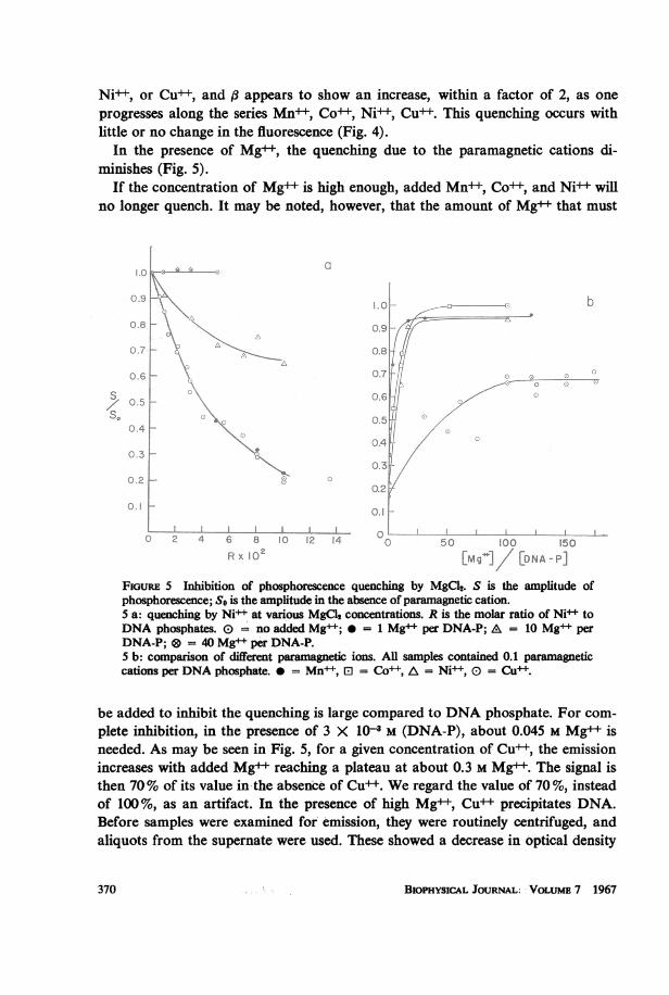
In the presence of Mg++, the quenching due to the paramagnetic cations di-minishes (Fig. 5).
If the concentration of Mg++ is high enough, added Mn+ , Co++, and Ni+ willno longer quench. It may be noted, however, that the amount of Mg++ that must
1.00
0.9 0
t '0.0 F .e Q ., ~~~~100.6
0.7 A 0.6
:0.6 ~~~~~~~~~~0.70s 0.6~~~~~~~~~~~~~
Z 0.6
/o 0.5
S
.I. I a*1---lA ,, I L I .......II 1 1I I I0 2 4 6 8 10 12 14 00 50 100 150
Rx 10 bEMg*]/ [DNA-P]FiGuRE 5 Inhibition of phosphorescence quenching by MgCl2. S is the amplitude ofphosphorescence; So is the amplitude in the absence of paramagnetic cation.5 a: quenching by Ni+ at various MgCl2 concentrations. R is the molar ratio of Ni toDNA phosphates. E = no added Mg+; = 1 Mg+ per DNA-P; A = 10 Mg+ perDNA-P; @ = 40 Mg+ per DNA-P.5 b: comparison of different paramagnetic ions. All samples contained 0.1 paramagnetccations per DNA phosphate. 0 = Mn+, El = Co++, A = Ni , 0 = Cu+.
be added to inhibit the quenching is large compared to DNA phosphate. For com-plete inhibition, in the presence of 3 X 10⁻⁸ M (DNA-P), about 0.045 M Mg++ isneeded. As may be seen in Fig. 5, for a given concentration of Cu++, the emissionincreases with added Mg++ reaching a plateau at about 0.3 M Mg. The signal isthen 70% of its value in the absence of Cu . We regard the value of 70 %, insteadof 100%, as an artifact. In the presence of high Mg++, Cu precipitates DNA.Before samples were examined for emission, they were routinely centrifuged, andaliquots from the supernate were used. These showed a decrease in optical density
BIOPHYSICAL JOURNALI VOLUME 7 1967370

which could account for the dimunition of phosphorescence from that observedwithout Cu++. Furthermore, such curves varied appreciably from run to run. The70% plateau of Fig. 5 b, therefore, does not demonstrate that the Cu+ bindingsite is different from that of the other cations.
It should be noted that Mg++ alone does not alter the emission of DNA evenwhen added in large concentrations. This has been checked up to a ratio of 130 Mg++ions per DNA phosphate.
DISCUSSION
As noted in the introduction, the accumulated evidence indicates that the phospho-rescence ofDNA is from the thymines. The results reported here are consistent withthis interpretation. The similarity between the emissions of the three polymers, ofwidely different base ratios, indicates that the emission is due to the same source.However, the emission does not appear to be simply proportional to the amount ofthymine present, although the intensity is larger for higher A-T content.The observation that one added paramagnetic cation quenches a section of salmon
sperm DNA of 10-20 phosphates led (4, 8) to a model in which the triplet excitationwandered from one position to another, being quenched only when in the vicinityof a paramagnetic cation. However, if triplet wandering does occur, the resultsreported here indicate that such migration is not a simple function of the amountof thymine present. The wandering must also depend on the order of the bases, atleast in a statistical sense. If the delocalization were only a function of the averagethymine content, perhaps being greatest for polynucleotides with highest thyminecontent, one would expect the emission from poly dAT to be quenched most effi-ciently. Instead, the quenching efficiency, using poly dAT, in which an A regularlyalternates with a T along the chain, is smaller than that of salmon sperm DNA. Infact, it is about the same as that for M. lysodeikticus which has only 28% A-T.Therefore, the extent of triplet wandering is not dependent alone on thymine con-tent, but probably depends on the sequence of bases along the chain. In salmonsperm DNA, one might expect a larger number of thymines close to one anotherthan in either of the other two DNA's. It may be expected that a high efficiency oftransfer from one thymine to another will occur if the thymines are adjacent to oneanother. Such a high efficiency of transfer has been observed in single-strandedpoly A where j8 has been found to be large, of the order of 100 (20). Our resultssuggest that even one adenine between two thymines serves to sharply reduce theefficiency of transfer of excitation energy between the thymines.The low quenching efficiency of Mn"+ when added to M. lysodeikticus can be
understood on the same model. The low thymine content makes it likely that be-tween most nearest pairs of thymines, there will be a number of nonthymine basesserving as barriers to the transfer of triplet excitation.
This model predicts that the quenching efficiency should be large when using
ISENBERG, ROsENBLUTH, AND BAIRD Phosphorescence Quenching ofDNA 371

natural DNA's of high A-T content, but not having a regular alternation of basesas in poly dAT.A complete characterization of barriers to triplet migration must await further
experimental information. An understanding of the range of migration not only in-volves the question of what constitutes an effective barrier but also depends on anunderstanding of the quenching range of a paramagnetic cation. Any ultimatetheory must also explain the observations (2, 20) that the lifetime of the residualluminescence of calf thymus DNA or of the neutral pH form of poly A is inde-pendent of the degree of quenching. These observations suggest that absolutebarriers to migration exist, and if a quencher is complexed between such barriers,the luminescence of the region is quenched completely, perhaps because migrationbetween barriers is very rapid.A related question involves the site of binding of the quenching cations. Even
though a wide variety of evidence has accumulated over the past 10 years that Mg++,Mn , Co++, Ni++ site bind to the phosphates of polynucleotides, (13-18), studiesof triplet quenching in liquid media at room temperature have indicated that forquenching to occur, the quenching cation must complex directly with the emitter.Furthermore, theoretical discussions of such quenching have invoked mechanismsinvolving wave function overlap between the emitting species and the quenchingcation.The experimental demonstrations of site binding at phosphates all have sufficient
experimental errors, so that one could raise the possibility that a minority speciesof cation existed, bound directly to bases, this species alone being responsible forquenching, while the majority of bound cations at phosphate sites do not quench(4). We believe that the experiments reported here rule out the existence of such apresumed minority component.The strongest argument in favor of cations bound to phosphates being the
quenchers lies with the general shape of the quenching curve. If extremely smallamounts of cations are added to DNA, a presumed minority fraction should be-come negligibly small, since the binding energy to phosphates would certainly bemuch larger than that to the presumed base sites. If cations bound to phosphatesdid not quench, the initial portion of the quenching curve would therefore have asmall slope. Experimentally one finds that this region has the largest slope.The inhibition of quenching by Mg++ may also be interpreted most simply as
indicating that it is the cations bound to phosphates that are the quenchers. Anypresumed base binding should certainly be weak. Consequently, the electrostaticpotential attributed to the over-all charge of the DNA would contribute a majorfraction of the binding energy. Effects of this type, in which the charge on one siteeffects the binding to another have been amply demonstrated by Felsenfeld andHuang (19). Therefore, if one were to have quenching only by a presumed fractionof cations bound directly to bases, the quenching would be quite sensitive to small
BIOPHYSICAL JouRNAL VOLUME 7 1967372
amounts ofMg. We do not find this. On the contrary, only rather large quantitiesof Mg++ inhibit quenching. Such inhibition may be most simply attributed to com-petition for phosphate sites with both paramagnetic cations and Mg++ binding tophosphate. The quenching inhibition is in the sequence Mn , Co++, Ni , Cu(Fig. 5). This is the order commonly found for increasing binding energy of thesecations for ligands (21, 22).
Since all evidence indicates that quenching is due to phosphate-bound paramag-netic cations, the mechanism of quenching must be regarded as unknown. However,space-filling models of DNA show that the location of a cation bound to a phos-phate site may actually be fairly close to the bases, depending, in large part, on theexact orientation of the phosphate oxygens and the cation.
We would like to thank Dr. Thomas M. Jovin for a gift of poly dAT.This work was supported by National Science Foundation Grant Number GB-3861.
Receivedfor publication 30 January 1967.
REFERENCES
1. RAHN, R. O., R. G. SHULMAN, and J. W. LONGWORTH. 1965. Proc. Natl. Acad. Sci. 53:893.2. RAHN, R. 0., R. G. SHULMAN, and J. W. LONGWORTH. 1966. J. Chem. Phys. 45:2955.3. LONGWORTH, J. W. 1962. Biochem. J. 84:104P.4. BERSOHN, R., and I. ISENBERG. 1964. J. Chem. Phys. 40:3175.5. RAHN, R. 0., J. W. LONGWORTH, J. EISINGER, and R. G. SHULMAN. 1964. Proc. Natl. Acad. Sci.
51:1299.6. RAHN, R. O., T. YAMANE, J. EISINGER, J. W. LONGWORTH, and R. G. SHULMAN. 1966. J. Chem.
Phys. 45:2947.7. LONGWORTH, J. W., R. 0. RAHN, and R. G. SHULMAN. 1966. J. Chem. Phys. 45:2930.8. ISENBERG, I., S. L. BAIRD, JR., and R. ROSENBLUTH. 1965. Science. 150:1179.9. NIEMAN, G. C., and G. W. ROBINSON. 1963. J. Chem. Phys. 38:1928.
10. ISENBERG, I., S. L. BAIRD, JR., and R. ROSENBLUTH. 1965. Anal. Biochem. 13:229.11. MARMUR, J. 1961. J. Mol. Biol. 3:208.12. INMAN, R. B., and R. BALDWIN. 1962. J. Mol. Biol. 5:172.13. FELSENFLD, G., and S. HUANG. 1959. Biochim. Biophys. Acta. 34:234.14. EISINGER, J., R. G. SHLLMAN, and B. M. SZYMANSKI. 1962. J. Chem. Phys. 36:1721.15. SHULMAN, R. G., STERNLICHT, H., and B. J. WYLUDA. 1965. J. Chem. Phys. 43:3116.16. DoVw, W. F., and N. DAVIDSON. 1962. J. Mol. Biol. 5:467.17. VENNER, H., and C. ZIMMER. 1966. Biopolymers. 4:321.18. EICHHORN, G. L. 1962. Nature. 194:474.19. FELSENFED, G., and S. HUANG. 1961. Biochim. Biophys. Acta. 51:19.20. E1SINGER, J., and R. G. SHULMAN. 1966. Proc. Natl. Acad. Sci. 55:1387.21. CoTrON, A. F., and G. WILKINSON. 1962. Advanced Inorganic Chemistry. Interscience Publish-
ers, Inc., New York.22. MARTN, R. B. 1964. Introduction to Biophysical Chemistry. McGraw-Hill Book Company,
New York.
ISENBERG, RosENBLuTH, AND BAIRD Phosphorescence Quenching ofDNA 373