
Comparative Evaluation of Microleakage with Different Composite Placement …€¦ · ·...
8
Open Access Journal of Dental Sciences ISSN: 2573-8771 Comparative Evaluation of Microleakage with Different Composite Placement Technique in Preparation with High C Factor-An In vitro Study J Dental Sci Comparative Evaluation of Microleakage with Different Composite Placement Technique in Preparation with High C Factor-An In vitro Study Dhanashri B*, Manwar NU and Manoj C Department of Conservative dentistry and Endodontics, Sharad Pawar Dental College Sawangi, India *Corresponding author: Dhanashri Bambal, Private practioner, Maharashtra, India, Tel: 00919422917271; E-mail: [email protected] Abstract Objective: To evaluate and compare the effect of different composite placement technique on microleakage in class I composite resin restoration with high C factor. Method: Standardized class-I cavities were prepared on 90 caries-free, extracted human premolars and were randomly assigned to three groups: Horizontal incremental curing was done; each increment of thickness 1.5 mm was cured one after the other using curing unit (Rotex, Taiwan). Concave surface was obtained with a ball burnisher on the first increment and cured for 20 seconds; subsequently, the next increment was placed and similarly cured. Cavities were filled with resin, short of the occlusal surface; two cuts (mesiodistal and buccolingual) were made through the condensed resin and cured for 20 seconds, followed by addition of resin in the gaps created by the cuts and additional curing for 20 seconds. The specimens were stored in distilled water for three months and then subjected to thermocycling, followed by immersion in 0.5% methylene blue dye for 24 hours. The teeth were sectioned longitudinally and evaluated for microleakage under stereomicroscope, and the scores obtained were analysed with Mann whitney test and Kruskal-Wallis nonparametric test. Result: The comparison of groups was statistically significant between Group I, Group II and Group III. Conclusion: split increment horizontal placement technique were capable of generating an effective bonding at the tooth/restoration interface followed by incremental horizontal placement technique and scoop shaped (concavity) technique. Keywords: Microleakage; C Factor; Polymerization shrinkage; Composite placement technique Research Article Volume 1 Issue 3 Received Date: October 01, 2016 Published Date: December 15, 2016 DOI: 10.23880/oajds-16000117
Transcript of Comparative Evaluation of Microleakage with Different Composite Placement …€¦ · ·...
Open Access Journal of Dental Sciences
ISSN: 2573-8771
Comparative Evaluation of Microleakage with Different Composite Placement Technique in Preparation with High C Factor-An In vitro Study J Dental Sci
Comparative Evaluation of Microleakage with Different
Composite Placement Technique in Preparation with High C
Factor-An In vitro Study
Dhanashri B*, Manwar NU and Manoj C
Department of Conservative dentistry and Endodontics, Sharad Pawar Dental College
Sawangi, India
*Corresponding author: Dhanashri Bambal, Private practioner, Maharashtra, India,
Tel: 00919422917271; E-mail: [email protected]
Abstract
Objective: To evaluate and compare the effect of different composite placement technique on microleakage in class I composite
resin restoration with high C factor.
Method: Standardized class-I cavities were prepared on 90 caries-free, extracted human premolars and were randomly
assigned to three groups:
Horizontal incremental curing was done; each increment of thickness 1.5 mm was cured one after the other using curing
unit (Rotex, Taiwan).
Concave surface was obtained with a ball burnisher on the first increment and cured for 20 seconds; subsequently, the next
increment was placed and similarly cured.
Cavities were filled with resin, short of the occlusal surface; two cuts (mesiodistal and buccolingual) were made through
the condensed resin and cured for 20 seconds, followed by addition of resin in the gaps created by the cuts and additional
curing for 20 seconds. The specimens were stored in distilled water for three months and then subjected to thermocycling,
followed by immersion in 0.5% methylene blue dye for 24 hours. The teeth were sectioned longitudinally and evaluated for
microleakage under stereomicroscope, and the scores obtained were analysed with Mann whitney test and Kruskal-Wallis
nonparametric test.
Result: The comparison of groups was statistically significant between Group I, Group II and Group III.
Conclusion: split increment horizontal placement technique were capable of generating an effective bonding at the
tooth/restoration interface followed by incremental horizontal placement technique and scoop shaped (concavity) technique.
Keywords: Microleakage; C Factor; Polymerization shrinkage; Composite placement technique
Research Article
Volume 1 Issue 3
Received Date: October 01, 2016
Published Date: December 15, 2016
DOI: 10.23880/oajds-16000117
Open Access Journal of Dental Sciences
Dhanashri B, et al. Comparative Evaluation of Microleakage with Different Composite Placement Technique in Preparation with High C Factor-An In vitro Study . J Dental Sci 2016, 1(3): 000117.
Copyright© Dhanashri B, et al.
2
Introduction
Amalgam was the material of choice worldwide for class I and class II restorations for more than a century. Declining acceptance of amalgam and patients interest in dental esthetics resulted in the development of new tooth colored restoratives and techniques [1]. Dental composite restorative materials have been available since the early 1960s [2]. Their use in posterior teeth has been recommended for more than 20 years [3]. In recent years, the demand for posterior resin composite restorations has dramatically increased because of their ability to match tooth color, absence of mercury, biocompatibility and bond to tooth structure [4]. Modern posterior resin composites undergo 2.6 to 7.1 % volumetric contraction during polymerization [5]. This shrinkage can result in a gap formation between the composite material and tooth structure, particularly if the restoration margin is placed in dentine or cementum [6]. Bacteria, fluids, molecules, or ions can pass through this gap between the resin composite and the cavity wall, a process called microleakage3. Microleakage is thought to be responsible for hypersensitivity, secondary caries, pulpal pathoses and failure of restorations [7]. Microleakage can lead to staining around the margins of restoration, post operative sensitivity, secondary caries, restoration failure, pulpal pathology or pulpal death, partial or total loss of restoration. Microleakage is usually associated with invasion from the external environment through the margins of the restoration, but microleakage can also occur internally [8]. When a resin composite restoration is cured, it bonds to the walls and the floor of the cavity preparation. During polymerization the restorative resin shrinks and pulls the opposing walls and floor of the cavity closer together. The magnitude of this phenomenon depends upon the configuration of the cavity and, hence, is called the cavity configuration factor or C-factor [9]. The configuration factor has been defined as the ratio of the bonded surface area to the free surface area of the cavity [7]. Higher C-factors have been reported to produce higher contraction stresses by limiting the flow capacity of the resin composites. Class I cavities have high C-factor values with possible high contraction stresses [10]. Seventh-generation systems were introduced in late 2002. All-in-one adhesives are user- friendly in that fewer steps are required for the bonding protocol. The elimination of separate etching and rinsing steps simplified the bonding technique, making these systems more popular in daily practice [11]. The use of flowable composites as a liner in restoration appeared to reduce, but did not completely eliminate, the microleakage at the gingival margins to the cement-enamel junction [12]. The use of an incremental placement technique has been
reported to reduce microleakage with resin based composite restoration [13]. Recently used LED curing lights offer many advantages than conventional halogen curing units as most of the energy radiated from the LED light falls within the absorption spectrum of champhoroquinone photoinitiators and they are more effective for polymerizing composite resins and they emit less heat and have longer life with minimal decrease in output overtime [14]. A dye penetration measurement on sections through restored teeth is one of the most common techniques used for microleakage evaluation because it is simple and fast .This method allows the production of sections showing leakage in contrasting colors to both tooth and restoration without the need for further chemical reaction or exposure to potentially hazardous radiation [15].
Methods and Materials
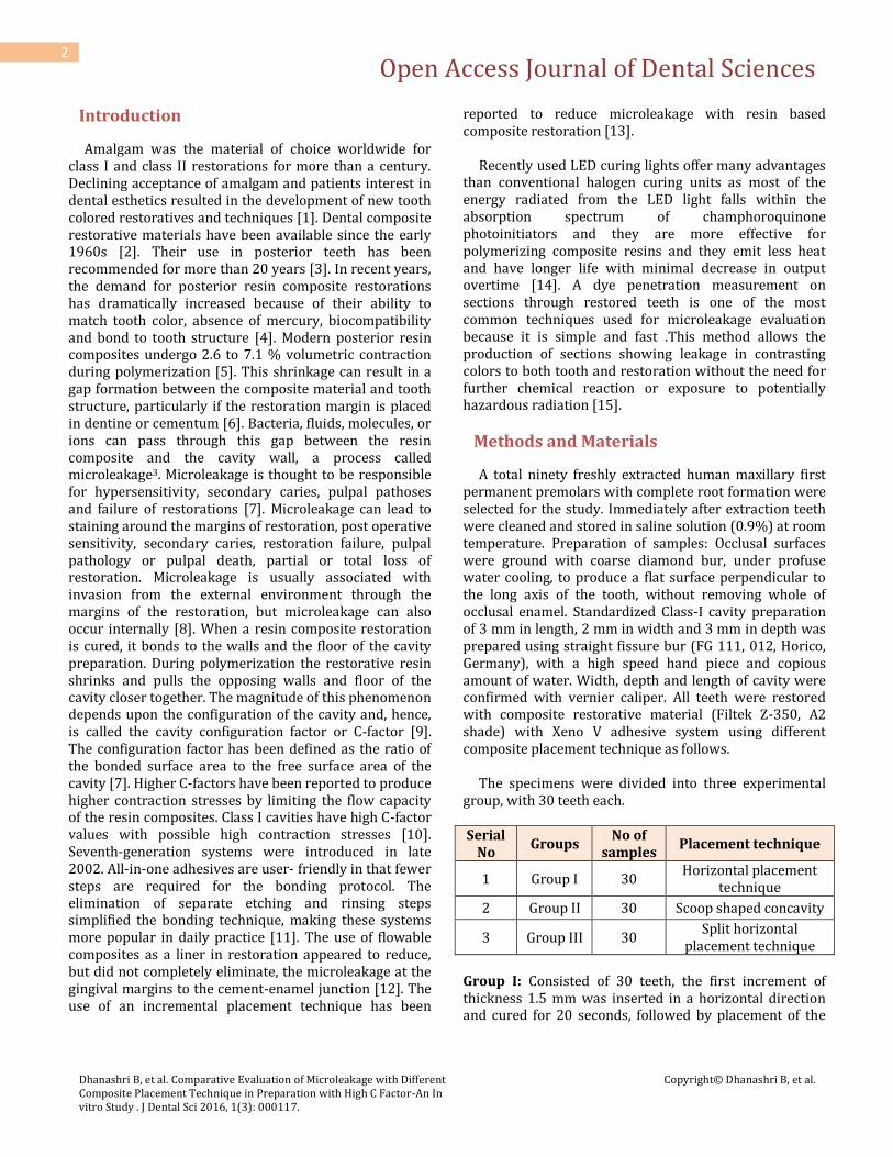
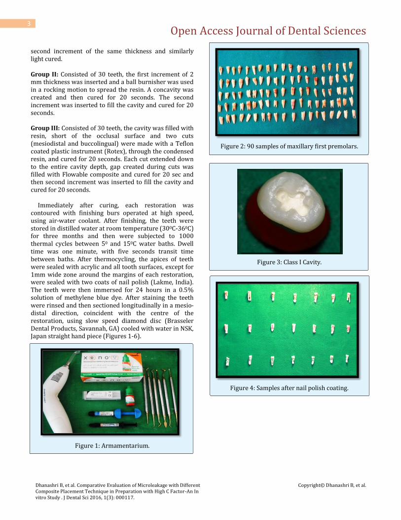
A total ninety freshly extracted human maxillary first permanent premolars with complete root formation were selected for the study. Immediately after extraction teeth were cleaned and stored in saline solution (0.9%) at room temperature. Preparation of samples: Occlusal surfaces were ground with coarse diamond bur, under profuse water cooling, to produce a flat surface perpendicular to the long axis of the tooth, without removing whole of occlusal enamel. Standardized Class-I cavity preparation of 3 mm in length, 2 mm in width and 3 mm in depth was prepared using straight fissure bur (FG 111, 012, Horico, Germany), with a high speed hand piece and copious amount of water. Width, depth and length of cavity were confirmed with vernier caliper. All teeth were restored with composite restorative material (Filtek Z-350, A2 shade) with Xeno V adhesive system using different composite placement technique as follows. The specimens were divided into three experimental group, with 30 teeth each. Serial
No Groups
No of samples
Placement technique
1 Group I 30 Horizontal placement
technique
2 Group II 30 Scoop shaped concavity
3 Group III 30 Split horizontal
placement technique Group I: Consisted of 30 teeth, the first increment of thickness 1.5 mm was inserted in a horizontal direction and cured for 20 seconds, followed by placement of the
Open Access Journal of Dental Sciences
Dhanashri B, et al. Comparative Evaluation of Microleakage with Different Composite Placement Technique in Preparation with High C Factor-An In vitro Study . J Dental Sci 2016, 1(3): 000117.
Copyright© Dhanashri B, et al.
3
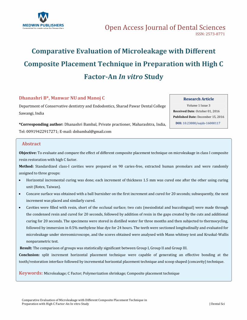
second increment of the same thickness and similarly light cured. Group II: Consisted of 30 teeth, the first increment of 2 mm thickness was inserted and a ball burnisher was used in a rocking motion to spread the resin. A concavity was created and then cured for 20 seconds. The second increment was inserted to fill the cavity and cured for 20 seconds. Group III: Consisted of 30 teeth, the cavity was filled with resin, short of the occlusal surface and two cuts (mesiodistal and buccolingual) were made with a Teflon coated plastic instrument (Rotex), through the condensed resin, and cured for 20 seconds. Each cut extended down to the entire cavity depth, gap created during cuts was filled with Flowable composite and cured for 20 sec and then second increment was inserted to fill the cavity and cured for 20 seconds. Immediately after curing, each restoration was contoured with finishing burs operated at high speed, using air-water coolant. After finishing, the teeth were stored in distilled water at room temperature (300C-360C) for three months and then were subjected to 1000 thermal cycles between 50 and 150C water baths. Dwell time was one minute, with five seconds transit time between baths. After thermocycling, the apices of teeth were sealed with acrylic and all tooth surfaces, except for 1mm wide zone around the margins of each restoration, were sealed with two coats of nail polish (Lakme, India). The teeth were then immersed for 24 hours in a 0.5% solution of methylene blue dye. After staining the teeth were rinsed and then sectioned longitudinally in a mesio-distal direction, coincident with the centre of the restoration, using slow speed diamond disc (Brasseler Dental Products, Savannah, GA) cooled with water in NSK, Japan straight hand piece (Figures 1-6).
Figure 1: Armamentarium.
Figure 2: 90 samples of maxillary first premolars.
Figure 3: Class I Cavity.
Figure 4: Samples after nail polish coating.
Open Access Journal of Dental Sciences
Dhanashri B, et al. Comparative Evaluation of Microleakage with Different Composite Placement Technique in Preparation with High C Factor-An In vitro Study . J Dental Sci 2016, 1(3): 000117.
Copyright© Dhanashri B, et al.
4
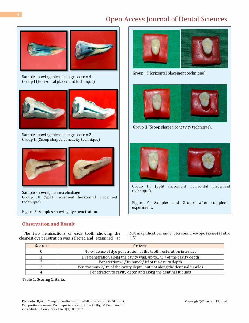
Sample showing microleakage score = 4 Group I (Horizontal placement technique)
Sample showing microleakage score = 2 Group II (Scoop shaped concavity technique)
Sample showing no microleakage Group III (Split increment horizontal placement technique)
Figure 5: Samples showing dye penetration.
Group I (Horizontal placement technique).
Group II (Scoop shaped concavity technique).
Group III (Split increment horizontal placement technique).
Figure 6: Samples and Groups after complete experiment.
Observation and Result
The two hemisections of each tooth showing the cleanest dye penetration was selected and examined at
20X magnification, under stereomicroscope (Zeiss) (Table 1-3).
Table 1: Scoring Criteria.
Scores Criteria
0 No evidence of dye penetration at the tooth restoration interface
1 Dye penetration along the cavity wall, up to1/3rd of the cavity depth 2 Penetration>1/3rd but<2/3rd of the cavity depth 3 Penetration>2/3rd of the cavity depth, but not along the dentinal tubules 4 Penetration to cavity depth and along the dentinal tubules
Open Access Journal of Dental Sciences
Dhanashri B, et al. Comparative Evaluation of Microleakage with Different Composite Placement Technique in Preparation with High C Factor-An In vitro Study . J Dental Sci 2016, 1(3): 000117.
Copyright© Dhanashri B, et al.
5
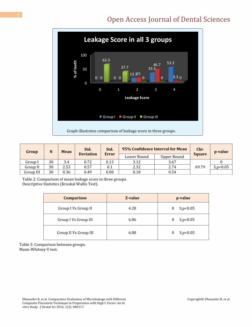
Graph illustrates comparison of leakage score in three groups.
Table 2: Comparison of mean leakage score in three groups. Descriptive Statistics (Kruskal Wallis Test).
Comparison Z-value p-value
Group I Vs Group II 4.28 0 S,p<0.05
Group I Vs Group III 6.86 0 S,p<0.05
Group II Vs Group III 6.88 0 S,p<0.05
Table 3: Comparison between groups. Mann-Whitney U test.
0
50
100
0 1 2 3 4
0 013.3
33.3
53.3
0 015
46.7
3.3
63.3
37.7
0 0 0% o
f te
eth
Leakage Score
Leakage Score in all 3 groups
Group I Group II Group III
Group N Mean Std.
Deviation Std.
Error 95% Confidence Interval for Mean Chi-
Square p-value
Lower Bound Upper Bound
Group I 30 3.4 0.72 0.13 3.12 3.67 69.79
0 Group II 30 2.53 0.57 0.1 2.32 2.74 S,p<0.05 Group III 30 0.36 0.49 0.08 0.18 0.54
Open Access Journal of Dental Sciences
Dhanashri B, et al. Comparative Evaluation of Microleakage with Different Composite Placement Technique in Preparation with High C Factor-An In vitro Study . J Dental Sci 2016, 1(3): 000117.
Copyright© Dhanashri B, et al.
6
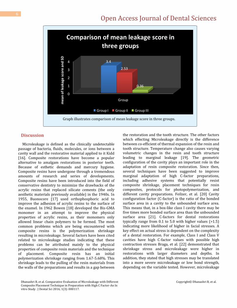
Graph illustrates comparison of mean leakage score in three groups.
Discussion
Microleakage is defined as the clinically undetectable passage of bacteria, fluids, molecules, or ions between a cavity wall and the restorative material applied to it Kidd [16]. Composite restorations have become a popular alternative to amalgam restorations in posterior teeth. Because of esthetic demands and mercury hygiene. Composite resins have undergone through a tremendous amounts of research and series of developments. Composite resins have been introduced into the field of conservative dentistry to minimize the drawbacks of the acrylic resins that replaced silicate cements (the only aesthetic materials previously available) in the 1940s. In 1955, Buonocore [17] used orthophosphoric acid to improve the adhesion of acrylic resins to the surface of the enamel. In 1962 Bowen [18] developed the Bis-GMA monomer in an attempt to improve the physical properties of acrylic resins, as their monomers only allowed linear chain polymers to be formed. The most common problems which are being encountered with composite resins is the polymerization shrinkage resulting in microleakage. Several factors have been cited related to microleakage studies indicating that these problems can be attributed mainly to the physical properties of composite resin materials and the technique of placement. Composite resin has an initial polymerization shrinkage ranging from 1.67-5.68%. This shrinkage leads to the pulling of the resin materials from the walls of the preparations and results in a gap between
the restoration and the tooth structure. The other factors which effecting Microleakage directly is the difference between co-efficient of thermal expansion of the resin and tooth structure. Temperature change also causes varying volumetric changes in the resin and tooth structure leading to marginal leakage [19]. The geometric configuration of the cavity plays an important role in the adaptation of resin composite restoration. Since then, several techniques have been suggested to improve marginal adaptation of high C-factor preparations, including adhesive systems that potentially resist composite shrinkage, placement techniques for resin composites, protocols for photopolymerization, and different cavity preparations. Feilzer, et al. [20] Cavity configuration factor (C-factor) is the ratio of the bonded surface area in a cavity to the unbounded surface area. This means that, in a box-like class I cavity there may be five times more bonded surface area than the unbounded surface area [21]. C-factors for dental restorations typically range from 0.1 to 5.0 with higher values (>1.5) indicating more likelihood of higher in facial stresses. A key effect on actual stress is dependent on the complexity of a dental restoration. For example, Class I and Class V cavities have high C-factor values with possible high contraction stresses Braga, et al. [22] demonstrated that shrinkage stress and microleakage were higher in restorations with larger diameters and depths. In addition, they stated that high stresses may be translated into a more severe microleakage or low bond strength, depending on the variable tested. However, microleakage
0
1
2
3
4
Group
3.4
2.53
0.36
me
an o
f le
akag
e s
core
an
d S
D
Comparison of mean leakage score in three groups
Group I Group II Group III
Open Access Journal of Dental Sciences
Dhanashri B, et al. Comparative Evaluation of Microleakage with Different Composite Placement Technique in Preparation with High C Factor-An In vitro Study . J Dental Sci 2016, 1(3): 000117.
Copyright© Dhanashri B, et al.
7
seemed to be related to restorations volume, but not to its C factor. After cavity preparation self etch adhesive-Xeno V was applied to the cavity wall and cure with LED for 20 sec. Various generation of adhesive system available in market. Seventh-generation systems were introduced in late 2002 in which etchant, primer, and adhesive are combined in a single bottle, eliminating an additional mixing and/or placement step over the sixth-generation systems. It also known as “all-in-one” and “self etch adhesive”. Xeno V has the lowest pH among the three self-etch adhesives. Xeno V has pH <2 which makes it “intermediatory strong” self-etch adhesive Vineeta Nikhil [23]. After the application of adhesive bond 20 sec curing was done with blue light LED light. In recent years, several new polymerization techniques and curing units have been introduced in an attempt to affect polymerization shrinkage. Conventional quartz halogen curing lights with higher intensities, plasma arc curing lights, blue light emitting diode curing lights, and argon lasers are used for polymerization of direct resin-based restorative materials Manhart, et al. [24]. The thermo cycling and dwell times in microleakage evaluation of bonded restorations. They stated that although a simple review of the literature in the last few years would tend to support limited or no effects of the thermal insult for composite restorations thermocycled with short dwell times, evaluation of microleakage must include thermocycling in order to simulate intraoral conditions. However, the relationship between thermal expansion and the duration of the temperature exposure is an important factor in evaluating the Microleakage potential of a restorative material [25]. After application of xenoV cavity was restored with Filtek Z-350XT (3M ESPE, USA). Filtek™ Z350 XT Nano Hybrid Universal Restorative is a visible light-activated nanohybrid composite.The resin technology is based on this restorative resin, replacing some of the TEGDMA with PEGDMA to moderate shrinkage so it exhibits a low shrinkage relative to competitive composites in this class of materials. Thermal stresses can be pathologic in two ways. Firstly, differential thermal changes induce mechanical stresses that can cause crack propagation through the bonded interface. Secondly, gap volume changes associated with changing gap dimensions pump pathogenic oral fluids in and out of the gaps with possible pulpal complications Gale & Darvell [26]. Dye penetration method was used to evaluate the Microleakage because ethylene blue dye penetration method provides the evaluators with a perfect and easy visualization of the prepared cavity in the digital images which provide the evaluators with a clear reference point from which to score. The dye also
provides an excellent contrast with the surrounding environment.
Conclusion
a. Split horizontal placement technique scored the lowest mean Microleakage value and was comparatively better than horizontal placement technique and was statistically significant.
b. Scoop shaped concavity technique scored the less mean Microleakage value, but less effective than split increment horizontal technique and was statistically significant.
c. Horizontal placement technique scored highest mean Microleakage value among the experimental groups. It was less effective composite placement technique and was statistically significant.
References
1. Radhika M, Girija S, Sajjan KBN, Neetu M (2010) Effect of different placement techniques on marginal microleakage of deep class- II cavities restored with two composite resin formulations. Journal of conservative dentistry 13(1): 9-15.
2. Loguercio AD, de Oliveira RBJ, Reis A, Miranda GRH (2004) In vitro microleakage of packable composites in Class II restorations. Quintessence International 35(1): 29-34.
3. Turkun LS, Aktener BO, Ateş M (2003) Clinical evalutation of different posterior resin composite Materials: A 7-year report. Quintessence International 34: 418-426.
4. Herrero AA, Yaman P, Dennison JB (2005) Polymerization shrinkage and depth of cure of packable composites. Quintessence International 36: 25-31.
5. Hilton TJ, Schwartz RS, Ferracane J (1997) Microleakage of four classes II resin composite insertion techniques at intraoral temperature. Quintessence International 28(2): 135-145.
6. Yazici RA, Celik C, Ozgunaltay G (2004) Microleakage of different resin composite types. Quintessence International 23(10): 790-794.
7. Franco EB, Lopes LG, Lia Mondelli RF, Da Silva ESMH, Lauris JRP (2003) Effect of the cavity configuration factor on the marginal microleakage of esthetic

Open Access Journal of Dental Sciences
Dhanashri B, et al. Comparative Evaluation of Microleakage with Different Composite Placement Technique in Preparation with High C Factor-An In vitro Study . J Dental Sci 2016, 1(3): 000117.
Copyright© Dhanashri B, et al.
8
restorative materials. American Journal of Dentistry 1: 211-214.
8. Andrea Fabinelli, Sarah Pollington, Carel LD, Maria CC, Cecilia G The Relence Of Microleakage Studies International Dentistry SA 9(3): 64-74.
9. Choi KK, Ferracane JL, Ryu GJ, Choi SM, Lee MJ, et al. (2004) Effects of cavity configuration on composite restoration. Operative Dentistry 29(4): 462-469.
10. Hashimoto M, Ohno H, Kaga M, Endo K, Sano H, et al. (2000) In vivo degradation of resin—dentin bonds over 1 to 3 years. J Dent Res 79: 1385-1391.
11. Jorge Perdiga (2007)New Developments in Dental Adhesion. Dental Clinic North America 51: 333-357.
12. Leevailoj C, Cochran MA, Matis BA, Moore BK, Platt JA (2001) Microleakage of posterior packable resin composite with and without flowable liners. Operative Dentistry 26: 302-307.
13. Poskus LT, Placido E, Cardoso PE (2004) Influence of adhesive system and placement technique on microleakage of resin-based composite restorations. J Adhesive Dent 227-232.
14. Yazici R, Cigdem C, Berrin D (2008) Effect of different light curing units on the microleakage of flowable composite resin. Eur J Dent 2: 240-246.
15. Dejou J, Sindres V, Camps J (1996) Influence of criteria on the results of in vitro evaluation of microleakage. Dent Mater 12(6): 342-349.
16. Kidd EAM (1976) Microleakage a review. J Dent 4(5): 199-206.
17. Buonocore MG (1955) A simple method of increasing the adhesion of acrylic filling materials to enamel surfaces. J Dent Res 34(6): 849-853.
18. Bowen RL (1996) Dental filling material comprising vinyl-silane treated fused silica and a binder consisting of the reaction product of bisphenol and glycidyl methacrylate. US Patent 3: 066 -112.
19. Tung FF, Hsieh WW, Estafan D (2000) In vitro microleakage study of a condensable and flowable composite resin. General Dentistry 48 (6): 711-715.
20. Feilzer AJ, De Gee AJ, Davidson CL (1987) Setting stress in composite resin in relation to configuration of the restoration. Journal Dental Research 66: 1636-1639.
21. Sillas D, Welingtom D, Maria H (2007) Influence of resin composite insertion technique in preparations with a high C-factor. Quintessence International 38: 829-835.
22. Braga RR, Ferracane JL (2004) Alternatives in polymerization contraction stress management. Critical Reveive Oral Biology Medicine 15(3): 176-184.
23. Vineeta N, Vijay S (2011) Comparative evaluation of bond strength of three contemporary self etch adhesive-An in vivo study. Contemporary clinical dentistry 2(2): 94-97.
24. Manhart J, Garcia GF, Hickel R (2002) Direct posterior restorations: clinical results and new developments. Dental Clinic North America 46(2): 303-339.
25. Kristi J. Rossomando, Stanely LW (1995) Thermocycling and dwell time in microleakage evaluation for bonded restoration. Dent Mater 11(1): 47-51.
26. Gale MS, Darvell BW (1999) Thermal cycling procedures for laboratory testing of dental restorations. J Dentistry 27(2): 89-99.


![Research Article A Comparative Evaluation of Microleakage ...pass through this gap between the resin composite and the cavity wall, a process called microleakage [ ]. Microleakage](https://static.fdocuments.net/doc/165x107/6101fac525ffb1579b200818/research-article-a-comparative-evaluation-of-microleakage-pass-through-this.jpg)

