Subjective and Objective Quality Assessment of Image: A Survey

501
Abstract: Since the introduction of cone-beam computed tomography (CBCT), several novel systems with different technical specifications and settings have become commercially available. Therefore, it is essential to evaluate CBCT systems for differences in the subjective quality of images obtained for various dental procedures. We evaluated the subjective image quality of cross-sectional scans obtained from various CBCT systems. Images of three cadaver mandibles were obtained from four different CBCT units: 1) Veraviewepocs 3D 40 × 40 mm field of view (FOV) (voxel size: 0.125 × 0.125 × 0.125 mm), 2) Iluma, low-resolution (voxel size: 0.3 × 0.3 × 0.3 mm), 3) Kodak, 50 × 3.7 cm FOV (voxel size: 0.076 × 0.076 × 0.076 mm), and 4) Vatech 12 × 8.5cm FOV (voxel size: 0.160 × 0.160 × 0.160 mm). We assessed subjective image quality and the visibility of 10 specific features, namely, caries, amalgam restoration, final implant drill, root canal filling, metal crown, mandibular canal, mental foramen, tooth (periodontal ligament space and lamina dura), trabecular pattern, and soft tissue. Images were viewed and scored by five calibrated observers, and image quality was ranked from best to worst. The Veraviewepocs 3D had the highest quality images for most of the assessed features, whereas the
Iluma low-resolution scans were rated as the lowest quality images. (J Oral Sci 53, 501-508, 2011)
Keywords: CBCT; subjective image quality; radiology; dentistry.
IntroductionCone-beam computerized tomography has started to
gain broad acceptance in dentomaxillofacial imaging and has largely replaced conventional tomography for most diagnostic tasks in dentistry (1). CBCT is now commonly used for a variety of purposes in implanto-logy, dentomaxillofacial surgery, image-guided surgical procedures, orthodontics, periodontics, endodontics, and cariology (2-4). CBCT systems focus a cone-shaped x-ray beam on a two-dimensional (2D) detector—either an amorphous silicon flat panel or an image intensifier/CCD/CMOS—that performs one pass or less around the patient’s head to produce a series of 2D images. The data set is then processed with a cone-beam algorithm, which allows the operator to extract planar and curved reconstructions of varying thicknesses in any orientation and to generate accurate three-dimensional (3D) images of bone and soft tissue surfaces. Voxels are isotropic and can be as small as 0.076 × 0.076 × 0.076 mm (2-6). The use of CBCT in clinical practice has a number of poten-tial advantages over conventional tomography, such as easier image acquisition, greater image accuracy, lower effective radiation dose, faster scan time, and greater cost-effectiveness (7-10). Disadvantages associated with dental CBCT include scatter radiation, limited dynamic
Correspondence to Dr. Kıvanç Kamburoğlu, Department of Dentomaxillofacial Radiology, Faculty of Dentistry, Ankara University, Ankara, TurkeyTel: +90-3122965632Fax: +90-3122123954Email: [email protected] & [email protected].
edu.tr
Journal of Oral Science, Vol. 53, No. 4, 501-508, 2011
Original
Comparative assessment of subjective image quality ofcross-sectional cone-beam computed tomography scans
Kıvanç Kamburoğlu1), Sema Murat2), Eray Kolsuz1), Hakan Kurt1),Selcen Yüksel3) and Candan Paksoy1)
1)Department of Dentomaxillofacial Radiology, Faculty of Dentistry, Ankara University, Ankara, Turkey2)Department of Prosthodontics, Faculty of Dentistry, Ankara University, Ankara, Turkey
3)Department of Biostatistics, Faculty of Medicine, Ankara University, Ankara, Turkey
(Received 31 August and accepted 5 November 2011)

502
range, minimal soft-tissue detail, and beam-hardening artifacts caused by dental-care material and implants (11-13).
A CBCT system was found to be superior to a multide-tector helical CT system in displaying hard tissues in the dental area while substantially decreasing patient radia-tion dose exposure (14). Since the introduction of CBCT, several novel systems with different technical specifica-tions and settings have become commercially available. Therefore, it is essential to evaluate CBCT systems for differences in the subjective quality of images obtained for various dental procedures. We assessed the subjective image quality of cross-sectional scans obtained from four CBCT systems.
Materials and MethodsThis study was conducted using 3 mandibles from
human cadavers obtained from the Department of Anatomy at Gulhane Academy. While alive, all subjects had given informed consent to donate their body for
medical research and teaching. Two of the mandibles were edentulous. In one of these mandibles, two implant locations were drilled (MIS, Implants Technologies Ltd., Shlomi, Israel), and final drills were inserted. No opera-tion was performed on the other edentulous mandible. The third mandible was dentulous and had one amalgam filling, one root canal filling, and one metal crown.
Images of the three cadaver mandibles were obtained using four different CBCT units:
1) Veraviewepocs 3D model X550 (J Morita Mfg. Corp., Kyoto, Japan) with a flat-panel detector offering digital 3D, panoramic, and cephalometric imaging options. With this system, images were obtained at a tube voltage of 60-90 kV and a tube current of 3 mA. The exposure time was 9.4 s and the field of view (FOV) was 40 × 40 mm (voxel size: 0.125 × 0.125 × 0.125 mm).
2) Iluma Ultra Cone-beam CT Scanner (3M Imtec, Ardmore, OK, USA) with a 24.4 × 19.5 cm amorphous silicon flat-panel image detector and a cylindrical volume of reconstruction up to 21.1 × 14.2 cm. With the Iluma
Fig. 1 Cross-sectional images of a trabecular pattern, from different CBCT units. a: Veraviewepocs 3D 40 × 40 mm FOV (0.125 × 0.125 × 0.125 mm voxel size); b: Iluma, low-resolution (0.3 × 0.3 × 0.3 mm voxel size); c: Kodak 9000, 50 × 3.7 cm FOV (0.076 × 0.076 × 0.076 mm voxel size); d: Vatech PanX-Duo3D 12 × 8.5 cm FOV (0.160 × 0.160 × 0.160 mm voxel size).
503
system, images were obtained at tube voltage of 120 kVp and a tube current of 3.8 mA. The exposure time was 40 s and images were reconstructed at low-resolution (voxel size: 0.3 × 0.3 × 0.3 mm).
3) Kodak 9000 Extra-oral imaging system (Eastman Kodak Co, Rochester, NY, USA). Images were obtained at tube voltage of 60 kVp and a tube current of 3 mA. The exposure time was 13.2 s and the FOV was 50 × 37 mm (voxel size: 0.076 × 0.076 × 0.076 mm).
4) Vatech PanX-Duo3D_Pano/CBCT (Vatech, Seoul, Korea) system with a 12 × 8.5 cm amorphous silicon flat-panel image detector. Images were obtained at tube voltage of 85 kVp and a tube current of 5.7 mA (voxel size: 0.160 × 0.160 × 0.160 mm). The exposure time was 11 s.
Axial scans and multiplanar reconstructions were obtained, and volumetric data were reconstructed using
the respective software programs of the systems to provide serial cross-sectional views. Suitable cross-sectional images were selected and exported for viewing as TIFF files to assess the subjective quality of the different CBCT units. A total of four image sets were obtained, as follows: Veraviewepocs 3D 40 × 40 mm FOV; Iluma, low-resolution; Kodak, 50 × 3.7 cm FOV; and Vatech 12 × 8.5 cm FOV.
Subjective image quality and visibility of the 10 specific features were assessed, namely, caries, amalgam restoration, final implant drill, root canal filling, metal crown, mandibular canal, mental foramen, tooth (peri-odontal ligament space and lamina dura), trabecular pattern, and soft tissue. The image quality of all cross-sectional images obtained from the four different CBCT units was assessed and ranked from best to worst for each feature: 4 for the best and 1 for the worst quality images.
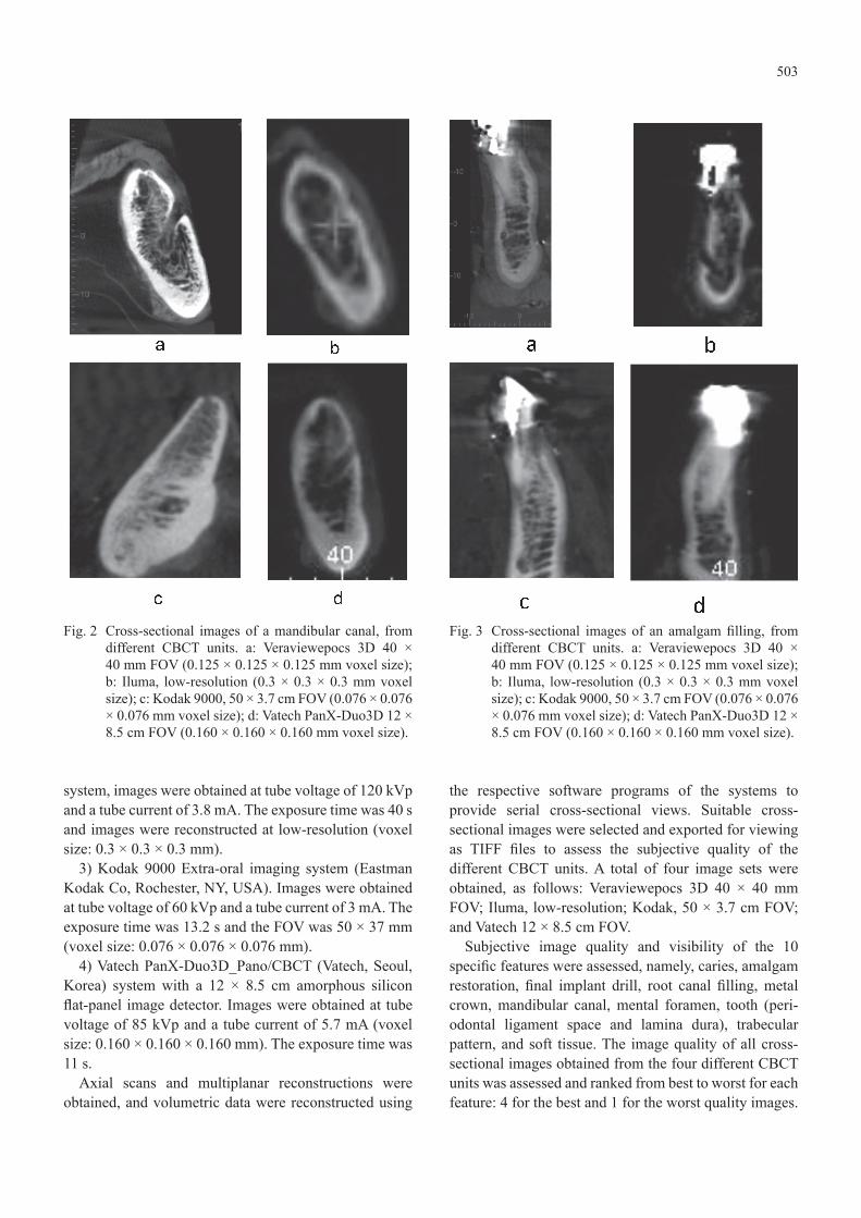
Fig. 2 Cross-sectional images of a mandibular canal, from different CBCT units. a: Veraviewepocs 3D 40 × 40 mm FOV (0.125 × 0.125 × 0.125 mm voxel size); b: Iluma, low-resolution (0.3 × 0.3 × 0.3 mm voxel size); c: Kodak 9000, 50 × 3.7 cm FOV (0.076 × 0.076 × 0.076 mm voxel size); d: Vatech PanX-Duo3D 12 × 8.5 cm FOV (0.160 × 0.160 × 0.160 mm voxel size).
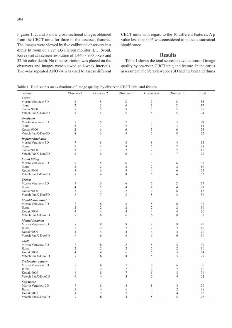
Fig. 3 Cross-sectional images of an amalgam filling, from different CBCT units. a: Veraviewepocs 3D 40 × 40 mm FOV (0.125 × 0.125 × 0.125 mm voxel size); b: Iluma, low-resolution (0.3 × 0.3 × 0.3 mm voxel size); c: Kodak 9000, 50 × 3.7 cm FOV (0.076 × 0.076 × 0.076 mm voxel size); d: Vatech PanX-Duo3D 12 × 8.5 cm FOV (0.160 × 0.160 × 0.160 mm voxel size).
504
Figures 1, 2, and 3 show cross-sectional images obtained from the CBCT units for three of the assessed features. The images were viewed by five calibrated observers in a dimly lit room on a 22" LG Flatron monitor (LG, Seoul, Korea) set at a screen resolution of 1,440 × 900 pixels and 32-bit color depth. No time restriction was placed on the observers and images were viewed at 1-week intervals. Two-way repeated ANOVA was used to assess different
CBCT units with regard to the 10 different features. A p value less than 0.05 was considered to indicate statistical significance.
ResultsTable 1 shows the total scores on evaluations of image
quality by observer, CBCT unit, and feature. In the caries assessment, the Veraviewepocs 3D had the best and Iluma
Table 1 Total scores on evaluations of image quality, by observer, CBCT unit, and feature Feature Observer 1 Observer 2 Observer 3 Observer 4 Observer 5 TotalCaries Morita Veraview 3D 8 8 8 2 8 34Iluma 2 2 4 7 2 17Kodak 9000 5 6 3 6 5 25Vatech PanX-Duo3D 5 4 5 5 5 24Amalgam Morita Veraview 3D 5 6 7 8 3 29Iluma 5 2 2 2 3 14Kodak 9000 2 6 6 5 6 25Vatech PanX-Duo3D 8 6 5 5 8 32Implant final drillMorita Veraview 3D 7 8 8 8 4 35Iluma 7 4 4 6 7 28Kodak 9000 3 2 2 2 2 11Vatech PanX-Duo3D 3 6 6 4 7 26Canal fillingMorita Veraview 3D 5 8 6 8 6 33Iluma 2 2 2 2 2 10Kodak 9000 5 6 4 4 6 25Vatech PanX-Duo3D 8 4 8 6 6 32CrownMorita Veraview 3D 5 6 6 6 2 25Iluma 4 5 4 4 4 21Kodak 9000 3 2 2 2 6 15Vatech PanX-Duo3D 8 7 8 8 8 39Mandibular canalMorita Veraview 3D 7 8 8 8 6 37Iluma 2 2 2 2 2 10Kodak 9000 4 4 4 4 4 20Vatech PanX-Duo3D 7 6 6 6 8 33Mental foramenMorita Veraview 3D 8 8 8 8 8 40Iluma 2 2 2 2 2 10Kodak 9000 4 4 4 4 4 20Vatech PanX-Duo3D 6 6 6 6 6 30ToothMorita Veraview 3D 7 8 8 8 8 39Iluma 2 2 2 2 2 10Kodak 9000 4 4 6 5 5 24Vatech PanX-Duo3D 7 6 4 5 5 27Trabecular patternMorita Veraview 3D 8 6 7 8 6 35Iluma 2 2 2 2 2 10Kodak 9000 6 8 7 5 8 34Vatech PanX-Duo3D 4 4 4 5 4 21Soft tissueMorita Veraview 3D 7 8 8 8 8 39Iluma 2 4 6 4 2 18Kodak 9000 4 2 2 3 4 15Vatech PanX-Duo3D 7 6 4 5 6 28

505
had the worst score (Veraviewepocs 3D > Kodak 9000 > Vatech > Iluma). A statistically significant difference was found between the Veraviewepocs 3D and Iluma systems (P = 0.003). In the amalgam assessment, the Vatech had the best and Iluma had the worst score (Vatech > Veraviewepocs 3D > Kodak 9000 > Iluma). Statistically significant differences were found between the Vatech and Iluma systems (P = 0.001) and between the Veraviewe-pocs 3D and Iluma (P = 0.006). In the assessment of final implant drill, the Veraviewepocs 3D had the best and the Kodak 9000 had the worst score (Veraviewepocs 3D > Vatech > Iluma > Kodak 9000). A statistically significant difference was found between the Kodak 9000 and the other three units (P < 0.001). In the assessment of root canal filling, the Veraviewepocs 3D had the best and the Iluma had the worst score (Veraviewepocs 3D > Vatech > Kodak 9000 > Iluma). A statistically significant differ-ence was found between the Iluma and the other three units (P < 0.001). In the assessment of the metal crown, the Vatech had the best and the Kodak 9000 had the worst score (Vatech > Veraviewepocs 3D > Iluma > Kodak 9000). Statistically significant differences were found
between the Vatech and the other three units (P < 0.001) and between the Veraviewepocs 3D and Kodak 9000 (P = 0.023). In the assessment of the mandibular canal, the Veraviewepocs 3D had the best and the Iluma had the worst score (Veraviewepocs 3D > Vatech > Kodak 9000 > Iluma). Statistically significant differences were found between the Veraviewepocs 3D and Iluma (P < 0.001), the Veraviewepocs 3D and Kodak (P < 0.001), the Iluma and the other three units (P < 0.001), the Kodak 9000 and the other three units (P < 0.001), the Vatech and Iluma, (P < 0.001), and the Vatech and Kodak (P < 0.001). In the assessment of the mental foramen, the Veraviewe-pocs 3D had the best and Iluma had the worst score (Veraviewepocs 3D > Vatech > Kodak 9000 > Iluma). There were statistically significant differences among all systems (P < 0.001). In assessment of the tooth, the Vera-viewepocs 3D had the best and Iluma had the worst score (Veraviewepocs 3D > Vatech > Kodak 9000 > Iluma). There were statistically significant differences among all systems (P < 0.001) except between the Vatech and Vera-viewepocs 3D systems (P = 0.886). In the assessment of the trabecular pattern, the Veraviewepocs 3D had the best and Iluma had the worst score (Veraviewepocs 3D > Kodak 9000 > Vatech > Iluma). There were statistically significant differences between the Iluma and the other three systems (P < 0.001) and between the Vatech and the other three systems (P < 0.001). In the assessment of soft tissue, the Veraviewepocs 3D had the best and Kodak 9000 had the worst scores (Veraviewepocs 3D > Vatech > Iluma > Kodak 9000). There were statistically significant differences between the Veraviewepocs 3D and the other three systems (P < 0.001) and between the Vatech and the other three systems (P < 0.001). No statistically signifi-cant difference was found between the Iluma and Kodak 9000 (P = 1). Figure 4 shows the estimated marginal means for the 10 features and 4 CBCT units.
DiscussionUnder clinical conditions, the subjective image quality
of CBCT scans and the capability of CBCT to assess different features is influenced by a number of variables, including device, FOV, voxel size, tube voltage and current, and other technical factors. Because of its small FOV and high-resolution images, the Veraviewepocs 3D produced the highest quality images for most of the assessed features. The Iluma produced the worst images, probably because of the large voxel size selected for the Iluma scanner. We used the 0.3 mm voxel size for the Iluma because the 0.1 mm and 0.2 mm voxel sizes require reconstruction times that are too long for use in routine clinical dental imaging. With CBCT, clinical
Fig.4 Estimated marginal means for the 10 features and 4 CBCT units.

506
assessment of different features might also be affected by observer performance and viewing conditions, as well as by hardware and software specifications and beam-hardening artifacts. In this ex vivo study, patient motion, which is an important factor in CBCT image quality, was not a concern. In addition, observers were calibrated and trained in interpreting CBCT images. We used cross-sectional images since that is the preferred view for most diagnostic tasks.
Beam-hardening artifacts due to dental implants are seen as dark and light streaks in CBCT images and can seriously degrade the visual quality and interpretability of CBCT images. It has been suggested that meaningful artifact reduction must be based on greater sophistication in mathematical modeling of the actual image-acquisition process rather than on postprocessing of the erroneous results obtained from the rather crude reconstruction algorithms used presently (15). In this study, the Vera-viewepocs 3D had the highest and the Kodak 9000 the lowest scores in the assessment of final implant drills. A statistically significant difference was found between the Kodak 9000 and the other three units (P < 0.001). These differences might be due to sensor type, the artifact reduction algorithms used, or the hardware and software capabilities of the CBCT systems.
In one study (16), a dry mandible was scanned with five CBCT scanners (Accuitomo 3D, i-CAT, NewTom 3G, Galileos, Scanora 3D) and one multislice spiral computed tomography (MSCT) system (Somatom Sensation 16), using 13 different scan protocols. The visibility of 11 anatomical structures and overall image noise were compared between the CBCT and MSCT systems. Five independent observers reviewed the images and assessed image quality on a five-point scale. Significant differences were found in the visibility of different anatomical structures and the image noise level between the MSCT and CBCT systems and among the five CBCT systems (P = 0.0001) (16). The Accuitomo system was superior to the MSCT and all other CBCT systems in depicting anatomical structures, while the MSCT was superior to all the CBCT systems with regard to reduced image noise. In the present study, a different version of the Accuitomo system—the Veraviewepocs 3D with a flat-panel detector—had the best results for most of the assessed features.
Subjective image quality for periapical diagnosis and implant planning was assessed using CBCT with different exposure parameters and FOVs. Examinations were performed of the posterior part of the jaws on a skull phantom with the 3D Accuitomo (FOV 3 cm × 4 cm) and 3D Accuitomo FPD (FOVs 4 cm × 4 cm and 6 cm × 6
cm). The overall ranking of FOVs was 4 cm × 4 cm and 6 cm × 6 cm, followed by 3 cm × 4 cm (17). We found that smaller FOVs were associated with higher quality images. Another study (18) evaluated image quality (by examining segmentation accuracy) and radiation dose of CBCT scanners. The i-CAT scanner had the best ratio of radiation dose to image quality, but was not evaluated in the present study. In addition, our study was not designed to measure the effective radiation doses derived from the different FOVs in the systems tested.
Soğur et al. (19) compared the subjective quality of limited cone-beam computed tomography (LCBCT) scans, storage phosphor plate (SPP) images, and F-speed film images in the evaluation of the length and homo-geneity of root fillings. Root canals of 17 extracted permanent mandibular incisors were filled. Images were obtained with the Accu-I-Tomo LCBCT, Digora Optime image plate system, and F-speed film. The image quality of the SPP images was subjectively as good as conven-tional film images, and superior to LCBCT images, in the evaluation of both the homogeneity and length of root fillings in single-rooted teeth (19). In the present study, the Veraviewepocs 3D and Vatech systems received the highest scores in the assessment of root canal filling. However, we did not compare the subjective image quality of CBCT and intraoral images.
CBCT systems offer different sensor types, FOVs, and exposure settings. CBCT systems with small FOVs were well suited for periapical diagnosis and implant planning (20). A recent study found that the use of smaller FOVs resulted in lower effective radiation doses. The authors suggested that, as a rule, smaller FOVs should be used for dental imaging and that systems with larger FOVs should be restricted to cases where such FOVs are required (21).
Radiation exposure is another concern in radiographic imaging, and doses from CBCT vary substantially by device, FOV, and other technical factors. Although CBCT is an innovative and promising technology, effec-tive radiation doses are still higher than in conventional panoramic and intraoral imaging, and the use of CBCT in routine clinical practice is impractical. In view of the concerns regarding radiation exposure, it is essential that new CBCT units offering higher-definition images with similar or lower effective doses than film are introduced into routine use in clinical dentistry (22,23).
In conclusion, the Veraviewepocs 3D system produced the highest quality images for most of the assessed features, whereas the low-resolution scans from the Iluma system were rated as the lowest quality images.

507
AcknowledgmentsWe are grateful to Dentistomo Dentomaxillofacial
Imaging Center, Ankara, Turkey and Ayrıntı Dentomax-illofacial Imaging Center, Ankara, Turkey for providing the CBCT units.
References 1. Scarfe WC, Farman AG, Levin MD, Gane D (2010)
Essentials of maxillofacial cone beam computed tomography. Alpha Omegan 103, 62-67.
2. White SC (2008) Cone-beam imaging in dentistry. Health Phys 95, 628-637.
3. Kamburoğlu K, Kolsuz E, Kurt H, Kiliç C, Özen T, Paksoy CS (2011) Accuracy of CBCT measure-ments of a human skull. J Digit Imaging 24, 787-793.
4. Kamburoğlu K, Kurt H, Kolsuz E, Öztaş B, Tatar İ, Çelik HH (2011) Occlusal caries depth measurements obtained by five different imaging modalities. J Digit Imaging 24, 804-813.
5. Feldkamp LA, Davis LC, Kress JW (1984) Practical cone-beam algorithm. J Opt Soc Am 1, 612-619.
6. De Vos W, Casselman J, Swennen GR (2009) Cone-beam computerized tomography (CBCT) imaging of the oral and maxillofacial region: a systematic review of the literature. Int J Oral Maxillofac Surg 38, 609-625.
7. White SC, Pharoah MJ (2008) The evolution and application of dental maxillofacial imaging modalities. Dent Clin North Am 52, 689-705.
8. Scarfe WC, Farman AG (2008) What is cone-beam CT and how does it work? Dent Clin North Am 52, 707-730.
9. Scarfe WC, Farman AG, Sukovic P (2006) Clinical applications of cone-beam computed tomography in dental practice. J Can Dent Assoc 72, 75-80.
10. Patel S (2009) New dimensions in endodontic imaging: Part 2. Cone beam computed tomog-raphy. Int Endod J 42, 463-475.
11. Ritter L, Mischkowski RA, Neugebauer J, Dreiseidler T, Scheer M, Keeve E, Zöller JE (2009) The influence of body mass index, age, implants, and dental restorations on image quality of cone beam computed tomography. Oral Surg Oral Med Oral Pathol Oral Radiol Endod 108, e108-116.
12. Draenert FG, Coppenrath E, Herzog P, Müller S, Mueller-Lisse UG (2007) Beam hardening artefacts occur in dental implant scans with the NewTom cone beam CT but not with the dental
4-row multidetector CT. Dentomaxillofac Radiol 36, 198-203.
13. Carrafiello G, Dizonno M, Colli V, Strocchi S, Pozzi Taubert S, Leonardi A, Giorgianni A, Barresi M, Macchi A, Bracchi E, Conte L, Fugazzola C (2010) Comparative study of jaws with multislice computed tomography and cone-beam computed tomography. Radiol Med 115, 600-611.
14. Hashimoto K, Arai Y, Iwai K, Araki M, Kawashima S, Terakado M (2003) A comparison of a new limited cone beam computed tomog-raphy machine for dental use with a multidetector row helical CT machine. Oral Surg Oral Med Oral Pathol Oral Radiol Endod 95, 371-377.
15. Schulze RK, Berndt D, d’Hoedt B (2010) On cone-beam computed tomography artifacts induced by titanium implants. Clin Oral Implants Res 21, 100-107.
16. Liang X, Jacobs R, Hassan B, Li L, Pauwels R, Corpas L, Souza PC, Martens W, Shahbazian M, Alonso A, Lambrichts I (2010) A comparative evaluation of Cone Beam Computed Tomog-raphy (CBCT) and Multi-Slice CT (MSCT) Part I. On subjective image quality. Eur J Radiol 75, 265-269.
17. Lofthag-Hansen S, Thilander-Klang A, Gröndahl K (2011) Evaluation of subjective image quality in relation to diagnostic task for cone beam computed tomography with different fields of view. Eur J Radiol 80, 483-488.
18. Loubele M, Jacobs R, Maes F, Denis K, White S, Coudyzer W, Lambrichts I, van Steenberghe D, Suetens P (2008) Image quality vs radiation dose of four cone beam computed tomography scanners. Dentomaxillofac Radiol 37, 309-318.
19. Soğur E, Baksi BG, Gröndahl HG (2007) Imaging of root canal fillings: a comparison of subjective image quality between limited cone-beam CT, storage phosphor and film radiography. Int Endod J 40, 179-185.
20. Lofthag-Hansen S (2009) Cone beam computed tomography radiation dose and image quality assessments. Swed Dent J Suppl, 4-55.
21. Hirsch E, Wolf U, Heinicke F, Silva MA (2008) Dosimetry of the cone beam computed tomog-raphy Veraviewepocs 3D compared with the 3D Accuitomo in different fields of view. Dentomax-illofac Radiol 37, 268-273.
22. Ludlow JB, Davies-Ludlow LE, Brooks SL, Howerton WB (2006) Dosimetry of 3 CBCT

508
devices for oral and maxillofacial radiology. CB Mercuray, NewTom 3G and i-CAT. Dentomaxil-lofac Radiol 35, 219-226.
23. Ludlow JB, Ivanovic M (2008) Comparative
dosimetry of dental CBCT devices and 64-slice CT for oral and maxillofacial radiology. Oral Surg Oral Med Oral Pathol Oral Radiol Endod 106, 106-114.