Comparative analysis and molecular characterization of ...
13
©FUNPEC-RP www.funpecrp.com.br Genetics and Molecular Research 13 (1): 992-1004 (2014) Comparative analysis and molecular characterization of genomic sequences and proteins of FABP4 and FABP5 from the giant panda (Ailuropoda melanoleuca) B. Song 1,2 , Y.L. Hou 2 , X. Ding 2 , T. Wang 2 , F. Wang 2 , J.C. Zhong 2 , T. Xu 2 , J. Zhong 2 , W.R. Hou 2 and S.R. Shuai 1 1 Institute of Animal Breeding and Genetics, Sichuan Agricultural University, Ya’an, China 2 Key Laboratory of Southwest China Wildlife Resources Conservation (Ministry of Education), College of Life Science, China West Normal University, Nanchong, China Corresponding authors: W.R. Hou / S.R. Shuai E-mail: [email protected] Genet. Mol. Res. 13 (1): 992-1004 (2014) Received January 8, 2013 Accepted July 18, 2013 Published February 20, 2014 DOI http://dx.doi.org/10.4238/2014.February.20.1 ABSTRACT. Fatty acid binding proteins (FABPs) are a family of small, highly conserved cytoplasmic proteins that bind long-chain fatty acids and other hydrophobic ligands. In this study, cDNA and genomic sequences of FABP4 and FABP5 were cloned successfully from the giant panda (Ailuropoda melanoleuca) using reverse transcription polymerase chain reaction (RT-PCR) technology and touchdown-PCR. The cDNAs of FABP4 and FABP5 cloned from the giant panda were 400 and 413 bp in length, containing an open reading frame of 399 and 408 bp, encoding 132 and 135 amino acids, respectively. The genomic sequences of FABP4 and FABP5 were 3976 and 3962 bp, respectively, which each contained four exons and three introns. Sequence alignment indicated a high degree of homology
Transcript of Comparative analysis and molecular characterization of ...

©FUNPEC-RP www.funpecrp.com.brGenetics and Molecular Research 13 (1): 992-1004 (2014)
Comparative analysis and molecular characterization of genomic sequences and proteins of FABP4 and FABP5 from the giant panda (Ailuropoda melanoleuca)
B. Song1,2, Y.L. Hou2, X. Ding2, T. Wang2, F. Wang2, J.C. Zhong2, T. Xu2, J. Zhong2, W.R. Hou2 and S.R. Shuai1
1Institute of Animal Breeding and Genetics, Sichuan Agricultural University, Ya’an, China2Key Laboratory of Southwest China Wildlife Resources Conservation (Ministry of Education), College of Life Science, China West Normal University, Nanchong, China
Corresponding authors: W.R. Hou / S.R. ShuaiE-mail: [email protected]
Genet. Mol. Res. 13 (1): 992-1004 (2014)Received January 8, 2013Accepted July 18, 2013Published February 20, 2014DOI http://dx.doi.org/10.4238/2014.February.20.1
ABSTRACT. Fatty acid binding proteins (FABPs) are a family of small, highly conserved cytoplasmic proteins that bind long-chain fatty acids and other hydrophobic ligands. In this study, cDNA and genomic sequences of FABP4 and FABP5 were cloned successfully from the giant panda (Ailuropoda melanoleuca) using reverse transcription polymerase chain reaction (RT-PCR) technology and touchdown-PCR. The cDNAs of FABP4 and FABP5 cloned from the giant panda were 400 and 413 bp in length, containing an open reading frame of 399 and 408 bp, encoding 132 and 135 amino acids, respectively. The genomic sequences of FABP4 and FABP5 were 3976 and 3962 bp, respectively, which each contained four exons and three introns. Sequence alignment indicated a high degree of homology

993
©FUNPEC-RP www.funpecrp.com.brGenetics and Molecular Research 13 (1): 992-1004 (2014)
Characterization of giant panda FABP4 and FABP5
with reported FABP sequences of other mammals at both the amino acid and DNA levels. Topology prediction revealed seven protein kinase C phosphorylation sites, two casein kinase II phosphorylation sites, two N-myristoylation sites, and one cytosolic fatty acid-binding protein signature in the FABP4 protein, and three N-glycosylation sites, three protein kinase C phosphorylation sites, one casein kinase II phosphorylation site, one N-myristoylation site, one amidation site, and one cytosolic fatty acid-binding protein signature in the FABP5 protein. The FABP4 and FABP5 genes were overexpressed in Escherichia coli BL21 and they produced the expected 16.8- and 17.0-kDa polypeptides. The results obtained in this study provide information for further in-depth research of this system, which has great value of both theoretical and practical significance.
Key words: Genomic sequence; Overexpression; FABP4 and FABP5; Cloning; Giant panda; Ailuropoda melanoleuca
INTRODUCTION
Fatty acid-binding proteins (FABPs) are abundant intracellular proteins that bind long-chain fatty acids with high affinity. Nine separate mammalian FABPs have been identified to date, whose tertiary structures are highly conserved (Choromańska et al., 2011). FABPs have unique tissue-specific distributions that have long suggested functional differences among them. In the last decade, considerable progress has been made in understanding the specific functions of FABPs and, in some cases, their mechanisms of action at the molecular level (Storch and Thumser, 2010; Smathers and Petersen, 2011). FABPs appear to be involved in the extranuclear compartments of the cell by trafficking their ligands within the cytosol via interactions with organelle membranes and specific proteins (Storch and McDermott, 2009; Monaco, 2009). Several members of the FABP family have been shown to function directly in the regulation of cognate nuclear transcription factor activity via ligand-dependent translo-cation to the nucleus. It is thought that FABPs’ roles include fatty acid uptake, transport, and metabolism (Uysal et al., 2000; Veerkamp and Zimmerman, 2001; Makowski and Hotamisligil, 2005; Maeda et al., 2005; Xu et al., 2007; Storch and Corsico, 2008). FABP4 encodes the fatty acid-binding protein found in adipocytes, while FABP5 encodes the fatty acid-binding protein found in epidermal cells (Boord et al., 2002; Makowski and Hotamisligil, 2004). FABP4 pre-dicts the development of the metabolic syndrome independently of pubertal status, adiposity, and insulin resistance (Grundy et al., 2005; Tuncman et al., 2006), whereas FABP5, first identi-fied as being upregulated in psoriasis tissue, is associated with type 2 diabetes (Hotamisligil, 2005; Krusinová and Pelikánová, 2008; Bu et al., 2011), oral squamous cell carcinoma (Fang et al., 2010), and some neurological diseases, such as autism, schizophrenia (Maekawa et al., 2010), and bipolar disorder (Iwayama et al., 2010). For these reasons, FABPs have been re-cently suggested as a potential therapeutic target of these abnormalities in animal models (Gong et al., 2010; Choi et al., 2011).
The giant panda (Ailuropoda melanoleuca) is a rare species that is currently found only in China. It is at high risk of extinction due to human population expansion and conse-

994
©FUNPEC-RP www.funpecrp.com.brGenetics and Molecular Research 13 (1): 992-1004 (2014)
B. Song et al.
quent destruction of its habitat. At present, there is very little genetic information about the panda (Du et al., 2007; Hou et al., 2008a,b; Zhang et al., 2009), which is an essential tool for obtaining a detailed understanding of the biology of this organism. Recently, the giant panda’s draft genome sequence, which has good coverage and completeness for genes with unique sequences, was generated and assembled (Li et al., 2010); however, some genes are still in-complete in this project.
In this study, reverse transcription polymerase chain reaction (RT-PCR) was used to amplify the cDNA of FABP4 and FABP5 from the total RNAs, which were extracted from the skeleton muscle of the giant panda. The genomic sequences of FABP4 and FABP5 were cloned successfully from the giant panda using touchdown-PCR. We also analyzed the sequence characteristics of the protein encoded by the cDNA and compared it with those reported for humans and other mammalian species. This study provides information regarding structure and function of FABP4 and FABP5 and characteristics of the panda genome, and contributes a solid foundation for developing deeper molecular research, formulating a protective strategy, and making proper use of the panda’s genetic resources.
MATERIAL AND METHODS
Material
Skeletal muscle was collected from a dead giant panda at the Wolong Conservation Center of the Giant Panda, Sichuan, China. The collected skeletal muscle was frozen in liquid nitrogen and then used for DNA and RNA isolation.
RNA and DNA isolation
Total RNAs were isolated from approximately 400 mg muscle tissue using the Total Tissue/Cell RNA Extraction Kits (Waton Inc., Shanghai, China) according to manufacturer instructions, and was then dissolved in RNase-free ddH2O and stored at -70°C. Genomic DNA was isolated from the muscle tissue of the giant panda, according to previously described methods (Hou et al., 2008a,b). The cDNA obtained was dissolved in TE buffer and kept at -20°C. DNA and RNA sample quality was checked using Experion (Bio-Rad, China) and quantification was performed spectrophotometrically.
Primer design, RT-PCR, cloning of cDNA sequences, and sequencing
Premier 5.0 was used to design primers. The primers of FABP4 were designed based on mRNA sequences from Homo sapiens (BC003672), Bos taurus (X89244), Ovis aries (EU301804), and Rattus norvegicus (BC084721). The primers of FABP5 were designed based on mRNA sequences from H. sapiens (BC019385), B. taurus (BT020981), O. aries (FJ546079), and R. norvegicus (S83247). The primer sequences were as follows: FABP4-F: 5'-TTCTCACCTTGAAGAATAAT-3'; FABP4-R: 5'-GTTCGATGAAAACTTCAGCT-3'; FABP5-F: 5'-CCGCACCCACCATGGCCACC-3'; FABP5-R: 5'-AGTATGGAGATTTGCTCATT-3′.
First-stranded cDNAs were synthesized using a reverse transcription kit with oligo dT as the primers, followed by PCR amplification, according to manufacturer instructions (Pro-
995
©FUNPEC-RP www.funpecrp.com.brGenetics and Molecular Research 13 (1): 992-1004 (2014)
Characterization of giant panda FABP4 and FABP5
mega, Shanghai, China). Reverse transcription reactions were performed in duplicate. Lack of genomic DNA contamination was confirmed by PCR amplification of RNA samples in the absence of cDNA synthesis. After amplification, PCR products were separated by electropho-resis on 1.5% agarose gel with 1X TAE buffer, stained with ethidium bromide, and visualized under ultraviolet light. The expected fragments of PCR products were harvested and purified from the gel using a DNA harvesting kit (Omega Bio-Tek, USA), and were then ligated into a pMD19-T vector (TaKaRa, Dalian, China) at 16°C for 2 h. The recombinant molecules were transformed into Escherichia coli competent cells (DH5a) and then spread on the Luria-Ber-tani plate containing 50 mg/mL ampicillin, 200 mg/mL isopropyl-b-d-1-thiogalactopyranoside (IPTG), and 20 mg/mL X-gal. Plasmid DNA was isolated and digested by PstI and ScaII to verify the insert size. Plasmid DNA was sequenced by the Huada Zhongsheng Scientific Cor-poration (Beijing, China).
Cloning the genomic sequences of FABP4 and FABP5
The genomic DNA of FABP4 was amplified using primers FABP4-F and FABP4-R by touchdown-PCR with the following conditions: 94°C for 30 s, 48°C for 45 s, and 72°C for 2 min in the first cycle, and the annealing temperature deceased by 0.2°C per cycle; after 20 cycles, conditions changed to 94°C for 30 s, 52°C for 45 s, and 72°C for 2 min for another 20 cycles.
The genomic DNA of FABP5 was amplified using primers FABP5-F and FABP5-R2 (5'-TGATGATGGA ATTTTTACTC-3') also by touchdown-PCR with the same conditions de-scribed above, except that the initial annealing temperature was 50°C.
Next, the amplified fragment was also purified, ligated into the clone vector, and trans-formed into the E. coli competent cells. Finally, the recombinant fragment was sequenced by the Huada Zhongsheng Scientific Corporation.
Construction of the expression vector and overexpression of recombinant FABP4 and FABP5
The PCR fragment corresponding to the FABP4 polypeptide was amplified from the FABP4 cDNA clone with the forward primer: 5'-GCGGATCCATGTGTGATGCCTTTG-3' (BamHI) and the reverse primer: 5'-GCGAATTCTTATGCTCTCTCATAA-3' (EcoRI). PCR was performed at 94°C for 3 min; 30 cycles of 30 s at 94°C, 45 s at 52°C, 1 min at 72°C, and 10 min at 72°C.
The PCR fragment corresponding to the FABP5 polypeptide was amplified from the FABP5 cDNA clone with the forward primer: 5'-GCGGATTCATGGCCACCATTCAGC-3' (BamHI) and the reverse primer: 5'-GCGAATTCTTACTCTACTTTTTCA-3' (EcoRI). PCR was performed under the same conditions as above except that the annealing temperature was 49°C.
The amplified PCR product was cut and ligated into the corresponding site of the pET28a vector (Stratagene, USA). The resulting construct was transformed into the E. coli BL21 (DE3) strain (Novagen, USA) and used for induction by adding IPTG at an OD600 of 0.6, and culturing further for 4 h at 37°C using the empty vector transformed BL21 (DE3) as a control. The recombinant protein samples were induced after 0, 0.5, 1, 2, 2.5, 3, 3.5, and 4
996
©FUNPEC-RP www.funpecrp.com.brGenetics and Molecular Research 13 (1): 992-1004 (2014)
B. Song et al.
h, and then separated by sodium dodecyl sulfate-polyacrylamide gel electrophoresis (SDS-PAGE) and stained with Coomassie blue R 250.
Data analysis
The genomic DNA and cDNA sequences were analyzed using GenScan. The ho-mology search was performed using Blast 2.1. Multiple sequence alignment was performed with DNAMAN6.0. Base bias and substitution was analyzed with MEGA 3.1. The open reading frame (ORF) of the DNA sequence was searched using the ORF Finder software. The protein structure of the cloned FABP4 and FABP5 sequences was deduced using the PredictProtein software. The advanced protein structure was analyzed using Jpred and SWISS-MODEL.
RESULTS
Analysis of the genomic sequences of FABP4 and FABP5 from the giant panda
DNA fragments of nearly 4000 bp were amplified with primers FABP4-F, FABP4-R, FABP5-F, and FABP4-R. The length of the DNA fragments cloned from the giant panda were 3976 and 3962 bp, which were both found to possess four exons and three introns. Comparisons between the cDNA sequence and the DNA fragment sequences of FABP4 and FABP5 amplified from the giant panda were performed using DNAMAN version 6.0. The result indicated that the cDNA sequences were in full accordance with four fragments in the DNA fragment, suggesting that these amplified DNA fragments were the genomic sequences of FABP4 and FABP5 from the giant panda. The genomic sequences of FABP4 and FABP5 of the giant panda have been submitted to GenBank (accession Nos. JN008913 and JN008914).
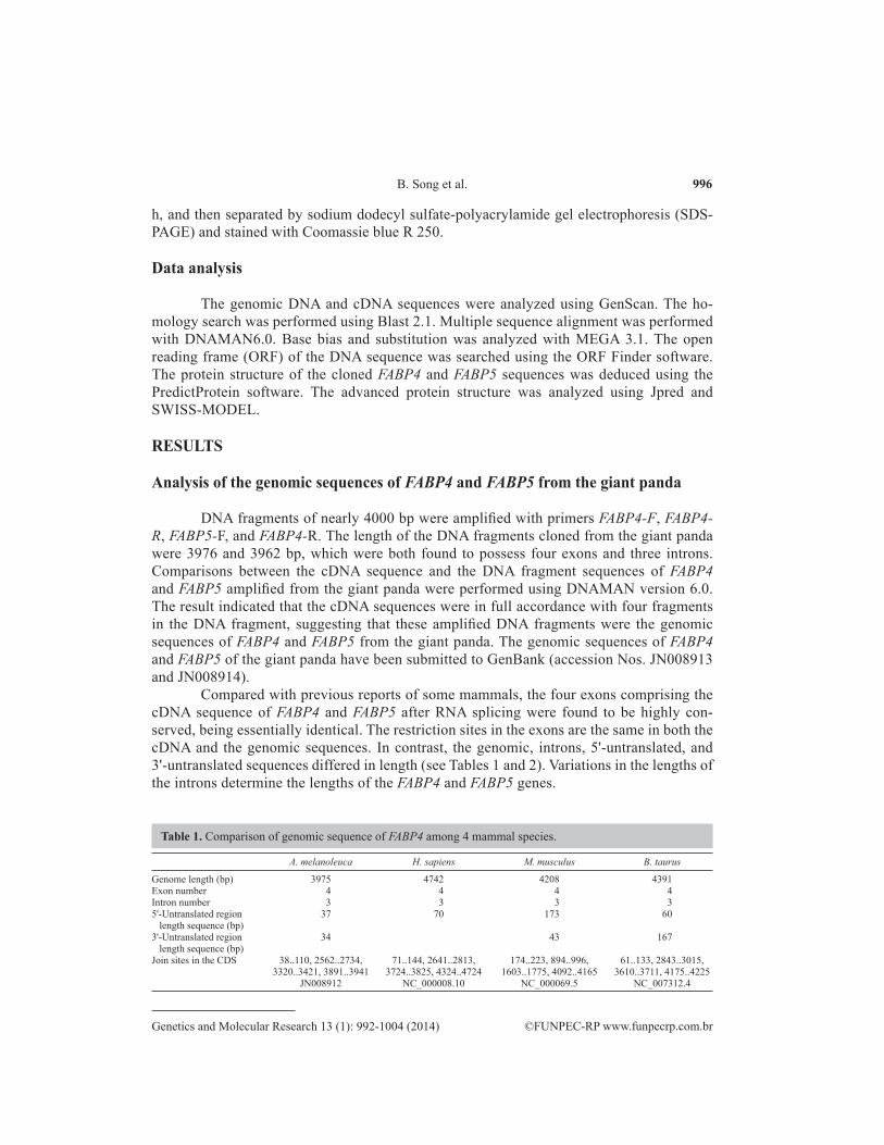
Compared with previous reports of some mammals, the four exons comprising the cDNA sequence of FABP4 and FABP5 after RNA splicing were found to be highly con-served, being essentially identical. The restriction sites in the exons are the same in both the cDNA and the genomic sequences. In contrast, the genomic, introns, 5'-untranslated, and 3'-untranslated sequences differed in length (see Tables 1 and 2). Variations in the lengths of the introns determine the lengths of the FABP4 and FABP5 genes.
A. melanoleuca H. sapiens M. musculus B. taurus
Genome length (bp) 3975 4742 4208 4391Exon number 4 4 4 4Intron number 3 3 3 35'-Untranslated region 37 70 173 60 length sequence (bp)3'-Untranslated region 34 43 167 length sequence (bp)Join sites in the CDS 38..110, 2562..2734, 71..144, 2641..2813, 174..223, 894..996, 61..133, 2843..3015, 3320..3421, 3891..3941 3724..3825, 4324..4724 1603..1775, 4092..4165 3610..3711, 4175..4225 JN008912 NC_000008.10 NC_000069.5 NC_007312.4
Table 1. Comparison of genomic sequence of FABP4 among 4 mammal species.
997
©FUNPEC-RP www.funpecrp.com.brGenetics and Molecular Research 13 (1): 992-1004 (2014)
Characterization of giant panda FABP4 and FABP5
Analysis of the cDNA of FABP4 and FABP5 from the giant panda
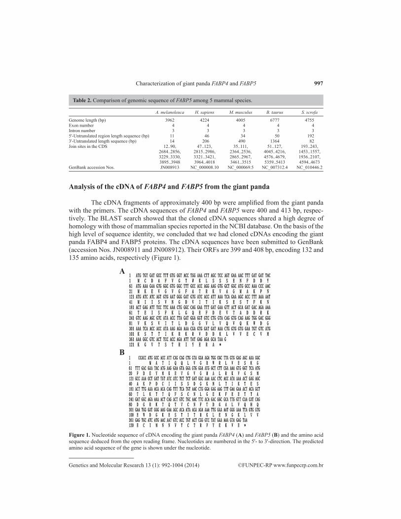
The cDNA fragments of approximately 400 bp were amplified from the giant panda with the primers. The cDNA sequences of FABP4 and FABP5 were 400 and 413 bp, respec-tively. The BLAST search showed that the cloned cDNA sequences shared a high degree of homology with those of mammalian species reported in the NCBI database. On the basis of the high level of sequence identity, we concluded that we had cloned cDNAs encoding the giant panda FABP4 and FABP5 proteins. The cDNA sequences have been submitted to GenBank (accession Nos. JN008911 and JN008912). Their ORFs are 399 and 408 bp, encoding 132 and 135 amino acids, respectively (Figure 1).
A. melanoleuca H. sapiens M. musculus B. taurus S. scrofa
Genome length (bp) 3962 4224 4005 6777 4755Exon number 4 4 4 4 4Intron number 3 3 3 3 35'-Untranslated region length sequence (bp) 11 46 34 50 1923'-Untranslated length sequence (bp) 14 206 490 1364 82Join sites in the CDS 12..90, 47..123, 35..111, 51..127, 193..243, 2684..2856, 2815..2986, 2364..2536, 4045..4216, 1453..1557, 3229..3330, 3321..3421, 2865..2967, 4576..4679, 1936..2107, 3895..3948 3964..4018 3461..3515 5359..5413 4594..4673GenBank accession Nos. JN008913 NC_000008.10 NC_000069.5 NC_007312.4 NC_010446.2
Table 2. Comparison of genomic sequence of FABP5 among 5 mammal species.
Figure 1. Nucleotide sequence of cDNA encoding the giant panda FABP4 (A) and FABP5 (B) and the amino acid sequence deduced from the open reading frame. Nucleotides are numbered in the 5'- to 3'-direction. The predicted amino acid sequence of the gene is shown under the nucleotide.
A
B
998
©FUNPEC-RP www.funpecrp.com.brGenetics and Molecular Research 13 (1): 992-1004 (2014)
B. Song et al.
Base bias analysis showed that the content of individual bases differed in different co-don positions; nucleotide G was the highest in the first position, T was the highest in the second position, while in the third position, the contents of T, G, and C were very similar and only A was the lowest. Furthermore, base substitution statistics suggested that the number of transitions was obviously higher than that of transversions, which is in accordance with the general rule that nucleotide substitution is the main mechanism of DNA evolution, and that nucleotide transition is the main mechanism of nucleotide substitution. In addition, variable sites and nucleotide sub-stitutions in the third codon position were far higher than those in the first or second positions of codons, which suggested a high rate of nucleotide substitution in the third positions of codons during evolution.
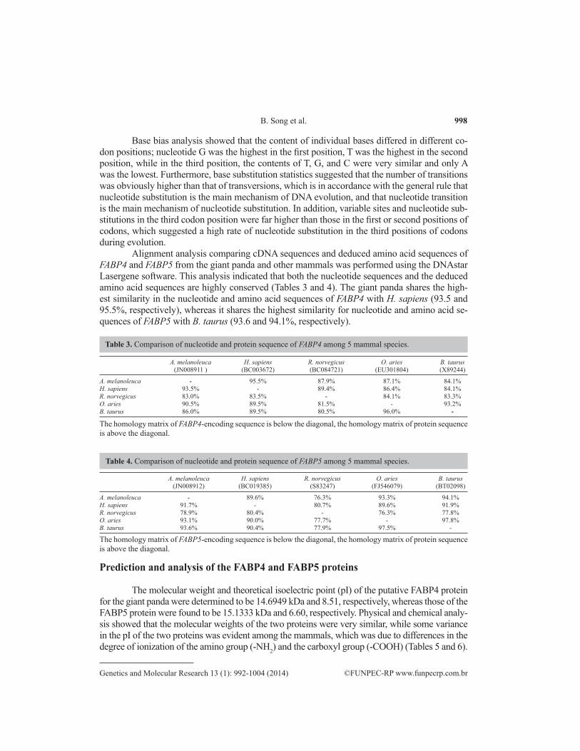
Alignment analysis comparing cDNA sequences and deduced amino acid sequences of FABP4 and FABP5 from the giant panda and other mammals was performed using the DNAstar Lasergene software. This analysis indicated that both the nucleotide sequences and the deduced amino acid sequences are highly conserved (Tables 3 and 4). The giant panda shares the high-est similarity in the nucleotide and amino acid sequences of FABP4 with H. sapiens (93.5 and 95.5%, respectively), whereas it shares the highest similarity for nucleotide and amino acid se-quences of FABP5 with B. taurus (93.6 and 94.1%, respectively).
A. melanoleuca H. sapiens R. norvegicus O. aries B. taurus (JN008911 ) (BC003672) (BC084721) (EU301804) (X89244)
A. melanoleuca - 95.5% 87.9% 87.1% 84.1%H. sapiens 93.5% - 89.4% 86.4% 84.1%R. norvegicus 83.0% 83.5% - 84.1% 83.3%O. aries 90.5% 89.5% 81.5% - 93.2%B. taurus 86.0% 89.5% 80.5% 96.0% -
The homology matrix of FABP4-encoding sequence is below the diagonal, the homology matrix of protein sequence is above the diagonal.
Table 3. Comparison of nucleotide and protein sequence of FABP4 among 5 mammal species.
A. melanoleuca H. sapiens R. norvegicus O. aries B. taurus (JN008912) (BC019385) (S83247) (FJ546079) (BT02098)
A. melanoleuca - 89.6% 76.3% 93.3% 94.1%H. sapiens 91.7% - 80.7% 89.6% 91.9%R. norvegicus 78.9% 80.4% - 76.3% 77.8%O. aries 93.1% 90.0% 77.7% - 97.8%B. taurus 93.6% 90.4% 77.9% 97.5% -
The homology matrix of FABP5-encoding sequence is below the diagonal, the homology matrix of protein sequence is above the diagonal.
Table 4. Comparison of nucleotide and protein sequence of FABP5 among 5 mammal species.
Prediction and analysis of the FABP4 and FABP5 proteins
The molecular weight and theoretical isoelectric point (pI) of the putative FABP4 protein for the giant panda were determined to be 14.6949 kDa and 8.51, respectively, whereas those of the FABP5 protein were found to be 15.1333 kDa and 6.60, respectively. Physical and chemical analy-sis showed that the molecular weights of the two proteins were very similar, while some variance in the pI of the two proteins was evident among the mammals, which was due to differences in the degree of ionization of the amino group (-NH2) and the carboxyl group (-COOH) (Tables 5 and 6).

999
©FUNPEC-RP www.funpecrp.com.brGenetics and Molecular Research 13 (1): 992-1004 (2014)
Characterization of giant panda FABP4 and FABP5
A. melanoleuca H. sapiens M. musculus B. taurus O. aries
Molecular weight (kDa) 14.6949 14.7188 14.6498 14.6778 14.6888Isoelectric point 8.51 6.59 8.53 5.52 5.22
Table 5. Molecular weight and isoelectric point of the FABP4 protein of giant panda and other mammal species.
A. melanoleuca H. sapiens M. musculus B. taurus O. aries
Molecular weight (kDa) 15.1333 15.1644 15.1374 15.0743 15.1033Isoelectric point 6.60 6.60 6.14 7.58 7.58
Table 6. Molecular weight and isoelectric point of the FABP5 protein of giant panda and other mammal species.
Topology prediction revealed seven protein kinase C phosphorylation sites, two casein ki-nase II phosphorylation sites, two N-myristoylation sites, and one cytosolic fatty acid-binding protein signature in the FABP4 protein, and three N-glycosylation sites, three protein kinase C phosphoryla-tion sites, one casein kinase II phosphorylation site, one N-myristoylation site, one amidation site, and one cytosolic fatty acid-binding protein signature in the FABP5 protein (Figure 2). Our analysis indicated minute differences in the functional sites among the reported mammalian proteins, but without exception. One cytosolic fatty acid-binding protein signature, which spans 19 amino acid residues, was found in the giant panda and other mammalian proteins, since the primary and most important role of this protein family is to bind long-chain fatty acids and other hydrophobic ligands. Although the 19 amino acid signature sequence is not exactly identical between the giant panda and other mammals, none of the changes result in alterations in the function or structure of the proteins.
Figure 2. Comparison of the amino acid sequence of FABP4 (A) and FABP5 (B) among the giant panda and mammalians reported. Panda = the giant panda; homo = Homo sapiens; bos = Bos taurus; ovis = Ovis aries; mus = Mus musculus; rat = Rattus norvegicus; cavia = Cavia porcellus; sus = Sus scrofa; bold = N-glycosylation site; italic = N-myristoylation site; solid underline = protein kinase C phosphorylation site; shaded = casein kinase II phosphorylation site; wave line = amidation site; dotted underline = cytosolic fatty acid-binding protein signature.
A
B

1000
©FUNPEC-RP www.funpecrp.com.brGenetics and Molecular Research 13 (1): 992-1004 (2014)
B. Song et al.
Overexpression of FABP4 and FABP5 in E. coli
FABP4 was overexpressed in E. coli using pET28a plasmids carrying strong promoter and terminator sequences derived from phage T7. For this purpose, the FABP4 gene was am-plified by PCR and cloned in a pET28a plasmid, resulting in a fusion gene coding for a protein bearing a His-tag extension at the N-terminus. Expression was tested by SDS-PAGE analysis of protein extracts from recombinant E. coli BL21 strains (Figure 3). Results indicated that the FABP4 protein fused with the N-terminal His-tagged form gave rise to the accumula-tion of an expected 16.8-kDa polypeptide that formed inclusion bodies. Expression of the recombinant protein was evident after 30 min of induction and reached its highest level after 2 h. The FABP5 gene was overexpressed by the same procedure, which produced a 17-kDa polypeptide (Figure 3).
These results suggested that the proteins were active and were the only proteins en-coded by FABP4 and FABP5 from the giant panda. The expression products obtained could be used to purify these proteins in order to study their functions in more detail.
Figure 3. FABP4 (A) and FABP5 (B) proteins extracted from recombinant Escherichia coli strains were analyzed by SDS-PAGE stained with Commassie blue R 250. Numbers on right show the molecular weight and arrows indicate the recombinant protein bands induced by IPTG with 0, 0.5, 1, 1.5, 2, 2.5, 3, 3.5, and 4 h (lanes 2-10) and 0.5, 1, 1.5, 2, 2.5, 3, 3.5, and 4 h (lanes 2-9), respectively; lane 1 = control.
A
B

1001
©FUNPEC-RP www.funpecrp.com.brGenetics and Molecular Research 13 (1): 992-1004 (2014)
Characterization of giant panda FABP4 and FABP5
DISCUSSION
FABPs belong to the conserved multigene family of intracellular lipid-binding proteins (Choromańska et al., 2011). These proteins are ubiquitously expressed in vertebrate tissues, with each FABP showing distinct expression patterns (Storch and McDermott, 2009). Various func-tions have been proposed for these proteins, including the promotion of cellular uptake and transport of fatty acids, the targeting of fatty acids to specific metabolic pathways, and participa-tion in the regulation of gene expression and cell growth (Uysal et al., 2000).
FABP4 and FABP5 are two closely related fatty acid-binding proteins that are expressed primarily in adipose tissue and/or macrophages. Alignment analysis of FABP4 and FABP5 of the giant panda and other mammalian species indicated that both the nucleotide and the deduced amino acid sequences are highly conserved. No deletions or insertions of nucleotides or amino acid residues were detected. Nonetheless, some polymorphic sites were identified in the deduced amino acid sequences of the FABP4 and FABP5 proteins. These polymorphic sites are located irregularly throughout the amino acid sequences, and have all resulted from the transversion or transition of the corresponding codons, without any deletion and insertion of bases. It is note-worthy that polymorphic sites located within the cytosolic fatty acid-binding protein signature sequences have not resulted in any changes in the functional role of binding long-chain fatty acids and other hydrophobic ligands.
Although some polymorphic sites are located within functional sites, indicating that these sites are not conserved, secondary and tertiary structure analysis showed that all amino acid sequences formed the same main structures (Figures 4, 5, and 6). For FABP4, residues spanning from 7 to 131 formed the same main structure: two helices (15-24, 26-36), and ten beta strands (6-14, 38-45, 54-47, 60-66, 70-74, 78-87, 90-97, 100-109, 112-119, 122-131). For FABP5, resi-dues spanning from 2 to 133 formed the same main structure: three helices (3-8, 18-27, 29-39) and ten beta strands (9-17, 41-48, 50-58, 61-69, 71-77, 81-90, 92-100, 103-112, 114-122, 125-134). This fact shows that variation among sites has no effect on the structure and function of the FABP4 and FABP5 proteins. This may reflect an ancient evolutionary relationship between these proteins and the primary, ancient structural and functional feature of the FABP protein family.
Figure 4. Secondary structure of FABP4 of the giant panda. A. β = beta turn; H = helixes; γ = gamma turn; hairpin loop = beta hairpin; A = beta strands. B. Pink thick arrow = beta strands; red cylinder = helixes.
A B

1002
©FUNPEC-RP www.funpecrp.com.brGenetics and Molecular Research 13 (1): 992-1004 (2014)
B. Song et al.
In summary, the cDNA and complete coding sequences of giant panda FABP4 and FABP5 were cloned, and their cDNAs were efficiently expressed in a prokaryotic organism using pET28a plasmids. The obtained fusion proteins were in accordance with the expected 16.8- and 17-kDa polypeptides, respectively. These results suggest that the proteins were ac-tive and were the respective proteins encoded by FABP4 and FABP5 from the giant panda. Determining the more complex molecular mechanism and genetic polymorphisms of these genes will be addressed in future study.
ACKNOWLEDGMENTS
Research supported by the National Natural Science Foundation of China (#31200012), the Application Foundation Project of Sichuan Province (#2013JY0094), the Science and Technology Support Project of Sichuan Province (#2014SZ0020 and #2014FZ0024), The Cultivate Major Projects of Sichuan Province (#14CZ0016), and the Doctor Startup Founda-tion Project of China West Normal University (#11B019 and #11B020).
Figure 5. Secondary structure of FABP5 of the giant panda. A. β = beta turn; H = helixes; γ = gamma turn; hairpin loop = beta hairpin; A = beta strands. B. Pink thick arrow = beta strands; red cylinder = helixes.
A B
Figure 6. Tertiary structure of FABP4 (A) and FABP5 (B) of the giant panda.
BA

1003
©FUNPEC-RP www.funpecrp.com.brGenetics and Molecular Research 13 (1): 992-1004 (2014)
Characterization of giant panda FABP4 and FABP5
REFERENCES
Boord JB, Fazio S and Linton MF (2002). Cytoplasmic fatty acid-binding proteins: emerging roles in metabolism and atherosclerosis. Curr. Opin. Lipidol. 13: 141-147.
Bu L, Salto LM, De Leon KJ and De Leon M (2011). Polymorphisms in fatty acid binding protein 5 show association with type 2 diabetes. Diabetes Res. Clin. Pract. 92: 82-91.
Choi KM, Yannakoulia M, Park MS, Cho GJ, et al. (2011). Serum adipocyte fatty acid-binding protein, retinol-binding protein 4, and adiponectin concentrations in relation to the development of the metabolic syndrome in Korean boys: a 3-y prospective cohort study. Am. J. Clin. Nutr. 93: 19-26.
Choromańska B, Myśliwiec P, Dadan J, Hady HR, et al. (2011). The clinical significance of fatty acid binding proteins. Postepy Hig. Med. Dosw. 65: 759-763.
Du YJ, Luo XY, Hao YZ, Zhang T, et al. (2007). cDNA cloning and overexpression of acidic ribosomal phosphoprotein P1 gene (RPLP1) from the giant panda. Int. J. Biol. Sci. 3: 428-433.
Fang LY, Wong TY, Chiang WF and Chen YL (2010). Fatty-acid-binding protein 5 promotes cell proliferation and invasion in oral squamous cell carcinoma. J. Oral. Pathol. Med. 39: 342-348.
Gong YN, Li WW, Sun JL, Ren F, et al. (2010). Molecular cloning and tissue expression of the fatty acid-binding protein (Es-FABP) gene in female Chinese mitten crab (Eriocheir sinensis). BMC Mol. Biol. 11: 71.
Grundy SM, Cleeman JI, Daniels SR, Donato KA, et al. (2005). Diagnosis and management of the metabolic syndrome: an American Heart Association/National Heart, Lung, and Blood Institute Scientific Statement. Circulation 112: 2735-2752.
Hotamisligil GS (2005). Role of endoplasmic reticulum stress and c-Jun NH2-terminal kinase pathways in inflammation and origin of obesity and diabetes. Diabetes 54 (Suppl 2): S73-S78.
Hou WR, Sun GL, Chen Y and Wu X (2008a). Molecular cloning of ribosomal protein L26 (RPL26) cDNA from Ailuropoda melanoleuca and its potential value in phylogenetic study. Biochem. Syst. Ecol. 136: 194-200.
Hou YL, Hou WR, Ren ZL, Hao YZ, et al. (2008b). cDNA, genomic sequence and overexpression of crystallin alpha-B gene (CRYAB) of the giant panda. Int. J. Biol. Sci. 4: 415-421.
Iwayama Y, Hattori E, Maekawa M, Yamada K, et al. (2010). Molecular cloning and tissue expression of the fatty acid-binding protein (Es-FABP) gene in female Chinese mitten crab (Eriocheir sinensis). BMC Mol. Biol. 11: 71.
Krusinová E and Pelikánová T (2008). Fatty acid binding proteins in adipose tissue: a promising link between metabolic syndrome and atherosclerosis? Diabetes Res. Clin. Pract. 82 (Suppl 2): S127-S134.
Li R, Fan W, Tian G, Zhu H, et al. (2010). The sequence and de novo assembly of the giant panda genome. Nature 463: 311-317.
Maeda K, Cao H, Kono K, Gorgun CZ, et al. (2005). Adipocyte/macrophage fatty acid binding proteins control integrated metabolic responses in obesity and diabetes. Cell Metab. 1: 107-119.
Maekawa M, Iwayama Y, Arai R, Nakamura K, et al. (2010). Polymorphism screening of brain-expressed FABP7, 5 and 3 genes and association studies in autism and schizophrenia in Japanese subjects. J. Hum. Genet. 55: 127-130.
Makowski L and Hotamisligil GS (2004). Fatty acid binding proteins - the evolutionary crossroads of inflammatory and metabolic responses. J. Nutr. 134: 2464S-2468S.
Makowski L and Hotamisligil GS (2005). The role of fatty acid binding proteins in metabolic syndrome and atherosclerosis. Curr. Opin. Lipidol. 16: 543-548.
Monaco HL (2009). Review: the liver bile acid-binding proteins. Biopolymers 91: 1196-1202.Smathers RL and Petersen DR (2011). The human fatty acid-binding protein family: evolutionary divergences and
functions. Hum. Genomics 5: 170-191.Storch J and Corsico B (2008). The emerging functions and mechanisms of mammalian fatty acid-binding proteins. Annu.
Rev. Nutr. 28: 73-95.Storch J and McDermott L (2009). Structural and functional analysis of fatty acid-binding proteins. J. Lipid. Res. 50
(Suppl): S126-S131.Storch J and Thumser AE (2010). Tissue-specific functions in the fatty acid-binding protein family. J. Biol. Chem. 285:
32679-32683.Tuncman G, Erbay E, Hom X, De Vivo I, et al. (2006). A genetic variant at the fatty acid-binding protein aP2 locus reduces
the risk for hypertriglyceridemia, type 2 diabetes, and cardiovascular disease. Proc. Natl. Acad. Sci. U. S. A. 103: 6970-6975.
Uysal KT, Scheja L, Wiesbrock SM, Bonner-Weir S, et al. (2000). Improved glucose and lipid metabolism in genetically obese mice lacking aP2. Endocrinology 141: 3388-3396.
1004
©FUNPEC-RP www.funpecrp.com.brGenetics and Molecular Research 13 (1): 992-1004 (2014)
B. Song et al.
Veerkamp JH and Zimmerman AW (2001). Fatty acid-binding proteins of nervous tissue. J. Mol. Neurosci. 16: 133-142.Xu A, Tso AW, Cheung BM, Wang Y, et al. (2007). Circulating adipocyte-fatty acid binding protein levels predict the
development of the metabolic syndrome: a 5-year prospective study. Circulation 115: 1537-1543.Zhang T, Hou WR, Hou YL and Hao YZ (2009). cDNA, genomic sequence cloning and overexpression of ribosomal
protein s20 gene (RPS20) from the giant panda (Ailuropoda melanoleuca). Afr. J. Biotechnol. 8: 5627-5632.