Communication Vol. 263, 25, Issue THE OF 5, 12175 … · Communication Vol. 263, No. 25, Issue of...
4
Communication Vol. 263, No. 25, Issue of September 5, pp. 12175-1217S,1988 THE JOURNAL OF BIOLOGICAL CHEMISTRY 0 1988 by The American Society for Biochemistry and Molecular Biology, Inc. Printed in U.S.A. The Central Helix of Calmodulin Functions as a Flexible Tether* (Received for publication, June 6, 1988) Anthony Persechini and Robert H. Kretsinger From the Department of Bwbgy, University of Virginia, Charlottesuille, Virginia 22901 Using site-directed mutagenesis we have created an altered calmodulin in which Gln-3 and Thr-146 have both been replaced by cysteines. We have reacted this protein with the bifunctional reagent, bismaleimido- hexane, forming an intramolecular cross-link between the two cysteines. In the crystal structure of nativp calmodulin a-carbonsat positions 3 and146 are 37 A apart. In the bismaleimidohexane crps-linked protein these atoms can be no more than 19 A apart, and model building studies indicate that there is probably a bend in the central helix of calmodulin. A second modified calmodulinwas generated by cleaving the central helix of the cross-linked protein at Lys-77 with trypsin. In this molecule, the two lobes of calmodulin are joined solely by thebismaleimidohexane cross-link, which bridges Cys-3 and Cys-146. V, and K,, valuesfor activation of myosin light chain kinase activity by the cross-linked and cross-linked/trypsinized proteins are not significantly different from those for the control protein. This result indicates that one role for the central helix may be to serve as a flexible tether be- tween the calmodulin lobes. This is consistent with a model calmodulin-enzyme complex in which the cen- tral helix is bent, and thetwo lobes exert a concerted effect. A detailed model of this type has been proposed for the calmodulin-myosin light chain kinase complex (Persechini, A. and Kretsinger, R. H. (1988) J. Cardi- oua8c. Pharrnacol., in press). Calmodulin is found in all eucaryotic cells, where it plays a key role in orchestrating the response of the cell to stimulus- induced changes in the intracellular Ca2+ion concentration (Manalan andKlee, 1984). Babu et al. (1985) and Kretsinger et al. (1986) have determined the crystal structure of Ca2+- saturated calmodulin to 3-A resolution. The molecule is 65 A long, with an overall dumbbell shape. There is no contact betyeen the two lobes, which have dimensions of 25 X 20 X 20 A. Each lobe consists of a pairof EF-hand (Kretsinger and Nockolds, 1973) Ca2+-binding domains, joined by a short segment of polypeptide. The most striking aspect of the struFture is the central helix that bridges the two lobes. It is 40 A long with the central third exposed to solvent. Trypsi- nolysis at Lys-77, within the solvent-exposed region of the central helix, abolishes activation of myosin light chain ki- nase, phosphodiesterase, and calcineurin activities (Newton * This work was supported by Grants IN-149D and JFRA-191 (to A. P.) from the American Cancer Society and Grant DMB-8608878 (to R. H. K.) from the National Science Foundation. The costs of publication of this article were defrayed in part by the payment of page charges. This article must therefore be hereby marked “aduer- tisement” in accordance with 18 U.S.C. Section 1734 solely to indicate this fact. et al., 1984; Newton et al., 1985). This result indicates the possibility of a concerted action of the two lobes of calmodulin in the calmodulin-enzyme complex. If this is true, then the role of the central helix is of particular interest. Here there are two general possibilities; the helix is maintained or it is not. If it is maintained, then the interaction between calmod- ulin and target is approximated by a “lock and key” analogy. If the central helix is bent, then rotations about the dihedral angles, 4 and I ) , allow considerable variation in the relative positions of the two lobes. We have begun to investigate the role of the central helix in formation of the active calmodulin-enzyme complex. Our approach has been to use site-directed mutagenesis to make specific changes in a calmodulin-encoding template, followed by expression in Escherichia coli. In this paper we describe the properties of a modified calmodulin in which we have cross-linked the two lobes by first introducing a cysteine pair, followed by reaction with bismaleimidohexane. We also de- scribe the effect of tryptic cleavage at Lys-77 on the properties of this molecule. EXPERIMENTAL PROCEDURES Myosin light chain kinase was purified from rabbit skeletalmuscle as described by Nunnally et al. (1985). Mixed rabbit skeletal muscle light chains were isolated as described by Blumenthal and Stull (1980). Calmodulin was isolated from bovine brain as described by Gopalakrishna and Anderson (1982). Calmodulin expressed in E. coli was isolated as described by Craig et al. (1987).Protein concentrations were determined as described by Bradford (1976),using bovine serum albumin and bovine calmodulin standards. [Q3C, T146C]calmodulin’ was converted to the reduced form by incubation with 5 mM DTT at 30 “C for 1 h, followed by addition of potassium acetate (pH 5.1) to a final concentration of 40 mM. DTT was removed by dialysis against degassed 5 mM potassium acetate (pH 5.1). Reactions with bismaleim- idohexane were performed under NZ in a buffer containing 0.2 M sodium phosphate (pH 7.1), 0.2 mM bismaleimidohexane (Pierce Chemical Co.), and 0.02 mM reduced [Q3C, T146Clcal- modulin. Reactions with N-ethylmaleimide were performed under identical conditions, except 0.4 mM N-ethylmaleimide replaced bis- maleimidohexane. After incubation for 1.5 h at 30 ”C, reactions were terminated by the addition of 30 mM 8-mercaptoethanol. Trypsino- lysis of BMHCM was performed essentially as described by Newton et al. (1984) for vertebrate calmodulin. Digestion mixtures contained 33 FM BMHCM and 5 pg/ml trypsin. Protein bands corresponding to BMHCM cleaved at Lys-77 (TBMHCM) were excised from a lightly stained urea/glycerol polyacrylamide gel. After electroeleution from gel slices, TBMHCM was further purified by performing chro- matography on phenyl-Sepharose (Gopalakrishna and Anderson, 1982). SDS-gel electrophoresis was performed in the presence of 0.1 mM EDTA in 15% polyacrylamide slabs, as described by Laemmli (1970). Preparation of samples for electrophoresis was performed by incubation for 1 min at 100 “C in SDS sample buffer plus or minus 10 mM DTT. Urea/glycerol polyacrylamide gel electrophoresis was performed in the presence of 0.2 mM EGTA as described by Persechini et al. (1986). Data from myosin light chain kinase assays (Blumenthal and Stull, 1982) were fit to the Eadie-Hofstee form of the Michaelis equation. The concentrations of myosin light chain kinase and phos- phorylatable light chain in enzyme assays were 0.2 nM and 8 PM, respectively. Concentrations of added calmodulin ranged from 1.0 to 50 nM. Reactions were initiated by the addition of [Y-~’P]ATP (300 ’ The abbreviations used are: [Q3C, T146C]calmodulin, calmodulin in which Gln-3 and Thr-146 have been replaced by Cys; BMHCM and NEMCM, [Q3C, T146C]calmodulin reacted with bismaleimido- hexane or N-ethylmaleimide, respectively; TBMHCM, BMHCM after trypsinolysis at Lys-77; DTT, dithiothreitol; EGTA, [ethylene- bis(oxyethylenenitri1o)l tetraacetic acid SDS, sodium dodecyl sulfate. 12175
Transcript of Communication Vol. 263, 25, Issue THE OF 5, 12175 … · Communication Vol. 263, No. 25, Issue of...

Communication Vol. 263, No. 25, Issue of September 5, pp. 12175-1217S,1988 THE JOURNAL OF BIOLOGICAL CHEMISTRY
0 1988 by The American Society for Biochemistry and Molecular Biology, Inc. Printed in U.S.A.
The Central Helix of Calmodulin Functions as a Flexible Tether*
(Received for publication, June 6, 1988) Anthony Persechini and Robert H. Kretsinger From the Department of Bwbgy, University of Virginia, Charlottesuille, Virginia 22901
Using site-directed mutagenesis we have created an altered calmodulin in which Gln-3 and Thr-146 have both been replaced by cysteines. We have reacted this protein with the bifunctional reagent, bismaleimido- hexane, forming an intramolecular cross-link between the two cysteines. In the crystal structure of nativp calmodulin a-carbons at positions 3 and 146 are 37 A apart. In the bismaleimidohexane crps-linked protein these atoms can be no more than 19 A apart, and model building studies indicate that there is probably a bend in the central helix of calmodulin. A second modified calmodulin was generated by cleaving the central helix of the cross-linked protein at Lys-77 with trypsin. In this molecule, the two lobes of calmodulin are joined solely by the bismaleimidohexane cross-link, which bridges Cys-3 and Cys-146. V, and K,, values for activation of myosin light chain kinase activity by the cross-linked and cross-linked/trypsinized proteins are not significantly different from those for the control protein. This result indicates that one role for the central helix may be to serve as a flexible tether be- tween the calmodulin lobes. This is consistent with a model calmodulin-enzyme complex in which the cen- tral helix is bent, and the two lobes exert a concerted effect. A detailed model of this type has been proposed for the calmodulin-myosin light chain kinase complex (Persechini, A. and Kretsinger, R. H. (1988) J. Cardi- oua8c. Pharrnacol., in press).
Calmodulin is found in all eucaryotic cells, where it plays a key role in orchestrating the response of the cell to stimulus- induced changes in the intracellular Ca2+ ion concentration (Manalan and Klee, 1984). Babu et al. (1985) and Kretsinger et al. (1986) have determined the crystal structure of Ca2+- saturated calmodulin to 3-A resolution. The molecule is 65 A long, with an overall dumbbell shape. There is no contact betyeen the two lobes, which have dimensions of 25 X 20 X 20 A. Each lobe consists of a pair of EF-hand (Kretsinger and Nockolds, 1973) Ca2+-binding domains, joined by a short segment of polypeptide. The most striking aspect of the struFture is the central helix that bridges the two lobes. It is 40 A long with the central third exposed to solvent. Trypsi- nolysis at Lys-77, within the solvent-exposed region of the central helix, abolishes activation of myosin light chain ki- nase, phosphodiesterase, and calcineurin activities (Newton
* This work was supported by Grants IN-149D and JFRA-191 (to A. P.) from the American Cancer Society and Grant DMB-8608878 (to R. H. K.) from the National Science Foundation. The costs of publication of this article were defrayed in part by the payment of page charges. This article must therefore be hereby marked “aduer- tisement” in accordance with 18 U.S.C. Section 1734 solely to indicate this fact.
et al., 1984; Newton et al., 1985). This result indicates the possibility of a concerted action of the two lobes of calmodulin in the calmodulin-enzyme complex. If this is true, then the role of the central helix is of particular interest. Here there are two general possibilities; the helix is maintained or it is not. If it is maintained, then the interaction between calmod- ulin and target is approximated by a “lock and key” analogy. If the central helix is bent, then rotations about the dihedral angles, 4 and I), allow considerable variation in the relative positions of the two lobes.
We have begun to investigate the role of the central helix in formation of the active calmodulin-enzyme complex. Our approach has been to use site-directed mutagenesis to make specific changes in a calmodulin-encoding template, followed by expression in Escherichia coli. In this paper we describe the properties of a modified calmodulin in which we have cross-linked the two lobes by first introducing a cysteine pair, followed by reaction with bismaleimidohexane. We also de- scribe the effect of tryptic cleavage at Lys-77 on the properties of this molecule.
EXPERIMENTAL PROCEDURES
Myosin light chain kinase was purified from rabbit skeletal muscle as described by Nunnally et al. (1985). Mixed rabbit skeletal muscle light chains were isolated as described by Blumenthal and Stull (1980). Calmodulin was isolated from bovine brain as described by Gopalakrishna and Anderson (1982). Calmodulin expressed in E. coli was isolated as described by Craig et al. (1987). Protein concentrations were determined as described by Bradford (1976), using bovine serum albumin and bovine calmodulin standards. [Q3C, T146C]calmodulin’ was converted to the reduced form by incubation with 5 mM DTT at 30 “C for 1 h, followed by addition of potassium acetate (pH 5.1) to a final concentration of 40 mM. DTT was removed by dialysis against degassed 5 mM potassium acetate (pH 5.1). Reactions with bismaleim- idohexane were performed under NZ in a buffer containing 0.2 M sodium phosphate (pH 7.1), 0.2 mM bismaleimidohexane (Pierce Chemical Co.), and 0.02 mM reduced [Q3C, T146Clcal- modulin. Reactions with N-ethylmaleimide were performed under identical conditions, except 0.4 mM N-ethylmaleimide replaced bis- maleimidohexane. After incubation for 1.5 h a t 30 ”C, reactions were terminated by the addition of 30 mM 8-mercaptoethanol. Trypsino- lysis of BMHCM was performed essentially as described by Newton et al. (1984) for vertebrate calmodulin. Digestion mixtures contained 33 FM BMHCM and 5 pg/ml trypsin. Protein bands corresponding to BMHCM cleaved at Lys-77 (TBMHCM) were excised from a lightly stained urea/glycerol polyacrylamide gel. After electroeleution from gel slices, TBMHCM was further purified by performing chro- matography on phenyl-Sepharose (Gopalakrishna and Anderson, 1982). SDS-gel electrophoresis was performed in the presence of 0.1 mM EDTA in 15% polyacrylamide slabs, as described by Laemmli (1970). Preparation of samples for electrophoresis was performed by incubation for 1 min at 100 “C in SDS sample buffer plus or minus 10 mM DTT. Urea/glycerol polyacrylamide gel electrophoresis was performed in the presence of 0.2 mM EGTA as described by Persechini et al. (1986). Data from myosin light chain kinase assays (Blumenthal and Stull, 1982) were fit to the Eadie-Hofstee form of the Michaelis equation. The concentrations of myosin light chain kinase and phos- phorylatable light chain in enzyme assays were 0.2 nM and 8 PM, respectively. Concentrations of added calmodulin ranged from 1.0 to 50 nM. Reactions were initiated by the addition of [Y-~’P]ATP (300
’ The abbreviations used are: [Q3C, T146C]calmodulin, calmodulin in which Gln-3 and Thr-146 have been replaced by Cys; BMHCM and NEMCM, [Q3C, T146C]calmodulin reacted with bismaleimido- hexane or N-ethylmaleimide, respectively; TBMHCM, BMHCM after trypsinolysis at Lys-77; DTT, dithiothreitol; EGTA, [ethylene- bis(oxyethylenenitri1o)l tetraacetic acid SDS, sodium dodecyl sulfate.
12175
12176 The Calmodulin Central Helix cpm/pmol), and rates were determined by evaluating the time course of 32Pi incorporation.
A partial calmodulin-encoding cDNA was isolated from an Arbmia punctuluta X-gtll library as described by Hardy et al. (1988). A missing region, encoding the last 7 amino acids, was produced syn- thetically. A protein identical in sequence to vertebrate calmodulin is encoded. Site-directed changes in the calmodulin-encoding sequence were introduced using mismatched primer extension mutagenesis of M13mp18 (Norrander et al., 1983) single-stranded DNA as described by Zoller and Smith (1983), except duplex DNA was not enriched after in vitro primer extension. Calmodulin-encoding sequences were expressed using a pKK233-2 vector (Pharmacia LKB Biotechnology Inc.). All calmodulin-encoding cDNAs were sequenced in their en- tirety before expression (Biggin et al., 1981; Sanger and Coulsen, 1978). Calmodulin expressed in E. coli differs from the vertebrate protein in that it lacks an N-terminal blocking group and trimethyl- lysine-115 (Putkey et al., 1985,1986; Roberts et al., 1985). The identity of calmodulin expressed from the unaltered template was confirmed by amino acid analysis (data not shown). Routine cloning procedures were performed as described by Maniatis et al. (1982). E. coli JMlOl was the host strain used for all procedures. Oligonucleotides were synthesized with a Biosearch 8600 nucleic acid synthesizer, used as described by the manufacturer. Amino acid analysis was performed as described by Bidlingmeyer et al. (1984) and Heinrickson and Meredith (1984). Protein sequencing was performed with an Applied Biosystems 470A protein sequenator, used essentially as described by Hewick et al. (1981), and Hunkapiller et a1 (1984). Molecular modeling was performed using the University of California Molecular Modeling System run on an Iris 3040 and coordinates from the crystal structure of vertebrate calmodulin. This structure is currently being refined in our laboratory. The coordinates for bismaleimidohexane were gener- ated using Chem-X (Chemical Design Ltd.), run on an Evans and Sutherland PS390.
RESULTS AND DISCUSSION
We have made an altered protein, termed [Q3C, T146Cl calmodulin, in which Gln-3 and Thr-146 have been replaced by cysteines. Since these are the only cysteine residues in the molecule, they can be used to specifically cross-link the two calmodulin lobes. During dialysis against 10 mM ammonium bicarbonate, which is the final step in purification, this cys- teine pair is oxidized to an intramolecular disulfide bridge. This is indicated by the marked reduction in SDS-gel electro- phoretic mobility seen after incubating purified [Q3C, T146Cl calmodulin with dithiothreitol (Fig. 1). The mobility of re- duced [Q3C, T146C]calmodulin is identical to that of the vertebrate protein (not shown). Since enzyme assays must be performed under reducing conditions, we have not attempted to determine the effect of disulfide formation on calmodulin function. Instead, we have created an irreversible intramolec- ular cross-link by reacting reduced [Q3C, Tl46C]calmodulin with bismaleimidohexane, in which a pair of maleimide mol- ecules are bridged by hexane. As a control, we have also reacted reduced [Q3C, T146C]calmodulin with N-ethylmal- eimide. Reaction products were dialyzed against 10 mM am- monium bicarbonate. As seen in Fig. 1, the electrophoretic mobility of [Q3C, T146C]calmodulin reacted with N-ethyl- maleimide (NEMCM) is unaffected by dithiothreitol and is identical to that of reduced [Q3C, Tl46C]calmodulin. This confirms that the sulfhydryl groups are blocked, since oxidi- zation to a disulfide does not occur during dialysis. The electrophoretic mobility of the bismaleimidohexane reaction product (BMHCM) is also unaffected by dithiothreitol but is similar to that of oxidized [Q3C,~T146C]calmodulin (Fig. 1). This is consistent with formation of an irreversible intramo- lecular cross-link. As seen in the figure, a faint protein band, with an electrophoretic mobility identical to that of reduced [Q3C, Tl46C]calmodulin, appears after incubation of BMHCM with dithiothreitol. This probably represents a small amount of unreacted material.
In the crystal structure of native calmodulin, a-carbons at
-DTT +Dl-f ” A B C A B C
FIG. 1. The effect of dithiothreitol on the electrophoretic mobilities of [Q3C, T146C]calmodulin (A), NEMCM (B) , and BMHCM (0 on SDS-polyacrylamide gels. The electrophoretic mobilities of samples incubated in the presence or absence of DTT are i-tdicated in the figure. In [Q3C, T146C]calmodulin, Gln-3 and Thr-146 have been replaced by cysteines. NEMCM and BMHCM refer to [Q3C, T146C]calmodulin after reaction with N-ethylmaleim- ide or bismaleimidohexane, respectively.
positions 3 and 146 are 37 A apart. In BMHCM these atoms can be no further than 19 A apart. This clearly requires a change in protein conformation. Model building studies indi- cate that in order to avoid distorting the pair of EF-hand domains that comprise each lobe, a bend must be introduced in the central helix. One such model is presented in Fig. 2. This class of model is consistent with small angle x-ray scattering investigations of calmodulin in solution, which indicate that although the average conformation is extended, as seen in the crystal structure, there is probably segmental flexibility within the central helix (Heidorn and Trewhella, 1988; Seaton et aL, 1985). Our observation of disulfide for- mation in [Q3C, T146C]calmodulin is also consistent with flexibility in this region, since the two sulfhydryl groups must come within 2 A of one another.
After mild trypsinolysis of BMHCM, the major digestion product has a mobility on urea/glycerol polyacrylamide gels which is slower than that of the starting material (not shown). We have purified this material, termed TBMHCM. As seen in Fig. 3, its mobility on SDS-polyacrylamide gels is inter- mediate to the mobilities of NEMCM and BMHCM. The first 10 residues of TBMHCM were determined by amino acid sequence analysis. Two N termini are indicated, one corre- sponding to the expected unblocked alanine residue at posi- tion 1 and the other to Asp-78. A cysteine residue is not seen at position 3, consistent with its participation in an intra- molecular cross-link. Additional cleavage sites are not indi- cated, and we conclude that TBMHCM consists of tryptic fragments 1-77 and 78-148, joined by a bismaleimidohexane cross-link between Cys-3 and Cys-146.
Steady-state kinetic evaluations of myosin light chain ki- nase activation by NEMCM, BMHCM, TBMHCM, bacterial control, and bovine calmodulins were performed. Typical values obtained for V,,, and K,,,, the concentration for half- maximal activation, are presented in Table I. Values for the modified calmodulins are not significantly different from those for the bacterial control. We have observed that V, values obtained for all the bacterial proteins, including the

The Calmodulin Central Helix 12177
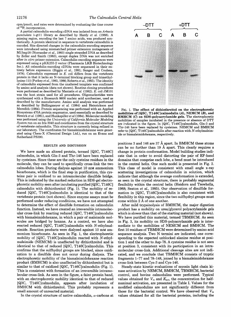
FIG. 2. A model structure for BMHCM. This model was gen- erated as described under “Experimental Procedures.” The model is seen as an a-carbon tracing, except for Cys-3, Cys-146, and the bismaleimidohexane cross-link, for which all atoms except hydrogens are shown. The positions of the N and C termini are indicated, as is the position of Lys-77, near the center of the central helix. In building this model, it was assumed that the region from Ala-1 through Thr- 5 is free to bend in any direction, since it is not helical and is poorly localized in the crystal structure (Babu et al., 1985; Kretsinger et al., 1986). It was also assumed that the structures of the EF-hand domain pairs are unaltered. Cys-3 and Cys-146 were brought as close together as possible by adjusting the @ and I) dihedral angles in residues 1-5. The bismaleimidohexane cross-link was kept fully extended. This was done to minimize the extent to which the central helix had to be altered. In the model, the helix is disrupted at a single residue, Thr- 79, with the axes of helical regions on either side intersecting at an angle of 110”.
A B C
(A), TBMHCM (B) and BMHCM (C). NEMCM and BMHCM are FIG. 3. SDS-polyacrylamide gel electrophoresis of NEMCM
defined in the legend to Fig. 1. In TBMHCM the central helix of BMHCM has been cleaved at Lys-77 by trypsin. The position of Lys- 77 in a model structure for BMHCM is indicated in Fig. 2.
TABLE I Steady-state kinetic parameters for activation of myosin light chain
kinase by control and modified calmodulins Assays were performed as described under “Experimental Proce-
dures.” K,, is the concentration of calmodulin that gives half-maxi- mal enzyme activation.
Activator K& Vlll
nM incorporatedfminlmg kinase pmol Pi
NEMCM 2.9 6.6 BMHCM 2.8 6.9 TBMHCM 3.5 6.3 Bacterial control 3.4 6.7 Bovine calmodulin 2.6 8.3
control, appear to be 20% less than for vertebrate calmodulin (Table I). This small difference may be due to the lack of an N-terminal blocking group or trimethyllysine-115 in the bac- terial proteins (Putkey et al., 1986; Roberts et al., 1985). It is not associated with any difference in values for K,,,
Newton et al. (1984, 1985) have shown that activation by native calmodulin of many enzymes, including myosin light chain kinase, is abolished by trypsinolysis at Lys-77, which cleaves the central helix and separates the two lobes. Our results with TBMHCM suggest that this apparent loss of function is probably not due to any conformational changes associated with proteolysis. Since the fragments are fully active when attached by bismaleimidohexane, loss of function is most likely due to a requirement for the concerted action of both calmodulin lobes. If each lobe interacts with a distinct region of the target and there are no cooperative effects, then the overall affinity of the native calmodulin-target complex, in which the lobes are attached by the central helix, would be proportional to the product of the affinities for the interaction with each isolated lobe (Crothers and Metzger, 1972). Theory does not allow calculation of the precise extent to which binding affinities are enhanced by bivalency (Crothers and Metzger, 1972). However, our results indicate that as long as the necessary geometries are attained in the complex, the nature of the attachment between the two lobes is not critical.
Our results indicate that activation of myosin light chain kinase requires the concerted action of both calmodulin lobes. Given this requirement, the behavior of TBMHCM and BMHCM in enzyme assays suggests that in the native cal- modulin-enzyme complex the central helix is probably bent, with the relative position of the two lobes quite different from that seen in the crystal structure. Hence, the central helix serves as a flexible tether between target binding domains on the lobes. In addition, residues of the central helix probably contribute some important side chain interactions. Craig et al. (1987) have replaced a stretch of three glutamates, com- prising residues 81-84 in the central helix, with three lysines. This protein can activate myosin light chain kinase to only 30% of its maximal activity. The importance of specific resi- dues in the central helix is also indicated by the high degree to which the calmodulin amino acid sequence has been con- served (Manalan and Klee, 1984).
We have recently presented a detailed model (Persechini and Kretsinger, 1988) for the complex between calmodulin and M13, a 27-residue peptide containing the presumptive calmodulin binding domain of myosin light chain kinase (Blumenthal el dl., 1985). In our model, the central helix of calmodulin is bent, and hydrophobic surfaces on the two lobes are juxtaposed with the hydrophobic surfaces on an M13 helix. Calmodulin then forms a channel which contains M13, providing a large contact surface. The distance between Cys- 3 and Cys-146 in the model complex can be bridged by

12178 The Calmodulin
bismaleimidohexane without changing the relationship be- tween calmodulin and M13 (not shown). Small adjustments in the central helix alter the channel to accommodate a range of different peptides. This is consistent with the ability of calmodulin to bind with high affinity a variety of peptides that conform only to the general pattern of a basic amphiphilic helix (Anderson and Malencik, 1986). The data we present here clearly support a class of calmodulin-target complex, of which our model is one example, in which the central helix functions as a flexible tether, with a concerted action of the two lobes.
Acknowledgments-We would like to thank Martha J. Spano for expert technical assistance, and Robert J. Cordaro and Paul S. Yadlowsky for assistance with computer applications. We would also like to acknowledge the Commonwealth of Virginia Protein and Nucleic Acid Sequencing Facility for performing amino acid analysis, protein sequencing, and oligonucleotide synthesis.
REFERENCES Anderson, S. R., and Malencik, D. A. (1986) in Calcium and Cell
Function (Cheung, W. Y., ed) pp. 1-42, Academic Press, New York Babu, Y. S., Sack, J. S., Greenhough, T. J., Bugg, C. E., Means, A.
R., and Cook, W. J. (1984) Nature 315,37-40 Bidlingmeyer, B. A., Cohen, S. A., and Tarvin, T. L. (1984) J.
Chromatogr. 336,93-104 Biggin, M. D., Gibson, T. J., and Hong, G. F. (1981) Proc. Natl. Acad.
Sci. U. S. A. 80,3963-3965 Blumenthal, D. K., and Stull, J. T. (1980) Biochemistry 19, 5608- 5614
Blumenthal, D. K., and Stull, J. T. (1982) Biochemistry 21, 2386- 2391
Blumenthal, D. K., Takio, K., Edelman, A. M., Charbonneau, H., Titani, K., Walsh, K. A., and Krebs, E. G. (1985) Proc. Natl. Acad. Sci. U. S. A. 82,3187-3191
Bradford, M. M. (1976) Anal. Biochem. 72,248-254 Craig, T. A., Watterson, D. M., Prendergast, F. G., Haiech, J., and
Crothers, D. M., and Metzger, H. (1972) Zmmumchemistry 9, 341- Roberts, D. M. (1987) J. Biol. Chem. 262,3278-3284
357
1 Central Helix Gopalakrishna, R., and Anderson, W. B. (1982) Biochem. Biophys.
Hardy, D. O., Bender, P. K., and Kretsinger, R. H. (1988) J. Mol.
Heidorn, D. B., and Trewhella, J. (1988) Biochemistry 27,909-915 Heinrikson, R. L., and Meredith, S. C. (1984) Anal. Biochem. 136,
Hewick, R. M., Hunkapiller, M. W., Hood, L. E., and Dreyer, W. J.
Hunkapiller, M. W., Hewick, R. M., Dreyer, W. J., and Hood, L. E.
Kretsinger, R. H., and Nockolds, C. E. (1973) J. Biol. Chem. 248,
Kretsinger, R. H., Rudnick, S. E., and Weissman, L. J. (1986) J.
Laemmli, U. K. (1970) Nature 157,221-231 Manalan, A. S., and Klee, C. B. (1984) Adu. Cyclic Nucleotide Res.
Maniatis, T., Fritsch, E. F., and Sambrook, J. (1982) Molecular Cloning, A Laboratory Manual, Cold Spring Harbor Laboratory, Cold Spring Harbor, NY
Newton, D. L., Oldewurtel, M. D., Krinks, M. H., Shiloach, J., and Klee, C. B. (1984) J. Biol. Chem. 269,4419-4426
Newton, D., Klee, C., Woodget, J., and Cohen, P. (1985) Biochim.
Norrander, J., Kempe, T., and Messing, J. (1983) Gene (Amst.) 26,
Nunnallv. M. H.. Rvbicki. S. B.. and Stull, J. T. (1985) J. Biol. Chem.
Res. Commun. 104,830-836.
Biol. 199,223-227
65-74
(1981) J. Biol. Chem. 266,7990-7997
(1984) Methods Enzymol. 91, 399-413
3313-3326
Inorg. Biochem. 28,289-302
1a,227-277
Biophys. Acta 845,533-539
101-106
260, iO20-1026 " Persechini, A., Kamm, K. E., and Stull, J. T. (1986) J. Biol. Chem.
Persechini, A., and Kretsinger, R. H. (1988) J. Cardiouasc. Phurrnacol, 261,6293-6299
Putkey, J. A., Slaughter, G. R., and Means, A. R. (1985) J. Biol. in press
Chem. 260,4704-4712 Putkey, J. A., Draetta, G. F., Slaughter, G. R., Klee, C. B., Cohen, P.,
9903 Stull, J. T., and Means, A. R. (1986) J. Biol. Chem. 261, 9896-
Roberts, D. M., Crea, R., Malecha, M., Alvarado-Urbina, G., Chi- arello, R. H., and Watterson, D. M. (1985) Biochemistry 24,5090- 5098
Sanger, R., and Coulsen, A. R. (1978) FEBS Lett. 87, 107-110 Seaton. B. A.. Head. J. F.. Emelman. D. M.. and Richards, F. M.
~ (1985) Biochemist& 24,6746-6743 ' Zoller, M. J., and Smith, M. (1983) Methods Enzymol. 100,486-487