Communicable Disease Sudan
130
WHO/CDS/2005.26 COMMUNICABLE DISEASE TOOLKIT SUDAN 1. COMMUNICABLE DISEASE PROFILE World Health Organization 2005 Communicable Disease Working Group on Emergencies, WHO/HQ WHO Regional Office for the Eastern Mediterranean, EMR0 WHO Country Office, Khartoum
Transcript of Communicable Disease Sudan

WHO/CDS/2005.26
COMMUNICABLE DISEASE TOOLKIT
SUDAN
1. COMMUNICABLE DISEASE PROFILE
World Health Organization
2005
Communicable Disease Working Group on Emergencies, WHO/HQ WHO Regional Office for the Eastern Mediterranean, EMR0
WHO Country Office, Khartoum

Communicable Disease Toolkit for SUDAN: Communicable Disease Profile 2005
World Health Organization
© World Health Organization 2005 All rights reserved.
The designations employed and the presentation of the material in this publication do not imply the expression of any opinion whatsoever on the part of the World Health Organization concerning the legal status of any country, territory, city or area or of its authorities, or concerning the delimitation of its frontiers or boundaries. Dotted lines on maps represent approximate border lines for which there may not yet be full agreement.
The mention of specific companies or of certain manufacturers’ products does not imply that they are endorsed or recommended by the World Health Organization in preference to others of a similar nature that are not mentioned. Errors and omissions excepted, the names of proprietary products are distinguished by initial capital letters. All reasonable precautions have been taken by WHO to verify the information contained in this publication. However, the published material is being distributed without warranty of any kind, either express or implied. The responsibility for the interpretation and use of the material lies with the reader. In no event shall the World Health Organization be liable for damages arising from its use. The named authors alone are responsible for the views expressed in this publication. Further information is available at: CDS Information Resource Centre, World Health Organization, 1211 Geneva 27, Switzerland; fax: (+41) 22 791 4285, e-mail: [email protected]

Communicable Disease Toolkit for SUDAN: Communicable Disease Profile 2005
World Health Organization iii
CONTENTS Contents ..............................................................................................................................................................iii Acknowledgements ............................................................................................................................................iv Introduction ..........................................................................................................................................................v 1. Acute lower respiratory infections (ALRI) ........................................................................................1 2. African trypanosomiasis (African sleeping sickness).....................................................................4 3. Bacillary dysentry (shigellosis) .........................................................................................................9 4. Cholera ...............................................................................................................................................11 5. Diarrhoeal diseases (others) ............................................................................................................15 6. Diphtheria ...........................................................................................................................................18 7. Dracunculiasis (guinea-worm disease)...........................................................................................22 8. Ebola Haemorrhagic Fever ...............................................................................................................26 9. HIV/AIDS .............................................................................................................................................30 10. Leishmaniasis (cutaneous and mucosal) .......................................................................................38 11. Visceral leishmaniasis (kala azar) ...................................................................................................42 12. Leprosy...............................................................................................................................................46 13. Lymphatic filariasis ...........................................................................................................................49 14. Malaria ................................................................................................................................................53 15. Measles...............................................................................................................................................58 16. Meningococcal disease (meningitis and septicaemic form).........................................................62 17. Onchocerciasis (river blindness).....................................................................................................68 18. Pertussis (whooping cough) ............................................................................................................73 19. Poliomyelitis.......................................................................................................................................77 20. Rabies.................................................................................................................................................81 21. Schistosomiasis ................................................................................................................................85 22. Soil-transmitted helminthiases.........................................................................................................89 23. Tuberculosis ......................................................................................................................................93 24. Typhoid fever ...................................................................................................................................103 25. Yellow fever......................................................................................................................................105
APPENDIX 1: Flowcharts for the diagnosis of communicable diseases ...................................................108 APPENDIX 2: Steps in outbreak management..............................................................................................113 APPENDIX 3: Safe water and sanitation ........................................................................................................114 APPENDIX 4: Injection safety .........................................................................................................................115 APPENDIX 5: Key contacts for Sudan ...........................................................................................................116 APPENDIX 6: List of WHO guidelines on communicable diseases.............................................................119 APPENDIX 7: Immunization schedule for Sudan..........................................................................................122 APPENDIX 8: Map of Sudan............................................................................................................................123 APPENDIX 9: Population of Sudan, 2000 ......................................................................................................124 APPENDIX 10: Basic health indicators in Sudan..........................................................................................125

Communicable Disease Toolkit for SUDAN: Communicable Disease Profile 2005
World Health Organization iv
ACKNOWLEDGEMENTS Edited by Dr Michelle Gayer, Dr Pamela Mbabazi, Dr Máire Connolly and Dr Albis Gabrielli of the Programme on Communicable Diseases in Complex Emergencies, WHO/CDS. This Profile is a collaboration between the Communicable Disease Working Group on Emergencies (CD-WGE) at WHO/HQ, the Division of Communicable Disease Prevention and Control (DCD) at WHO/EMRO and the Office of the WHO Representative for Sudan. The CD-WGE provides technical and operational support on communicable disease issues to WHO Regional and Country Offices, ministries of health, other United Nations agencies, and nongovernmental and international organizations. This Working Group includes the Departments of Control, Prevention and Eradication (CPE), Surveillance and Response (CSR) in Communicable Diseases (CDS), Roll Back Malaria (RBM), Stop TB (STB) and HIV/AIDS (HIV) in HTM; and the Departments of Child and Adolescent Health and Development (CAH), Immunization, Vaccines and Biologicals (IVB) and Health and Action in Crisis (HAC).
The following individuals at HQ, EMRO and the WHO Country Office in Khartoum contributed to the development of this document, and their technical input is gratefully acknowledged:
Dr Samira Aboubaker (FCH/CAH), Dr Roberta Andraghetti (CDS/CSR), Dr Hoda Atta (EMRO/DCD), Dr Samiha Baghdadi (EMRO/DCD), Dr Andrew Ball (FCH/HIV), Ms Rachel Bauquerez (CDS/CSR), Dr Claudio Beltramello (CDS/CPE), Dr Eric Bertherat (CDS/CSR), Dr Julian Bilous (HTP/IVB), Dr Sylvie Briand (CDS/CPE), Dr Philippe Calain (CDS/CPE), Dr Claire-Lise Chaignat (CDS/CPE), Dr Kabir Cham (CDS/IVB), Ms Claire Chauvin (HTP/IVB), Dr Ottorino Cosivi (CDS/CSR), Dr Denis Coulombier (CDS/CSR), Dr Philippe Desjeux (CDS/CPE), Dr Dirk Engels (CDS/CPE), Dr Suzanne Farhoud (EMRO/DHP), Dr Pierre Formenty (CDS/CSR), Dr Malgosia Grzemska (CDS/STB), Dr Zoheir Hallaj (EMRO/DCD), Dr Bradley Hersh (HTP/IVB), Prof. Martin Hugh-Jones (WHO Collaborating Centre for Remote Sensing and Geographic Information Systems for Public Health, Louisiana State University, USA), Dr Yvan Hutin (HTP/BCT), Dr Frédérique Marodon (CDS/CPE), Mrs Gill Mayers (HTP/VAB), Dr François-Xavier Meslin (CDS/CPE), Dr Abraham Mnzava (EMRO/DCD), Dr Ezzeddine Mohsni (EMRO/DCD), Dr Antonio Montresor (CDS/CPE), Mr Altaf Musani (EMRO/EHA), Ms Kathy O’Neill (CDS/CSR), Dr Salah-Eddine Ottmani (HTM/STB), Dr Brian Pazvakavambwa (FCH/HIV), Dr William Perea (CDS/CSR), Dr Tailhades Michel (HTM/HIV), Dr Sergio Pièche (EMRO/DHP), Ms Claire Preaud (CDS/CSR), Dr Aafje Rietveld (HTM/RBM), Dr Mike Ryan (CDS/CSR), Dr Guido Sabatinelli (WHO/Khartoum), Dr Maria Santamaria (CDS/CSR), Dr Lorenzo Savioli (CDS/CPE), Dr Khalid Shibib (SDE/HAC), Dr Nadia Teleb (EMRO/DCD), Dr Williamina Wilson (CDS/CSR), Dr Nevio Zagaria (CDS/CPE). We would like to thank the Government of Ireland and the Office of Foreign Disaster Assistance (OFDA) of the United States Agency for International Development (USAID) for their support in the development of this document.

Communicable Disease Toolkit for SUDAN: Communicable Disease Profile 2005
World Health Organization v
INTRODUCTION
The purpose of this document is to provide public health professionals working in Sudan with up-to-date information on the major communicable disease threats faced by the population. The list of endemic and epidemic-prone diseases has been selected on the basis of the burden of morbidity and mortality. Diseases for which there are global eradication or elimination goals are also included. The document outlines the burden of communicable diseases in Sudan for which data are available, provides data on recent outbreaks in the country and presents disease-specific guidelines on the prevention and control of these diseases. The control of communicable diseases represents a major challenge to those providing health care services in Sudan. It is hoped that this document will facilitate the coordination of communicable disease control activities among all agencies working in the country.

Communicable Disease Toolkit for SUDAN: Communicable Disease Profile 2005
World Health Organization 1
1. ACUTE LOWER RESPIRATORY INFECTIONS (ALRI) – CHILDREN AGED UNDER 5 YEARS
DESCRIPTION Infectious agent Bacteria: the most common are likely to be Streptococcus pneumoniae and
Haemophilus influenzae (and, to a lesser extent, Staphylococcus aureus). Several respiratory viruses.
Case definition Clinical case definition "Pneumonia" is used at government health facilities as an action-oriented classification for management purposes according to both the ALRI and IMCI guidelines. It is therefore likely to include lower ARI clinically presenting with similar signs and symptoms, such as pneumonia, bronchiolitis and bronchopneumonia.
The classification of cases aged under 5 years according to the national IMCI guidelines, which differ slightly from the ALRI guidelines, is as indicated below.
Children aged 2 months up to 5 years:
• Pneumonia Symptoms: Cough or difficult breathing; and Signs: 50 or more breaths per minute for infants aged 2 months up to 1 year, or
40 or more breaths per minute for children aged 1 up to 5 years old; and No general danger signs, chest indrawing or stridor in a calm child.
• Severe pneumonia or very severe disease Symptoms: Cough or difficult breathing and any general danger signs or chest indrawing or stridor in a calm child. General danger signs: unable to drink or breastfeed; vomits everything; convulsions; lethargic or unconscious.
Infants aged under 2 months:
Severe cases in young infants are classified broadly as "Possible serious bacterial infection", based on the presence of any of 16 signs or symptoms, among which are also respiratory signs such as fast breathing (60 or more breaths per minute), severe chest indrawing, nasal flaring, grunting and wheezing. Other signs include also fever or low body temperature, typical signs of infection (ear and skin), danger signs and feeding problems.
General danger signs: unable to drink or breastfeed; vomits everything; convulsions; lethargic or unconscious.
Source: National guidelines on Integrated Management of Childhood Illness – IMCI (revised in 2001).
Mode of transmission
Airborne by droplet spread.
Incubation Depends on the infective agent; usually 2–5 days.
Period of communicability
Depends on the infective agent; usually during the symptomatic phase.

Communicable Disease Toolkit for SUDAN: Communicable Disease Profile 2005
World Health Organization 2
EPIDEMIOLOGY Burden Pneumonia is reported as one of the leading causes of death in children aged under
5 years throughout the country.
ALRI represented 20% of outpatient visits in the under-fives and were responsible for 41% of hospital admissions for the same age group in 1997, according to data from the Federal Ministry of Health. Pneumonia caused 16% of deaths in paediatric hospitals in 1996; acute respiratory infections were responsible for 19% of hospital deaths in the under-fives in 1997.
Source: Report on IMCI early implementation phase in Sudan. Khartoum, Primary Health Care, Federal Ministry of Health, November 1999.
Geographical distribution
Throughout the country.
Seasonality An ARI peak is likely to occur in the colder months (December–February).
Alert threshold An increase in the number of cases above the level expected for the time of the year
Recent epidemics No data available.
RISK FACTORS FOR INCREASED BURDEN Population movement
Yes Influx of non-immune population into areas of new pathogen strains. War in Sudan has caused the displacement of significant numbers of people from war-torn areas to safer areas, including Khartoum. Crowded living conditions of internally displaced populations (IDPs) in the new areas may put them at higher risk of developing ARI.
Overcrowding Yes Overcrowding increases the risk of developing ALRI.
Poor access to health services
Yes Access to services and drugs varies considerably between areas, especially in rural areas. High attrition rates of government health care providers, including those trained in child health (IMCI), are high and represent a major concern. Immunization coverage is low, with rates of 51% for measles, less than 50% for DPT3 and 27% for a fully immunized child in 2001 (Source for immunization rates: Multiple indicator cluster survey, Sudan, 2000).
Prompt identification and treatment of cases by appropriate providers is the most important control measure.
Without proper treatment, the case-fatality rate is high (20% or more in emergency situations).
Food shortages Yes Food insecurity is likely to occur in war-torn areas and among IDPs. Additional risk factors include: poor breastfeeding practices (less than 20% of infants aged under 4 months are exclusively breastfed), likely high malnutrition indicators (low birth weight, malnutrition, vitamin A deficiency) and poor feeding practices during illness (Source:Multiple indicator cluster survey, Sudan, 2000).
Lack of safe water and poor sanitation
Yes Increased risk of ARLI is linked to inadequate hygiene and inadequate handwashing: 34% of the total population is using proper sanitary means of excreta disposal, the percentage being less than 25% for the poor households. Access to sources of safe water varies considerably, especially by standards of living, with the poor having very limited access to them. (Source : WHO/UNICEF Multiple indicator cluster and Demographic and Health Surveys. Sudan 2000: http://www.unicef.org/infobycountry/sudan_statistics.html ).

Communicable Disease Toolkit for SUDAN: Communicable Disease Profile 2005
World Health Organization 3
Others Yes Indoor air pollution. Low temperatures may increase the relative risk of children's acquiring pneumonia.
Risk assessment conclusions
ALRI represent one of the major leading causes of death in children aged under 5 years in Sudan. Inadequate feeding practices, food insecurity and overcrowding among IDPs, low immunization coverage, limited access to high-quality health care (trained staff and drugs) are likely to increase children's risk to illness and death, especially among rural populations.
PREVENTION AND CONTROL MEASURES Case management The priority is early recognition and adequate treatment of cases.
The first-line antibiotic for cases aged under 5 years classified as pneumonia is co-trimoxazole; the second-line antibiotic is amoxicillin.
Pre-referral antibiotics for severe cases that cannot tolerate oral antibiotics or for treatment of severe cases that cannot be referred are:
− intramuscular chloramphenicol for children aged 2 months up to 5 years; and
− intramuscular benzylpenicillin and gentamicin for infants aged under 2 months.
Children with fever, in addition to cough or difficult breathing, may also be treated for malaria according to their exposure to malaria risk (high vs low malaria risk areas) and laboratory results (blood film) if these services are available.
Supportive measures such as continued feeding to avoid malnutrition, vitamin A if indicated, antipyretics to reduce high fever and protection from cold (especially keeping young infants warm) are part of integrated case management. Prevention of low blood glucose is carried out for severe cases.
Integrated management of illness is practised in any sick child seen by a provider trained in IMCI.
Proper advice is given to caretakers of non-severe cases on home care, including compliance with antibiotic treatment instructions.
Signs of malnutrition are assessed as this increases the risk of death due to pneumonia. Severely malnourished children (weight-for-height index <70%) must be referred to hospital.
Source: National guidelines on Integrated Management of Childhood Illness – IMCI (revised in 2001).
Prevention Health education on early danger signs for prompt care-seeking, good ventilation of housing and avoiding overcrowding.
Adequate feeding, including exclusive breastfeeding, to avoid malnutrition.
Improved immunization coverage.
Immunization Measles, diphtheria and pertussis (whooping cough) immunization is effective in reducing the impact of ALRI. Immunization coverage rates for these antigens are currently suboptimal in Sudan.

Communicable Disease Toolkit for SUDAN: Communicable Disease Profile 2005
World Health Organization 4
2. AFRICAN TRYPANOSOMIASIS (AFRICAN SLEEPING SICKNESS) DESCRIPTION Infectious agent Protozoan: Trypanosoma brucei gambiense and Trypanosoma brucei rhodesiense
Case definition Clinical description 1st stage (haemolymphatic involvement): • A painful chancre (papular or nodular) at the primary site of tsetse fly bite (rare in T.b. gambiense infection). • Possibly fever, intense headache, insomnia, painless lymphadenopathy, anaemia,
local oedema and rash.
2nd stage (neurological involvement): • Parasites cross the blood–brain barrier and attack the central nervous system. • Cachexia, somnolence and signs of central nervous system involvement.
− Possible protracted course of several years in T.b. gambiense infection.
− Rapid and acute evolution in T.b. rhodesiense infection.
− Both diseases are always fatal without treatment.
Laboratory tests available
• Serological: − Card Agglutination Trypanosomiasis Test (CATT): for T.b. gambiense only. − Immunofluorescent assay: for T.b. rhodesiense mainly; possibly for T.b.
gambiense.
• Parasitological: − Detection (microscopy) of trypanosomes in blood, lymph node aspirates or
cerebrospinal fluid (CSF).
Case classification
• Suspected*: any case without direct demonstration of the parasite − that is compatible with the clinical description
and/or − with a positive serology.
• Confirmed: a case with direct demonstration of the parasite, compatible or not with the clinical description. − 1st stage: parasite seen in blood and/or lymph nodes, with CSF containing no
detectable trypanosomes and a leukocyte count <5/µl. − 2nd stage: CSF containing trypanosomes and/or a leukocyte count >5/µl.
* In the 1st stage or early in the 2nd stage of the disease there are often no clinical signs or symptoms classically associated with the disease. Suspicion is then based on local risk of contracting the disease and local disease history.
Mode of transmission
The disease is transmitted primarily by the bites from infected tsetse flies (Glossina spp.). Transmission is also possible through contamination with infected blood or through the placenta (congenital).
Incubation − In T.b. rhodesiense infection: 3 days to a few weeks. − In T.b. gambiense infection: longer incubation period that can take several months
or even years.

Communicable Disease Toolkit for SUDAN: Communicable Disease Profile 2005
World Health Organization 5
Period of communicability
The disease is communicable to the tsetse fly as long as the parasite is present in the blood of the infected person or animal (from 5 to 21 days after the infecting bite). Parasitaemia occurs in waves of varying intensity in untreated cases during all stages of the disease. Once infected, the tsetse fly remains infective for life (lifespan: 1–6 months).
EPIDEMIOLOGY Burden From January 2000 to November 2002, the figures collected from various and incomplete
sources show that about 138 800 people were screened and about 6155 cases identified, of which about 48% were already in the neurological phase of the disease. The average prevalence rate calculated for these figures is 3.7%. The prevalence can reach more than 20% in some areas such as Ibba village in Kotobi, South Sudan. About 5 million people are at risk from African trypanosomiasis in Sudan, and 50 000 are estimated to be already infected.
Given the focal nature of the disease, prevalence should refer only to the areas at risk. Aggregation of data at the national level is misleading and obscures the problem. It is almost impossible to measure incidence rates of T.b. gambiense sleeping sickness because the variable and long asymptomatic period of the disease make it impossible to predict with any accuracy when infection begins. Scant information on mortality exists outside hospital records, since most deaths occur in rural areas with poor or non-existing civil registration systems. Mortality in infants is particularly difficult to measure, even with systematic screening, because of the well known systematic underreporting of infant deaths. In addition, it is very difficult to obtain breakdowns by age or sex.
A seroprevalence of 10–30% has been found in certain villages of southern Sudan.
Geographical distribution
Foci of T.b. gambiense are located in the southern part of Sudan, west of the Nile, within 100 km of the borders with Central African Republic, Democratic Republic of the Congo and Uganda. The main foci are Juba, Kajo Keji and Yei counties in Bahr Al Jebel State, and Maridi, Tambura, Yambio county in Western Equatoria State.
Very little information is available on the current status of African trypanosomiasis in Bahr AI Ghazal and Eastern Equatoria states, but the area around Torit (Eastern Equatoria) is known to be heavily affected.
Small foci of T.b. rhodesiense are located in southern Sudan on the east side of the Nile river, along the border with Ethiopia.
An important feature of African trypanosomiasis is its focal nature. It tends to occur in circumscribed zones, and observed prevalence rates vary greatly from one geographical area to another, and even between villages within the same area.
Seasonality The disease has no obvious seasonal pattern.
Recent epidemics in the country
Several major outbreaks have been observed periodically in southern Sudan since the early part of the 20th century. The first outbreak lasted from 1920 to 1929, the second from 1953 to 1961 and the third from 1975 to 1985. The 1975 epidemic primarily affected the Li Rangu, Yambio and N'zara areas. In 1977, Yambio district alone reported 614 new cases, all of which were self-reporting.
After the resurgence of the disease in the late 1970s, a bilateral Sudanese–Belgian Sleeping Sickness Control Programme limited incidence of the disease, which had been virtually eliminated by 1983. However, control activities collapsed in 1990 when fighting in the civil war intensified. The disease has gained epidemic proportions since the mid-1990s; today, Sudan is included among the four worst-hit countries (with Angola, Democratic Republic of the Congo and Uganda), where African trypanosomiasis is epidemic due to high prevalence and an important transmission level.

Communicable Disease Toolkit for SUDAN: Communicable Disease Profile 2005
World Health Organization 6
RISK FACTORS FOR INCREASED BURDEN Population movement Yes Risk of settlement in a high-transmission area. Uncultivated land often
becomes resettlement area for displaced populations.
Overcrowding No Tsetse density is not related to the density of the human population.
Poor access to health services
Yes The complex nature of the disease requires efficient health structures and trained personnel for diagnosis and treatment.
Food shortages No
Lack of safe water and poor sanitation
No The tsetse fly is not attracted by dirty water.
Others Yes It is a neglected disease.
Risk assessment conclusions
Southern Sudan is experiencing a resurgence of epidemic sleeping sickness: the transmission rate and prevalence are increasing rapidly. War has exacerbated the breakdown of surveillance, case detection and treatment. Access to populations in epidemic areas has so far been extremely difficult. The health infrastructure and services capacity has almost collapsed.
The number of people living in areas at risk for sleeping sickness in southern Sudan can be estimated at 1–2 million, but reliable data are not available.
Prevalence of confirmed T.b. gambiense infection in humans now exceeds 5% in several foci.

Communicable Disease Toolkit for SUDAN: Communicable Disease Profile 2005
World Health Organization 7
PREVENTION AND CONTROL MEASURES Case management Early screening and diagnosis are essential, as treatment is easier in the 1st
stage of the disease (fewer injections required, no psychiatric symptoms, has lower risk and can be administered on an outpatient basis.
Diagnosis and treatment require trained personnel; self-treatment is not possible. All confirmed cases must be treated immediately.
Most available drugs are old, difficult to administer in suboptimal conditions and frequently unsuccessful.
T.b. gambiense infection:
• 1st stage:
− Pentamidine (4 mg/kg per day) IM for 7 consecutive days on an outpatient basis.
• 2nd stage:
− Melarsoprol. Hospitalization with three series of injections administered with a rest period of 8–10 days between each series. A series consists of one daily injection of 3.6 mg/kg melarsoprol IV for 3 consecutive days.
− If melarsoprol treatment failure occurs, use eflornithine 400 mg/kg per day administered in four daily slow infusions (lasting approximately 2 hours). Infusions are given every 6 hours, representing a dose of 100 mg/kg per infusion.
T.b. rhodesiense infection:
• 1st stage:
− Suramin. The recommended dosage is 20 mg/kg per day with a maximum dose of 1 g per injection. The drug is administered intravenously at the rate of one weekly injection. The treatment course is 5 weeks for a total of five injections.
• 2nd stage:
− Melarsoprol. Hospitalization with three series of injections administered with a rest period of 8–10 days between each series. A series consists of one daily injection of 3.6 mg/kg melarsoprol IV for 3 consecutive days.
Note: Melarsoprol causes reactive encephalopathy in 5–10% of patients, with fatal outcome in about 50% of cases. The treatment has a 10–30% failure rate, probably due to pharmacological resistance. Increasing rates of resistance to melarsoprol (as high as 25%) have been reported from various countries.
A Human African Trypanosomiasis Treatment and Drug Resistance Network has been established by WHO. Four working groups deal with: (a) Drug availability and accessibility; (b) Coordination of drug development and clinical trials; (c) Research on resistance and treatment schedules; and (d) Surveillance of resistance.
Procurement of drugs
Since 2001, a public – private partnership signed by WHO has made all drugs widely available. The drugs are donated to WHO. Requests for supplies are made to WHO by governments of disease-endemic countries and organizations working in association with these governments. Stock control and delivery of the drugs are undertaken by Médecins sans Frontières in accordance with WHO instructions. All the drugs are provided free of charge: recipient countries pay only for transport costs and customs charges.

Communicable Disease Toolkit for SUDAN: Communicable Disease Profile 2005
World Health Organization 8
Prevention • Routine preventive measures through public education on the following
should be encouraged:
− Avoidance of known foci of sleeping sickness and/or tsetse infestation
− Wearing suitable clothing (including long sleeves and long trousers) in endemic areas
− Routine use of insect repellents and mosquito nets.
• Case detection through containment of the human reservoirs through periodical population screening and chemotherapy of cases remains the cornerstone of disease control for gambiense sleeping sickness. Active periodical screening (active case-finding) of the population of endemic foci by mobile screening teams is the best option, since infected subjects can remain asymptomatic and contagious for months or years before developing overt symptomatology. Screening usually comprises CATT testing of the entire population visited by teams.
Because rhodesiense sleeping sickness is an acute disease, passive case-finding by fixed posts is more appropriate, since symptoms are severe and patients will tend to seek health care voluntarily.
• Vector control through tsetse fly control programmes:
− Application of residual insecticides or aerosol insecticides
− Use of insecticide-impregnated traps and screens
− Destruction of tsetse habitats by selective clearing of the vegetation: clearing bushes and tall grasses around villages is useful when peridomestic transmission occurs. Indiscriminate destruction of vegetation is NOT recommended.
Since 1997, a community-based vector trapping project has been implemented in Tambura county (Western Equatoria): more than 3000 pyramidal traps made locally and maintained by volunteers have been placed at sites where people are likely to come into contact with tsetse flies. Between 1997 and 1999, the seroprevalence of African trypanosomiasis in Tambura county villages in which screening, drug treatment and vector control activities were being conducted dropped from almost 9% to less than 2%.
• Prohibition of blood donation from those who live (or have stayed) in endemic areas.
The Government of Sudan has established a National Committee for Tsetse and Trypanosomiasis Control (NCTTC) to enhance human trypanosomiasis management activities and to effectively mobilize and manage resources allocated for the control or eradication of the disease from Sudan. The NCTTC includes the Federal Ministry of Health, the Bahr Al Jebel Regional Ministry of Health, the Tropical Medicine Research Institute and the Central Veterinary Research Laboratories. These institutions are involved in surveillance and case detection activities, hospitalization of cases, drug resistance monitoring, training of sleeping sickness staff, vector surveys and studies on the animal reservoir.
Unfortunately, control activities are currently hampered by lack of adequate funding.
Epidemic control Mass surveys to identify affected areas.
Early identification of infection in the community, followed by treatment.
Urgent implementation of tsetse fly control measures (e.g. aerosol insecticides sprayed by helicopter and fixed-wing aircraft).

Communicable Disease Toolkit for SUDAN: Communicable Disease Profile 2005
World Health Organization 9
3. BACILLARY DYSENTRY (SHIGELLOSIS)
DESCRIPTION Infectious agent Bacterium: genus Shigella, of which Shigella dysenteriae type 1 (Sd1) causes the
most severe disease and is the only strain responsible for epidemics.
Case definition Case classification
Suspected: Diarrhoea with visible blood in the stools. Confirmed: A case corresponding to the clinical case definition with isolation of Shigella from stools.
Mode of transmission
Faecal–oral route, particularly through contaminated water and food.
Incubation Incubation period is usually 1–3 days; may be up to 1 week for S. dysenteriae type 1.
Period of communicability
During acute infection and until 4 weeks after illness (without treatment). With appropriate treatment 2–3 days. Asymptomatic carriers exist.
EPIDEMIOLOGY Burden Although many suspected cases exist, most cases have not been confirmed. Geographical distribution
Diffuse distribution with no foci.
Seasonality Cases occur throughout the year. Seasonal incidence patterns are not constant over years.
Alert threshold Five or more linked cases must be investigated further.
Recent epidemics in the country
2004 March–June. An average of 50 cases per week was reported in Darfur. Sd1 was confirmed by laboratory. In Week 25, 100 cases and 2 deaths were reported.
2001 February. 7 deaths were reported from Acumcum (Western Bahr Al Ghazal). Many cases were also reported but figures were not available. S. dysenteriae type 1 (Sd 1) was isolated from stool samples.
1999 March–April. During an outbreak of relapsing fever in Rumbek county (Lakes State), cases of bloody diarrhoea were observed and confirmed as shigellosis.
RISK FACTORS FOR INCREASED BURDEN Population movement
Yes Spread of the infectious agent.
Overcrowding Yes Very important for transmission of the disease.
Poor access to health services
Yes Early detection and containment of cases are paramount in reducing transmission.
Without proper treatment, the case-fatality rate of S. dysenteriae type 1 can be as high as 10% in children aged under 10 years.
Food shortages No However, malnutrition increases gastrointestinal tract susceptibility to invasiveness of the organism and severity of disease.
Lack of safe water and poor sanitation
Yes The most important risk factor.

Communicable Disease Toolkit for SUDAN: Communicable Disease Profile 2005
World Health Organization 10
Others No Contaminated food, lack of soap and poor hygiene are also very important risk factors.
Risk assessment conclusions
Overcrowding, lack of safe water, and inadequate sanitation promote the risk of infection.
The risk of epidemics of S. dysenteriae type 1 is high in camp settings (up to one-third of the population at risk may be affected).
Early detection of cases and institution of antibiotic therapy is essential.
PREVENTION AND CONTROL MEASURES Case management Early and appropriate therapy is very important: treatment with an effective
antimicrobial can reduce the severity and duration of shigellosis. Selection depends on resistance patterns of the bacteria and drug availability.
The problem of rapid acquisition of antimicrobial resistance in treating Shigella dysentery in Africa is a cause of concern. It is therefore important to confirm the sensitivity of S. dysenteriae to antibiotics in the early stages of an outbreak of shigellosis. Resistance patterns may vary during the course of an outbreak and regaular stool sampling is required. Ciprofloxacin is the current first-line antibiotic of choice recommended for treatment of S .dysenteriae type 1.
Supportive treatment using oral rehydration salts (ORS), continued feeding (frequent small meals) and antipyretics to reduce high fever is also essential.
S. dysenteriae type 1 is often more severe or fatal in young children, the elderly and the malnourished, and prompt treatment with antibiotics is essential. If in short supply, antibiotics should be reserved for such high-risk groups.
See: Annex 6 of this Toolkit - Case Management of epidemic-prone diseases.
Epidemic control Inform the health authorities when one or more suspected cases are identified. Early detection and notification of epidemic dysentery, especially among adults, enables timely mobilization of resources for appropriate Case management and control.
Confirm the outbreak in accordance with WHO guidelines. See: Annex 6 of this Toolkit - Case Management of epidemic-prone diseases.
Rectal swabs from suspected cases should be collected and shipped refrigerated to laboratories in an appropriate medium (e.g. Cary-Blair medium) for culture to confirm the diagnosis of Sd1. It is recommended that at least 10 cases be used to confirm the cause, identify antibiotic sensitivity and verify the outbreak. Once confirmed, it is not necessary to obtain laboratory confirmation for every patient.
Testing of Sd1 isolates for antimicrobial sensitivity should be done at regular intervals to determine whether treatment guidelines remain appropriate. International referral laboratories are available to assist in identification of the organism and confirmation of the antimicrobial resistance pattern.
Do not wait for laboratory results before starting treatment/control activities.
Prevention See:
─ Diarrhoeal diseases (others) and Appendix 3: Safe water and sanitation in this Profile.
─ Guidelines for the control of epidemics due to Shigella dysenteriae type 1. Geneva, WHO, 1995 (WHO/CDR/95.4 available at: http://www.who.int/emc-documents/cholera/whocdr954c.html).

Communicable Disease Toolkit for SUDAN: Communicable Disease Profile 2005
World Health Organization 11
4. CHOLERA DESCRIPTION
Infectious agent Bacterium: Vibrio cholerae
Case definition A cholera outbreak should be suspected if:
A person aged older than 5 years develops severe dehydration or dies from acute watery diarrhoea (clinical case definition);
or
There is a sudden increase in the daily number of patients with acute watery diarrhoea, especially patients who pass the "rice water" stools typical of cholera.
Confirmed case: Isolation of Vibrio cholerae O1 or O139 from stools in any patient with diarrhoea.
Mode of transmission
Faecal – oral route
1. Person-to-person transmission – when taking care of cholera patients. – through direct contact with the bodies of deceased cholera patients (e.g. washing and preparing the body for funeral ceremonies).
2. Drinking contaminated water
3. Eating food (fruits and vegetables) contaminated through – water – soil – contamination during preparation (rice, millet, food from street vendors) – contaminated seafood.
4. Indirect contamination (hands)
Incubation Incubation period is usually a few hours to 5 days.
Period of communicability
During the symptomatic phase until 2–3 days after recovery; very rarely for months. Asymptomatic carriers are common.
EPIDEMIOLOGY Burden Although no official data are available, cases of cholera are known to occur in the
country.
Geographical distribution
There is no definite geographical distribution of the disease.
Seasonality All of the outbreaks mentioned below occurred between March and June.
Alert threshold Any suspected case must be investigated.

Communicable Disease Toolkit for SUDAN: Communicable Disease Profile 2005
World Health Organization 12
Recent epidemics in the country
No official data are available.
2002 February–April. As of 18 April, 109 cases of "acute watery diarrhoea" including 1 death had been reported from Kerker in the Nuba mountains area of Southern Kordofan State. All cases were children aged under 5 years.
2001 April–May. A total of 65 cases of "acute watery diarrhoea" including 5 deaths were reported from Wudier and Beih (Upper Nile). (Source: WHO SS Health Update)
1999 – Since early March, Padak, Mading, Wanding, Lankien, Akobo and Burmat areas have reported a total of 892 cases of "acute watery diarrhoea” with 24 deaths up to 27 April. The outbreak affected mainly Jongli State in areas south of river Sobat.(Source: WHO)
1996 – In April, an outbreak of cholera and severe diarrhoeal diseases spread rapidly through rebel-held areas in southern Sudan, with more than 12 000 cases reported in 6 weeks. The outbreak resulted in at least 1800 deaths. Although the exact numbers are unknown, because several locations reporting outbreaks were inaccessible, case-fatality rates were extremely high in those locations where data could be confirmed. (Source: United Nations)
1985 May–June. 1175 cases of cholera with 41 presumed home deaths and 13 inpatient deaths were registered among displaced populations from Ethiopia settled in two adjacent camps near Khashm el Girba in eastern Sudan (Kassala State).
RISK FACTORS FOR INCREASED BURDEN Population movement
Yes Spread of the infectious agent.
Overcrowding Yes Very important.
Poor access to health services
Yes Early detection and containment of cases are paramount in reducing transmission.
Food shortages No Lack of safe water and poor sanitation
Yes The most important risk factor.
Others No
Risk assessment conclusions
The high prevalence of acute and chronic malnutrition could also lead to increased susceptibility to severe disease. Cholera can result in severe dehydration within a few hours. The case fatality rate may surpass 50% in those presenting with severe dehyadration if untreated. With good case management case fatality rate should be below 1%.
Risk remains high while there is inadequate water and sanitation, population displacement and overcrowding. Without adequate access to appropriate health care, case fatality is very high.

Communicable Disease Toolkit for SUDAN: Communicable Disease Profile 2005
World Health Organization 13
PREVENTION AND CONTROL MEASURES Case management The mainstay of case management for cholera is the treatment of dehydration
using ORS or IV fluids (Ringer’s lactate).
Use of antibiotics (doxycycline/tetracycline) is not essential for disease treatment but may be used to reduce the volume of diarrhoea (and of the rehydration solutions required), shorten its duration and the period of vibrio excretion. Antimicrobial sensitivity patterns should be assessed in order to select the appropriate antibiotic.
The case-fatality rate can be extremely high (from 5% up to 40%) without proper treatment. With appropriate case management, it is less than 1%.
Epidemic control Inform the health authorities immediately if one or more suspected cases are identified.
Confirm the outbreak in accordance with WHO guidelines. Stool samples must be taken with a rectal swab and transported in Cary-Blair medium. If a transport medium is not available, a cotton-tipped rectal swab can be soaked in the liquid stool, placed in a sterile plastic bag, tightly sealed and sent to the laboratory. It is recommended that at least 10 cases be used to confirm the cause, identify antibiotic sensitivity and verify the outbreak. Once confirmed, it is not necessary to obtain laboratory confirmation for every patient.
Do not wait for laboratory results before starting treatment/control activities:
− Ensure prompt treatment and confirm the diagnosis − Isolate cases in cholera treatment centres − Provide adequate health education − Ensure access to safe water and proper sanitation.
Prevention See: “Prevention” in Diarrhoeal diseases (others) and Appendix 3: Safe water and sanitation in this Communicable Disease Profile.
Immunization The use of oral cholera vaccine (OCV) is considered an additional public health tool to recommended cholera control measures such as provision of safe water and adequate sanitation.
OCV is recommended for populations to limit the risk of :
- occurrence of cholera outbreaks in endemic areas.
- spread and incidence of cholera during an outbreak.
Two oral vaccines are currently available: the killed cholera vaccine (WC/rBS; 2 doses) and the attenuated live vaccine (CVD103-HgR; single dose). Both vaccines have been licensed in some countries.
Use of the single dose OCV is possible once an outbreak has started. The two dose OCV cannot be used once an outbreak has started (See: Joint WHO-UNICEF statement fro Cholera vaccine use in tsunami affected areas. http://www.who.int/cholera/tsunami_choleravaccine/en/index.html)
For more specific information on cholera vaccines and their use, contact the Global Task Force on Cholera Control at WHO Geneva: [email protected]

Communicable Disease Toolkit for SUDAN: Communicable Disease Profile 2005
World Health Organization 14
References See: − Leaflet, First steps for managing an outbreak of acute diarrhoea. Geneva, WHO,
2003 (WHO/CDS/CSR/NCS/2003.7 available at www.who.int/csr/diseases/cholera).
− Guidelines for collection of specimens for laboratory testing in this Toolkit (Document 7).
− Cholera Outbreak: Assessing the outbreak response and improving preparedness. WHO/CDS/CPE/ZFK/2004.4
− Cholera vaccines: a new public health tool? Report, WHO meeting, 10–11 December 2002, Geneva, Switzerland. Geneva, WHO, 2004 (WHO/CDS/CPE/ZFK/2004.5).

Communicable Disease Toolkit for SUDAN: Communicable Disease Profile 2005
World Health Organization 15
5. DIARRHOEAL DISEASES (OTHERS) DESCRIPTION
Infectious agent Bacteria: such as Salmonellae (commonly S. enteritidis, S. typhimurium) and Escherichia coli. The bacteria that cause the most severe outbreaks are Shigella dysenteriae type 1 and Vibrio cholerae (see Bacillary dysentery and Cholera).
Protozoa: such as Entamoeba histolytica, Giardia lamblia and Cryptosporidium parvum.
Viruses: such as Rotavirus and Norwalk virus.
Case definition Clinical case definition Three or more abnormally loose or fluid stools over a period of 24 hours.
Mode of transmission
Faecal–oral route, particularly through contaminated water and food.
Incubation Salmonella generally requires an 8–48 hour incubation period, whereas that for E. coli is typically longer at 2–8 days (median of 3–4 days); both usually last between 2–5 days.
The average incubation period is 2–4 weeks for E. histolytica, 7–10 days for G. lamblia and 7 days for C. parvum.
The incubation period for Rotavirus is about 48 hours, and symptoms may last for up to 1 week.
Period of communicability
During the acute stage of the disease and for the duration of faecal excretion. Temporary Salmonella carriers can continue to exist for several months.
EPIDEMIOLOGY Burden Year Number of cases reported nationally
2000 32 48423 2001 27 09955 2002 10 66893 (Data source: WHO Sudan Country Office, 2004)
Geographical distribution
Throughout the country.
Seasonality Diarrhoeal disease rates are higher in summer than in winter.
Alert threshold An increase in the number of cases above what is expected compared with previous years.
Recent epidemics in the country
1999. An outbreak in Maywut (Upper Nile) caused 65 cases and one death. A non-typhoid Salmonella was found to be the responsible microorganism.
RISK FACTORS FOR INCREASED BURDEN Population movement
Yes Can import cases.
Overcrowding Yes Very important.
Poor access to health services
Yes Early detection and containment of the cases are paramount in reducing transmission.

Communicable Disease Toolkit for SUDAN: Communicable Disease Profile 2005
World Health Organization 16
Food shortages No However, malnutrition increases gastrointestinal tract susceptibility to invasiveness of the organism and severity of disease.
Lack of safe water and poor sanitation
Yes The most important risk factor: prevention of diarrhoeal diseases depends on the provision and use of safe water, adequate sanitation and health education. The supply of adequate quantities of water should be one of the highest priorities for camp planners. The emergency requirement is 20 litres/person per day.
Common sources of infection in emergency situations are:
– Contaminated water sources (e.g. by faecally-contaminated surface water entering an incompletely sealed well) or during storage (e.g. by contact with hands soiled by faeces).
– Shared water containers and cooking pots.
Others Yes Poor hygiene, lack of soap, contaminated food items.
Risk assessment conclusions
In camp situations, diarrhoeal diseases can account for 25–40% of deaths in the acute phase of an emergency. More than 80% of deaths usually occur in children aged under 2 years.
PREVENTION AND CONTROL MEASURES Case management • Prevention – using home made fluids and ORS – and treatment of dehydration –
with ORS or IV fluids (Ringer’s lactate) for severely dehydrated patients – is the mainstay of case management of diarrhoeal illness, together with continuing feeding especially in children.
− Reduction of mortality due to diarrhoeal diseases is primarily related to effective management of dehydration particularly in children.
• Use of antibiotics is dependent on the infectious agent.
• Resume feeding with a normal diet when vomiting has stopped. It is important to separate those who are eating from those who are not. Food should be cooked on site. Continue breastfeeding infants and young children.
Epidemic control • Inform the health authorities immediately if an increase in the number of cases above what is expected is identified.
• Confirm the diagnosis and ensure prompt treatment.
• Confirm the outbreak in accordance with WHO guidelines.

Communicable Disease Toolkit for SUDAN: Communicable Disease Profile 2005
World Health Organization 17
Prevention Safe drinking-water
Provision of an adequate supply, collection and storage system.
Provision of information on the importance of clean water, also covering system maintenance and household storage. (See Appendix 3: Safe water and sanitation).
Safe disposal of human excreta
Provision of adequate facilities for the disposal of human waste.
Provision of information on the importance of human waste disposal, also covering use and maintenance of facilities.
Food safety
Provision of adequate storage facilities for food (both uncooked and cooked), cooking utensils, adequate quantity of water and fuel to allow for cooking and reheating.
Health education on the importance of food safety and safe food handling.
Hand-washing with soap
Provision of soap in sufficient quantities for hand-washing, bathing and laundry.
Health education on the relationship between disease spread and lack of or poor hand-washing practices. Demonstration on the importance of thorough hand-washing.
Breastfeeding
Provision of information on the protective qualities of breastfeeding and the importance of breastfeeding ill children.
Practical support for breastfeeding ill children.

Communicable Disease Toolkit for SUDAN: Communicable Disease Profile 2005
World Health Organization 18
6. DIPHTHERIA DESCRIPTION
Infectious agent Bacterium: Corynebacterium diphtheriae
Case definition Clinical description
Upper respiratory tract illness with laryngitis or pharyngitis or tonsillitis plus adherent membranes of tonsils or nasopharynx.
Laboratory confirmation: isolation of C. diphtheriae from a clinical specimen.
Case classification Suspected case: not applicable. Probable case: a case that meets the clinical description. Confirmed case: a probable case confirmed by laboratory or epidemiologically linked to a laboratory-confirmed case. Carrier: presence of C. diphtheriae in nasopharynx, no symptoms.
Note: Persons with positive C. diphtheriae identification who do not meet the clinical description (e.g. asymptomatic carriers) should not be reported as probable or confirmed cases.
Mode of transmission
Contact (usually direct, rarely indirect) with the respiratory droplets of a case or carrier.
In rare cases, the disease may be transmitted through foodstuffs (raw milk has served as a vehicle).
Incubation Usually 2–5 days; occasionally longer.
Period of communicability
Until virulent bacilli have disappeared from discharges and lesions; usually 2 weeks or less and seldom more than 4 weeks. The rare chronic carrier can shed bacilli for 6 months or more. The disease is usually not contagious 48 hours after antibiotics are instituted.
EPIDEMIOLOGY Burden Number of cases reported nationally
2003: 156 cases 1997: 15 cases 2002: 26 cases 1990: 1342 cases 2001: 28 cases 1980: 587 cases 2000: 26 cases 1999: 21 cases 1998: 67 cases
(Data source: WHO/IVB data, 2004)
Geographical distribution
Throughout the country.
Seasonality Seasonal incidence patterns are not constant over years.
Alert threshold Once suspected, a probable or confirmed case must be investigated.
Recent epidemics No outbreaks have been reported recently.

Communicable Disease Toolkit for SUDAN: Communicable Disease Profile 2005
World Health Organization 19
RISK FACTORS FOR INCREASED BURDEN Population movement
Yes Importation.
Overcrowding Yes Crowded conditions facilitate transmission.
Poor access to health services
Yes No access to routine immunization services.
Early detection and containment of cases are paramount to reduce transmission.
Food shortages No
Lack of safe water and poor sanitation
No
Others Yes Low DPT3 coverage (<80%).
DTP3 coverage
2001: 71% (46% by WHO–UNICEF estimates) 2000: 65% 1999: 79% 1998: 70% 1997: 79% 1990: 62% 1980: 1% (Data source: WHO/Sudan country estimates)
Risk assessment conclusions
Given that DPT3 coverage is below the recommended standard, outbreaks can be expected. Detection of outbreaks may be hampered due to poor access to health centres and poorly trained personnel. Additionally, outbreaks occur when social or natural conditions lead to overcrowding of susceptible groups, especially infants and children. This frequently occurs when there are large-scale movements of non-immunized populations.
PREVENTION AND CONTROL MEASURES Introduction The control of diphtheria is based on three measures:
− Ensuring high population immunity through vaccination (primary prevention). − Rapid investigation and treatment of contacts (secondary prevention of spread). − Early diagnosis and proper case management (tertiary prevention of
complications and deaths).
Immunization Immunize the population at risk as soon as possible. In an epidemic involving adults, immunize groups that are most affected and at highest risk. Repeat immunization procedures 1 month later to provide at least 2 doses to recipients.
Diphtheria–toxoid-containing vaccine (preferably the adult form of tetanus toxoid with reduced amount of diphtheria toxoid – Td) should be given.
To ensure injection safety during immunization, auto-disable syringes and safety boxes are recommended. Safe disposal of used sharps should be ensured.

Communicable Disease Toolkit for SUDAN: Communicable Disease Profile 2005
World Health Organization 20
Case management Diphtheria antitoxin and antibiotic therapy are the cornerstones of therapy for diphtheria.
The antibodies neutralize toxin only before its entry into cells, and it is therefore critical that diphtheria antitoxin be administered s soon as a presumptive diagnosis has been made.
• Antibiotic therapy, by killing the organism, has three benefits: − Termination of toxin production − Improvement of local infection − Prevention of spread of the organism to uninfected persons.
Do not wait for laboratory results before starting treatment/control activities.
Patients
Diphtheria antitoxin IM (20 000–100 000 units) in a single dose, immediately after throat swabs have been taken plus Procaine penicillin IM (25 000–50 000 units/kg per day for children; 1.2 million units/kg per day for adults in 2 divided doses), or parenteral erythromycin (40–50 mg/kg per day with a maximum of 2 g per day) until the patient can swallow then Oral phenoxymethylpenicillin (125–250 mg) in 4 doses per day, or oral erythromycin (40–50 mg/kg per day with a maximum of 2 g per day) in 4 divided doses.
Antibiotic treatment should be continued for a total period of 14 days.
Isolation: strict isolation for pharyngeal diphtheria, or contact isolation only for cutaneous diphtheria for a total of 14 days.
Close contacts1
Surveillance for 7 days for close contacts, regardless of vaccination status, and throat cultures.
All close contacts must receive a single dose of benzathine benzylpenicillin G IM (600 000 units for children aged under 6 years; 1.2 million units for those aged 6 years or older). Erythromycin can be used also as second choice. If culture is positive, give antibiotics as for patients above.
Carriers
All carriers must receive a single dose of benzathine benzylpenicillin G IM (600 000 units for children aged under 6 years; 1.2 million units for those aged 6 years and older). Note: Clinical diphtheria does not necessarily confer natural immunity, and patients should therefore be vaccinated before discharge from a health facility.
1 Close contacts include household members and other persons with a history of direct contact with a case, as well as health care workers exposed to oral or respiratory secretions of a case.

Communicable Disease Toolkit for SUDAN: Communicable Disease Profile 2005
World Health Organization 21
Epidemic control Inform the health authorities when one or more suspected cases are identified.
Confirm the suspected outbreak, following WHO guidelines. Investigate any probable case: check whether it fulfils the case definition, record date of onset, age and vaccination status.
Investigate any probable case: check whether it fulfils the case definition, record date of onset, age and vaccination status.
Confirm the diagnosis: collect both nasal and pharyngeal swabs for culture and swabs from any wounds or skin lesions. If appropriate facilities are available, determine the biotype and toxigenicity of C. diphtheriae.
Identify close contacts and define population groups at high risk. Adult contacts must avoid contact with children and must not be allowed to undertake food handling until proven not to be carriers.
Implement outbreak response measures. Give priority to case management and immunization of population in areas not yet affected where the outbreak is likely to spread.
Immunize the population at risk as soon as possible, especially children. In an epidemic involving adults, immunize groups that are most affected and at highest risk. Repeat immunization procedures 1 month later to provide at least 2 doses to recipients.
In endemic situations, Td vaccine (a combination of diphtheria and tetanus toxoids with reduced diphtheria content) should preferably be given.
To ensure safety of injection during immunization, auto-disable syringes and safety boxes are recommended. Safe disposal of used sharps should be ensured.

Communicable Disease Toolkit for SUDAN: Communicable Disease Profile 2005
World Health Organization 22
7. DRACUNCULIASIS (GUINEA-WORM DISEASE) DESCRIPTION
Infectious agent Nematode: Dracunculus medinensis
Case definition Clinical description: Diagnosis is usually easy and unambiguous: the gravid female worm (up to 1 m long) emerges through the skin of any part of the body about 1 year after infection. When the anterior part of the worm reaches the surface of the skin, an intensely painful oedema and a papule are formed. The papule is succeeded (within 1–3 days) by a blister that ruptures (after 3–5 days) leaving a small superficial ulcer. Systemic symptoms include fever, nausea and vomiting. Functional lesions of the affected limb are frequent. Lower extremities are involved in 90% of cases, with resulting crippling. Secondary bacterial infections are also of major concern. No immunity to infection develops, and people in endemic areas suffer from infection year after year. Each infection lasts about 1 year.
Case definition: Anyone exhibiting or having a history of a skin lesion with the emergence of a guinea worm within the current year.
Mode of transmission
Swallowing of water containing minute crustacean copepods (Cyclops or "water fleas", which measure 1–2 mm) that have ingested larvae of D. medinensis discharged by the adult female worm into stagnant water bodies. There is no known animal reservoir of the infection.
Incubation The female gravid worm emerges through the skin (most frequently of the legs) about 12 months after larvae have been introduced into the human body.
Period of communicability
12–50 days after rupture of vesicle. This results from the sum of the following periods:
─ 2–3 weeks: the period from rupture of vesicle until larvae have been completely evacuated from the uterus of the gravid worm.
─ About 5 days: the period during which larvae are infective for the copepods in water.
─ 12–14 days to about 3 weeks after ingestion by copepods: the period during which the larvae become infective for humans (at temperatures exceeding 25 °C).
EPIDEMIOLOGY Burden 2001. Sudan reported 49 471 cases, equivalent to 78.0% of all cases reported
worldwide (63 717).
2000. Sudan reported 54 890 cases, equivalent to 72.9% of all cases reported worldwide (75 223).
1999. Sudan reported 66 097 cases, equivalent to 68.6% of all cases reported worldwide (96 293).
1998. Sudan reported 47 977 cases, equivalent to 61.0% of all cases reported worldwide (78 557).
1997. Sudan reported 43 596 cases, equivalent to of 55.9% of all cases reported worldwide (77 863)
Note: Dracunculiasis transmission has been confined to Africa since 1998. (The last indigenous cases outside Africa were reported from Yemen in September 1997.)
Communicable Disease Toolkit for SUDAN: Communicable Disease Profile 2005
World Health Organization 23
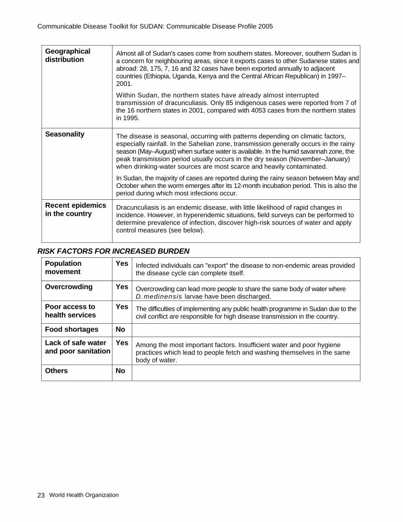
Geographical distribution
Almost all of Sudan's cases come from southern states. Moreover, southern Sudan is a concern for neighbouring areas, since it exports cases to other Sudanese states and abroad: 28, 175, 7, 16 and 32 cases have been exported annually to adjacent countries (Ethiopia, Uganda, Kenya and the Central African Republican) in 1997–2001.
Within Sudan, the northern states have already almost interrupted transmission of dracunculiasis. Only 85 indigenous cases were reported from 7 of the 16 northern states in 2001, compared with 4053 cases from the northern states in 1995.
Seasonality The disease is seasonal, occurring with patterns depending on climatic factors, especially rainfall. In the Sahelian zone, transmission generally occurs in the rainy season (May–August) when surface water is available. In the humid savannah zone, the peak transmission period usually occurs in the dry season (November–January) when drinking-water sources are most scarce and heavily contaminated.
In Sudan, the majority of cases are reported during the rainy season between May and October when the worm emerges after its 12-month incubation period. This is also the period during which most infections occur.
Recent epidemics in the country
Dracunculiasis is an endemic disease, with little likelihood of rapid changes in incidence. However, in hyperendemic situations, field surveys can be performed to determine prevalence of infection, discover high-risk sources of water and apply control measures (see below).
RISK FACTORS FOR INCREASED BURDEN Population movement
Yes Infected individuals can "export" the disease to non-endemic areas provided the disease cycle can complete itself.
Overcrowding Yes Overcrowding can lead more people to share the same body of water where D. medinensis larvae have been discharged.
Poor access to health services
Yes The difficulties of implementing any public health programme in Sudan due to the civil conflict are responsible for high disease transmission in the country.
Food shortages No
Lack of safe water and poor sanitation
Yes Among the most important factors. Insufficient water and poor hygiene practices which lead to people fetch and washing themselves in the same body of water.
Others No

Communicable Disease Toolkit for SUDAN: Communicable Disease Profile 2005
World Health Organization 24
Risk assessment conclusions
Sudan reported 78% of all dracunculiasis cases reported globally in 2001. Almost all of Sudan's cases are in the southern states, where the civil war has limited for a long time accessibility to endemic areas.
The country's proportion of global dracunculiasis cases has steadily increased during the past 7 years as the number of cases is reduced in all other endemic countries. The number of reported cases has decreased during the past 3 years despite intensive campaigns to reduce under-reporting. However, this decrease is not considered wholly representative because many endemic villages in the south are inaccessible as a result of civil disturbance.
Although rarely fatal, dracunculiasis is of great socioeconomic importance. Persons with this disease are incapacitated as a result of pain caused by the primary wound at the exit point of the worms and by associated secondary infections. Temporary disability usually lasts for periods averaging almost 3 months (usually 10–11 weeks), mainly because:
• several worms can be expelled successively,
• migration and emergence of the worms occur in sensitive parts of the body, e.g. the sole of the feet,
• serious secondary bacterial infection frequently sets in subsequent to the accidental rupture of the worm.
The emerging of the worm often happens at the busiest time of the year when people need to plant or harvest their crops, and half or more of a village population may be affected simultaneously. In addition to its impact on agricultural productivity, dracunculiasis is also a major cause of absenteeism from school. Moreover, it has been observed that, when disabled adult members of a household are prevented from fully performing their agricultural or domestic activities as a result of dracunculiasis, the nutritional status of children in the same household will deteriorate in the following year due to both lack of food and negligence in the care of children.
Man-made water-catchment ponds such as haffir, shallow wells and ponds are the main source of transmission in Sudan, and the epidemiology of the disease is determined largely by the use of these open water sources.
Sudan currently has the highest disease burden of dracunculiasis in the world. Vigilant surveillance and appropriate public health interventions are key for the global effort to eliminate the disease.

Communicable Disease Toolkit for SUDAN: Communicable Disease Profile 2005
World Health Organization 25
PREVENTION AND CONTROL MEASURES Case management No drugs are currently available to kill the adult worm. Slow extraction of the emergent
guinea worm, with appropriate antibiotic cover, is the most effective measure.
Once a worm emerges, use a matchstick to roll it out gently a few centimetres a day until the worm has been removed. Never break the worm, and never pull it. Bandage the wound after applying an antibiotic ointment to prevent superinfection of the lesion; 24 hours later, remove the bandages and roll the part of the worm that has emerged. Repeat this procedure until the whole worm has been removed (usually 10–20 days). Care must be taken to ensure that the worm does not break during the course of extraction.
Although practised in certain endemic countries, surgical extraction is NOT recommended.
Prevention Provision of safe water sources: this is the most expensive, and the most durable, intervention. It also has the advantage of providing other important benefits besides eliminating the guinea worm.
Control of copepod populations in ponds, tanks, reservoirs and step wells.
─ Insecticide of choice for stagnant sources of water: temephos (Abate®), which is effective and safe.
─ Formulation and dosage: based on the estimated amount of water present.
─ Time of application: the insecticide must be applied regularly, with a maximum interval of 28 days between applications to ponds less than 500 m in diameter during transmission season of known endemic villages and in villages newly reporting cases. The application is most effective after a flood has receded.
Health education: programmes should be focused on the following two messages:
1. Villagers with blisters or ulcers should not enter any source of drinking-water. It is well known that infected persons try to relieve the burning sensation by immersing the affected part of the body in local water sources. This should be discouraged.
2. Guinea-worm infection comes from drinking-water. Water should therefore be:
− boiled (this is usually impractical given the scarcity or high cost of wood or other fuel); or
− chlorinated; or
− filtered to remove copepods. Systematic filtering of drinking-water derived from ponds, shallow unprotected wells or from surface water should be encouraged: use of finely-meshed cloth filter, straw filter, or, preferably a filter made from a 0.15 mm nylon mesh, is the recommended option.
Immunization No vaccine is available. However, populations at high risk should be immunized against tetanus.

Communicable Disease Toolkit for SUDAN: Communicable Disease Profile 2005
World Health Organization 26
8. EBOLA HAEMORRHAGIC FEVER DESCRIPTION
Infectious agent Ebola virus, belonging to the genus Filovirus.
Case definition Clinical description
Presentation may be very nonspecific. Initial symptoms include acute fever, diarrhoea that can be bloody (referred to as diarrhee rouge in francophone Africa) and vomiting. Headache, nausea and abdominal pain are common. Conjunctival injection, dysphagia and haemorrhagic symptoms (nosebleeds, bleeding gums, vomiting of blood, blood in stools, purpura) may further develop. Some patients may show a maculopapular rash on the trunk. Dehydration and significant wasting occur as the disease progresses. At a later stage, frequent involvement of the central nervous system occurs, manifested by somnolence, delirium or coma. The case-fatality rate ranges from 50% to 90%.
Laboratory criteria: Confirmation
− Positive ELISA antigen detection or IgM capture, or − Positive virus isolation (only in a laboratory of Biosafety Level 4), or − Positive skin biopsy (immunohistochemistry), or − Positive PCR with sequence confirmation.
Case classification*: Suspected: a case that is compatible with the clinical description. Probable (in epidemic situation):
− Any person having had contact with a clinical case and presenting with acute fever, or
− Any person presenting with acute fever and three of the following symptoms: headache, vomiting/nausea, loss of appetite, diarrhoea, intense fatigue, abdominal pain, general or articular pain, difficulty in swallowing, difficulty in breathing, hiccups, or
− Any unexplained death. Confirmed: Any suspected or probable case that is laboratory-confirmed. Contact (in epidemic situation): An asymptomatic person having had physical contact within the past 21 days with a confirmed or probable case or his/her body fluids (e.g. care for patient, participation in a burial ceremony, handling of potentially infected laboratory specimens).
* Case classification should be tailored according to circumstances locally identified in the field (e.g. including contact with sick animals or animals with abnormal behaviour).
Mode of transmission
Person-to-person transmission by direct contact (spread of droplets onto mucous membranes) or indirectly by infected blood, secretions, organs, semen and fomites.
Risk is highest during the late stages of illness when the patient is vomiting, having diarrhoea or haemorrhaging. Risk during the incubation period is low. Under natural conditions, airborne transmission among humans has not been documented. Nosocomial infections have been frequent.
Incubation Incubation period is usually 2–21 days.
Period of communicability
As long as blood, saliva, faeces and other secretions contain virus, which can be up to 6 months.

Communicable Disease Toolkit for SUDAN: Communicable Disease Profile 2005
World Health Organization 27
EPIDEMIOLOGY Burden Occurs in epidemics (see below).
Geographical distribution
Ebola outbreaks in Sudan have occurred in the southernmost area of the country, close to the border with Democratic Republic of the Congo.
Seasonality No clearly evident seasonal pattern.
Alert threshold One suspect case must lead to an alert.
Recent epidemics in the country
2004 May–July/August. Cases of acute haemorrhagic fever syndrome in Hai-Cuba, Yambio county, Western Equatoria, southern Sudan. A rapid assessment team is in the field to investigate the situation. The cases were confirmed to be of Ebola Haemorrhagic fever (EHF). A total of 17 cases and 7 deaths of EHF were reported, of which 13 were laboratory-confirmed and 4 epidemiologically linked. The last death was reported on 26 June in the Yambio hospital isolation ward. After a mandatory 42 days of viglant surveillance the outbreak was officially declared over on 7 August 2004.
1979 July–October. On 2 August, a 45 year-old man was admitted to the N'zara hospital with a fever that had lasted for 3 days and recent onset of diarrhoea and vomiting. While at the N'zara hospital, he developed gastrointestinal haemorrhaging and died on 5 August. No precautionary isolation measures were taken or barrier-nursing techniques practised. Three of his relatives who had cared for him during his illness developed haemorrhagic fever and were hospitalized. All cases occurred among five families in a rural district in the remote savannah of southern Sudan. The district was later quarantined. The total number of cases was 34, of which 22 were fatal (CFR=65%). All cases were directly linked to the index case who was employed at the N'zara Cotton Manufacturing Factory.
1976 June–November. The first case of EHF in Sudan was detected in N'zara (Western Equatoria, close to the border with Democratic Republic of the Congo) and then spread to Maridi, Tambura and Juba. On 27 June, a N'zara Cotton Manufacturing Factory cloth-room worker became ill with a haemorrhagic febrile disease and died in the N'zara hospital on 6 July. The disease was introduced to Maridi, 128 km away, by a case admitted to Maridi hospital. Spreading occurred mainly through close personal contact within the hospital. Several medical care personnel were infected, as transmission was usually associated with the act of nursing a patient. The viral subtype identified was named Ebola-Sudan (EBO-S). The total number of cases was 284 (the largest part in Maridi); the percentage of deaths among cases (case-fatality rate) was 53%.
RISK FACTORS FOR INCREASED BURDEN Population movement
Yes In case of outbreak, population movement can contribute to the spread of infection to non-affected areas. Contacts under daily follow-up should be encouraged to limit their movements through community sensitization and social mobilization.
Overcrowding Yes Prompt isolation of a suspect case is a key control strategy.
All conditions favouring contact with sick persons, their cloths and bedding constitute a risk factor for increased transmission.
Poor access to health services
Yes Health centres are essential as an alert network, not for providing treatment. Prompt identification of cases is paramount to rapidly implement control measures.
Food shortages No

Communicable Disease Toolkit for SUDAN: Communicable Disease Profile 2005
World Health Organization 28
Lack of safe water and poor sanitation
No
Others No Activities related to hunting have been indicated as a risk factor for acquiring the infection on several occasions.
Risk assessment conclusions
The reservoir of Ebola haemorrhagic fever is not known, and it is therefore difficult to evaluate the risk of transmission. Implementation of control measures can also be difficult given cultural practices such as the custom of eating primate meat.
With the exception of Uganda, Ebola outbreaks have always occurred in ecologically similar areas: these areas could represent the biotope of the reservoir. Moreover, there are indications that similar climatic patterns are associated with Ebola outbreaks. Monitoring climatic variables could therefore help to identify high-risk areas.
Future priorities include identification of the reservoir in order to better target public health measures.
PREVENTION AND CONTROL MEASURES Case management Specific therapy: not currently available for filoviral infections.
Supportive treatment: − Analgesic drugs − Antimicrobial drugs (to avoid secondary infections) − Antimalarials (if clinically indicated) − Fluid replacement, with careful oral and less intravenous rehydration.
Implementation of barrier-nursing practices:
In order to prevent secondary infections, contact with the patient's lesions and body fluids should be minimized using standard isolation precautions:
− Isolation of patients − Restriction of access to patients wards − Use of protective clothing − Safe disposal of waste − Disinfection of all non-disposable supplies and equipment − Safe burial practices.
All the above measures can be implemented despite problems due to limited resources (see WHO/CDC. Infection control for viral haemorrhagic fevers in the African care setting. Geneva, WHO, 1998; WHO/EMC/EST/98.2).
Epidemic control Epidemics of the disease in health care institutions with poor hygiene standards can be dramatically amplified through contact with patients or body fluids from infected patients (blood, vomitus, urine, stools, semen, saliva). The potential for explosive nosocomial infections constitutes the main threat to public health posed by the disease. Strict adherence to isolation precautions with all patients has been shown to reduce the risk of transmission. During the 1995 Ebola haemorrhagic fever outbreak in Kikwit (Democratic Republic of the Congo, 1995), no new cases were reported among health workers who used these precautions consistently.

Communicable Disease Toolkit for SUDAN: Communicable Disease Profile 2005
World Health Organization 29
Prevention The following key elements are essential in the prevention of explosive epidemics in areas potentially subject to EHF:
1. Social mobilization and health education of the community, emphasizing:
− Avoiding contact with body fluids of an EHF patient.
− Seeking treatment early and avoiding harmful funeral practices.
− Boiling and burning all clothing of an Ebola patient.
− Use of protective methods when handling the patient and EHF patient’s articles.
− Avoiding consumption of dead animal meat found in the forest.
2. Correct case management, including barrier nursing and appropriate funeral practices.
3. Effective and efficient coordination of interventions.
4. Good logistics and security.
5. Vigilant surveillance, standard epidemiological practice and laboratory services.
Health workers in EHF-prone regions should receive advance training in the use of isolation procedures and universal isolation precautions.

Communicable Disease Toolkit for SUDAN: Communicable Disease Profile 2005
World Health Organization 30
9. HIV/AIDS DESCRIPTION
Infectious agent Human immunodeficiency virus (HIV). Two types have been identified: HIV-1 and HIV-2; both have similar epidemiological characteristics. HIV-2 is less pathogenic than HIV-1.
Case definition AIDS case definition Acquired immunodeficiency syndrome (AIDS) is the late clinical stage of HIV infection, defined as an illness characterized by one or more indicator diseases.
WHO staging system for HIV infection and disease in adults and adolescents
Stage 1 1. Asymptomatic. 2. Persistent generalized lymphadenopathy (PGL). Performance Scale 1: asymptomatic, normal activity. Stage 2 3. Weight loss, <10% of body weight. 4. Minor mucocutaneous manifestations (seborrheic dermatitis, prurigo, fungal nail infections, recurrent oral ulcerations, angular cheilitis). 5. Herpes zoster within the past 5 years. 6. Recurrent upper respiratory tract infections (e.g. bacterial sinusitis), And/or Performance Scale 2: symptomatic, normal activity. Stage 3 7. Weight loss, >10% of body weight. 8. Unexplained chronic diarrhoea, >1 month. 9. Unexplained prolonged fever (intermittent or constant), >1 month. 10. Oral candidiasis (thrush). 11. Oral hairy leukoplakia. 12. Pulmonary tuberculosis within the past year. 13. Severe bacterial infections (i.e. pneumonia, pyomyositis), And/or Performance Scale 3: bedridden, <50% of the day during the past month. Stage 4 14. HIV wasting syndrome, as defined by the US Centers for Disease Control and Prevention (CDC).a 15. Pneumocystis carinii pneumonia. 16. Toxoplasmosis of the brain. 17. Cryptosporidiosis with diarrhoea >1 month. 18. Cryptococcosis, extrapulmonary. 19. Cytomegalovirus (CMV) disease of an organ other than liver, spleen or lymph nodes.20. Herpes simplex virus (HSV) infection, mucocutaneous >1 month, or visceral any duration. 21. Progressive multifocal leukoencephalopathy (PML). 22. Any disseminated endemic mycosis (e.g. histoplasmosis, coccidiomycosis). 23. Candidiasis of the oesophagus, trachea, bronchi or lungs. 24. Atypical mycobacteriosis, disseminated. 25. Non-typhoid Salmonella septicaemia. 26. Extrapulmonary tuberculosis. 27. Lymphoma. 28. Kaposi sarcoma. 29. HIV encephalopathy, as defined by CDC.b Note: Both definitive and presumptive diagnoses are acceptable. (a) HIV wasting syndrome: weight loss of >10% of body weight, plus either unexplained chronic diarrhoea (>1 month) or chronic weakness and unexplained prolonged fever (>1 month); (b) HIV encephalopathy: clinical finding of disabling cognitive and/or motor dysfunction interfering with activities of daily living, progressing over weeks tomonths, without a concurrent illness or condition other than HIV infection that could explain the findings.

Communicable Disease Toolkit for SUDAN: Communicable Disease Profile 2005
World Health Organization 31
Expanded WHO case definition for AIDS surveillance* An adult or adolescent ( aged >12 years) is considered to have AIDS if a test for HIV antibody gives a positive result, and one or more of the following conditions are present:
1. >10% body weight loss or cachexia, with diarrhoea or fever, or both, intermittent or constant, for at least 1 month, not known to be due to a condition unrelated to HIV.
2. Cryptococcal meningitis.
3. Pulmonary or extrapulmonary tuberculosis.
4. Kaposi sarcoma.
5. Neurological impairment that is sufficient to prevent independent daily activities, not known to be due to a condition unrelated to HIV infection (e.g. trauma or cerebrovascular accident).
6. Candidiasis of the oesophagus (which may be presumptively diagnosed based on the presence of oral candidiasis accompanied by dysphagia).
7. Clinically diagnosed life-threatening or recurrent episodes of pneumonia, with or without etiological confirmation.
8. Invasive cervical cancer.
* WHO. Weekly Epidemiological Record, 1994, 69:273-275.
Laboratory evidence of HIV
This is most commonly based on detection of HIV antibody in serum samples using enzyme-linked immunosorbent assay (ELISA or EIA). When positive, this test must be confirmed with another test of higher specificity such as the Western blot, the indirect fluorescent antibody (IFA) test or a second ELISA test that is methodologically and/or antigenically independent.
The rapid tests that are recommended by WHO have been evaluated at WHO collaborating centres and have levels of sensitivity and specificity comparable with WHO-recommended ELISA tests. The use of rapid HIV tests may afford several advantages in emergency and disaster settings, including:
− Rapid tests that do not require refrigeration will be more suitable for remote and rural areas and sites without a guaranteed electricity supply. Long shelf-life is also important, especially for remote areas and sites performing smaller numbers of tests.
− Many rapid tests require no laboratory equipment and can be performed in settings where electrical and water supplies need not be guaranteed.
− Rapid tests can detect HIV antibodies in whole blood (finger-prick samples) as well as in serum/plasma, and testing may therefore be performed by non-laboratory personnel with adequate training and supervision.

Communicable Disease Toolkit for SUDAN: Communicable Disease Profile 2005
World Health Organization 32
Mode of transmission
Sexual intercourse (vaginal or anal) with an infected partner, especially in the presence of a concurrent ulcerative or non-ulcerative sexually transmitted infection (STI).
Contaminated needles, syringes, other injecting equipment and injecting solutions (contamination often occurs when drug solutions are mixed or when multiple users draw up solutions from a single container).
Transfusion of infected blood or blood products.
Infected mother to her child during pregnancy, labour and delivery or through breastfeeding.
Incubation Variable. On average, the time from HIV infection to clinical AIDS is 8–10 years, although AIDS may be manifested in less than 2 years or be delayed in onset beyond 10 years.
Incubation times are shortened in resource-poor settings and in older patients. They can be prolonged by provision of primary prophylaxis for opportunistic infections or by antiretroviral treatment.
Period of communicability
Any person who is infected with HIV may pass the infection to another through the routes of transmission described above.
Infectiousness is observed to be high during the initial period after infection. Studies suggest it increases further with increasing immune deficiency, clinical symptoms and presence of other STIs.
EPIDEMIOLOGY Burden Estimated number of adults and children living with HIV/AIDS, end of 2001:
(including all people with HIV infection, whether or not they have developed symptoms of AIDS).
Adults (15–49) 410 000 (2.6% of all adults) Women (15–49) 230 000 Children (0–15) 30 000
Estimated number of deaths due to AIDS in 2001: 23 000 Reported AIDS cases in 2001: 492 (Mode of transmission: heterosexual, 348; perinatal, 6; unknown, 138) Estimated number of living orphans (2001): 62 000 (Data source: WHO Sudan, 2004)
In 2002, the results of an epidemiological survery conducted among 7, 385 individuals in 11 of the 16 sates, involving a number of groups at varying risk of infection were reported. Persons tested included Sudanese and nonSudanese. The seroprevalence among Sudanese was 1.6% . Of the 3, 355 women in ANC care, 30% (1.0%) were infected. Of 367 Sudanese sex workers, 16% (4.4%) were infected. Lesser prevalences were found among prisoners (4 of 200, 2.0%), soilders (2 of 377, 0.5%), those with STDs (4 of 362, 1.1%), university students (4 of 369, 1.1%) and TB patients (6 of 367, 1.6%). The Sudan National AIDS Control Programme HIV surveillance system indicates the large majority of finfections are acquired via heterosexual transmission, and this survey showed levels of awareness of HIV/AIDS and means to protect oneself from becoming infected were poor.
See: UNAIDS - Sudan, Epidemiological fact sheet. 2004 update. http://www.unaids.org/html/pub/publications/fact-sheets01/sudan_en_pdf.pdf
Geographical distribution
No data available; HIV median prevalence among ANC attendees in 1998 was about 0.5% in urban areas and about 3.75% in rural areas.

Communicable Disease Toolkit for SUDAN: Communicable Disease Profile 2005
World Health Organization 33
Seasonality Not applicable.
Alert threshold One suspected case must be investigated.
Recent epidemics in the country
Number of AIDS cases by year of reporting 2003 : no data available 1994: 201 1985: 0
2002: no data available 1993: 191 1984: 0
2001 : 492 1992: 184 1983: 0
2000 : 652 1991: 188 1982: 0
1999 : 517 1990: 130 1981: 0
1998 : 511 1989: 122 1980: 0
1997: 270 1988: 64 1979: 0
1996 : 221 1987: 2
1995 : 257 1986: 2
Total (end 2001): 10959 (Data source: UNAIDS, 2004 update).
RISK FACTORS FOR INCREASED BURDEN Population movement
Yes In emergency situations, population movement can:
─ Cause breakdown in family and social ties.
─ Erode traditional values and coping strategies. This can result in higher-risk sexual behavior, which increases the risk of HIV spread.
─ Influence illicit drug trafficking and drug use, which increases the risk of HIV transmission through injecting drug use.
Overcrowding Yes Groups with differing levels of HIV awareness, and differing rates of infection, are often placed together in temporary locations, such as refugee camps, where there is greater potential for sexual contact.
Overcrowding can also influence injecting drug use patterns and result in increased risk of sharing contaminated injecting equipment (this has been noted in refugee camps).
Poor access to health services
Yes Without adequate medical services, STIs, if left untreated in either partner, greatly increase the risk of acquiring HIV. Important materials for HIV prevention, particularly condoms, are likely to be lacking in an emergency situation. In emergency situations, services for drug dependence treatment usually do not exist. It is more likely to be difficult to access sterile injecting equipment.
Food shortages Yes The need for food is paramount in emergency situations, and exchanging sex for money to buy food and other essentials can occur (see "Sex work", below).
Lack of safe water and poor sanitation
No

Communicable Disease Toolkit for SUDAN: Communicable Disease Profile 2005
World Health Organization 34
Others Yes Sexual violence
Refugees and IDPs are often physically and socially powerless, with women and children at particular risk of sexual coercion, abuse or rape.
Sexual violence carries a higher risk of infection because the person violated cannot protect herself or himself from unsafe sex, and because the virus can be transmitted more easily if bodily tissues are torn during violent sex.
Sex work
Exchange of sexual favours for basic needs such as money, shelter and security is common in or around refugee camps, and inevitably involves both the refugee and host communities. Both sex workers and clients are at risk of HIV infection if unprotected sex is practised.
Injecting drug use
In Sudan, no AIDS cases officially reported from the beginning of the epidemics to the end of 2001 had contracted the disease by injecting drugs.
In typical emergency conditions, it is highly likely that drug injectors will be sharing needles, a practice that carries a very high risk of HIV transmission if one of the people sharing is infected.
Unsafe blood transfusions
Transfusion with HIV-infected blood is a highly efficient means of transmitting the virus. In emergency situations, when regular transfusion services have broken down, it is particularly difficult to ensure blood safety.
Adolescent health
Children in refugee settings may have little to occupy themselves with, which may lead them to experiment with sex earlier than children in other situations.
Lack of regular supplies
Lack of laboratory reagents for screening and testing, particularly for blood transfusions.
Lack of condoms.

Communicable Disease Toolkit for SUDAN: Communicable Disease Profile 2005
World Health Organization 35
Risk assessment conclusions
HIV/AIDS is becoming an increasing problem in Sudan. Although the surveillance system is limited to four sites only, it is feared that Sudan could be facing a generalized epidemic.
HIV prevalence among pregnant women was reported to be 2.94% in 1997.
Based on recent HIV surveillance data in Khartoum State in 2000, 2.5% of women attending gynaecological clinics were found to be HIV seropositive, compared with HIV seropositivity rates of 1.86% in pregnant women attending antenatal clinics.
HIV rates among blood donors increased from 0.15% in 1993 to 1.4% in 1999–2000.
There is a clear link between HIV and tuberculosis (TB). In 1999, HIV prevalence among TB patients varied between 7.7% and 20% depending on the regions.
Women attending both gynaecological and antenatal clinics in Khartoum State showed STI incidence of 10.5% and 34% respectively.
Reported AIDS cases by mode of transmission (from the beginning of the epidemic* up to end 2001):
Heterosexual contacts 3758 (93.9%) Blood and blood products 12 (0.3%) Perinatal 96 (2.4%) Unknown 138 (3.4%) All AIDS cases reported 4004 (100%) * The earliest AIDS cases in Sudan were reported in 1986 (Source: UN AIDS Sudan epidemiological profile fact sheet on HIV/AIDS and sexually transmitted diseases, 1 September 2004).
All stakeholders involved in humanitarian activities must be sensitized to the importance of addressing HIV in tandem with all other activities. Activities should include HIV prevention (promotion of safer sexual behaviours, treatment of STIs, blood safety) and care and support for people living with HIV/AIDS (PLWHA). They must reach vulnerable populations and address the needs of women and children.
All stakeholders must also be sensitized about HIV risks associated with injecting drug users and the need for drug dependence treatment and risk reduction education and counselling.

Communicable Disease Toolkit for SUDAN: Communicable Disease Profile 2005
World Health Organization 36
PREVENTION AND CONTROL MEASURES Case management Provide high-quality care and support to all PLWHA, which includes counselling,
psychosocial support, treatment for opportunistic infections (e.g. TB), palliative care and access to antiretroviral therapy where feasible.
Support PLWHA to live normal and productive lives that are free of stigmatization and discrimination.
Prevention Reduce sexual and mother-to-child transmission
Awareness and life skills education, especially among youth, to ensure that all people are well informed of what does, and does not, constitute a mode of transmission; of how and where to acquire free condoms and medical attention if necessary; and information on basic personal hygiene.
Condom promotion to ensure that good-quality condoms are freely available to those who need them, using culturally sensitive instructions and distribution mechanisms.
STI control, including for sex workers, using the syndromic STI management approach, with partner notification and promotion of safer sex.
Reduce mother-to-child transmission of HIV by:
− the primary prevention of HIV among women, especially young women
− avoiding unintended pregnancies among HIV-infected women and promoting family planning methods, particularly in women who are infected with HIV
− preventing HIV transmission from infected pregnant women to their infants by:
− using an antiretroviral prophylaxis regimen;
− avoiding unnecessary and invasive obstetrical procedures such as artificial rupture of membranes or episiotomy; and
− modifying infant feeding practices (replacement feeding given with a cup when acceptable, feasible, affordable, sustainable and safe; otherwise exclusive breastfeeding for the first six months of life is recommended: SeeThe optimal duration of exclusive breastfeeding - A systematic review, WHO/FCH/CAH/01.23).
Blood safety
HIV testing of all transfused blood. Avoid non-essential blood transfusion
− Recruitment of safe blood donor pool.
Prevention among injecting drug users
Ready access to sterile needles, syringes and other injecting equipment (and disposal of used equipment).
HIV risk reduction education and counselling for injecting drug users (including peer outreach when possible).
Drug dependence treatment services, including substitution treatment (e.g. methadone) where possible.
Access to STI and HIV/AIDS treatment for injecting drug users.

Communicable Disease Toolkit for SUDAN: Communicable Disease Profile 2005
World Health Organization 37
Universal precautions
Washing hands thoroughly with soap and water, especially after contact with body fluids or wounds.
Using protective gloves and clothing when there is risk of contact with blood or other potentially infected body fluids.
Safe handling and disposal of waste material, needles and other sharp instruments. Proper cleaning and disinfection of medical instruments between patients.
Physical protection
The protection of the most vulnerable, especially women and children, from violence and abuse is not only an important principle of human rights but is also essential for reducing the risk of HIV infection.
Protecting health care workers
In order to reduce nosocomial transmission, health workers must strictly adhere to universal precautions with all patients and laboratory samples – whether or not known to be infected with HIV.
Health care workers should have access to voluntary counselling, testing and care; those deployed in complex emergencies frequently experience significant occupational stress, and those tested as part of the management of occupational exposures will require additional support.
Counselling and voluntary testing programmes
The establishment of voluntary counselling and testing services to help individuals make informed decisions about HIV testing should be considered when relative stability has been restored. Displaced populations are often coerced into testing or are required to make decisions about testing when they are suffering acute or post-traumatic stress disorders.
As displaced populations are often tested before resettlement in other countries, it is critical that they receive counselling on the legal and social implications of the test. Often, migration or temporary residency status is contingent on the applicant’s having HIV antibody seronegative status.
Post-test counselling is essential for both seronegative and seropositive results. Displaced populations and conflict survivors who are already traumatized will require additional psychosocial support if they test seropositive. Typically, the support networks of displaced persons are disrupted, and suicide risk assessment forms an important part of post-test counselling in a refugee or conflict context.
Testing of orphaned minors should be done with the consent of their official guardians only where there is an immediate health concern or benefit to the child.
No mandatory screening should take place before admittance to substitute care.
Immunization Asymptomatic HIV-infected children should be immunized with the EPI vaccines.
Symptomatic HIV-infected children should NOT receive BCG or yellow fever vaccine.

Communicable Disease Toolkit for SUDAN: Communicable Disease Profile 2005
World Health Organization 38
10. LEISHMANIASIS (CUTANEOUS AND MUCOSAL) DESCRIPTION Infectious agent Protozoan, belonging to the genus Leishmania:
• L. major, agent of cutaneous leishmaniasis (and, less frequently, of mucosal leishmaniasis)
• L. donovani, agent of mucosal leishmaniasis (see Visceral leishmaniasis).
Case definition Clinical description Cutaneous leishmaniasis is characterized by the appearance of one or more skin lesions, typically on uncovered parts of the body; the face, neck, arms and legs are the most common sites. A nodule may appear at the site of inoculation and may enlarge to become an indolent ulcer. The sore may remain at this stage for a variable time before healing, typically leaving a depressed scar. Other atypical forms may occur. In some individuals, certain strains can disseminate and cause mucosal lesions. These sequelae involve nasopharyngeal tissues and can be very disfiguring with major psychological consequences (see below).
Sudanese mucosal leishmaniasis is a chronic infection of the upper respiratory tract and/or oral mucosa caused mainly by L.donovani or, less frequently, by L.major. The disease occurs in areas of the country endemic for visceral leishmaniasis. The condition may develop during or after an attack of visceral leishmaniasis, but in most cases it is a primary mucosal disease. It is not preceded or accompanied by a cutaneous lesion. The duration of the disease can vary between a few months and several years.
Laboratory criteria • Positive parasitology (stained smear or culture from the lesion) Positive serology (immunofluorescent assay, ELISA, Direct Agglutination Test) for mucosal leishmaniasis only. WHO operational definitions
• A case of cutaneous leishmaniasis can be defined as a person showing clinical signs (skin lesions) with parasitological confirmation of the diagnosis (positive smear or culture).
• A case of mucosal leishmaniasis can be defined as a person showing clinical signs (mucosal lesions) with parasitological confirmation of the diagnosis and/or
Mode of transmission
From the reservoir host through the bite of infective female phlebotomines (sandflies).
Phlebotomus papatasi is the vector of L. major in Sudan. The highest vector population density is usually found when the temperature is high, humidity is medium and rainfall is low. The vector is domestic and peridomestic in the villages. Humans are the preferred hosts, and daily biting activity is highest in the evening.
There is limited information on the animal reservoir of cutaneous leishmaniasis in Sudan. It is probably represented by the Nile rat Arvicanthis niloticus.
Phlebotomus orientalis, the vector of L. donovani in Sudan, is most abundant where Acacia seyal and Balanites aegyptica vegetation is common and where the soil is rich in clay.

Communicable Disease Toolkit for SUDAN: Communicable Disease Profile 2005
World Health Organization 39
Incubation Cutaneous leishmaniasis: usually 2–4 weeks but may be longer. The incubation period is inversely proportional to the size of the inoculum.
Mucosal leishmaniasis: difficult to establish. Patients usually provide no history of cutaneous leishmaniasis. Some patients give a history of treated visceral leishmaniasis; others may have had mucosal and visceral leishmaniasis concurrently.
Period of communicability
An infected subject is susceptible to transmit the parasite as long as it remains in lesions; in untreated cases, usually a few months to 2 years.
EPIDEMIOLOGY Burden Year Number of cases Deaths
2000 204 2
2001 351 32
2002 258 --- (Data source: WHO/Sudan, 2004)
Geographical distribution
Darfur and Kordofan provinces are known to be endemic for zoonotic cutaneous leishmaniasis. Epidemics in recent years have occurred in Northern, Eastern, Khartoum and Central provinces.
Most patients with mucosal leishmaniasis come from areas also endemic for visceral leishmaniasis.
Seasonality During recent epidemics in Sudan, the peak of infection was believed to occur in August and December, dropping sharply in March, April and May.
Recent epidemics in the country
1976 Shendi–Atbara area (Northern Province). 1985–1987 Khartoum Province: about 10 000 recorded cases. Peak incidence in September 1986. 1990–1992 Dongola and Mahas areas (Northern Province).
RISK FACTORS FOR INCREASED BURDEN Population movement
Yes Movements of population such as displaced populations contribute to the maintenance of epidemics, bringing non-immune people into endemic areas and infected people into non-endemic areas where the vector is widespread.
The characteristics of epidemics in Sudan are typical of a disease newly introduced into a previously non-immune population, in that all age groups have been affected.
Migrants from western Sudan (endemic for zoonotic cutaneous leishmaniasis) to Khartoum Province probably contributed to the epidemic that affected this area between 1985 and 1987.
Overcrowding Yes Overcrowding can increase the risk of contact with animal reservoir.
Poor access to health services
Yes Lack of treatment increases the number of infected individuals in the population.
Food shortages Yes However, malnourished people are more susceptible to the infection due to a weakened immune response. Many of the patients seeking treatment are also malnourished.

Communicable Disease Toolkit for SUDAN: Communicable Disease Profile 2005
World Health Organization 40
Lack of safe water and poor sanitation
Yes Open sewage systems and lack of garbage or rubble collection favour the proliferation of vector breeding sites.
Others Yes Rapid expansion of towns and villages, establishment of new settlements and consequent development of previously uninhabited areas.
Interruption of control measures: discontinuation of insecticide spraying for malaria control in Sudan in the years preceding the epidemics may have contributed to an increase in the vector population.
Increase in the rodent population: coinciding, in 1976 and 1986, with the two epidemics of cutaneous leishmaniasis in Sudan.
Prolonged low rainfall: the dry soil cracks and becomes waterlogged, creating ideal breeding conditions for the sandfly.
Risk assessment conclusions
The disease is known as hashara ("insect") in Sudan.
PREVENTION AND CONTROL MEASURES Case management Cutaneous leishmaniasis is a self-limiting disease. Self-healing usually occurs
within 6 months, but skin scarring and changes in pigmentation always follow.
There is currently no uniform protocol for treating cutaneous leishmaniasis in Sudan. Patients with minor lesions are usually reassured and left to heal spontaneously. Patients with severe or multiple lesions (>5), diabetics with lesions and patients who acquired infection in geographic regions where mucosal disease has been reported should be treated promptly.
Treatment is based on:
Pentavalent antimonials (the drug of choice in Sudan is sodium stibogluconate) as a first-line drug, except when resistance develops. The drugs can be administered systemically (IM or IV), or locally (intralesional infiltrations). WHO recommends the following course: 20 mg/kg per day for 20 days.
In the presence of resistance, second-line drugs must be used. Standard amphotericin B, aminosidine plus pentavalent antimonials or pentamidine isetionate are the main alternatives.
Other therapeutic options are available: − Antifungal drugs (e.g. ketoconazole). − Cryotherapy.
Patients with mucosal leishmaniasis respond well to treatment with pentavalent antimony compounds (sodium stibogluconate).
Epidemic control Epidemics of cutaneous leishmaniasis can be controlled by an integrated, feasible and efficient strategy based on:
─ Provision of first-line drug (pentavalent antimonials) to improve cure rate.
─ Provision of long-lasting bednets (insecticide-treated nets – ITNs) to limit contact between human and vector.
─ Health education and social interventions to increase awareness and improve early diagnosis.

Communicable Disease Toolkit for SUDAN: Communicable Disease Profile 2005
World Health Organization 41
Prevention Personal protective measures are effective in preventing contact between
sandflies and humans. Such measures include skin repellents, vaporizing liquids, bednets impregnated or sprayed with pyrethroids, and screened doors and windows.
Vector control: application of residual insecticides on surfaces where sandflies rest, such as indoor and outdoor walls, tree trunks, rock crevices, water wells and flowering plants may be effective in reducing the size of the sandfly population over time, thus decreasing the risk of infection. However, this measure is not recommended since it produces a transient effect only.
Reservoir control: control methods must be adapted to the biology of the reservoir species (anticoagulants, poison baits, deep ploughing to eliminate plants on which the rodents feed, use of artificial canals or barriers to prevent colonization or reinvasion). No definitive control method against Arvicanthis is currently known.
Many field activities for control of cutaneous leishmaniasis are integrated with those for malaria control. Work is in progress to evaluate the use of mosquito nets impregnated with insecticide to reduce human–fly contact.

Communicable Disease Toolkit for SUDAN: Communicable Disease Profile 2005
World Health Organization 42
11. VISCERAL LEISHMANIASIS (KALA AZAR) DESCRIPTION
Infectious agent Protozoan: Leishmania donovani
Case definition Clinical description
An illness with prolonged irregular fever, splenomegaly and weight loss as its main symptoms.
• Post–kala-azar dermal leishmaniasis (PKDL) is increasingly recognized in Sudan as a complication of visceral leishmaniasis, occurring in about 55% of patients during treatment or within 0–6 months after treatment. It is characterized by a rash that may be macular, maculopapular, nodular or plaque-like.
• Sudanese mucosal leishmaniasis is a chronic infection of the upper respiratory tract and/or oral mucosa caused mainly by L. donovani (see Cutaneous leishmaniasis). The disease occurs in areas of the country endemic for visceral leishmaniasis. In most cases it is a primary mucosal disease, not preceded or accompanied by a cutaneous lesion, but less frequently the condition may develop during or after an attack of visceral leishmaniasis. In this case the disease represents a phenomenon similar to PKDL.
Laboratory criteria • Positive parasitology. − stained smears from bone marrow, spleen, liver, lymph node, blood or, − culture of the organism from a biopsy or aspirated material.
• Positive serology (immunofluorescent assay, ELISA, Direct Agglutination Test).
WHO operational definition
• A case of visceral leishmaniasis (VL) is a person showing clinical signs (prolonged irregular fever, splenomegaly and weight loss) with serological (at peripheral geographical level) and/or (when feasible at central level) parasitological confirmation of the diagnosis. The main differential diagnosis is malaria. In endemic malarious areas, VL must be suspected when fever lasts for more than 2 weeks and no response has been achieved with antimalarial drugs (assuming drug-resistant malaria has also been considered).
Mode of transmission
Vector-borne, through the bite of infective female phlebotomines (sandflies).
Phlebotomus orientalis, the vector of L. donovani in Sudan, is most abundant where Acacia seyal and Balanites aegyptica vegetation is common and where the soil is rich in clay.
Transmission dynamics have not been elucidated fully; the large numbers of patients with PKDL in heavily affected villages indicate a human reservoir and anthroponotic transmission, whereas heavy transmission in scarcely populated areas suggests zoonotic transmission.
Incubation Usually 2–6 months. Intensity of infection, partial immunity resulting from previous exposure, intercurrent illness, malnutrition and other factors may play a role in determining the acuteness or slowness of the course.
Period of communicability
An infected subject is susceptible to transmit the parasite to sandflies as long as it persists in the circulating blood or skin. Infectivity for sandflies may persist even after clinical recovery of human patients.

Communicable Disease Toolkit for SUDAN: Communicable Disease Profile 2005
World Health Organization 43
EPIDEMIOLOGY Burden Number of cases reported (July–June):
2001–2002: 2413 2000–2001: 2308 1999–2000: 3922 1998–1999: 4804 1997–1998: 6182 (Data Source: WHO/Sudan, 2004)
Geographical distribution
The disease is historically endemic in a wide belt extending from the western bank of the White Nile and the Sudan–Ethiopian border. This area includes the southern areas of Central and Eastern provinces and the north-eastern part of Upper Nile Province. Since 1989, visceral leishmaniasis has spread in the western part of Upper Nile Province, where visceral leishmaniasis has never been reported before.
Cases have also been reported from Darfur Province in west Sudan, the Nuba mountains area (Kordofan Province) and the Kapoeta area in Eastern Equatoria.
Seasonality It is likely that the peak of transmission occurs towards the end of the dry season (March–June), especially just before the rainy season begins (May, June), when large numbers of P. Orientalis appear. Most individuals report with illness after the rains in October and November.
Number of cases reported in the period 1997–2002 (monthly basis): January: 2335 July: 934 February: 2094 August: 870 March: 1883 September: 1083 April: 1554 October: 1856 May: 1376 November: 2384 June: 1122 December: 2138 (Data source: WHO/Sudan)
Recent epidemics in the country
2002. A severe increase in the number of VL cases was reported in October and November from communities in southern Sudan. The overlap of areas affected by VL and areas of conflict suggests that insecurity, malnutrition and poor access to health care lower people's natural resistance, creating an epidemic-prone environment.
1997–1998. A dramatic upsurge in the number of VL cases was reported from eastern Sudan (Atbara river area): more than 2500 confirmed cases were registered in a MSF-run treatment centre in Gedaref State from October to December 1997 (+439% compared with the same period in 1996). Cumulative factors played a role: influx of displaced and non-immune population from southern states and overall decline in the nutritional status of the population.
1984–1994.Upper Nile Province (southern Sudan): because of the isolation of the area caused by the war, the epidemics went unnoticed until 1988. Between 1984 and 1994, an estimated 100 000 died. A number of causes were proposed for this outbreak in a previously non-endemic area: • Regeneration of Acacia seyal and Balanites aegyptica forests after they
were destroyed in the 1960s. • Introduction of the parasite from VL-endemic areas along the Ethiopian border
by movement of military personnel. • Discontinuation of residual insecticide spraying for malaria because of the war. • Malnutrition of the people.
From 1990 onwards, the epidemics reached the southern part of Kordofan Province and, in 1995–1996 the eastern part of Upper Nile Province.

Communicable Disease Toolkit for SUDAN: Communicable Disease Profile 2005
World Health Organization 44
RISK FACTORS FOR INCREASED BURDEN Population movement
Yes Movements of displaced populations contribute to the maintenance of epidemics, bringing non immune people into endemic areas and infected people into non-endemic areas where the vector is widespread.
The characteristics of epidemics in Sudan are typical of a disease newly introduced into a previously non-immune population: all age groups are affected.
Migrants from western Sudan (endemic for zoonotic cutaneous leishmaniasis) to Khartoum Province probably contributed to the epidemics.
Overcrowding Yes Persons with PKDL act as reservoirs of Leishmania parasites in anthroponotic foci.
Poor access to health services
Yes As a result of many factors (geographical, economic and cultural, poor transportation). Additionally, since most services are not free of charge, most patients seek traditional treatment measures.
Food shortages Yes Poor nutritional status increases susceptibility to VL infection and disease.
Lack of safe water and poor sanitation
No
Others Yes Acacia seyal and Balanites aegyptica woodland: the disease vector is found only in forests where these trees are present and in villages in or near these forests.
Black cotton soils: this clay-rich soil shrinks and swells with drying and wetting to the extent that very few woody plants can survive in it. However, certain trees survive and even thrive in it, including A. seyal and B. aegyptica.
Termitaria are thought to provide the insects with a relatively low, stable temperature and high humidity during the heat of the day. Termitaria have been found to be resting sites for P. martini in south-eastern Sudan, but many localities harbouring P. orientalis have no or small termitaria.
Interruption of control measures: discontinuation of insecticide spraying for malaria control in Sudan may have led to an increase in the vector population and contributed to the VL epidemics in southern Sudan.
Prolonged low rainfall: the dry soil cracks and becomes waterlogged, creating ideal breeding/resting sites for the sandfly. P. orientalis is most abundant towards the end of the dry season (March–June).
Risk assessment conclusions
Visceral leishmaniasis is endemic in Sudan: the first case was reported in 1904. High mortality in the country is mainly due to the absence of diagnostic facilities, the unavailability of first-line drugs at the peripheral level and increasing resistance to pentavalent antimonials.
The majority of cases are found in children and teenagers (up to 75%) as transmission occurs early in life. Prognosis is usually severe due to high prevalence of malnutrition and associated diseases - such as TB and respiratory and/or intestinal infections.

Communicable Disease Toolkit for SUDAN: Communicable Disease Profile 2005
World Health Organization 45
PREVENTION AND CONTROL MEASURES Case management Visceral leishmaniasis is a severe and fatal disease without treatment.
First-line treatment: pentavalent antimonials (Sodium stibogluconate was introduced in Sudan in 1947). WHO recommends the following course: 20 mg/kg per day for 30 days.
Second-line treatment: liposomal amphotericin B, aminosidine.
Three lipid-associated amphotericin B formulations (liposomal, colloidal dispersion, lipid complex) are highly effective against visceral leishmaniasis and better tolerated than the conventional preparation. Liposomal amphotericin B is an expensive drug and cannot therefore be recommended as a first-line drug; if available, it should be the drug of choice in cases resistant or unresponsive to antimonials due to its high effectiveness and lower toxicity.
Treatment for PKDL is needed only for those who have severe and prolonged disease; sodium stibogluconate is usually sufficient. Liposomal amphotericin B is also effective.
Patients with mucosal leishmaniasis respond well to treatment with pentavalent antimony compounds (sodium stibogluconate).
Resistance to pentavalent antimonials has been reported from eastern Sudan, but data are fragmentary. It has not been reported from southern Sudan.
Epidemic control Epidemics of visceral leishmaiasis can be controlled by an integrated, feasible and efficient strategy based on:
─ Provision of first-line drug (pentavalent antimonials) to improve cure rate and, in zoonotic foci, reduce transmission.
─ Provision of insecticide-treated nets (ITNs) to limit contact between human and vector. Long-lasting ITNs are now available.
─ Health education and social interventions to increase awareness and improve early diagnosis, early health-seeking and good treatment compliance.
Visceral leishmaniasis control programmes may be integrated with malaria control programmes.
Prevention Personal protective measures are effective in preventing contact of sandflies and humans. Such measures include skin repellents, vaporizing liquids, bednets impregnated or sprayed with pyrethroids, and screened doors and windows. Usually, the mesh used for leishmaniasis control is the same as that used for malaria control: it therefore has to be impregnated with insecticide, otherwise sandflies will pass through.
Vector control: application of residual insecticides on surfaces where sandflies rest, such as indoor and outdoor walls and tree trunks, could be effective in reducing the size of the sandfly population over time and thus decrease the risk of infection, but is not recommended due to its high cost, low sustainability and logistic constraints.
Reservoir control: The animal reservoir for L. donovani has not yet been identified.
Systematic case detection and rapid treatment: this applies to anthroponotic foci.
Many programmes and field activities are integrated with those for malaria control. Work is in progress to evaluate the use of vaccine against L. donovani and of mosquito nets impregnated with insecticide to reduce human–fly contact.

Communicable Disease Toolkit for SUDAN: Communicable Disease Profile 2005
World Health Organization 46
12. LEPROSY DESCRIPTION
Infectious agent Bacterium: Mycobacterium leprae.
Case definition WHO operational definition:
A case of leprosy is defined as a person showing hypopigmented or reddish skin lesion(s) with definite loss of sensation.
The operational case-definition includes: • Retrieved defaulters with signs of active disease. • Relapsed cases who have previously completed a full course of treatment.
Case classification (clinical): Paucibacillary leprosy: 1–5 patches or lesions on the skin. Multibacillary leprosy: more than 5 patches or lesions on the skin.
Laboratory criteria for confirmation: In practice, laboratories are not essential for the diagnosis of leprosy.
Mode of transmission
Not clearly established: organisms probably enter the human body through the mucous membranes of the upper respiratory tract and possibly through broken skin during close and frequent contact with untreated, infected persons.
Incubation 9 months to 40 years; on average 3–4 years.
Period of communicability
• If not treated: infectivity is possible, the risk being higher for contacts of multibacillary cases than for paucibacillary cases.
• Treated: infectivity vanishes within a few doses of treatment with multidrug therapy (MDT).

Communicable Disease Toolkit for SUDAN: Communicable Disease Profile 2005
World Health Organization 47
EPIDEMIOLOGY Burden Registered cases at the end of 2003 (point prevalence): 999
New cases detected (2003) = 906 (791 MB; 175 PB). Overall prevalence (2003) = 0.35/10 000 population. Year New cases Prevalence/10 000 population 1992 484 0.3 1995 3800 2.0 1996 4620 2.6 1997 3633 1.3 1998 2077 0.9 1999 2426 0.7 2001 1299 0.4 2002 1361 0.4 2003 906 0.35
Geographical distribution
Cases of leprosy are widespread throughout the country.
New cases (2001) Khartoum 175 (151 MB; 24 PB)
Northern 20 (13 MB; 7 PB)
Eastern 38 (18 MB; 20 PB)
Kordofan 110 (90 MB; 20 PB)
Darfur 263 (168 MB; 95 PB)
Central 395 (220 MB; 175 PB)
Bahr Al Ghazal 234 (180 MB; 54 PB)
Equatoria 36 (32 MB; 4 PB)
Upper Nile 28 (20 MB; 8 PB) The highest prevalance occurs in Darfur Province, with approximately 0.9 per 10 000 population in 2002, followed by the central zone, Bahr Al Ghazal, Equatoria/Kordofan zones and Khartoum. The northern and southern zones have the lowest prevalence. However, lower prevalence in the southern states reflects weaker leprosy programme activities due to insecurity, while that in the northern zones is realistic.
Seasonality No seasonality registered.
Recent epidemics in the country
The disease has no epidemic potential.
RISK FACTORS FOR INCREASED BURDEN Population movement
No
Overcrowding Yes Close contact facilitates transmission. However, reducing physical contact is of dubious value and can lead to stigmatization.
Poor access to health services
Yes Lack of treatment increases the number of infected individuals in the population.
Food shortages No

Communicable Disease Toolkit for SUDAN: Communicable Disease Profile 2005
World Health Organization 48
Lack of safe water and poor sanitation
No
Others No
Risk assessment conclusions
At the end of 2003, with a prevalence rate of 0.5/10.000, Sudan was not among the countries in which leprosy is considered a public health problem (prevalence rate >1 per 10 000 and population more than 1 million).
However, Sudan is the country with the second highest leprosy burden in EMRO after Egypt.
Data are likely to be incomplete due to lack of coverage in southern Sudan. In the effort to eliminate leprosy, a particular approach is needed for southern states of Sudan because of the ongoing complex emergency situation, with implementation of MDT in as many areas as possible, sound involvement of NGOs, community sensibilization and direct involvement in case-finding and case management. Differential diagnosis between leprosy and post–kala-azar dermal leishmaniasis should always be considered, since any form of leprosy may be confused with PKDL. However, if the three cardinal signs of leprosy are always kept in mind (anaesthetic lesions, nerve enlargement and the demonstration of M. leprae), no confusion between PKDL and leprosy should arise.
PREVENTION AND CONTROL MEASURES Case management
Treatment by multidrug therapy (MDT) according to case classification:
Multibacillary leprosy: the standard regime is a combination of the following for 12 months:
Adults:
− Rifampicin: 600 mg once a month.
− Dapsone: 100 mg once a day.
− Clofazimine: 50 mg once a day and 300 mg once a month.
Children must receive appropriately scaled-down doses (in child blister-packs).
Paucibacillary leprosy: the standard regimen is a combination of the following for 6 months:
Adults:
− Rifampicin: 600 mg once a month.
− Dapsone: 100 mg once a day.
Children must receive appropriately scaled-down doses (in child blister-packs).
A core element of the elimination strategy is to make leprosy diagnosis and MDT available at all health centres, to all existing leprosy patients. MDT is provided free of charge by WHO.
Prevention Early detection and treatment of cases.
Immunization BCG vaccination can induce limited protection against the tuberculoid form of the disease in some populations. However, this is one of the control methods against tuberculosis and must not be undertaken specifically against leprosy.

Communicable Disease Toolkit for SUDAN: Communicable Disease Profile 2005
World Health Organization 49
13. LYMPHATIC FILARIASIS DESCRIPTION Infectious agent Helminth: Wuchereria bancrofti, a filarial worm belonging to the class Nematoda.
Case definition Clinical case definition: Hydrocele or lymphoedema in a resident of an endemic area for which other causes of these findings have been excluded.
Laboratory criteria for diagnosis: Positive parasite identification by: ─ Direct blood examination or ─ Ultrasound or ─ Positive antigen-detection test.
Case classification: ─ Suspected: Not applicable. ─ Probable: A case that meets the clinical case definition. ─ Confirmed: A person with positive laboratory criteria even if he or she does not
meet the clinical case definition.
The burden of lymphatic filariasis, as measured in disability-adjusted life years (DALYs), is the highest of all tropical diseases after malaria.
Mode of transmission
Bite of infected blood-feeding female mosquitoes (mainly Anopheles spp.; also Culex spp.), which transmit immature larval forms of the parasitic worms from human to human.
Incubation 1 month to 1 year and more: recidivant attacks of "filarial fever" (pain and inflammation of lymph nodes and ducts, often accompanied by fever, nausea and vomiting).
5 to 20 years: chronic illness manifestations may include elephantiasis (massive swelling of limbs), hydrocele (swelling of the scrotum in males), enlarged breasts in females and chyluria.
Period of communicability
As long as microfilariae are present in the peripheral blood (from 6–12 months to 5–10 years after the infective bite).
EPIDEMIOLOGY Burden Sudan is included in the Afrotropical endemic region. The estimated population at risk is
12.2 million.
Geographical distribution
Lymphatic filariasis is endemic in the southern part of the country. It is not present in the northern part.
Lymphatic filariasis is a focal disease, and an important feature is that it tends to occur in circumscribed zones. Observed prevalence rates vary greatly from one geographical area to another, and even between villages within the same district.
Seasonality Seasonal pattern is not clearly evident.
Recent epidemics in the country
The disease is not epidemic-prone.

Communicable Disease Toolkit for SUDAN: Communicable Disease Profile 2005
World Health Organization 50
RISK FACTORS FOR INCREASED BURDEN
Population movement
Yes Disease-free population can be displaced in endemic areas.
Overcrowding Yes Population crowding increases the risk of transmission. Poor access to health services
Yes Increased risk of transmission, especially where no services exist for providing treatment for lymphatic filariasis.
Food shortages No
Lack of safe water and poor sanitation
Yes Providing safe water is a secondary preventive measure (prevention of the disease, not of the infection) since it enables some of the hygienic measures recommended for the affected body parts.
Poor sanitation may contribute to create breeding sites for mosquito vectors (especially Culex spp).
Others Yes Established link between the grade of poverty and the prevalence of LF.
Risk assessment conclusions
The complex emergency situation in Sudan is one of the reasons why this country's inclusion in the Global Programme to Eliminate Lymphatic Filariasis (GPELF) has been delayed. This poses a risk for elimination of lymphatic filariasis in neighbouring countries since Sudan can represent a source of transmission.
Health Mapping for lymphatic filariasis in order to localize precisely populations at risk in Sudan has begun and should be completed soon. It will then be possible to implement the control programme, monitor drug coverage over time and eliminate the disease in space and time.
GPELF in Sudan will bring “beyond filariasis" benefits: for example, albendazole is also an effective and safe drug for treating soil-transmitted helminth infections; ivermectin is also effective against many intestinal parasites, scabies and lice.

Communicable Disease Toolkit for SUDAN: Communicable Disease Profile 2005
World Health Organization 51
PREVENTION AND CONTROL MEASURES Case management
• Hygiene measures for the affected body parts (and, when necessary, antibiotics and antifungal agents) can decrease the risk of adenolymphangitis:
− Washing the affected parts twice daily with soap and water. − Raising the affected limb at night. − Exercising to promote lymph flow. − Keeping nails short and clean. − Wearing comfortable footwear. − Using antiseptic or antibiotic creams to treat small wounds or abrasions or, in
severe cases, systemic antibiotics.
• Drug regimen
− Diethylcarbamazine (DEC) 6 mg/kg single dose for 12 days. However, a single 6 mg/kg dose is equally effective in killing the adult worm and in reducing microfilaraemia.
− DEC 6–8 mg/kg per day for 2 days each month for 12 months.
Since the use of DEC in patients with either onchocerciasis or loiasis can be unsafe, it is important that patients with bancroftian filariasis who live in areas endemic for these other infections be examined for coinfection with these parasites before being treated with DEC.
Alternatively, ivermectin and albendazole can be used: ivermectin, though very effective in decreasing microfilaraemia, appears not to kill adult worms (i.e. it is not macrofilaricidal) and thus does not cure infection completely. Albendazole can be macrofilaricidal for W. bancrofti, but optimization of its usage has not been attempted.
Prevention and Control
Prevention of infection can be achieved only by reducing contact between humans and vectors or by decreasing the amount of infection the vector can acquire. A – Population level:
Even when effective mosquito control can be implemented, the long lifespan of the parasite (4–8 years) means that the infection remains in the community for a long period of time, generally longer than the period over which intensive vector control efforts can be sustained.
The recent advent of the extremely effective single-dose, once-yearly drug regimen has permitted an alternative approach – and the launch of GPELF in 1998.
When a country is included in the GPELF, the following steps are undertaken:
1. The national territory is divided into areas called implementation units (IUs).
2. In lUs known to be endemic, mass drug administration (MDA) is implemented if the prevalence by antigenaemia in the IU exceeds 1%.
3. In each IU where lymphatic filariasis status is uncertain, a village will be selected that has the greatest likelihood of transmission (or will be randomly selected if no information available). − In the selected villages, a sample of 250 persons aged 15 and older should be
examined using the ICT card test. If any person has a positive result, the IU should be classified as endemic.
− For each village, the number of persons examined and the number of persons positive is required for calculation of the prevalence.
− MDA will be implemented if the prevalence in the IU exceeds 1%.
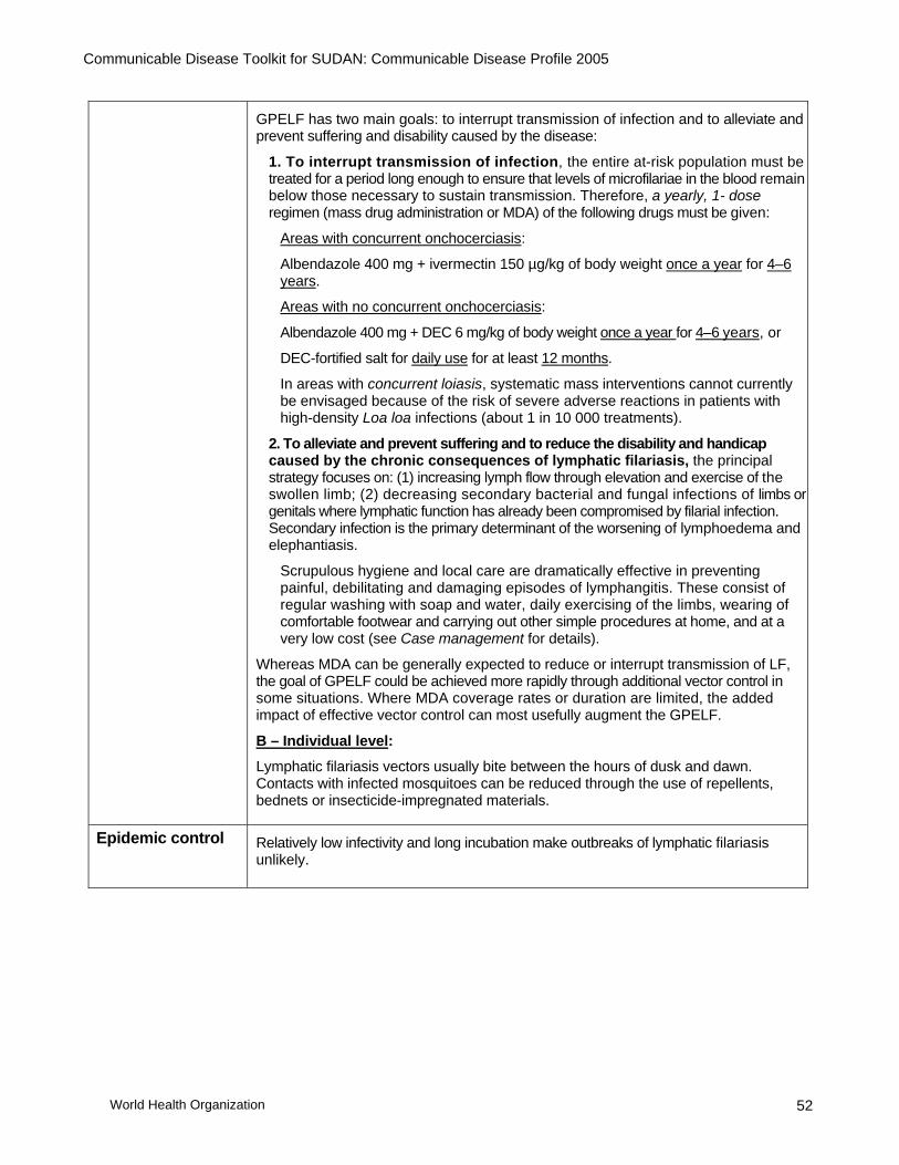
Communicable Disease Toolkit for SUDAN: Communicable Disease Profile 2005
World Health Organization 52
GPELF has two main goals: to interrupt transmission of infection and to alleviate and
prevent suffering and disability caused by the disease:
1. To interrupt transmission of infection, the entire at-risk population must be treated for a period long enough to ensure that levels of microfilariae in the blood remain below those necessary to sustain transmission. Therefore, a yearly, 1- dose regimen (mass drug administration or MDA) of the following drugs must be given:
Areas with concurrent onchocerciasis:
Albendazole 400 mg + ivermectin 150 µg/kg of body weight once a year for 4–6 years.
Areas with no concurrent onchocerciasis:
Albendazole 400 mg + DEC 6 mg/kg of body weight once a year for 4–6 years, or
DEC-fortified salt for daily use for at least 12 months.
In areas with concurrent loiasis, systematic mass interventions cannot currently be envisaged because of the risk of severe adverse reactions in patients with high-density Loa loa infections (about 1 in 10 000 treatments).
2. To alleviate and prevent suffering and to reduce the disability and handicap caused by the chronic consequences of lymphatic filariasis, the principal strategy focuses on: (1) increasing lymph flow through elevation and exercise of the swollen limb; (2) decreasing secondary bacterial and fungal infections of limbs or genitals where lymphatic function has already been compromised by filarial infection. Secondary infection is the primary determinant of the worsening of lymphoedema and elephantiasis.
Scrupulous hygiene and local care are dramatically effective in preventing painful, debilitating and damaging episodes of lymphangitis. These consist of regular washing with soap and water, daily exercising of the limbs, wearing of comfortable footwear and carrying out other simple procedures at home, and at a very low cost (see Case management for details).
Whereas MDA can be generally expected to reduce or interrupt transmission of LF, the goal of GPELF could be achieved more rapidly through additional vector control in some situations. Where MDA coverage rates or duration are limited, the added impact of effective vector control can most usefully augment the GPELF.
B – Individual level:
Lymphatic filariasis vectors usually bite between the hours of dusk and dawn. Contacts with infected mosquitoes can be reduced through the use of repellents, bednets or insecticide-impregnated materials.
Epidemic control Relatively low infectivity and long incubation make outbreaks of lymphatic filariasis unlikely.

Communicable Disease Toolkit for SUDAN: Communicable Disease Profile 2005
World Health Organization 53
14. MALARIA DESCRIPTION
Infectious agent In Sudan, about 90% of all malaria cases are caused by the protozoan parasite Plasmodium falciparum. This causes the most life-threatening form of the disease. P. vivax and P. ovale are responsible for the remaining malaria burden.
Case definition Clinical case definition:
Uncomplicated malaria
A patient with fever or history of fever within the past 48 hours (with or without other symptoms such as nausea, vomiting and diarrhoea, headache, back pain, chills, myalgia) in whom other obvious causes of fever have been excluded.
Severe malaria
A patient with symptoms as for uncomplicated malaria, plus drowsiness with extreme weakness and associated signs and symptoms related to organ failure (e.g. disorientation, loss of consciousness, convulsions, severe anaemia, jaundice, haemoglobinuria, spontaneous bleeding, pulmonary oedema and shock).
Confirmed case
Demonstration of malaria parasites in blood film by examining thick or thin smears, or by rapid diagnostic test for P. falciparum.
Mode of transmission
Vector-borne, through infective Anopheles mosquito bite.
In Sudan, the primary malaria vectors are An. arabiensis, An. gambiae and An. funestus.
Malaria may also be transmitted through blood transfusion of infected blood. Rarely, infants may contract malaria in utero through transplacental transfer of parasites, or during delivery.
Incubation The incubation period for mosquito-transmitted infection is approximately 7–14 days for P. falciparum, 8–14 days for P. vivax and 7–30 days for P. malariae.
However, malaria should be considered in all cases of unexplained fever that starts at any time between 1 week after the first possible exposure to malaria risk and 2 months (or even longer in rare cases) after the last possible exposure.
Period of communicability
Communicability is related to the presence of infective Anopheles mosquitoes and of infective gametocytes in the blood of patients. Untreated or insufficiently treated patients may be a source of mosquito infection for more than 3 years in P. malariae malaria, 1–2 years in P. vivax malaria and usually no longer than 1 year in P. falciparum malaria.

Communicable Disease Toolkit for SUDAN: Communicable Disease Profile 2005
World Health Organization 54
EPIDEMIOLOGY Burden Number of malaria cases (2003):
Reported: 3 084 320 (including 1 085 953 laboratory-confirmed cases). Number of malaria deaths reported: 2 479(2003) Estimated: 7 500 000 clinical malaria cases annually.
Estimated malaria deaths per year: 35 000.
Geographical distribution
Malaria risk is present throughout the country but is predominant in southern Sudan. Endemicity ranges from holoendemic in the south to hypoepidemic in the north, Epidemic-prone areas are confined to north/central Sudan.
Seasonality Risk is year round-in the southern part of the country. In the North, malaria risk exists from July to November with a peak just after the rains.
Alert threshold Any increase in the number of cases above what is expected for the time of the year in a defined area.
Recent epidemics An epidemic that resulted from the absence of sufficient prevention measures was reported in White Nile state (2003).
RISK FACTORS FOR INCREASED BURDEN Population movement
Yes The potential for epidemics can increase with the influx of non-immune populations moving from areas of no malaria/low transmission to highly endemic areas. Similarly, an influx of parasite carriers may trigger an epidemic in an epidemic-prone hypoendemic area.
Overcrowding Yes A consequence of increased population density and increased exposure to mosquito bites in temporary shelters.
Poor access to health services
Yes • Delays in access to effective treatment increase the likelihood of severe disease and death.
• Delays in access to effective treatment also increase the pool of malaria gametocyte carriers (the mature sexual stage of the parasite in humans that, once picked up in the blood-meal of a mosquito, develops into the infective stage for transmission to another human).
Food shortages No However, malnutrition increases vulnerability to severe malaria once infection has occurred. Case management also becomes more complicated, resulting in increased mortality.
Lack of safe water and poor sanitation
No However, temporary surface-water bodies may increase breeding opportunities for the malaria vector.
Others Yes • Breakdown of control measures, and lack of preventive interventions (insecticide-treated materials such as bednets, sheeting, etc.) and indoor residual spraying of shelters with residual insecticide.
• Changes in agricultural practice leading to extensive mosquito breeding sites.
• Usually heavy rains and flooding.

Communicable Disease Toolkit for SUDAN: Communicable Disease Profile 2005
World Health Organization 55
Risk assessment conclusions
Epidemiological situation
Malaria is the major health problem in Sudan, and the whole country is now considered endemic, with varying degrees. Malaria endemicity ranges from holo-endemic in the south to hypoendemic in the north with epidemic outbreaks. P. falciparum infection is overwhelmingly predominant (90%); An. arabiensis is the main vector; Country-wide 32% of outpatient attendance and 20% of all hospital deaths are due to malaria, which causes an estimated total of 7.5 million cases per year (Ministry of Health data, 1998). The situation is further aggravated by the spread of chloroquine-resistant P. falciparum, increasing insecticide resistance of vectors and inaccessibility of many areas, particularly in the south and west.
It is hoped that the malaria situation will improve as a result of strong political commitment from the country towards that end, as well as the support generated from the RBM partnership.
Training of health workers in case management and vector control is resulting in an improvement in malaria control activities. Epidemic preparedness and malaria information systems have markedly improved and helped in early detection and abortion of the expected malaria epidemic following the floods of 1998. Since 2002, a start has been made with the distribution of insecticide-treated nets (ITNs) were distributed throughout the country.
The management system for malaria control in Sudan includes national malaria administration (NMCP) at central level and malaria units active in the majority of states.
Constraints − Expansion of irrigation schemes and poor maintenance of drainage systems. − Lack of intersectoral and intrasectoral cooperation. − Massive population movement within the country. − Complex emergency situation in the south and west. − Inadequate surveillance systems. − Frequent turnover and emigration of technical staff to Arabian Gulf countries.
Priority actions adopted by the NMA − Intensify malaria control in selected states (initiative in Gezira, Khartoum and
Sinnar States). − Reduce malaria mortality in priority States. − Prevent epidemics in the country as a whole. − Support malaria control in complex emergency situation territories. − Prevent and control malaria in pregnancy through intermittent treatment in
holoendemic stable transmission and irrigated areas. − Protect high-risk groups by insecticide-treated material.
National programme objectives − To reduce incidence and morbidity of severe and complicated malaria cases,
and prevent mortality. − To prevent, detect early and contain malaria epidemics.

Communicable Disease Toolkit for SUDAN: Communicable Disease Profile 2005
World Health Organization 56
PREVENTION AND CONTROL MEASURES Case management The new treatment protocol for malaria recommended by the Federal Ministry of Health
and WHO/Sudan (Khartoum, June 2004) is summarized below:
Treatment of uncomplicated malaria
First-line treatment: (artesunate (AS) + sulfadoxine–pyrimethamine (SP). Day 1 Day 2 Day 3 Age
(years) Weight
(kg) SP
(500 S + 25 P
tablets)
AS
(50 mg tablets)
AS
(50 mg tablets)
AS
(50 mg tablets)
<1 <10 ½ ½ ½ ½
1 – <7 10 <20 1 1 1 1
7–13 20 <40 2 2 2 2
14+ 40+ 3 4 4 4
Second-line treatment: (artemether 20 mg + lumefantrine 120 mg )=Coartem® Day 1 Day 2 Day 3 Age
(years) Weight
(kg) 0
time 8
hours AM PM AM PM
Total no. of tablets
<10 Use is not recommended; give oral quinine instead.
<1 – <3 10–14 1 1 1 1 1 1 6
3 – <7 15–24 2 2 2 2 2 2 12
7–11 25–34 3 3 3 3 3 3 18
12+ 35+ 4 4 4 4 4 4 24
Treatment of malaria in pregnancy
Weeks of amenorrhoea
Uncomplicated malaria
Severe malaria Prevention
0–12 Quinine (Q) Quinine (Q) –
13–36
1st opition:
Quinine or 3 days Q followed by SP
2nd Option
AS+ SP
Artemether or quinine
SP in two treatment doses, >1 month apart
37 until delivery Quinine Artemether or quinine
–
Puerperium AS + SP Artemether or quinine
–

Communicable Disease Toolkit for SUDAN: Communicable Disease Profile 2005
World Health Organization 57
Treatment of severe malaria Quinine dihyrochloride, quinine hydrochloride or quinine sulfate 20 mg salt/kg (loading dose) diluted in 10ml/kg isotonic fluid by intravenous (IV) infusion over 4 hours. Eight hours after the start of the loading dose, give 10mg/kg over 4 hours. Repeat dose every 8 hours and shift to oral quinine as soon as the patient can tolerate oral medication. Quinine can be given intramuscularly (IM) in divided doses, with the same doses as above, diluted to 60mg/ml. or Quinine same as above (IV or IM) for at least 3 days and then shift to the first-line treatment (AS + SP). or Artemether IM 3.2mg/kg divided into 2 doses on the first day followed by 1.6mg/kg daily for the next 6 days. or Artemether IM 3.2/kg divided into 2 doses on the first day followed by 1.6mg/kg daily for at least 3 days and then shift to the first-line treatment (AS + SP). Pre-referral treatment of severe malaria Artesunate rectal capsules/suppositories, 10mg/kg, should be given as soon as possible once a diagnosis of severe malaria is made. If the rectal capsule is expelled within the first hour, another rectal capsule should be inserted immediately. A second dose can be repeated after 12 hours. THE PATIENT MUST BE TRANSPORTED WITHIN 24 HOURS. or Quinine 10 mg salt/kg IM in the standard dose. Repeat dose every 8 hours if referral is delayed. THE PATIENT MUST BE TRANSPORTED WITHIN 24 HOURS.
Prevention and Control
At present, malaria prevention measures in Sudan include distribution of insecticide treated mosquito nets, environmental management, distribution of Gambusia fish, larviciding and indoor residual spraying with pyrethroids. Free-of-charge drugs are provided in some endemic areas.
Chemoprophylaxis: WHO recommends mefloquine, doxycycline or atovaquone/proguanil prophylaxis for expatriate staff traveling to Sudan. However according to new treatment guidelines, doxycline is not recommended.
Chemoprophylaxis must be complemented by personal protection. It is not recommended on a population wide basis because it is extremely difficult to implement and assure compliance. Additionally, non-compliance to prophylaxis guidelines can accelerate the development of drug resistance. Intermittent presumptive treatment (IPT) at least twice during pregnancy (2nd and 3rd trimester) is advisable for pregnant women living in areas where transmission is high. National policy has recently changed to IPT in pregnancy with sulfadoxine–pyrimethamine, once in the 2nd trimester and again in the 3rd trimester.
Vigorous health education at community level to improve rapid treatment-seeking behaviour for fever cases during the transmission season.

Communicable Disease Toolkit for SUDAN: Communicable Disease Profile 2005
World Health Organization 58
15. MEASLES DESCRIPTION Infectious agent Measles virus (genus Morbillivirus, family Paramyxoviridae)
Case definition Clinical case definition:
Any person with: − Fever and − Maculopapular (i.e. non-vesicular) rash, and − Cough or coryza (i.e. runny nose) or conjunctivitis (i.e. red eyes); or
Any person in whom a clinical health worker suspects measles infection.
Laboratory criteria: Presence of measles-specific IgM antibodies.
Case classification: Clinically confirmed: A case that meets the clinical case definition. Laboratory-confirmed (only for outbreak confirmation and during the outbreak prevention/elimination phase):
− A case that meets the clinical case definition and is laboratory-confirmed. or
− A case meeting clinical definition and epidemiologically linked by direct contact to a laboratory-confirmed case in which rash onset occurred 7–18 days earlier.
Mode of transmission
Airborne by droplet spread; or
Direct contact with the nasal and throat secretions of infected persons or via objects (e.g. toys) that have been in close contact with an infected person.
Incubation After infection there is an asymptomatic incubation period of 10–12 days, with a range from 7 to 18 days from exposure to the onset of fever.
Period of communicability
Measles is most infectious from 4 days before the rash until 1–2 days after rash onset.
EPIDEMIOLOGY
Number of cases reported:
1997: 350 cases 1990: 14 075 cases 1980: 50 168 cases
Burden
2003: 2002: 2001: 2000: 1999: 1998:
4381 cases 4529 cases 4362 cases 2875 cases 3347 cases 550 cases
(Data source: WHO–UNICEF estimates, 2004)
Geographical distribution
Measles is highly endemic throughout Sudan, and the expected number of measles cases is high.
Seasonality Higher incidence during the colder months. Some states experience two peaks (February–April and September–December).

Communicable Disease Toolkit for SUDAN: Communicable Disease Profile 2005
World Health Organization 59
Alert threshold One case in a state that has conducted a measles catch-up campaign (e.g Northern, River Nile, Red Sea and Kassala) must lead to an alert.
Laboratory confirmation of all cases is not required. Only 5–10 cases from each outbreak need to be laboratory-confirmed.
Recent epidemics It is reported that, in Sudan, one-third of all deaths in children aged under 3 years is due to measles.
October–November 2002. 40 cases with 3 deaths were reported from Nimule (Magwe county, Eastern Equatoria).
October–November 2002. 45 cases (no deaths) were reported from Gomjuer (Aweil West county, southern Bahr Al Ghazal).
September–October 2002. 118 cases and 3 deaths were reported from Labone and Yei (Bahr Al Jebel).
July–August 2002. 260 cases with 6 deaths were reported from areas of the Nuba mountains, including Jullud, Timen and Tima (Southern Kordofan).
March–April 2002. 13 cases with 3 deaths were reported from Ruweng county (southern Sudan).
March–April 2001. An outbreak involving 158 cases of measles was reported from Buoth and Mayoum (Upper Nile).
April–May 2001. 14 cases were reported from Niemni (Upper Nile).
January–February 2001: 101 cases and 26 deaths were reported from Nyimboli (Aweil West county, southern Bahr Al Ghazal). (Data source: WHO/Sudan)
RISK FACTORS FOR INCREASED BURDEN Population movement
Yes Importation of virus.
Overcrowding Yes Crowded conditions facilitate transmission.
Poor access to health services
Yes Case-fatality rates can be reduced by effective case management, including the administration of vitamin A supplements.
Food shortages Yes Disease is more severe among children with malnutrition and vitamin A deficiency, and increases need for hospital care.
Lack of safe water and poor sanitation
No
Others Yes Low immunization coverage in the area of origin of the refugees or internally displaced populations and/or in the host area.
MCV (measles-containing vaccine) coverage
2001: 80% (67% by WHO–UNICEF estimates) 2000: 60% 1999: 79% 1998: 62% 1997: 92% 1990: 57% 1980: NA (Data source: WHO/Sudan official country estimates)

Communicable Disease Toolkit for SUDAN: Communicable Disease Profile 2005
World Health Organization 60
Risk assessment conclusions
Routine immunization services have been hampered for several years due to civil unrest, leading to poor coverage rates, especially in the southern states.
Measles is still common in various areas of the country, including the capital's area: the disease is the most common diagnosis among vaccine-preventable diseases in febrile children who present at the emergency hospitals in Khartoum.
PREVENTION AND CONTROL MEASURES Introduction Sudan has a routine immunization policy that requires a dose of single-antigen measles
vaccine at 9 months of age (see Appendix 7: Immunization schedule for Sudan).
However, supplementary measles immunization activities are required in order to reduce the risk of a measles outbreak.
Routine Immunization
Immunize the population at risk as soon as possible. The priority is to immunize children aged 6 months to 15 years, regardless of vaccination status or history of disease. Expansion to older children is of lesser priority and should be based on evidence of high susceptibility among this age group.
Children who are vaccinated against measles before 9 months of age must receive a second measles vaccination. This should be given as soon as possible after 9 months, with an interval of at least 1 month between doses.
All children aged 6 months to 5 years should also receive prophylactic vitamin A supplementation. If there is evidence of clinical vitamin A deficiency in older age groups, treatment with vitamin A should be initiated as per WHO guidelines.
To ensure safety of injection during immunization, auto-disable syringes and safety boxes are recommended. Safe disposal of used sharps should be ensured.
Outbreak response Inform the health authorities immediately if one or more suspected cases are identified.
Confirm the suspected outbreak, following WHO guidelines.
Investigate suspected case: check if it fulfils the case definition, record date of onset, age and vaccination status.
Confirm the diagnosis: collect blood specimen from 3–5 initial reported cases.
Assess the extent of the outbreak and the population at risk.
Implement outbreak response measures as follows:
− Give priority to proper case management and immunization of groups at highest risk (e.g. children aged 6 months to 15 years) as soon as possible even in areas not yet affected but where the outbreak is likely to spread.
− Promote social mobilization of parents in order to ensure that previously unvaccinated children aged from 6 months to 5 years are immunized.
− The presence of several cases of measles in an emergency setting does not preclude a measles immunization campaign. Even among individuals who have already been exposed to, and are incubating, the natural virus, measles vaccine, if given within 3 days of exposure, may provide protection or modify the clinical severity of the illness.
− Isolation is not indicated and children should not be withdrawn from feeding programmes.

Communicable Disease Toolkit for SUDAN: Communicable Disease Profile 2005
World Health Organization 61
Case management For uncomplicated cases:
─ Give vitamin A immediately upon diagnosis and ensure the child receives a second dose the next day (can be given to parent to administer at home).
─ Advise the parent to treat the child at home (control fever and provide nutritional feeding).
For cases with non-severe eye, mouth or ear complications:
─ Children can be treated at home. ─ Give vitamin A immediately upon diagnosis and ensure that the child receives a
second dose the next day (can be given to parent to administer at home). ─ If pus draining from the eyes, clean eyes and treat with 1% tetracycline eye
ointment. ─ If mouth ulcers, treat with gentian violet. ─ If pus draining from the ear, clean ear discharge and treat with antibiotics for 5 days
(amoxicillin, first-line; or co-trimoxazole second-line-, as per national ARI policy and IMCI guidelines currently under development).
─ Treat malnutrition and diarrhoea, if present, with sufficient fluids and high-quality diet.
For cases with severe, complicated measles (any general danger signs*, clouding of cornea, deep or extensive mouth ulcers, pneumonia):
─ Refer urgently to hospital. ─ Treat pneumonia with an appropriate antibiotic. ─ If clouding of the cornea or pus draining from the eye, clean eyes and apply 1%
tetracycline eye ointment. ─ If the child has any eye signs indicating vitamin A deficiency (i.e. night blindness, Bitot
spots, conjunctival and corneal dryness, corneal clouding or corneal ulceration), he or she should receive a third dose of vitamin A 2–4 weeks later.
* Inability to drink or breastfeed, vomiting everything, convulsions, lethargy or unconsciousness.

Communicable Disease Toolkit for SUDAN: Communicable Disease Profile 2005
World Health Organization 62
16. MENINGOCOCCAL DISEASE (MENINGITIS AND SEPTICAEMIC FORM) DESCRIPTION
Infectious agent Bacterium: Neisseria meningitidis serogroups A, B, C, Y, W135
Case definition Clinical case definition:
An illness with sudden onset of fever (>38.5 °C rectal; >38.0 °C axillary) and one or more of the following: ─ neck stiffness ─ altered consciousness ─ other meningeal sign or petechial or purpural rash.
In patients aged under one year, suspect meningitis when fever is accompanied by bulging fontanelle.
Laboratory criteria:
Positive CSF antigen detection, or Positive culture.
Case classification:
Suspected: a case that meets the clinical case definition above. Probable: a suspected case as defined above and:
− Turbid CSF (with or without positive Gram-stain), or − Ongoing epidemic and epidemiological link to a confirmed case.
Confirmed: a suspected or probable case with laboratory confirmation.
Mode of transmission Direct contact with respiratory droplets.
Incubation Incubation period varies between 2–10 days; most commonly 4 days.
Period of communicability
From the onset of symptoms until 24 hours after institution of therapy, but the most important sources of infection are asymptomatic carriers.
EPIDEMIOLOGY Burden Cases and deaths of meningococcal meningitis reported to WHO/Sudan:
2002: no data reported 2001: no data reported 2000: 4031 cases, 328 deaths 1999: 33 313 cases, 2410 deaths 1998: 697 cases, 82 deaths 1997: 297 cases 1996: 340 cases
Geographical distribution
Epidemics occurred in Sudan between 1980–1999, affecting the following regions: Blue Nile, Darfur, Gezira, Kassala, Khartoum, Kordofan, Omdurman, Rumbek, Sinnar, White Nile.

Communicable Disease Toolkit for SUDAN: Communicable Disease Profile 2005
World Health Organization 63
Seasonality Outbreaks tend to occur during the dry season (December–January).
Alert threshold1 Population >30 000: 5 cases per 100 000 inhabitants per week or a cluster of cases in an area.
Population <30 000: 2 cases in 1 week or an increase in the number of cases compared with previous non-epidemic years.
Intervention: (1) inform authorities; (2) investigate; (3) confirm; (4) treat cases; (5) strengthen surveillance; (6) prepare.
Epidemic threshold Population >30 000:
− 10 cases per 100 000 inhabitants per week if no epidemic for 3 years and vaccination coverage <80% or alert threshold crossed early in the dry season.
− 15 cases per 100 000 inhabitants per week in other situations.
Population <30 000:
The population should be vaccinated if:
− 5 cases in 1 week or
− Doubling of the number of cases in a 3-week period or
− For mass gatherings and displaced populations, 2 confirmed cases in 1 week.
Other situations should be studied on a case-by-case basis.
Intervention: 1. Mass vaccination. 2. Distribute appropriate antibiotics and case management protocols to health centres. 3. Treat according to epidemic protocol. 4. Maintain surveillance to track epidemic. 5. Inform the public.
Current thresholds have been established from data in meningitis belt countries, which includes Sudan.
1 Detecting meningococcal meningitis epidemics in highly-endemic African countries. Weekly Epidemiological Record, 2000, 38: 306–309.

Communicable Disease Toolkit for SUDAN: Communicable Disease Profile 2005
World Health Organization 64
Recent epidemics in the country
2002. As of 11 February, a total of 330 cases including 49 deaths were reported from Limun, Kauda and Hieman, in the Nuba mountains (Southern Kordofan). Serogroup A was confirmed. (CFR=14.8%).
January–March 2002. As of 21 March 2003, 126 cases and 7 deaths were reported from Isoke (Torit county, Eastern Equatoria), and 104 cases and 14 deaths from Ikotos, in the same county
February–March 2002. As of 15 March 2003, 8 cases and 1 death were reported from Padak (Boma county, Eastern Equatoria).
March 2001. 42 admission cases and 2 deaths were reported from Jaibor (Keew county, Upper Nile).
February–March 2001. As of 26 March, 19 cases and 1 death were reported from Chuil (Latjor county, Upper Nile). N. meningitidis serogroup A was identified.
February–March 2001. As of 22 March, 67 cases and 13 deaths were reported from Paluer (Bor county, Upper Nile).
February 2001. 117 cases and 1 death were reported from Narus (Eastern Equatoria).
2000. A total of 2549 cases of meningococcal disease, of which 186 were fatal, were reported to the national health authorities between 1 January and 31 March 2000. Bahr Al Jebel State was the most affected, with 1437 cases (including 99 deaths) reported in the Juba city area. Other affected states included White Nile (197 cases, 15 deaths), Southern Kordofan and Sinnar (where incidence was lower). Epidemic response activities included vaccination of a total of 70 000 people in early March.
1998–1999. An outbreak of meningococcal meningitis was reported in the Northern Darfur region. An increase in the number of cases had already been observed in December 1998. As far as May 1999, about 22 000 cases of meningococcal disease had been notified from 19 of the 26 states of Sudan, of which 1600 had died. More than 10 million doses of meningococcal vaccine were distributed for mass vaccination campaigns. In 1999, a total of 33 313 cases and 2410 deaths were reported from Sudan to WHO.
1988. Following the return of pilgrims from Mecca (Hajj) in August 1987, many countries in EMR faced an unusual spread of meningococcal infection. In Sudan, the 1987 introduction developed into epidemic spread in the meningitis season of 1988, when 32 016 cases of meningococcal disease were reported to WHO.
RISK FACTORS FOR INCREASED BURDEN Population movement
Yes Travel, migration and displacement facilitate the circulation of virulent strains within a country or from country to country.
Overcrowding Yes High density of susceptible people is an important risk factor for outbreaks. Internally displaced populations and refugee camps, crowding because of cattle or fishing-related activities, military camps and schools facilitate spread of the disease.
Poor access to health services
Yes Case identification is crucial to rapidly implement control measures.
The case-fatality rate without treatment is very high . Food shortages No
Lack of safe water and poor sanitation
No
Others No Concurrent infections: upper respiratory tract infections may contribute to some meningococcal outbreaks.
Dry and windy/dusty conditions increase transmission of the disease.

Communicable Disease Toolkit for SUDAN: Communicable Disease Profile 2005
World Health Organization 65
Risk assessment conclusions
Central and southern Sudan are included in the African meningitis belt, which extends from Ethiopia in the east to Senegal in the west, mainly within the annual rainfall range of 300–1100 mm. In this area, sporadic infections occur in seasonal, annual cycles; large-scale epidemics occur at greater intervals with irregular patterns, usually beginning during the dry season (December–February), and sometimes lasting for more than a year.
Since the early 1990s, Sudan has been practising preventive vaccination, mainly directed to high-risk groups. However, this was not sufficient to prevent the 1999 epidemics due to mobility of populations.
There is a high risk of epidemics in overcrowded camps.
PREVENTION AND CONTROL MEASURES Case management Meningococcal disease (either meningitis or septicaemia) is potentially fatal and should
always be viewed as a medical emergency. NON-EPIDEMIC CONDITIONS:
• Admission to a hospital or health centre is necessary for diagnosis (lumbar puncture and CSF examination). As soon as mengitis is suspected, a lumbar puncture must be done before starting antibiotic treatment
• As infectivity of patients is moderate and disappears quickly following antimicrobial treatment, isolation of the patient is not necessary.
• Antimicrobic treatment must be instituted as soon as possible after lumbar puncture (without waiting for laboratory results), and should be combined with supportive treatment.
Initial antimicrobial therapy should be effective against the three major causes of bacterial meningitis until bacteriological results are available: AGE GROUP
PROBABLE PATHOGENS
ANTIBIOTIC THERAPY
FIRST CHOICE
ALTERNATIVE
Adults and children < 5years
S. pneumoniae
Benzylpenicillin
Ampicillin or amoxicillin Chloramphenicol Ceftriaxone or cefotaxime
Children 1 month - 5 years
H. Influenza S. pneumoniae N. meningitidis
Ampicillin or amoxicillina
Chloramphenicol Ceftriaxone or cefotaxime
Neonates
Gram-negative bacteria Group B streptococci Listeria
Ampicillin and gentamicin
Ceftriaxone or cefotaximeb
a If H. influenzae is highly resistant to ampicillin, chloramphenicol should be given with ampicillin. b No effect on listeria.
Once diagnosis of meningococcal disease has been established, many antimicrobials can be used:
─ either penicillin or ampicillin is the drug of choice.
─ Chloramphenicol is a good and inexpensive alternative.
─ The third-generation cefalosporins, ceftriaxone and cefotaxime, are excellent alternatives but are considerably more expensive.
─ A 7-day course is still the general rule for the treatment of meningococcal disease (except in the neonatal period where a 14-day course is given).

Communicable Disease Toolkit for SUDAN: Communicable Disease Profile 2005
World Health Organization 66
─ The long-acting (oily) form of chloramphenicol has also been shown to be effective.
The Sudan Federal Ministry of Health recommends injectable oily choloramphenicol and benzylpenicillin.
EPIDEMIC CONDITIONS:
During epidemics of confirmed meningococcal disease, case management needs to be simplified to permit the health system to respond to rapidly increasing numbers of cases.
• Diagnosis: as the flood of patients could make the routine use of lumbar puncture to confirm meningitis impossible, every suspected case of meningitis should be considered and treated as one of meningococcal meningitis.
• Treatment: simplified treatment protocols are appropriate: long-acting oily chloramphenicol (100 mg/kg up to 3 g in a single dose) IM is the drug of choice for all age groups, particularly in areas with limited health facilities. For patients who do not improve rapidly, an additional dose of the same antimicrobial is recommended 48 hours later.
Prevention NON-EPIDEMIC CONDITIONS:
• Vaccination: to prevent secondary cases around a sporadic case of meningococcal disease, vaccine can be used for close contacts of patients with meningococcal disease due to serogroup A, C or W135.
• Chemoprophylaxis: the aim of chemoprophylaxis is to prevent secondary cases by eliminating nasopharyngeal carriage. To be effective in preventing secondary cases, chemoprophylaxis must be initiated as soon as possible (i.e. not later than 48 hours after diagnosis of the case). Its use should be restricted to close contacts of a case, which are defined as:
− Household members (i.e. persons sleeping in the same dwelling as the case);
− Institutional contacts (i.e. persons who share sleeping quarters (i.e. roommates in boarding schools; persons sharing a barracks in military camps); nursery school or childcare centre contacts (i.e. children and teachers who share a classroom with the case);
− Other persons who have had contact with the patient's oral secretions through kissing or sharing of food and beverages.

Communicable Disease Toolkit for SUDAN: Communicable Disease Profile 2005
World Health Organization 67
EPIDEMIC CONDITIONS
• Vaccination: a mass vaccination campaign, if appropriately carried out, can halt an epidemic of meningococcal disease. Laboratory diagnosis and confirmation of epidemic serogroups will guide the type of vaccine needed, either meningococcal polysaccharide bivalent A/C (if serogroup A or C is confirmed as the epidemic serogroup), or meningococcal polysaccharide tetravalent vaccine A/C/Y/W135 (if serogroup Y or W135 is confirmed). Vaccination should be targeted to areas where the epidemic threshold is reached.
Camp population: Following confirmation (serogroup identified) of two cases, mass vaccination is recommended if the serogroup/s identified is/are included in either the bivalent (A/C) or tetravalent (A/C/Y/W135) vaccine. At-risk populations (e.g. aged 2–30 years) should be given priority.
− General population: If an outbreak is suspected, vaccination should only be considered after careful investigation (including confirmation and serogroup identification) and the assessment of the population group at highest risk.
• Chemoprophylaxis: chemoprophylaxis of contacts of meningitis patients is NOT warranted during an epidemic for several reasons. In small clusters or outbreaks among closed populations (e.g. extended household, boarding schools), chemoprophylaxis may still be appropriate.

Communicable Disease Toolkit for SUDAN: Communicable Disease Profile 2005
World Health Organization 68
17. ONCHOCERCIASIS (RIVER BLINDNESS) DESCRIPTION
Infectious agent Onchocerca volvulus, a filarial worm belonging to the class Nematoda.
Case definition Clinical description Persons suffering from onchocerciasis may experience:
(A) Skin lesions: dermal changes are secondary to tissue reaction to motile larvae as they migrate subcutaneously, or to their destruction in the skin.
− Itching: the pruritus of onchocerciasis is the most severe and intractable that is known. In lightly infected persons, this may persist as the only symptom.
− Rashes: the rash usually consists of many raised papules, which are due to microabscess formation, and may disappear within a few days or may spread. Sowda, from the Arabic for black or dark, is an intensely pruritic eruption usually limited to one limb and including oedema, hyperpigmented papules and regional lymphadenopathy. It is common in Yemen, but can also be found in Sudan.
− Depigmentation of the skin: areas of depigmentation over the anterior shin with islands of normally pigmented skin, commonly called "leopard skin", are found in advanced dermatitis.
− Subcutaneous nodules: these are 0.5–3.0 cm asymptomatic subcutaneous granulomas resulting from a tissue reaction around adult worms. They occur most often over bony prominences: in Africa the nodules are often located over the hips and lower limbs.
− Lymphadenopathy: it is frequently found in inguinal and femoral areas, and can result in "hanging groin" (especially when associated with skin atrophy and loss of elasticity) and elephantiasis of the genitalia.
(B) Eye lesions: ocular onchocerciasis is related to the presence of live or dead microfilariae. Involvement of all tissues of the eye has been described, and many changes in both anterior and posterior segments of the eye can occur. The more serious lesions lead to serious visual impairment, including blindness. (C) General debilitation: onchocerciasis has also been associated with weight loss and musculoskeletal pain.
Clinical case definition
In an endemic area, a person with fibrous nodules in subcutaneous tissues. These must be distinguished from lymph nodes or ganglia.
Laboratory criteria Presence of one or more of the following:
− Microfilariae in skin snips taken from the iliac crest (Africa) or scapula (Americas)
− Adult worms in excised nodules − Typical ocular manifestations, such as slit-lamp observations of microfilariae in
the cornea, the anterior chamber, or the vitreous body − Serology (especially for non-indigenous persons)
Case classification − Suspected: A case that meets the clinical case definition. − Probable: Not applicable. − Confirmed: A suspected case that is laboratory-confirmed.

Communicable Disease Toolkit for SUDAN: Communicable Disease Profile 2005
World Health Organization 69
Mode of transmission
Vector-borne, by the bite of infected female blackflies belonging to the genus Simulium (mainly S. damnosum complex). Larvae of the vector for onchocerciasis lay their eggs in the water of fast-flowing rivers – hence the name "river blindness".
Microfilariae are ingested by a blackfly feeding on an infected person, and penetrate thoracic muscles of the fly. Here, a few of them develop into infective larvae after several days, migrate to the cephalic capsule, are liberated onto the skin and enter the bite wound during a subsequent blood-meal. Infective larvae develop into adult parasites in the human body, where adult forms of O. volvulus can live for up to 14–15 years and are often found encased in fibrous subcutaneous nodules. Each adult female produces millions of microfilariae that migrate under the skin and through the eyes, giving rise to a variety of dermal and ocular symptoms.
Humans are the only reservoir. Other Onchocerca species found in animals cannot infect humans but may occur together with O. volvulus in the insect vector.
Incubation Larvae take at least 6–12 months to become adult worms. Adult worms are usually innocuous, apart from the production of the subcutaneous nodules (these can develop as early as 1 year after infection). The main pathologic sequelae of O. volvulus infection are caused by the microfilariae in skin and ocular tissue, where they can be found after a period of 7–34 months.
Microfilariae are found in the skin usually only 1 year or more after the time of the infective bite.
Period of communicability
Human blackfly Infected individuals can infect blackflies as long as living microfilariae occur in their skin. Microfilariae are continuously produced by adult female worms (about 700 per day), and can be found in the skin after a prepatent period of 7–34 months following introduction of infective larvae. They may persist for up to 2 years after the death of the adult worms.
Blackfly human Blackfly vectors become infective (i.e. able to transmit infective larvae) 7–9 days after the blood-meal.
EPIDEMIOLOGY Burden An estimated 2 million persons are at risk of onchocerciasis in Sudan, with 10 000
cases of onchocerciasis-related blindness.
Southern focus: in Western Bahr Al Ghazal, more than 80% of subjects in some villages had palpable nodules in 1998.
Northern focus: a rapid epidemiological assessment (REA) in 1995 revealed that 16% of local inhabitants had palpable nodules. Skin-snip positivity reached 33.6%.
Eastern focus: skin-snip positivity may reach 50% in some villages, but nodule rates were low (1998).
Western focus: an REA in 1996 revealed that 22% of subjects in this area had palpable nodules; 28% had onchocercal skin lesions or itching.

Communicable Disease Toolkit for SUDAN: Communicable Disease Profile 2005
World Health Organization 70
Geographical distribution
Infection with O. volvulus is known to occur in four main regions, known as the southern, northern, eastern and western foci.
Southern focus: this is the largest focus and includes all the southern states from Western Bahr Al Ghazal in the west to Upper Nile in the east. The rate of infection is highest in the south-west, in Western Bahr Al Ghazal State, especially among communities living along the Jur river (Nahr el Jur) and its tributaries.
Northern focus: the rocky ground and river bed between the fourth and fifth Nile cataracts create breeding sites for S. damnosum. The river level reaches its peak in late summer (August–September), and ideal conditions for Simulium breeding occur when the water recedes and rocks and vegetation emerge, creating surface turbulence. This focus is located in the Abu Hamad area of the Nubian desert in Northern State. This is the most northerly focus in Africa, and probably in the world.
Eastern focus: this focus is located in Gedaref State, along the upper Atbara river, close to the Ethiopian border.
Western focus: this focus is contiguous to the southern one and includes the communities living along the Umbellasha, Adda and Bahr El Arab River in Southern Darfur. This is a savannah region where seasonal rainfalls (May–September) give rise to fast-flowing rivers that, during the dry months, become parched or stagnant.
Seasonality Southern focus: transmission from July to October. Northern focus: transmission between November and January. Eastern focus: transmission from July to October. Western focus: transmission from May to September.
Recent epidemics No data available.
RISK FACTORS FOR INCREASED BURDEN Population movement
Yes Infections in the Bahr El Arab area (western focus) are probably the result of individuals from the southern focus migrating northwards during the civil war in the 1950s.
Overcrowding No
Poor access to health services
Yes Community-directed treatment with ivermectin (CDTI) is an effective tool for transmission control. People can infect flies as long as living microfilariae occur in their skin.
Food shortages No Lack of safe water and poor sanitation
No
Others No

Communicable Disease Toolkit for SUDAN: Communicable Disease Profile 2005
World Health Organization 71
Risk assessment conclusions
"Craw-craw" (itchy skin due to onchocerciasis) was reported for the first time in Sudan in 1908 from Bahr Al Ghazal.
The Global 2000 River Blindness Program of The Carter Center estimates the crude ultimate treatment goal for Sudan (total number of people in need of treatment) at 743 230. Of the several endemic foci, the southern focus is the most significant and is characterized by high prevalence of blinding onchocerciasis. Some of the highest rates of blindness due to onchocerciasis in the world occur in south-west Sudan.
The cost per treatment in 2001 was considerably above that recommended by APOC, and calculated at US$ 0.74. The high cost underscores the principle that distribution in conflict areas will be more expensive.
In general, communities are committed to the distribution of ivermectin. The onchocerciasis control programme is viewed at the highest governmental levels as an example of a successful health delivery system and is well integrated into the Sudanese primary health care system.
Among the constraints encountered in Sudan:
− Accessibility problems due to the civil unrest, flood, famine, drought and mass population displacement (in southern Sudan).
− Continuous reshaping of the population and the community-directed drug distributors.
− Impaired treatment activities in areas devoid of any health infrastructure, or in areas where the primary health care system is not operational.
− Coendemicity for O. volvulus and Loa loa in some areas of south-western Sudan. Guidelines for Loa loa coendemic areas still need to be implemented.
It seems likely that there has been a decrease in the prevalence of infection in the past one or two decades in the western focus. This reduction may be the result of mass population migration and reduced vector density following a period of drought in 1987, and also of annual ivermectin treatment. The prevalence of infection appeared to be reduced also in the northern focus in 1995 when compared with 1985, thanks to the five annual rounds of ivermectin treatment carried out in this area.
PREVENTION AND CONTROL MEASURES Case management
Administration of ivermectin once a year over a period of at least 15–20 years reduces infection to insignificant levels and prevents the appearance of clinical manifestations. The recommended dosage is equivalent to 150 µg/kg of body weight (in practice, dosage is according to height, using 1–4 tablets of 3 mg). Established clinical manifestations are also treated by ivermectin.
Treatment with ivermectin is contraindicated in: – Children under 5 years (age), less than 15 kg (weight), or less than 90 cm (height). – Pregnant women – Lactating mothers of infants less than 1 week old – Severely ill persons
NB: Ivermectin should be used with extreme caution in areas coendemic for Loa loa.
Epidemic control Recrudescence of transmission may occur and can be managed by the mass administration of ivermectin where programmes can maintain good treatment coverage.

Communicable Disease Toolkit for SUDAN: Communicable Disease Profile 2005
World Health Organization 72
Prevention • Vector control
Destruction of Simulium larvae by application of insecticides such as temephos (Abate®) through aerial spraying to breeding sites in fast-flowing rivers, in order to interrupt the cycle of disease transmission. Once the cycle has been interrupted for 14–15 years, the reservoir of adult worms dies out in the human population, thus eliminating the source of the disease. This was the basic strategy of the Onchocerciasis Control Programme (OCP) in west Africa.
In the west African savannah zone, onchocerciasis was a severely blinding disease. It was also responsible for the depopulation of fertile river valleys in OCP countries and was therefore a major impediment to economic development. For these reasons, the large-scale vector control operations of the OCP – based on the aerial application of insecticides and aiming at the virtual elimination of the disease – were considered economically justified. OCP has been successful in eliminating onchocerciasis as a public health and socioeconomic problem.
The African Programme for Onchocerciasis Control (APOC), which includes Sudan and other 18 African countries, uses focal vector eradication as a control option. This implies that the whole focus is covered at once, resulting in the total eradication of the vector over a very short timescale.
• Community-directed treatment with ivermectin (CDTI):
Course: Once-a-year administration of ivermectin 150 µg/kg of body weight
The introduction of ivermectin in 1987 provided for the first time a feasible chemotherapy regimen for large-scale treatment of onchocerciasis. Ivermectin is an effective microfilaricide that greatly reduces the numbers of skin microfilariae to low levels for up to a year, thus:
(1) alleviating many symptoms: since the microfilariae cause the severe morbidity of onchocerciasis, ivermectin treatment is an effective tool for morbidity control, able to prevent the development of ocular lesions and blindness; (2) making the recipient less infective for the vector: ivermectin is an effective tool for onchocerciasis transmission control.
Ivermectin treatment greatly reduces transmission of the parasite but does not halt it within the period of a decade or more, and the adult worm may live for as long as 14–15 years. Annual large-scale treatment will therefore have to continue for a very long time. Current predictions with a simulation model indicate that annual treatment at the current level of coverage may have to continue for at least two decades. The main challenge facing ivermectin-based control is therefore to develop and implement simple methods of ivermectin delivery that can be sustained by the communities themselves. The current APOC strategy consists of house to-house distribution or at central meeting points in villages. CDTI is the main strategy adopted by APOC. In the 19 countries included in this programme, onchocerciasis remains a major cause of blindness but does not appear to be the cause of major depopulation of fertile lands. Partly for this reason, large-scale vector control operations are not likely to be as cost-effective as they have been in the OCP area.

Communicable Disease Toolkit for SUDAN: Communicable Disease Profile 2005
World Health Organization 73
18. PERTUSSIS (WHOOPING COUGH) DESCRIPTION
Infectious agent Bordetella pertussis, the pertussis bacillus.
Case definition Clinical description:
The initial stage, the catarrhal stage, is characterized by the insidious onset of coryza (runny nose), sneezing, low-grade fever and a mild, occasional cough, similar to the commom cold. The cough gradually becomes more severe and irritating, and after 1–2 weeks the second stage, or paroxysmal stage, begins. The patient has bursts, or paroxysms, of numerous, rapid coughs, apparently due to difficulty in expelling thick mucus from the tracheobronchial tree. At the end of the paroxysm, a long inspiratory effort is usually accompanied by a characteristic whoop.
In younger infants, periods of apnoea may follow the coughing spasms, and the patient may become cyanotic (turn blue). Pneumonia is a relatively common complication (reported 21.7% of cases in developed countries); otitis, haemorrhages (subconjunctival petechiae and epistaxis), convulsions, encephalopathies and death occur more rarely). The disease lasts 4–8 weeks. Complications are more frequent and severe in younger infants. In developed countries, the case-fatality rate among infants aged less than 1 month has been reported to be around 1%. Older persons (adolescent and adults) and those partially protected by the vaccine may become infected with B. pertussis but usually have milder disease.
In the convalescent stage, recovery is gradual. The cough becomes less paroxysmal and disappears over 2–3 weeks. However, paroxysms often recur with subsequent respiratory infections for many months after the onset of pertussis. Fever is generally minimal throughout the course of pertussis.
Clinical case definition: A case diagnosed as pertussis by a physician, or A person with a cough lasting at least 2 weeks with at least one of the following symptoms:
− Paroxysms (i.e. fits) of coughing − Inspiratory "whooping" − Post-tussive vomiting (i.e. vomiting immediately after coughing).
Laboratory criteria: − Isolation of Bordetella pertussis, or − Detection of genomic sequences by polymerase chain reaction (PCR) − Positive paired serology.
Case classification: Clinical case: A case that meets the clinical case definition. Confirmed case: A clinical case that is laboratory-confirmed.
Mode of transmission
Primarily by direct contact with discharges from respiratory mucous membranes of infected persons via the airborne route. Humans are the only hosts.
Although the disease may be milder in older persons, these infected persons may transmit the disease to other susceptible persons, including non-immunized or under-immunized infants. An adult is often found to be the first case in a household with multiple pertussis cases.

Communicable Disease Toolkit for SUDAN: Communicable Disease Profile 2005
World Health Organization 74
Incubation The incubation period usually lasts 7–10 days; rarely more than 14 days.
Period of communicability
Pertussis is highly communicable in the early catarrhal stage. Communicability gradually decreases after onset of paroxysmal cough.
Untreated patients may be contagious for up to 3 weeks after onset of paroxysmal cough without treatment or for up to 5 days after onset of treatment.
EPIDEMIOLOGY Burden Number of cases reported
Year Cases
2003 232
2002 213
2001 645
2000 80
1999 51
1998 169
1997 418
1990: 566
1980 28 631 (Source: WHO–UNICEF data, 2004)
Geographical distribution
No data available.
Seasonality Pertussis has no distinct seasonal pattern, but activity may increase in the summer and autumn.
Alert threshold Recent epidemics in the country
2002 December: 5 cases (no deaths) were reported from Akob Payam (Tonj county, Lakes state).
2002 October–December: 127 cases with 2 deaths were reported from Akon (Gogrial county, Western Bahr Al Ghazal).
2002 August–September: 68 cases and 5 deaths were reported from several villages in Oriny and Shilluk counties (Upper Nile state). All cases, except 5, were children aged <5 years.
RISK FACTORS FOR INCREASED BURDEN Population movement
Yes Importation and spread of B. pertussis.
Overcrowding Yes Crowded conditions facilitate transmission. The disease is usually introduced into a household by an older sibling or a parent.
Poor access to health services
Yes No access to routine immunization services. Susceptibility of non-immunized individuals is universal, and vaccination is the mainstay of pertussis control.
Food shortages No

Communicable Disease Toolkit for SUDAN: Communicable Disease Profile 2005
World Health Organization 75
Lack of safe water and poor sanitation
No
Others Yes Low DTP3 coverage (<80%).
DTP3 coverage: 2001: 71% (46% by WHO–UNICEF estimates) 2000: 65% 1999: 79% 1998: 70% 1997: 79% 1990: 62% 1980: 1 % (Data source: WHO/Sudan official country estimates)
Risk assessment conclusions
Yearly fluctuations in the number of reported cases reflect a weak surveillance system.
Pertussis is a potential problem if introduced into crowded conditions with many non-immunized children. It is highly contagious. Children aged under 1 year and pregnant women are at greatest risk.
PREVENTION AND CONTROL MEASURES Case management • Erythromycin or erythromycin estolate or – in case of allergies to erythromycin –
trimethoprim–sulfamethoxazole (contraindicated during pregnancy) should be administered for 7–14 days to all cases and close contacts of persons with pertussis, regardless of age and vaccination status. Drug administration both (1) modifies the course of illness (if initiated early) and (2) eradicates the organism from secretions, thereby decreasing communicability.
• Symptomatic treatment and supportive case-management.
It is important that vaccination coverage is improved. Health workers should be trained to recognize and treat cases and contacts as indicated below.
Immunization Vaccination is the most effective way to control pertussis. Active primary immunization against B. pertussis infection with the whole-cell vaccine (wP) is recommended in association with the administration of diphtheria and tetanus toxoids (DTP). No single-antigen pertussis vaccine is available.
Although the use of acellular vaccines is less commonly associated with adverse reactions, price considerations affect their use, and wP vaccines are the vaccines of choice for most countries, including Sudan.
In general, pertussis vaccine (wP) is not given to persons aged 7 years or older, since local reactions (convulsions, collapse, high temperature) may be increased in older children and adults.
The efficacy of the vaccine in children who have received at least 3 doses is estimated to be 80%: protection is greater against severe disease and begins to wane after about 3 years.

Communicable Disease Toolkit for SUDAN: Communicable Disease Profile 2005
World Health Organization 76
Epidemic control The highly contagious nature of the disease leads to large numbers of secondary cases among non-immune contacts. Prophylactic antibiotic treatment (with erythromycin) in the early incubation period may prevent disease, but difficulties of early diagnosis, the costs involved and concerns related to the occurrence of drug resistance all limit prophylactic treatment to selected individual cases. Priority must be given to:
• Protecting children aged under 1 year and pregnant females in the last 3 weeks of pregnancy because of the risk of transmission to the newborn.
• Stopping infection among household members, particularly if the household includes children aged under 1 year and pregnant women in the last 3 weeks of pregnancy.
The strategy relies on chemoprophylaxis of contacts within a maximum delay of 14 days following the first contact with the index case. Index cases must avoid contact with childcare centres, schools and other places where susceptible individuals are grouped for up to 5 days after starting treatment, or for up to 3 weeks after onset of paroxysmal cough, or until the end of cough, whichever comes first. All contact cases must have their immunization status verified and brought up to date.

Communicable Disease Toolkit for SUDAN: Communicable Disease Profile 2005
World Health Organization 77
19. POLIOMYELITIS DESCRIPTION
Infectious agent Poliovirus (Enterovirus group): types 1, 2, 3.
Case definition and classification
Clinical description:
All three types of wild poliovirus may cause paralysis, although most infections (at least 95%) remain asymptomatic.
Most symptomatic cases report a nonspecific febrile illness lasting a few days, corresponding to the viraemic phase of the disease. In a few cases, fever can be followed by the abrupt onset of meningitic and neuromuscular symptoms such as stiffness in the neck and pain in the limbs. Initial symptoms may also include fatigue, headaches, vomiting, constipation (or, less commonly, diarrhoea). In a very small percentage of cases (<1 of 100 infected susceptible persons), this is followed by gradual onset (2–4 days) of flaccid paralysis. Paralytic disease usually affects the lower limbs and is typically asymmetric and more severe proximally. Bulbar (brainstem) paralysis may also occasionally occur, leading to respiratory muscle involvement and death unless artificial respiration can be applied. The mortality from paralytic poliomyelitis is 2–10%, mainly as a result of bulbar involvement and/or respiratory failure.
• Risk factors for paralytic disease are a large inoculum of virus, increasing age, pregnancy, recent tonsillectomy, strenuous exercise and intramuscular injections during the incubation period.
• After the acute illness there is often a degree of recovery of muscle function; 80% of eventual recovery occurs within 6 months, although recovery of muscle function may continue for up to 2 years.
• After many years of stable neurological impairment, new neuromuscular symptoms (weakness, pain and fatigue) develop (post-polio syndrome) in 25–40% of patients.
Clinical case definition: • Acute flaccid paralysis (AFP) in a child aged <15 years, including Guillain - Barré
syndrome*; or • Any paralytic illness in a person of any age when poliomyelitis is suspected. * For practical reasons, Guillain–Barré syndrome is considered as poliomyelitis until proven otherwise.
Case classification: • Suspected: A case that meets the clinical case definition. • Confirmed: AFP with laboratory-confirmed wild poliovirus in stool sample. • Polio-compatible: AFP clinically compatible with poliomyelitis, but without
adequate virological investigation.
Mode of transmission
Poliovirus is highly communicable. Transmission is primarily from person to person via the faecal–oral route.
Incubation The time between infection and onset of paralysis is 4–30 days.
Period of communicability
From 36 hours after infection, for 4–6 weeks.

Communicable Disease Toolkit for SUDAN: Communicable Disease Profile 2005
World Health Organization 78
EPIDEMIOLOGY Burden Number of confirmed wild polio virus cases reported:
2004: 1 2003: 0 2002: 0 2001: 1 2000: 4 1999: 10 1998: 8 1997: 13 1996: 6 (Source: WHO/Polio Eradication initiative, 2004)
Geographical distribution
Seasonality
Alert threshold Any AFP case must be notified and investigated.
Recent epidemics in the country
2004. A wild poliovirus case genetically linked to the genotypes circulating in Northern Nigeria was confirmed during the humanitarian crisis in Darfur.
1993. The earliest cases of a large outbreak were reported from Darfur State in May, reaching a peak in July, and spreading to eight of nine states (since then administrative division of the country has changed) by December 1993. The highest reported incidence was from Darfur and Equatoria, where infant coverage for the third dose of oral poliomyelitis vaccine was reported to be 29% and 23% respectively in 1993. A total of 252 cases were confirmed as poliomyelitis according to WHO criteria; 85% were aged under 5 years, 53% were male and 57% were from urban areas. Only 26% of cases had received the third dose of OPV, suggesting that the outbreak was largely due to an accumulation of susceptible children and was accelerated by low immunization coverage.
RISK FACTORS FOR INCREASED BURDEN Population movement
Yes Importation of virus.
Overcrowding Yes Very important in facilitating transmission.
Poor access to health services
Yes No access to routine immunization services.
Risk of undetected poliovirus circulation. Food shortages No
Lack of safe water and poor sanitation
Yes Generally poor sanitation.
Others Yes Low OPV3 coverage (<80%).
2001: 71% (47% by WHO–UNICEF estimates) 2000: 65% 1999: 78% 1998: 68% 1997: 78% 1990: 62% 1980: 1%
(Data source: WHO/Sudan official country estimates)

Communicable Disease Toolkit for SUDAN: Communicable Disease Profile 2005
World Health Organization 79
PREVENTION AND CONTROL MEASURES
Risk assessment conclusions
The civil war in southern Sudan has hampered routine immunization services for more than 20 years. However, high-quality supplementary immunization activities have been implemented in the country, especially in recent years. The recently confirmed case in Darfur emphasizes the need to maintain vigilant AFP surveillance and high OPV immunization coverage.
Sub-NIDs targeting children aged <5 years in southern Sudan held in October and November 2002 reached an unprecedented number of children. During the first round, 755 877 children were vaccinated (compared with 539 845 in 2001). In the second round, 1 108 316 children were vaccinated. Unimpeded access throughout southern Sudan allowed the polio programme to reach some areas for the first time.
Management of polio eradication efforts has been split between government-controlled zones and rebel-dominated territories.
The last reported wild poliovirus-positive case in Sudan occurred in May 2004.
Supplementary immunization activities began in 1994 in Sudan.
Case management
Management of the acute phase of paralytic poliomyelitis is supportive and symptomatic: ─ Bed rest. ─ Close monitoring of respiration: respiratory support in case of respiratory failure or
pooling of pharyngeal secretions. ─ Moist hot-packs for muscle pain and spasms. ─ Passive physical therapy to stimulate muscles and prevent contractures. ─ Anti-spasmodic drugs. ─ Frequent turning to prevent bedsores.
If hospitalization is required, the patient should be isolated.
Disinfection of discharges, faeces and soiled articles, and immediate reporting of further cases are essential.

Communicable Disease Toolkit for SUDAN: Communicable Disease Profile 2005
World Health Organization 80
Immunization Two types of poliovirus vaccine are available: • Oral poliovirus vaccine (OPV):
OPV is an orally administered vaccine that includes live attenuated strains of all three virus types. It is easily administered by health workers or volunteers, induces a good humoral (antibody) and mucosal (intestine) immune response and is four times cheaper than inactivated poliovirus vaccine (IPV). OPV is the only vaccine of choice for poliomyelitis eradication because it achieves much better mucosal immunity than IPV while limiting the dissemination of wild poliovirus in the community.
• Inactivated poliovirus vaccine (IPV): IPV can be given only by intramuscular injection and requires trained health workers.It elicits an excellent antibody response but only minimal intestinal mucosal response; it is much more expensive than IPV.
Sudan has a routine immunization policy that requires 4 doses of OPV (see Appendix7: Immunization schedule for Sudan). However, supplementary immunization activities are also conducted in the country in order to maximize immunization coverage: these consist of national immunization days (NlDs), sub-NIDs (mass campaigns similar to NIDs but confined to a smaller geographical area), and mop-up campaigns, during which 2 OPV doses are given at an interval of 1 month to all children aged under 5 years, preferably during the low transmission season for enteroviruses (the cooler season).
Supplementary immunization activities in Sudan: NIDs started in 1994 for polio eradication activity: - 11 national campaigns have been conducted, with two rounds for each. - 5 sub-NIDs in selected high-risk areas conducted two rounds for each. - MNT (maternal and neonatal tetanus elimination campaigns were conducted in high-risk localities (28) from 2000–2003.
Among displaced populations, all children aged 0–59 months should be vaccinated on arrival. Any AFP case must be notified and investigated.
Epidemic Control In case of a suspected outbreak: Investigation – Clinical and epidemiological investigation. – Rapid virological investigation (2 stool samples taken within 14 days of onset of paralysis should be sent to a WHO-accredited laboratory).
Outbreak confirmation will be based on the isolation of wild poliovirus.
Intervention
A house-to-house mop-up campaign with OPV should be conducted in a wide geographical area (at least province involved and relevant neighbours) if no NIDs or sub-NIDs are planned to cover the area within the next 3 months. If NIDs or sub-NIDs are planned, focus should be set on ensuring that high-quality immunization activities are implemented in the area of the outbreak and adjacent districts. Surveillance should be enhanced through intensive monitoring of all reporting units, ensuring active surveillance and zero reporting, extensive retrospective record reviews and active case-finding in surrounding areas.

Communicable Disease Toolkit for SUDAN: Communicable Disease Profile 2005
World Health Organization 81
20. RABIES DESCRIPTION
Infectious agent Rabies virus, a Rhabdovirus of the genus Lyssavirus.
Case definition Clinical description • Paresis or paralysis, delirium, convulsions. • Without medical attention, death in about 6 days, usually due to respiratory
paralysis.
Clinical case definition An acute neurological syndrome (encephalitis) dominated by forms of hyperactivity (furious rabies) or paralytic syndrome (dumb rabies) that progresses towards coma and death, usually from respiratory failure, within 7–10 days after the first symptom.
Laboratory criteria One or more of the following:
− Detection of rabies viral antigens by direct fluorescent antibody (FA) in clinical specimens, preferably brain tissue (collected post mortem).
− Detection by FA on skin or corneal smear (collected antemortem).
− FA positive after inoculation of brain tissue, saliva or CSF in cell culture, in mice or in suckling mice.
− Detectable rabies-neutralizing antibody titre in the CSF of an unvaccinated person.
− Identification of viral antigens by PCR on fixed tissue collected post mortem or in a clinical specimen (brain tissue or skin, saliva or urine).
− Isolation of rabies virus from clinical specimens and confirmation of rabies viral antigens.
Case classification
Human rabies: − Suspected: A case that is compatible with the clinical case definition. − Probable: A suspected case plus history of contact with a suspected rabid animal.− Confirmed: A suspected case that is laboratory-confirmed.
Human exposure to rabies: − Possibly exposed: A person who had close contact (usually a bite or a scratch)
with a rabies-susceptible animal in (or originating from) a rabies-infected area. − Exposed: A person who had close contact (usually a bite or a scratch) with a
laboratory-confirmed rabid animal. Mode of transmission
Usually through the bite of an infected mammalian species (dog, cat, fox, bats): bites or scratches introduce virus-laden saliva into the human body.
No human-to-human transmission has been documented. Incubation The incubation period usually ranges from 2 to 10 days but may be longer (up to 7
years). Period of communicability
In dogs and cats, usually for 3–7 days before onset of clinical signs (rarely more than 4 days) and throughout the course of the disease. Longer periods of excretion before onset of clinical signs have been observed in other animals.

Communicable Disease Toolkit for SUDAN: Communicable Disease Profile 2005
World Health Organization 82
EPIDEMIOLOGY Burden In 2000:
• Total number of rabies deaths in humans: 32 (diagnosed on clinical grounds only) − Human rabies diagnosis in Sudan is usually based on clinical grounds only.
Laboratory confirmation is very rare. − 8934 persons received post-exposure treatment; 100% of them received
vaccine alone.
• Total number of animal rabies cases: − 35 (confirmed by laboratory examination) − 189 (diagnosed on clinical grounds only)
In 2002, 42 unprovoked bites from suspected rabid dogs and 3 deaths from rabies were confirmed in southern Sudan. In Sudan, canine rabies remains the most important epidemiological type of the disease: dogs represent more than 90% of human exposure. The spread of rabies in Sudan increased in 2000 by more than 10% compared with 1999. Rabies foci occur in most parts of the country. Dog vaccination is optional in Sudan. The total number of dogs vaccinated is 2946. Estimated dog vaccination coverage is 5%. Vaccine is also applied to cats, monkeys and bovines.
Geographical distribution
Foci of human disease occur in limited areas within the country. Foci of animal disease occur in most parts of the country.
Seasonality No seasonality reported.
Alert threshold One case in a susceptible animal species and/or human must lead to an alert.
Recent epidemics August–October 2002: suspected rabies outbreak in Rumbek, Twic and Ikotos/Torit (Lakes and Western Equatoria states). A total of 38 unprovoked dog bites and 3 deaths with manifestations consistent with rabies among people were reported. Suspected rabid dogs were killed but no specimens were tested. Bitten people were vaccinated.
RISK FACTORS FOR INCREASED BURDEN Population movement
No
Overcrowding Yes An infected animal has the potential to bite more people due to an increased dog population density parallel to humans.
Poor access to health services
Yes Prompt administration of vaccine post exposure (plus immunoglobulin if heavy exposure) is the only way to avoid death of an infected person.
Food shortages No
Lack of safe water and poor sanitation
No
Others Yes Availability of food sources for the dogs and wild susceptible animals increases their number. Children aged 5–15 years are the group at major risk.
Risk assessment conclusions
Risk of epidemics for humans is significant if cases of rabies are reported in dogs or other susceptible animals in the same zone.

Communicable Disease Toolkit for SUDAN: Communicable Disease Profile 2005
World Health Organization 83
PREVENTION AND CONTROL MEASURES Case management There is no specific treatment for rabies, which is a fatal disease.
The most effective way to prevent rabies after exposure is to wash and flush the wound or point of contact with soap and water, detergent or plain water, then apply ethanol or tincture or aqueous solution of iodine. Anti-rabies vaccine should be given for Category II and III exposures as soon as possible, according to WHO recognized regimens (see below). Anti-rabies immunoglobulin should be applied for Category III exposures only. Suturing should be postponed if possible; if it is necessary, immunoglobulin must be applied first. Where indicated, antitetanus treatment, antimicrobials and drugs should be administered to control infections other than rabies.
Recommended treatments according to type of contact with suspect animal
Category Type of contact with a suspect or confirmed rabid domestic or wild animal, or animal unavailable for testing
Type of Exposure
Recommended treatment.
I Touching or feeding of animals; Licks on intact skin.
None None, if reliable case history is available.
II Nibbling of uncovered skin Minor scratches or abrasions without bleeding
Minor exposure
Administer vaccine immediately Stop treatment if animal remains healthy throughout an observation period of 10 days or if animal is humanely killed and proven to be negative for rabies by a reliable laboratory using appropriate diagnostic techniques.
III Single or multiple transdermal bites or scratches, licks on broken skin Contamination of mucous membrane with saliva (i.e. licks) Exposures to bats
Severe exposure
Administer rabies immunoglobulin and vaccine immediately. Stop treatment is animal remains healthy throughout an observation period of 10 days or if animal is humanely killed and found to be negative for rabies using appropriate diagnostic techniques
Epidemic control Immediate notification if one or more suspected cases are identified. Confirm the outbreak in accordance with WHO guidelines. Confirm diagnosis and ensure prompt management.
Prevention WHO promotes human rabies prevention through:
− Well-targeted post exposure treatment using modern vaccine types and, when appropriate, antirabies immunoglobulin
− Increased availability of modern rabies vaccine.
Elimination of dog rabies through mass vaccination of dogs and dog population management.

Communicable Disease Toolkit for SUDAN: Communicable Disease Profile 2005
World Health Organization 84
Immunization Preventive mass vaccination in humans is generally not recommended but can be considered under certain circumstances for the age group 5–15 years.

Communicable Disease Toolkit for SUDAN: Communicable Disease Profile 2005
World Health Organization 85
21. SCHISTOSOMIASIS DESCRIPTION
Infectious agent Helminths: Schistosoma haematobium (agent of urinary schistosomiasis) and Schistosoma mansoni (agent of intestinal schistosomiasis), blood fluke worms belonging to the class Trematoda.
Other Schistosoma species are not present in Sudan.
Case definition URINARY SCHISTOSOMIASIS
1. ENDEMIC AREAS (MODERATE OR HIGH PREVALENCE) Suspected: Not applicable. Probable: Not applicable. Confirmed: A person with:
− visible haematuria or − positive reagent strip for haematuria or − S. haematobium eggs in urine (microscopy).
2. NON-ENDEMIC AREAS AND AREAS OF LOW PREVALENCE Suspected: A person with:
− visible haematuria or − positive reagent strip for haematuria, and − possible contact with infective water.
Probable: Not applicable. Confirmed: A person with S. haematobium eggs in urine (microscopy).
INTESTINAL SCHISTOSOMIASIS
1. ENDEMIC AREAS (MODERATE OR HIGH PREVALENCE) Suspected: A person with nonspecific abdominal symptoms, blood in stool, hepato(spleno)megaly. Probable: A person who meets the criteria for presumptive treatment, according to the locally applicable diagnostic algorithms. Confirmed: A person with S. mansoni eggs in stools (microscopy).
2. NON-ENDEMIC AREAS AND AREAS OF LOW PREVALENCE Suspected: A person with nonspecific abdominal symptoms, blood in stool, hepatosplenomegaly and possible contact with infective water. Probable: Not applicable. Confirmed: A person with S. mansoni eggs in stools (microscopy).
Mode of transmission
Water-based disease:
Penetration of human skin by larval worms (cercariae) developed in snail after the eggs have been discharged in urine (S. haematobium) or faeces (S. mansoni) into a body of fresh water by patients with chronic schistosomiasis.
Incubation Within 4 days: localized dermatitis at the site of cercarial penetration.
Within 2–8 weeks: acute febrile reaction (Katayama fever; almost completely absent in S. haematobium infection).
From 3 months to several years: manifestations of chronic illness.
Period of communicability
As long as eggs are discharged by patients; may be 10–12 weeks to more than 10 years after infection.

Communicable Disease Toolkit for SUDAN: Communicable Disease Profile 2005
World Health Organization 86
EPIDEMIOLOGY Burden S. haematobium prevalence of infection:
─ Gezira: 0.5–30% ─ Khartoum: 10–24.9% ─ White Nile: 12–46% ─ Southern Sudan: a school-based survey conducted in Lui (Mundri county,
Western Equatoria) in 2002 on 200 schoolchildren, found no cases of S. haematobium infection. Another survey conducted in Nyal (Upper Nile) found that the prevalence among 200 schoolchildren was 73%.
─ Other regions: no data available
S. mansoni prevalence of infection:
– Gezira: 2.2–75% – Khartoum: 0–39.9% – River Nile: 25–49.9% – White Nile: 4.6–24.9%
Southern Sudan: − Eastern Equatoria: 0–9.9% − Bahr Al Jebel: in Juba area, a study conducted in 1998 among 2789 children
found that the prevalence of S. mansoni was 6.9%. − Western Equatoria: a school-based survey conducted in Lui, Mundri
county (2002) among 200 schoolchildren, obtained the following results: Prevalence of infection: 51.5% Patients with low intensity of infection (1–99 eggs/g): 52.5% Patients with moderate intensity of infection (100–399 eggs/g): 32% Patients with high intensity of infection (more than 399 eggs/g): 15.5%
− Upper Nile: a school-based survey conducted in Nyal (2002) found that the prevalence of S. mansoni among 200 schoolchildren was 70%.
Geographical distribution
The endemic area of S. haematobium is located in the central part of Sudan, between the ninth and the sixteenth parallels. Those areas most affected include:
– The southern two-thirds of Northern Darfur, except in the heart of the Jebel Marra mountains.
– Southern Darfur. – Southern Kordofan.
South of 9 °N latitude, foci of transmission are sporadic; foci north of 16 °N latitude are found only in the Nile valley.
S. mansoni is markedly present in the southern edge of Sudan, and in the area between the two branches of the Nile (Gezira El Manaqil area). In the east of the country, there are a few foci of transmission in the zone of Khasm el Girba. In the west, some foci are found in the Jebel Marra mountains. Foci of transmission are also present along the White Nile. Few data are available on the epidemiology of S. mansoni in southern Sudan, but prevalence is known to be high.
Generally speaking, the most humid and the coolest regions (River Nile tributaries and the irrigation canals appear to be more susceptible to the spread of intestinal schistosomiasis, and the arid areas to the spread of urinary schistosomiasis. The major foci of transmission are invariably in the savannah zone.
Seasonality Dry periods tend to increase transmission of the disease as a result of higher cercarial densities in bodies of water and to drying of wells with consequent increased use of unsafe water.

Communicable Disease Toolkit for SUDAN: Communicable Disease Profile 2005
World Health Organization 87
Recent epidemics in the country
Schistosomiasis is usually an endemic disease, with little likelihood of rapid changes in incidence. Surveys may identify areas of particularly high endemicity where mass treatment will be warranted.
RISK FACTORS FOR INCREASED BURDEN Population movement
Yes Population displacement in Somalia and Sudan is known to have led to the introduction of S. mansoni in areas previously endemic for S. haematobium only.
Overcrowding Yes Higher population densities increase the likelihood of snails being penetrated and colonized by miracidia.
Poor access to health services
Yes Regular treatment of cases has proved effective in reducing or preventing introduction of Schistosoma spp. into Schistosoma-free areas.
Food shortages No
Lack of safe water and poor sanitation
Yes Use of surface water infested by cercariae and contamination of water by urination/defecation are essential for transmission of schistosomiasis.
Others No
Risk assessment conclusions
In chronic complex emergencies such as the one affecting Sudan, case management and control of schistosomiasis should be a priority intervention due to the effect that this disease plays on the general status of infested individuals and on the increased severity of concomitant infections. Infected individuals are predisposed to more severe diarrhoea, anaemia and abdominal symptoms.
No large-scale programmes are currently implemented in Sudan. Praziquantel is locally produced and is available commercially. However, the quality of the drug is not always good, and should be tested before being used in control programmes.
PREVENTION AND CONTROL MEASURES Case management Praziquantel is the drug of choice against all schistosome parasites. A single oral
dose of 40 mg/kg is generally sufficient to produce cure rates of 80–90% and dramatic reductions in the average number of eggs excreted.
Praziquantel treatment for 1 person requires, on average, 3 tablets of 600 mg in 1 dose. The cost of a tablet is now less than US$ 0.10, bringing the total drug cost of a treatment to about US$ 0.35.
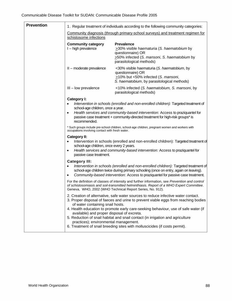
Communicable Disease Toolkit for SUDAN: Communicable Disease Profile 2005
World Health Organization 88
Prevention 1. Regular treatment of individuals according to the following community categories:
Community diagnosis (through primary-school surveys) and treatment regimen for schistosome infections
Community category Prevalence I – high prevalence >30% visible haematuria (S. haematobium by questionnaire) OR >50% infected (S. mansoni, S. haematobium by parasitological methods)
II – moderate prevalence <30% visible haematuria (S. haematobium, by questionnaire) OR >10% but <50% infected (S. mansoni, S. haematobium, by parasitological methods)
III – low prevalence <10% infected (S. haematobium, S. mansoni, by parasitological methods)
Category I: • Intervention in schools (enrolled and non-enrolled children): Targeted treatment of
school-age children, once a year. • Health services and community-based intervention: Access to praziquantel for
passive case treatment + community-directed treatment for high-risk groups* is recommended.
* Such groups include pre-school children, school-age children, pregnant women and workers with occupations involving contact with fresh water.
Category II: • Intervention in schools (enrolled and non-enrolled children): Targeted treatment of
school-age children, once every 2 years. • Health services and community-based intervention: Access to praziquantel for
passive case treatment.
Category III: • Intervention in schools (enrolled and non-enrolled children): Targeted treatment of
school-age children twice during primary schooling (once on entry, again on leaving). • Community-based intervention: Access to praziquantel for passive case treatment. For the definition of classes of intensity and further information, see Prevention and control of schistosomiasis and soil-transmitted helminthiasis. Report of a WHO Expert Committee. Geneva, WHO, 2002 (WHO Technical Report Series, No. 912).
2. Creation of alternative, safe water sources to reduce infective water contact. 3. Proper disposal of faeces and urine to prevent viable eggs from reaching bodies
of water containing snail hosts. 4. Health education to promote early care-seeking behaviour, use of safe water (if
available) and proper disposal of excreta. 5. Reduction of snail habitat and snail contact (in irrigation and agriculture
practices); environmental management. 6. Treatment of snail breeding sites with molluscicides (if costs permit).

Communicable Disease Toolkit for SUDAN: Communicable Disease Profile 2005
World Health Organization 89
22. SOIL-TRANSMITTED HELMINTHIASES (ascariasis, hookworm infection, trichuriasis) DESCRIPTION
Infectious agent Helminths: Ascaris lumbricoides, hookworm, Trichuris trichiura
Case definition • Ascariasis:
Suspected: Abdominal or respiratory symptoms and history of passing worms. Confirmed: Suspected case and passage of A. lumbricoides (anus, mouth, nose), or presence of A. lumbricoides eggs in stools (microscopy).
• Hookworm infection:
Suspected: Severe anaemia for which there is no other obvious cause. Confirmed: Suspected case and presence of hookworm eggs in stools (microscopy).
• Trichuriasis:
Suspected: Bloody, mucoid stools. Confirmed: Suspected case and presence of T. trichiura eggs in stools.
Mode of transmission
− Ingestion of eggs, mainly as a food contaminant: A. lumbricoides and T. trichiura − Active penetration of skin by larvae in the soil: Hookworm
Incubation − 4–8 weeks for A. lumbricoides − a few weeks to many months for hookworm − unspecified for T. trichiura.
Period of communicability
− A. lumbricoides: eggs appear in the faeces 45–75 days after ingestion and become infective in soil after 2–3 weeks. They can remain viable in soil for years. Infected people can contaminate soil as long as mature fertilized female worms live in the intestine (lifespan of adult worms can be 12–24 months).
− Hookworm: eggs appear in the faeces 6–7 weeks after infection. As larvae they become infective in soil after 7–10 days and can remain infective for several weeks. Infected people can contaminate soil for several years.
− T. trichiura: eggs appear in the faeces 70–90 days after ingestion and become infective in soil after 10–14 days. Infected people can contaminate soil for several years.

Communicable Disease Toolkit for SUDAN: Communicable Disease Profile 2005
World Health Organization 90
EPIDEMIOLOGY Burden 1992. A survey carried out in Juba (Bahr Al Jebel) obtained 241 faecal samples and
gave the following prevalence rates: Ascaris lumbricoides: 1.2%. Trichuris trichiura: 0.8%. Hookworm: 36%.
1994. A survey conducted in 6 different localities in Sudan covering all climatic conditions gave the following results (2489 faecal samples): Geohelminths: 53 cases (2.12%). 50 out of 53 cases were from Juba (Bahr Al Jebel).
Another survey conducted in Juba, Bahr Al Jebel (2789 stool samples) found a prevalence of geohelminthic infections of 20.6%.
1998. A study performed in Chukudum and Kimatong (Eastern Equatoria) gave the following results (274 faecal samples): Ascaris lumbricoides: not detected Trichuris trichiura: 5 cases (1.8%). Hookworm: 36 cases (13.1 %).
Geographical distribution
Soil-transmitted helminthiases are mainly present in southern Sudan.
Seasonality No data available.
Recent epidemics in the country
Soil-transmitted helminthiases are usually endemic diseases, with little likelihood of rapid changes in incidence. Surveys may identify areas of particularly high endemicity where mass treatment will be warranted.
RISK FACTORS FOR INCREASED BURDEN Population movement
Yes Strictly linked to insufficient sanitation resources. Not a risk factor if people remain in the same place for a period shorter than the time needed for eggs to be discharged by an infected patient and become infective themselves (at least 45–50 days).
Overcrowding Yes Linked to the number of people defecating and to unsafe faeces disposal.
Poor access to health services
Yes No treatment provided.
Food shortages No
Lack of safe water and poor sanitation
Yes The number of people relative to available sanitation facilities is the most important risk factor.
Others No

Communicable Disease Toolkit for SUDAN: Communicable Disease Profile 2005
World Health Organization 91
Risk assessment conclusions
Control of helminthic infestations can play a major role in reducing the communicable disease burden shouldered by populations in complex emergency situations.
Moreover, given their simplicity and feasibility, intestinal helminth control activities can represent a starting point for the reconstruction of health care systems in complex emergency-affected countries.
All soil-transmitted helminthiases compete with the host for nutrients, causing malabsorption of fats, proteins, carbohydrates and vitamins, and directly synergizing malnutrition. They can cause growth retardation.
A. lumbricoides infestation exacerbates vitamin A deficiency. Elimination of ascarids may therefore result in rapid clinical improvement in night blindness and dryness around the eye. Infection from measles in a patient already infected with A. lumbricoides can result in a very severe disease.
Hookworm infestation is strongly associated with chronic anaemia. Significant inverse correlations between intensity of worm infestation and haemoglobin level have been demonstrated.
Heavy T. trichiura infection may cause diarrhoea and severe malabsorption.
There is currently no national programme for the control of soil-transmitted helminthiases in Sudan.
Soil-transmitted helminths can be controlled with low-cost, highly effective interventions that can dramatically increase the quality of life of affected populations. The average cost in a school distribution campaign (including drugs, distribution and monitoring activities) is approximately US$ 0.05 per child.
PREVENTION AND CONTROL MEASURES Case management For treatment, WHO recommends the following four drugs:
albendazole 400 mg, or levamisole 2.5 mg/kg, or mebendazole 500 mg, or pyrantel 10 mg/kg (less commonly used because it is more difficult to administer) Note 1: These drugs must not be given during the first trimester of pregnancy. Note 2: Where mass treatment with albendazole for filariasis is envisaged, chemotherapy of intestinal helminths will take place as part of the antifilarial chemoprophylaxis.
Prevention and control
Overall:
Personal hygiene, disposal of faeces, hand-washing and clean food Improvements in sanitation standards (see Appendix 3: Safe water and sanitation) Community-wide treatment according to the following categories:
Community diagnosis (through primary-school surveys) and treatment regimen for STH:
Community category Prevalence % of moderate-to- of any infection heavy intensity infections I (high prevalence–high intensity) >70% >10% II (high prevalence-–low intensity) >50% but <70% <10% III (low prevalence–low intensity) <50% <10%

Communicable Disease Toolkit for SUDAN: Communicable Disease Profile 2005
World Health Organization 92
Category I:
• Intervention in schools (enrolled and non-enrolled children): Targeted treatment of school-age children, 2–3 times a year.
• Health services and community-based intervention: Systematic treatment of pre-school children and women of childbearing age in mother and child health programmes.
Category II:
• Intervention in schools (enrolled and non-enrolled children): Targeted treatment of school-age children, once a year.
• Health services and community-based intervention: Systematic treatment of pre-school children and women of childbearing age in mother and child health programmes.
Category III:
• Intervention in schools (enrolled and non-enrolled children): Selective treatment.
• Community-based intervention: Selective treatment.
For the definition of classes of intensity and further information, see: Prevention and control of schistosomiasis and soil-transmitted helminthiasis. Report of a WHO Expert Committee. Geneva, WHO, 2002 (WHO Technical Report Series, No. 912).
In case of suspected or confirmed hookworm infection, in addition:
• In highly endemic areas, wear shoes.
• Consider drug treatment and iron supplementation during pregnancy.

Communicable Disease Toolkit for SUDAN: Communicable Disease Profile 2005
World Health Organization 93
23. TUBERCULOSIS DESCRIPTION
Infectious agent Bacterium: Mycobacterium tuberculosis. This complex includes M. tuberculosis and M. africanum primarily from humans, and M. bovis primarily from cattle.
Diagnosis in Adults
Clinical description
The most important symptoms in the selection of tuberculosis (TB) suspects in adults (aged older than 15 years) are: ─ productive cough for more than 2 weeks, and/or ─ haemoptysis and ─ significant weight loss.
Patients with TB may also have other symptoms (which are more common, but less suggestive) such as: ─ chest pain ─ breathlessness ─ fever/night sweats ─ tiredness, and ─ loss of appetite.
Among refugee and internally displaced populations, it is unusual to have ready access to X-ray facilities. It is the priority of health services to detect the sources of infection by sputum microscopy, and cure them.
Clinical case definition
Tuberculosis suspect: Any person who presents with symptoms or signs suggestive of TB, in particular cough of long duration (more than 3 weeks)
Case of tuberculosis: A patient in whom TB has been bacteriologically confirmed or diagnosed by a clinician. Note: Any person given treatment for TB should be recorded as a case. Incomplete "trial" tuberculosis treatment should not be given as a method for diagnosis.
Definite case of tuberculosis: A patient with positive culture for the M. tuberculosis complex. (In countries where culture is not routinely available, a patient with two sputum smears positive for acid-fast bacilli (AFB) is also considered a "definite" case.)
Laboratory criteria for diagnosis
Each TB suspect should have three sputum samples examined by light binocular microscopy for AFB.
The chances of finding TB organisms are greater with three sputum samples than with one or two samples. Secretions build up in the airways overnight, so that an early-morning sputum sample is more likely to contain the TB organism than a sample taken later in the day. In practice, a suspect provides sputum samples in the following manner:

Communicable Disease Toolkit for SUDAN: Communicable Disease Profile 2005
World Health Organization 94
Day 1 Sample 1 – Person suspected of TB provides an “on-the-spot” sample under supervision on presentation to the health facility. He or she is given a sputum container to take home for an early-morning sample the following day.
Day 2 Sample 2 – Person suspected of TB brings an early-morning sputum sample collected just after waking up. Sample 3 – Person suspected of TB provides another “on-the-spot” sample.
At least two sputum smears are positive Smears should be stained using the Ziehl–Neelsen method. Any TB suspect with two positive smears is a smear-positive TB patient, who must then be registered and started on anti-TB treatment.
If only one initial sputum smear is positive A suggestive X-ray showing active pulmonary TB interpreted by an experienced medical officer may lead to a diagnosis of smear-positive TB. AFB microscopy may be repeated and, if at least one smear is again positive with compatible X-ray, the patient should be considered a smear-positive TB patient. In the absence of X-ray, one sputum smear with positive culture for M. tuberculosis is also classified as sputum-positive TB.
If all three sputum smears are negative If the initial three smears are negative, but pulmonary TB is still suspected because of persistent symptoms, the suspect should be treated for acute respiratory infection with broad-spectrum antibiotics (e.g. amoxicillin or co-trimoxazole, but not rifampicin or any other anti-TB drug) for at least 1 week. If there is no improvement, sputum samples must be re-examined 2 weeks after the first sputum examination.
Between 65–80% of all pulmonary TB cases are expected to be confirmed by positive sputum smear examination. X-ray lesions compatible with active TB should encourage further sputum examination if the three sputum smear examinations were negative. X-ray itself is not a diagnostic tool for pulmonary TB.
In some circumstances, a compatible X-ray together with symptoms consistent with TB will lead to the diagnosis of pulmonary TB in smear-negative cases. Thus, if all three samples are again negative after the trial of antibiotics, either a compatible X-ray interpreted by an experienced physician or, in the absence of X-ray facilities, the experienced physician’s judgement alone will decide whether a patient is categorized as having TB (classed as smear-negative TB).
Additional cases of TB may be found among close contacts of known smear-positive cases, either family members or persons sleeping in the same shelter. Symptomatic contacts should be screened using the procedures described above.
TB in HIV-positive patients
HIV-positive patients are more susceptible to TB infection, and HIV in a TB patient is a potent cause of progression of TB infection to disease. The principles of TB control are the same even when there are many HIV/TB patients. In HIV-infected patients, pulmonary TB is still the commonest form of TB. The clinical presentation of TB depends on the degree of immunosuppression.
Early in HIV infection, when immunity is good, the signs of TB are similar to those in an individual without HIV infection. As HIV infection progresses and immunity declines, the risk of TB dissemination increases. TB meningitis, miliary TB and widespread TB lymphadenopathy occur.
It is important to look systematically for signs or symptoms of TB in HIV-positive patients and to start treatment without delay based on clinical, bacteriological and, in some circumstances, radiological evidence.
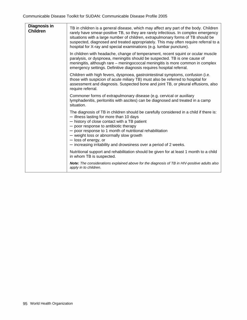
Communicable Disease Toolkit for SUDAN: Communicable Disease Profile 2005
World Health Organization 95
Diagnosis in Children
TB in children is a general disease, which may affect any part of the body. Children rarely have smear-positive TB, so they are rarely infectious. In complex emergency situations with a large number of children, extrapulmonary forms of TB should be suspected, diagnosed and treated appropriately. This may often require referral to a hospital for X-ray and special examinations (e.g. lumbar puncture).
In children with headache, change of temperament, recent squint or ocular muscle paralysis, or dyspnoea, meningitis should be suspected. TB is one cause of meningitis, although rare – meningococcal meningitis is more common in complex emergency settings. Definitive diagnosis requires hospital referral.
Children with high fevers, dyspnoea, gastrointestinal symptoms, confusion (i.e. those with suspicion of acute miliary TB) must also be referred to hospital for assessment and diagnosis. Suspected bone and joint TB, or pleural effusions, also require referral.
Commoner forms of extrapulmonary disease (e.g. cervical or auxiliary lymphadenitis, peritonitis with ascites) can be diagnosed and treated in a camp situation.
The diagnosis of TB in children should be carefully considered in a child if there is: ─ illness lasting for more than 10 days ─ history of close contact with a TB patient ─ poor response to antibiotic therapy ─ poor response to 1 month of nutritional rehabilitation ─ weight loss or abnormally slow growth ─ loss of energy, or ─ increasing irritability and drowsiness over a period of 2 weeks.
Nutritional support and rehabilitation should be given for at least 1 month to a child in whom TB is suspected. Note: The considerations explained above for the diagnosis of TB in HIV-positive adults also apply in to children.

Communicable Disease Toolkit for SUDAN: Communicable Disease Profile 2005
World Health Organization 96
Diagnostic criteria for classification of TB
Pulmonary tuberculosis (PTB)
Pulmonary TB refers to disease involving the lung parenchyma. Tuberculous intrathoracic lymphadenopathy (mediastinal and/or hilar) or tuberculous pleural effusion, without radiographic abnormalities in the lungs, therefore constitutes a case of extrapulmonary TB. A patient with both pulmonary and extrapulmonary TB should be classified as a case of pulmonary TB.
• Smear-positive pulmonary TB
Either: A patient with at least two sputum specimens positive for AFB by microscopy;
or: A patient with at least one sputum specimen positive for AFB by microscopy and radiographic abnormalities consistent with pulmonary TB; or: A patient with at least one sputum specimen positive for AFB by microscopy, which is culture-positive for M. tuberculosis.
• Smear-negative pulmonary TB
A case of PTB that does not meet the above definition for smear-positive TB. This group includes cases without smear result. This commonly occurs in children but is comparatively uncommon in adults.
Diagnostic criteria for PTB (which is also used to exclude sputum negative PTB) is based on the following criteria: – at least three sputum specimens negative for AFB, and – no clinical response to a one-week course of broad-spectrum antibiotics, and – radiographic abnormalities consistent with active PTB, and – decision by a clinician to treat with a full course of anti-TB chemotherapy. A patient whose initial sputum smears were negative and whose subsequent sputum culture result is positive is also considered to have smear-negative pulmonary TB.
Extrapulmonary tuberculosis (EPTB)
EPTB refers to TB of organs other than the lungs, e.g. pleura, lymph nodes, abdomen, genitourinary tract, skin, joints and bones, meninges. Diagnosis should be based on one culture-positive specimen, or on histological or strong clinical evidence consistent with active EPTB, followed by a decision by a clinician to treat with a full course of anti-TB chemotherapy.
The case definition of an EPTB case with several sites affected depends on the site representing the most severe form of disease.
Some cases will be easy to diagnose with peripheral lymphadenitis, swelling of cervical or axillary lymph nodes, chronic evolution and/or production of caseous discharge. Other cases, such as severe, life-threatening forms (e.g. miliary TB, TB meningitis), TB of bone joints, TB peritonitis, TB laryngitis, will be suspected but should be referred to a hospital for assessment.
Mode of transmission
Exposure to tubercle bacilli in airborne droplet nuclei produced by people with pulmonary or laryngeal TB during expiratory efforts such as coughing and sneezing. Extrapulmonary tuberculosis (other than laryngeal) is usually non-infectious. Bovine tuberculosis results from exposure to tuberculous cattle, usually by ingestion of unpasteurized milk or dairy products, and sometimes by airborne spread to farmers and animal handlers.

Communicable Disease Toolkit for SUDAN: Communicable Disease Profile 2005
World Health Organization 97
Progression to active disease
Progression to active disease can take weeks or years; latent infections may persist throughout life. The risk of TB occurrence is relatively high during the first year following TB infection, then progressively decreases by half within the next 4–5 years.
Only 10% of infected people with normal immune systems will develop clinically evident TB at some point in life; 5% will have an early progression of the disease (primary tuberculosis); the remaining 5% will have a late progression of the disease (post-primary tuberculosis) after a period of initial containment.
Period of communicability
As long as viable tuberculosis bacilli are being discharged in the sputum. Effective treatment usually eliminates communicability within 2 weeks.
EPIDEMIOLOGY Burden Estimated number of new cases of all forms of TB:
2002: 71 211 (of which 24 554 (34.4%) were notified) 2001: 59 897 (of which 23 997 (40.0%) were notified) 2000: 59 875 (of which 24 807 (41.4%) were notified)
Estimated number of new cases of smear positive (ss+) TB:
2002: 31 432 (of which 33.01%) were notified) 2001: 26 953 (of which 11 136 (41.3%) were notified) 2000: 26 944 (of which 12 311 (45.6%) were notified) (Data source: WHO/Sudan)
Geographical distribution
Tuberculosis is known to be widespread throughout the country.
Seasonality No specific seasonality is reported.
Alert threshold Not applicable.
Recent epidemics in the country
Not applicable.
RISK FACTORS FOR INCREASED BURDEN Population movement
Yes May generate conditions that disrupt TB treatment.
Overcrowding Yes Overcrowding is recognized as one of the most important factors leading to increased risk of transmission.
Poor access to health services
Yes − People affected by TB who cannot access health services and be treated remain infectious for a longer period.
− The case-fatality rate is high (about 50%) without proper treatment.
− Together with poor drug prescribing practices, the interruption of treatment is one of the most important causes of development of multidrug-resistant TB (MDR-TB).
Food shortages Yes Poor nutritional status increases vulnerability to TB infection and development of active disease.
Lack of safe water and poor sanitation
No
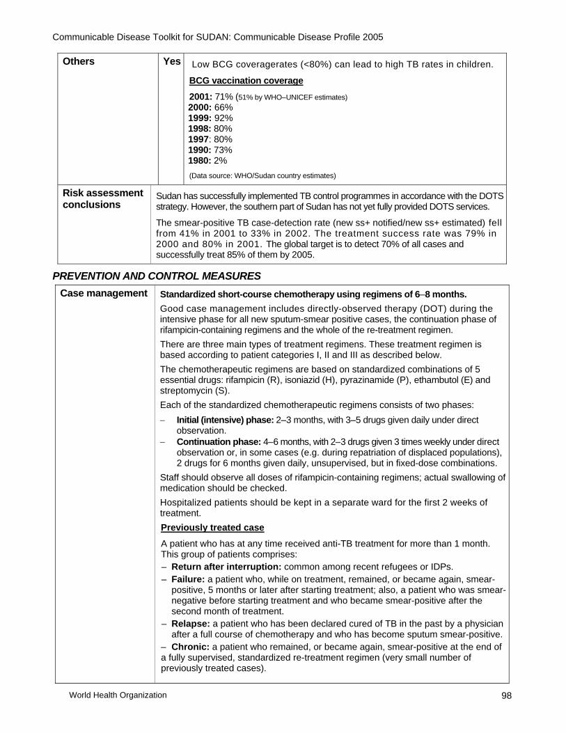
Communicable Disease Toolkit for SUDAN: Communicable Disease Profile 2005
World Health Organization 98
Others Yes Low BCG coveragerates (<80%) can lead to high TB rates in children.
BCG vaccination coverage
2001: 71% (51% by WHO–UNICEF estimates) 2000: 66% 1999: 92% 1998: 80% 1997: 80% 1990: 73% 1980: 2% (Data source: WHO/Sudan country estimates)
Risk assessment conclusions
Sudan has successfully implemented TB control programmes in accordance with the DOTS strategy. However, the southern part of Sudan has not yet fully provided DOTS services.
The smear-positive TB case-detection rate (new ss+ notified/new ss+ estimated) fell from 41% in 2001 to 33% in 2002. The treatment success rate was 79% in 2000 and 80% in 2001. The global target is to detect 70% of all cases and successfully treat 85% of them by 2005.
PREVENTION AND CONTROL MEASURES Case management Standardized short-course chemotherapy using regimens of 6–8 months.
Good case management includes directly-observed therapy (DOT) during the intensive phase for all new sputum-smear positive cases, the continuation phase of rifampicin-containing regimens and the whole of the re-treatment regimen. There are three main types of treatment regimens. These treatment regimen is based according to patient categories I, II and III as described below. The chemotherapeutic regimens are based on standardized combinations of 5 essential drugs: rifampicin (R), isoniazid (H), pyrazinamide (P), ethambutol (E) and streptomycin (S). Each of the standardized chemotherapeutic regimens consists of two phases: − Initial (intensive) phase: 2–3 months, with 3–5 drugs given daily under direct
observation. − Continuation phase: 4–6 months, with 2–3 drugs given 3 times weekly under direct
observation or, in some cases (e.g. during repatriation of displaced populations), 2 drugs for 6 months given daily, unsupervised, but in fixed-dose combinations.
Staff should observe all doses of rifampicin-containing regimens; actual swallowing of medication should be checked. Hospitalized patients should be kept in a separate ward for the first 2 weeks of treatment. Previously treated case
A patient who has at any time received anti-TB treatment for more than 1 month. This group of patients comprises: – Return after interruption: common among recent refugees or IDPs. – Failure: a patient who, while on treatment, remained, or became again, smear-
positive, 5 months or later after starting treatment; also, a patient who was smear-negative before starting treatment and who became smear-positive after the second month of treatment.
– Relapse: a patient who has been declared cured of TB in the past by a physician after a full course of chemotherapy and who has become sputum smear-positive.
– Chronic: a patient who remained, or became again, smear-positive at the end of a fully supervised, standardized re-treatment regimen (very small number of previously treated cases).

Communicable Disease Toolkit for SUDAN: Communicable Disease Profile 2005
World Health Organization 99
Treatment in Children
The drug regimens used for children are the same as for adults*
Drug dosages must be calculated according to the child’s weight. Adjustments may have to be made during the course of the treatment as the child may rapidly regain lost weight.
For infants of newly diagnosed smear-positive mothers, breastfeeding should continue. The infant should not be separated from the mother. Transmission is likely to have already occurred, and the infant is at greater risk of dying from other causes if breastfeeding is stopped. If the infant is well, isoniazid prophylaxis should be given for 6 months and requires regular follow-up, for example one in every two months.
See: Precautions for use of streptomycin and ethambutol in children. Geneva, WHO, 2003 (WHO/CDS/TB 2003.313; page 64).
Treatment categories
Treatment categories are essential for prioritization of TB treatment according to public health risk – Category I is the highest priority.
Category I These patients are:
– smear-positive persons who have never previously been treated or who have only received treatment for less than 1 month.
– severely ill patients with other forms of TB (new smear-negative pulmonary TB. with extensive parenchymal involvement, and new cases of severe forms of TB1).
The recommended regimen lasts 6 months. The initial (intensive) phase of treatment lasts for 2 months; rifampicin, isoniazid, pyrazinamide and ethambutol are given daily or 3 times weekly (streptomycin may be used as a subsititute for ethambutol), under direct supervision.
At the end of the second month of treament, most patients will have a negative result on sputum microscopy; they can then progress to the second stage of treatment – the continuation phase. This phase lasts for 4 months, with rifampicin and isoniazid given 3 times weekly, under direct supervision.2
If the sputum smear examination is positive at the end of the second month, for whatever reason, the initial phase is prolonged for a third month. The patient then starts the continuation phase irrespective of the results of the sputum examination at the end of the third month. If the sputum smears are still positive at the end of the fifth month or at the end of a treatment regimen, the patient is classified a treatment failure case. He or she is re-registered and starts a full course of the re-treatment regimen as a Category II patient.
Drug dose is adjusted for weight gain at the end of the initial phase (2nd or 3rd month). 1This category includes patients with TB meningitis, disseminated TB, pericarditis, peritonitis, bilateral or extensive pleurisy, vertebral disease with neurological complications, and intestinal and genitourinary disease. 2Daily self-administered ethambutol and isoniazid may be used in the continuation phase for 6 months, so this treatment regimen lasts a total of 8 months. However, this regimen is associated with a higher rate of failure and relapse.

Communicable Disease Toolkit for SUDAN: Communicable Disease Profile 2005
World Health Organization 100
Category II Patients who were previously treated and are now sputum smear-positive include:
– treatment after interruption; – treatment failure; and – relapse after treatment. These patients should receive a standardized re-treatment regimen, fully supervised throughout both phases of treatment.
The initial phase of treatment lasts for 3 months; rifampicin, isoniazid, pyrazinamide and ethambutol are given daily and supplemented by streptomycin daily for the first 2 months.
The continuation phase of this regimen constitutes 5 months of rifampicin, isoniazid and ethambutol given 3 times weekly. Sputum smear examination is performed at the end of the initial phase of treatment (i.e. at the end of 3 months), during the continuation phase of treatment (at the end of the fifth month) and at the end of treatment (at the end of the eighth month). If the patient is sputum smear-positive at the end of the third month, the initial phase of treatment is extended with rifampicin, isoniazid, pyrazinamide and ethambutol for one more month. Patients who are still positive at the end of the fourth month progress to the continuation phase, regardless of the results of the sputum examination.
Category III
These patients include: ─ smear-negative pulmonary patients (with limited parenchymal involvement) ─ adults and children with non-serious extrapulmonary disease (including symptomatic primary disease).
All Category III patients should receive 2 months of rifampicin, isoniazid and pyrazinamide daily, followed by 4 months of isoniazid and rifampicin every second day.
When the continuation phase cannot be carried out under direct observation, all patients should be given daily ethambutol and isoniazid in the continuation phase for 6 months.
HIV-positive patients Anti-TB drug treatment is the same for HIV-positive and HIV-negative patients, with one exception: thiacetazone should not be given to HIV-positive TB patients as there is increased risk of severe toxicity and sometimes fatal skin reactions.
Controlled clinical trial studies have shown that isoniazid preventive treatment (IPT) reduces the risk of TB disease in HIV-positive individuals with latent TB infection (shown by a positive tuberculin skin test).
The use of IPT has shown to be more effective than other regimens for prevention of latent TB infection. The decision to use IPT must be carefully evaluated, and requires first the exclusion of active TB in the patient.
To manage the problem of HIV/TB coinfection effectively, TB and HIV programmes should coordinate activities through a TB/HIV coordinating body. See: ─ An expanded DOTS framework for effective tuberculosis control. Geneva, WHO, 2002
(WHO/CDS/TB/2002.297). ─ Treatment of tuberculosis: guidelines for national programmes, 3rd ed. Geneva, WHO,
2003 (WHO/TB/2003.313). ─ Tuberculosis control in refugee situations: an inter-agency field manual. Geneva, WHO,
1997 (WHO/TB/97.221; to be updated in 2004).
Communicable Disease Toolkit for SUDAN: Communicable Disease Profile 2005
World Health Organization 101
Prevention and control
Detection and treatment of smear-positive (infectious) TB cases is the most effective preventive measure. To ensure the appropriate treatment and cure of TB patients, strict implementation of the DOTS strategy is important. DOTS is the internationally recommended strategy for TB control, and has the following components:
− Government commitment to ensuring sustained, comprehensive TB control activities.
− Case detection by sputum smear microscopy among symptomatic patients self-reporting to health services.
− Standardized short-course chemotherapy using regimens of 6–8 months, with direct observation of treatment at least during the intensive phase (or for as long as rifampicin is administered) for at least all confirmed smear-positive cases (see Case management).
− A regular, uninterrupted supply of all essential anti-TB drugs. − A standardized recording and reporting system that allows assessment of follow-
up and treatment results for each patient and of the TB control programme's overall performance.
Complementary control strategies:
− Health education to improve awareness and reduce stigma. − Maintaining good ventilation and reducing overcrowding in health clinics, and
ensuring hospitalized patients are kept in a separate ward for the first 2 weeks of treatment.
− Isoniazid prophylaxis is not recommended in refugee situations, except for children being breastfed by smear-positive mothers. If the child is well, BCG vaccination should be postponed and isoniazid given to the child for 6 months. In the event of a sudden disruption to the programme, isoniazid may be stopped and BCG given before the child leaves the refugee camp (preferably after a one-week interval).
Immunization BCG has been shown to be effective in preventing severe forms of TB such as TB meningitis and miliary TB in children. As overcrowding and malnutrition are common among many refugee and displaced populations, the risk of TB transmission to children is increased. BCG is strongly recommended for all newborn children and any children aged up to 5 years who have not already received it. The vaccination of newborns should be incorporated into routine immunization programmes for all children. Re-vaccination is not recommended.

Communicable Disease Toolkit for SUDAN: Communicable Disease Profile 2005
World Health Organization 102
Health education Key elements of community education: ─ avoiding stigmatization of TB patients. ─ curability of TB disease. ─ early (self) referral of TB suspects. ─ importance of adherence to treatment. ─ contact tracing. The most important messages to teach: • TB in an adult should be suspected when the person has a productive cough lasting
more than 2 weeks, and/or blood in the sputum, with significant weight loss. • Cover the mouth whenever coughing or sneezing to prevent the spread of lung
diseases. • Anyone may contract TB. • TB is curable. • Early treatment is important for best results and to prevent spread, especially to family
members. • Children are especially at risk if not treated and may develop severe, even fatal,
disease. • Good treatment is the best prevention. • All patients must take the full course of treatment. • Treatment makes patients non-infectious in 2 weeks, but cure takes 6–8 months. • Treatment must be completed even though the patient may feel better sooner. • Failure to complete the treatment may result in a recurrence that may be impossible to
treat and may spread serious disease to others, especially children. • All patients should be treated sympathetically and with respect. • Controlling TB is a community responsibility. Note: Diagrams should be used as much as possible – a high literacy level should not be assumed. Cured patients are often helpful teachers and supporters of new patients.

Communicable Disease Toolkit for SUDAN: Communicable Disease Profile 2005
World Health Organization 103
24. TYPHOID FEVER DESCRIPTION
Infectious agent Bacterium: Salmonella eneterica serovar Typhi (S.Typhi).
Case definition Clinical case definition Clinical diagnosis is difficult. In the absence of laboratory confirmation, any case with fever of at least 38 °C for 3 or more days is considered suspect if the epidemiological context is conducive.
Confirmed case Isolation of S. typhi from blood or stool cultures.
Mode of transmission
Faecal–oral route, particularly through contaminated water and food.
Incubation Incubation period is usually 8–14 days but may be from 3 days up to 1 month.
Period of communicability
From the symptomatic period for 2 weeks; 2–5% of infected cases remain carriers for several months. Chronic carriers contribute significantly to spread of the disease.
EPIDEMIOLOGY Burden No data available.
Geographical distribution
No data available.
Seasonality No data available.
Alert threshold Two or more linked cases.
Recent epidemics in the country
No data available.
RISK FACTORS FOR INCREASED BURDEN Population movement
Yes Dissemination of multidrug-resistant strains of S. typhi.
Overcrowding Yes Very important.
Poor access to health services
Yes Early detection and containment of cases are paramount in reducing dissemination. The case-fatality rate is high (10–20%) without proper treatment.
Food shortages No Lack of safe water and poor sanitation
Yes The most important risk factor.
Others Yes Multidrug-resistant strains of S. typhi, including resistance to ciprofloxacin. Milk and dairy products are an important source of infection.

Communicable Disease Toolkit for SUDAN: Communicable Disease Profile 2005
World Health Organization 104
Risk assessment conclusions
Among the general population the risk is related to the availability of safe food, clean water and sanitation. This must be ensured to prevent outbreaks. It is essential that health care workers are trained and have a reasonable degree of suspicion. Monitoring of antibiotic resistance is essential. Early and effective treatment is crucial for maintaining low case-fatality rates.
PREVENTION AND CONTROL MEASURES Case management Early antimicrobial treatment, selected according to the antimicrobial resistance
pattern of the strain. Quinolones (e.g. ciprofloxacin), co-trimoxazole, chloramphenicol and ampicillin are usually used for typhoid fever. Dehydration prevention and case management using ORS also play an important role.
Epidemic control Inform the health authorities when one or more suspected cases are identified. Confirm the outbreak in accordance with WHO guidelines. Confirm the diagnosis and ensure prompt treatment.
Prevention Good sanitation can markedly reduce the risk of transmission, especially where its absence may lead to contamination of clean water sources. High priority should be given to observing the basic principles of sanitary human waste disposal as well as to ensuring the availability of safe water supplies.
Appropriate facilities for human waste disposal are a basic need of all communities. The absence of such facilities creates a high risk for disease transmission. Sanitary systems that are appropriate for local conditions should be constructed with the cooperation of the community.
People will need to be taught how to use latrines, about the dangers of defecating on the ground, or in or near waters, and about the importance of thorough hand-washing with soap or ash after any contact with excreta. The disposal of children's excreta in latrines needs to be emphasized.
Immunization Mass immunization may be an adjunct for the control of typhoid fever during a sustained, high-incidence epidemic. This is especially true when access to well functioning medical services is not possible or in the case of a multidrug-resistant strain. A parenteral vaccine containing the polysaccharide Vi antigen is the vaccine of choice among displaced populations. An oral, live vaccine using S. typhi strain Ty21a is also available. Neither the polysaccharide vaccine nor the Ty21a vaccine is licensed for children aged under 2 years. The Ty21a vaccine should not be used in patients receiving antibiotics.
Communicable Disease Toolkit for SUDAN: Communicable Disease Profile 2005
World Health Organization 105
25. YELLOW FEVER DESCRIPTION
Infectious agent Yellow fever virus, belonging to the Flavivirus group.
Case definition Clinical description:
Characterized by acute onset of fever followed by jaundice within 2 weeks of onset of first symptoms. Haemorrhagic manifestations and signs of renal failure may occur.
There are two disease phases for yellow fever:
Acute phase: While some infections have no symptoms whatsoever, this first phase is normally characterized by fever, muscle pain (with prominent backache), headache, shivers, loss of appetite, nausea and/or vomiting. Often, the high fever is paradoxically associated with a slow pulse (Faget’s sign). Most patients improve after 3–4 days and their symptoms disappear, but 15% enter the toxic phase.
Toxic phase: Fever reappears; the patient rapidly develops jaundice and complains of abdominal pain with vomiting. Bleeding can occur from mouth, nose, eyes and/or stomach. Once this happens, blood appears in the vomit and faeces. Kidney function deteriorates; this can range from abnormal protein levels in the urine (albuminuria) to complete renal failure with no urine production (anuria). Half the patients in the toxic phase die within 10–14 days. The remainder recovers without significant organ damage.
Laboratory criteria: Isolation of yellow fever virus, or Presence of yellow-fever-specific IgM or a fourfold or greater rise in serum IgG levels in paired sera (acute and convalescent), or Positive postmortem liver histopathology, or Detection of yellow fever antigen in tissues by immunohistochemistry, or Detection of yellow fever virus genomic sequences in blood or organs by polymerase chain reaction.
Case classification: Suspected: a case that is compatible with the clinical description. Probable: not applicable Confirmed: a suspected case that is laboratory-confirmed (national reference laboratory) or epidemiologically linked to a confirmed case or outbreak.
Mode of transmission
Bite of infective mosquitoes.
The vectors of yellow fever in forest areas in Africa are Aedes africanus and other Aedes species. In urban areas, the vector is Ae. aegypti (all-day biting species).
Incubation From 3 to 6 days.
Period of communicability
Blood of patients is infective for mosquitoes shortly before onset of fever and for the first 3–5 days of illness. The disease is highly communicable where many susceptible people and abundant vector mosquitoes coexist; it is not communicable by contact or other common means of disease transmission. Once infected, mosquitoes remain so for life.
Communicable Disease Toolkit for SUDAN: Communicable Disease Profile 2005
World Health Organization 106
EPIDEMIOLOGY Burden 2001: 0 cases reported 1997: no data available
2000: no data available 1990: no data available 1999: no data available 1980: no data available 1998: no data available (Source: WHO/IVB data, 2004)
Geographical distribution
The southern half of the country was officially endemic in the year 2000.
Seasonality In forest areas, where the yellow fever virus circulates between mosquitoes and monkeys or chimpanzees, the disease is present throughout the year. In field or savannah areas outside the forest areas, the virus remains dormant in infected mosquito eggs throughout the dry season and emerges in the rainy season when eggs hatch.
Alert threshold One confirmed case must lead to alert. An outbreak of yellow fever is at least one confirmed case.
Recent epidemics in the country
Sudan is known to be endemic. 2003: From 11 to 15 May, 178 cases and 27 deaths occurred in Imatong and Ikotos districts, Torit county, in the south-eastern part of the country. International technical assistance, vaccine and vaccine supplies for a vaccination campaign targeting 100 000 people in the area were provided. No details of previous epidemics are available.
RISK FACTORS FOR INCREASED BURDEN Population movement
Yes Unvaccinated people moving to areas of endemicity are at risk. Changes in land use are a risk factor.
Overcrowding Yes Increased population density and increased exposure to mosquito bites in temporary shelters.
Poor access to health services
Yes Collapse of vaccination programmes. Increased fatality due to unavailability of case management.
Food shortages No
Lack of safe water and poor sanitation
No
Others Yes Open water storage provides a favourable habitat for Ae. Aegypti. Old tyres, old water containers, etc. increase vector breeding sites. Temporary surface-water bodies (poor drainage leading to pools and open channels of water) may increase vector breeding opportunities. Yellow fever virus vaccine (YFV) coverage: 2001: no data available 2000: no data available 1999: no data available 1998: no data available 1997: no data available 1990: no data available 1980: no data available (Data source: WHO/IVB data)

Communicable Disease Toolkit for SUDAN: Communicable Disease Profile 2005
World Health Organization 107
Risk assessment conclusions
Although no official cases are reported from Sudan, the risk of transmission exists. Reduced coverage rates for yellow fever immunizations and disruption of mosquito control programmes are likely to have increased this risk. Moreover, population movements from rural to urban areas have resulted in large numbers of people living in conditions of poverty, overcrowding and poor sanitation, all conditions that amplify the risk of transmission. The precise extent of illness and death due to yellow fever is unknown.
Disease surveillance is not adequate to detect cases of sylvatic yellow fever that often occur in remote areas. Moreover, an outbreak of yellow fever can go undetected because the signs and symptoms of yellow fever have a wide spectrum and overlap with those of many other diseases, making it difficult for health workers to make a definitive diagnosis on this basis alone. Mild cases can go undetected because patients are likely to be treated at home and may not seek care in a health facility.
PREVENTION AND CONTROL MEASURES Case management No specific treatment for yellow fever is available.
Dehydration and fever can be corrected with oral rehydration salts. Intensive supportive care may improve the outcome but is rarely available. See: Case management of epidemic-prone diseases in this Toolkit for Sudan (Document 6).
Epidemic control An infected mosquito spreads yellow fever when it bites non-infected humans. When human-to-human transmission is established, the conditions for an epidemic are in place. Depending on the travel patterns of infected humans or infected mosquitoes, the epidemic spreads from village to village and into cities.
Under epidemic conditions, the following must be implemented: − Mass vaccination with YFV − Emergency mosquito control measures:
• Eliminating potential mosquito breeding sites (the most important measure) • Spraying to kill adult mosquitoes (less important because of limited impact) • Use of insecticide-treated nets.
Prevention Vaccination is the single most important measure for preventing yellow fever. In endemic areas, vaccination must be given routinely through the incorporation of YFV in routine child immunization programmes and mass preventive campaigns. YFV is not recommended for symptomatic HIV-infected persons or other immunosuppressed individuals. For theoretical reasons, it is not recommended for pregnant women.
Recommended strategies: ─vaccinating the population aged older than 9 months in counties where coverage
in recent campaigns achieved less than 80%; ─if funds are limited, a cheaper intervention would be to vaccinate children aged
between 9 months and 14 years to reach at least 50% of the population; ─yellow fever vaccination should be integrated in routine EPI activities.
Routine mosquito control measures Potential mosquito breeding sites must be eliminated.

Communicable Disease Toolkit for SUDAN: Communicable Disease Profile 2005
World Health Organization 108
APPENDIX 1: FLOWCHARTS FOR THE DIAGNOSIS OF COMMUNICABLE DISEASES Suspected outbreak Definition of syndrome
Faeces
Possible diseases/pathogens Specimens required Laboratory studies *Ebola and other haemorrhagic fevers may initially present as bloody diarrhoea. If such an etiology is suspected, refer to "Acute haemorrhagic fever syndrome" for appropriate specimen collection guideline.
ACUTE DIARRHOEAL SYNDROME
Acute onset of diarrhoea AND severe illness AND absence of known predisposing factors
Watery Viral gastroenteritis
Cholera Enterotoxigenic E. coli
Giardiasis Cryptosporidium
Dysentery Shigellosis
Salmonellosis Campylobacteriosis Amoebic dysentery
Enterohaemorrhagic E. coli Clostridium difficile
Ebola and other haemorrhagic fevers*
Faeces
Bacterial: Faecal leukocytes
Culture Antimicrobial susceptibility
Serotyping Toxin identification
Viral: Culture
Antigen detection Genome detection
Parasitic: Macro and microscopic
examination

Communicable Disease Toolkit for SUDAN: Communicable Disease Profile 2005
World Health Organization 109
Suspected outbreak Definition of syndrome Possible diseases/pathogens Specimens required Laboratory studies
ACUTE HAEMORRHAGIC FEVER SYNDROME
Acute onset of fever of less than 3 weeks’ duration AND any two of the following:
• Haemorrahgic or purpuric rash • Epistaxis • Haemoptysis • Blood in stool • Other haemorrhagic symptom
AND absence of known predisposing factors
Dengue haemorrhagic fever and shock syndrome
Yellow fever Other arboviral haemorrhagic fevers (e.g. Rift Valley, Crimean–Congo,
Tick-borne flaviviruses) Lassa fever and other arenoviral haemorrhagic fevers
Ebola or Marburg haemorrhagic fevers Haemorrhagic syndrome (hantaviruses)
Malaria Relapsing fever
Blood Blood smear
Serum Postmortem tissue specimens (e.g. skin
biopsy and/or liver biopsy)
Viral: Culture
Antigen detection Antibody levels
Genome detection
Parasitic: Demonstration of
pathogen

Communicable Disease Toolkit for SUDAN: Communicable Disease Profile 2005
World Health Organization 110
Suspected outbreak Definition of syndrome Possible diseases/pathogens Specimens required Laboratory studies * Requires specialized media and handling procedures. See Annex 7 "Guidelines for collections of speciments for laboratory testing in this Toolkit..
ACUTE JAUNDICE SYNDROME
Acute onset of jaundice AND severe illness AND absence of known predisposing factors
Hepatitis A–E Leptospirosis and other spirochaetal diseases
Postmortem liver biopsy
Serum
Viral: Culture
Antigen detection Antibody levels
Genome analysis
Leptospiral: Culture
Antibody levels Serotyping
Blood culture (urine)*
Yellow fever

Communicable Disease Toolkit for SUDAN: Communicable Disease Profile 2005
World Health Organization 111
Suspected outbreak Definition of syndrome Possible diseases/ pathogens Specimens required Laboratory studies
ACUTE NEUROLOGICAL SYNDROME
Acute neurological dysfunction with one or more of the following: • Deterioration of mental function • Acute paralysis • Convulsions • Signs of meningeal irritation • Involuntary movements • Other neurological symptoms
AND severe illness AND absence of predisposing factors
Poliomyelitis or Guillain–Barré syndrome
Viral, bacterial, fungal or parasitic meningoencephalitis
Rabies
Faeces CSF Blood Culture Blood smears
Serum Throat swab
Serum Postmortem specimens (e.g. corneal impressions, brain
tissue, skin biopsy from neck)
Viral: Culture
Bacterial (including leptospiral): Gram stain and other
microscopic techniques Culture
Antimicrobial susceptibility Antigen detection
Serotyping
Viral: Culture
Antigen detection Antibody levels
Genome analysis

Communicable Disease Toolkit for SUDAN: Communicable Disease Profile 2005
World Health Organization 112
Suspected outbreak Definition of syndrome Possible diseases/pathogens Specimens required Laboratory studies
Adapted from: Guidelines for the collection of clinical specimens during field investigation of outbreaks. Geneva, WHO, 2000 (WHO/CDS/CSR/EDC/2000.4).
ACUTE RESPIRATORY SYNDROME
Acute onset of cough OR respiratory distress AND severe illness AND absence of known predisposing factors
Influenza Diphtheria
Streptococcal pharyngitis
Scarlet fever
Hantavirus pulmonary syndrome
Pertussis Respiratory syncytial virus
(RSV)
Bacterial pneumonia including:
Pneumococcal Legionellosis Haemophilus
influenzae Mycoplasma
Respiratory anthrax Pneumonic plague
Throat swab
Serum Nasopharyngeal swab
Blood culture Serum Sputum
Urine (for Legionella)
Bacterial or viral: Culture
Antimicrobial susceptibility (for bacteria) Antigen detection Antibody levels
Genome analysis Serotyping
Toxin identification

Communicable Disease Toolkit for SUDAN: Communicable Disease Profile 2005
World Health Organization 113
APPENDIX 2: STEPS IN OUTBREAK MANAGEMENT PREPARATION
• Health coordination meetings. • Surveillance system – weekly health reports to WHO. • Stockpiles – specimen kits, appropriate antibiotics, IV fluids. • Epidemic investigation kits. • Contingency plans for isolation wards in hospitals. • Laboratory support.
DETECTION If a certain number of cases of any of the following diseases/syndromes is diagnosed (i.e. alert threshold is passed):
• Acute watery diarrhoea in over-5-year-olds. • Bloody diarrhoea. • Suspected cholera. • Measles. • Meningitis. • Acute haemorrhagic fever syndrome. • Acute jaundice syndrome. • Suspected polio (acute flaccid paralysis). • Cluster of deaths of unknown origin. (Diseases/syndromes in list should be modified according to the country's most updated epidemiological profile.
Inform your health coordinator as soon as possible. The health coordinator should inform the Ministry of Health and WHO.
RESPONSE Confirmation
• The lead health agency should investigate reported cases to confirm the outbreak situation – number of cases higher than that expected for same period of year and population. Clinical specimens will be sent for testing.
• The lead health agency should activate an outbreak control team, with members drawn from relevant organizations: Ministry of Health, WHO and other United Nations organizations, nongovernmental organizations in the fields of health and water and sanitation, veterinary experts.
Investigation • Confirm diagnosis (laboratory testing of samples). • Define outbreak case definition. • Count number of cases and determine size of population (to calculate attack rate). • Collect/analyse descriptive data to date (e.g. time/date of onset, place/location of cases and individual characteristics
such as age/sex). • Follow up cases and contacts. • Determine the at-risk population. • Formulate hypothesis for pathogen/source/transmission. • Conduct further investigation/epidemiological studies (e.g. to clarify mode of transmission, carrier, infectious dose
required, better definition of risk factors for disease and at-risk groups. • Write an investigation report (investigation results and recommendations for action).
Control • Implement control measures specific for the disease and prevent exposure (e.g. isolation of cases in viral haemorrhagic
fever outbreak). • Prevent infection (e.g. immunization in measles outbreak). • Treat cases as recommended in WHO guidelines.
EVALUATION
• Assess timeliness of outbreak detection and response, cost. • Change public health policy if indicated (e.g. preparedness). • Write outbreak report and disseminate.

Communicable Disease Toolkit for SUDAN: Communicable Disease Profile 2005
World Health Organization 114
APPENDIX 3: SAFE WATER AND SANITATION The following are effective methods to obtain safe drinking-water:
Boiling To make water safe for drinking and hygiene purposes, bring it to a vigorous, rolling boil and keep it boiling for 1 minute. This will kill, or inactivate, most of the organisms that cause diarrhoea.
Household filtration Household filtration should considerably reduce the pathogens in the water. It should be followed by disinfection through chlorination or boiling.
Disinfection through chlorination The following guidelines should be translated into messages that take into account locally available products and measuring devices. To make water safe by chlorination, the first step is to make a stock solution of chlorine. A stock solution can be prepared by adding the following products to one litre of water:
Product (% concentration by weight of available chlorine) Amount for 1 litre
Calcium hypochlorite (70%); or 15 g Bleaching powder or chlorinated lime (30%); or 33 g Sodium hypochlorite (5%); or 250 ml Sodium hypochlorite (10%); or 110 ml
The stock solution must be stored in a closed container, in a cool dark place and used within 1 month. It should be used to prepare safe water as follows:
Stock solution Added volume of water
0.6 ml or 3 drops l litre 6 ml 10 litres 60 ml 100 litres
Mix by stirring, and allow the chlorinated water to stand for at least 30 minutes before using it. The free residual chlorine level after 30 minutes should be between 0.1 and 0.5 mg/litre. If the free residual chlorine is not within this range, the number of drops of the stock solution should be adjusted so that the final product falls within this range.
If the water is cloudy or turbid it must either be filtered before chlorination or boiled vigorously rather than chlorinated. Chlorination of turbid water might not make it safe.
Sanitation
Good sanitation can markedly reduce the risk of transmission of intestinal pathogens, especially where its absence may lead to contamination of clean water sources. High priority should be given to observing the basic principles of sanitary human waste disposal, as well as to ensuring the availability of safe water supplies.
Appropriate facilities for human waste disposal are a basic need of all communities; without such facilities there is a high risk of water-related diseases. Sanitary systems that are appropriate for the local conditions should be constructed with the cooperation of the community.
People will need to be taught how to use latrines, about the dangers of defecating on the ground, or in or near water, and about the importance of thorough hand-washing with soap or ash after any contact with excreta. The disposal of children's excreta in latrines needs to be emphasized.
See: - Franceys R, Pickford J, Reed R. A guide to the development of on-site sanitation. Geneva, WHO, 1992.
- Environmental health in emergencies and disasters: a practical guide http://www.who.int/water_sanitation_health/hygiene/emergencies/emergencies2002/en/ - Fact sheets on environmental sanitation WHO/EOS/96.4 http://www.who.int/water_sanitation_health/hygiene/emergencies/envsanfactsheets/en/

Communicable Disease Toolkit for SUDAN: Communicable Disease Profile 2005
World Health Organization 115
APPENDIX 4: INJECTION SAFETY Analysis of data collected as part of the Comparative Risk Assessment component of the Global Burden of Disease study suggests that the region that includes Sudan faces substantial challenges in terms of unsafe injection practices and transmission of blood-borne pathogens through injections. In this region, the proportion of new infections with hepatitis B, hepatitis C, and HIV that are attributable to unsafe injection practices are 58.3%, 81.7% and 7.1% respectively.
In Sudan, only 50% of EPI injections are administered safely (clean preparation, safe reconstitution and use of sterile syringe and needle), while therapeutic injections are safe in 30%. Sharps are presently collected in safety boxes in 130% of immunization and 0% of therapeutic settings, while they are found in open containers in 84% of health facilities.
Thus, in any relief efforts to assist the population and the displaced populations in this region of the world, safe and appropriate use of injections should be ensured through the following actions:
PATIENTS: • State a preference for oral medications when visiting health care facilities. • Demand a new, single-use syringe for every injection.
HEALTH WORKERS: • Avoid prescribing injectable medication whenever possible. • Use a new, single-use syringe for every injection. • Do not recap syringes; discard them immediately in a sharps box to prevent needlestick injury. • Dispose of by open-air incineration and burial of full sharps boxes.
IMMUNIZATION SERVICES: • Deliver vaccines with matching quantities of auto-disable syringes and sharps boxes. • Make sterile syringes and sharps boxes available in every health care facility.
ESSENTIAL DRUGS: • Build rational use of injections into the national drug policy. • Make single-use syringes available in quantities that match injectable drugs in every health care facility.
HIV-AIDS PREVENTION: • Communicate the risk of HIV infection associated with unsafe injections.
HEALTH CARE SYSTEM: • Monitor safety of injections as a critical quality indicator for health care delivery.
MINISTRY OF HEALTH: • Coordinate safe and appropriate national policies with appropriate costing, budgeting and financing.
REMEMBER: Observe the "ONE SYRINGE, ONE NEEDLE SET, ONE INJECTION" rule
A safe injection is one that: • Does no harm to the recipient. • Does not expose the health worker to avoidable risk. • Does not result in waste that puts other people at risk.
An unsterile injection is usually caused by: • Reusable syringes that are not properly sterilized before use. • Single-use syringes that are used more than once. • Used syringes and needles that are not disposed of properly.

Communicable Disease Toolkit for SUDAN: Communicable Disease Profile 2005
World Health Organization 116
APPENDIX 5: KEY CONTACTS FOR SUDAN Table 1: World Health Organization – Sudan
Office of the WHO Representative PO Box 2234 Khartoum – Sudan
Dr Guido Sabatinelli The WHO Representative ([email protected])
Location Federal Ministry of Health Nile Avenue, Eastern Gate, Khartoum, Sudan Tel: +249 (11) 776 471 (office) +249 (11) 780 190 (direct) +249 (11) 781 707 Fax:+249 (11) 776 282 +873-382-420-336 (UNDP satellite in case of need) E-mail: [email protected]
Table 2: Relevant WHO Regional Offices and Headquarters Technical Staff
Area of work EMRO contact HQ contact Communicable disease control in complex emergencies
Dr Ezzedine Mohsni [email protected] Dr Máire Connolly
Dr Michelle Gayer [email protected]
Dr Pamela Mbabazi [email protected]
Outbreak alert and response Dr Nadia Teleb [email protected]
Dr Mike Ryan [email protected]
Dr Tom Grein [email protected]
Mr Pat Drury [email protected]
Acute lower respiratory infections
Dr Suzanne Farhoud [email protected] Dr Shamim Qazi
[email protected] African trypanosomiasis Dr Jean Jannin
[email protected] Bacillary dysentery – Cholera Typhoid Fever – Other Diarrhoeal diseases
Dr Nadia Teleb [email protected]
Dr Suzanne Farhoud [email protected]
Dr Claire-Lise Chaignat [email protected]
Diphtheria Dr Ezzedine Mohsni [email protected]
Dr Julian Bilous [email protected]
Dracunculiasis Dr Ahmed Tayeh [email protected]
HIV/AIDS Dr Jihane Tawilah [email protected]
Dr Hany Ziady [email protected]
Dr Andrew Ball [email protected]
Dr Brian Pazvakavambwa [email protected]

Communicable Disease Toolkit for SUDAN: Communicable Disease Profile 2005
World Health Organization 117
Leishmaniasis
Dr Riadh Ben-Ismail [email protected]
Dr François-Xavier Meslin [email protected]
Leprosy Dr Pannikar Vijaykumar [email protected]
Dr Myo Thet Htoon [email protected]
Lymphatic filariasis Dr Francesco Rio [email protected]
Dr Sergio Yactayo [email protected]
Malaria Dr Hoda Atta [email protected]
Dr Suzanne Farhoud [email protected]
Dr Aafje Rietveld [email protected]
Dr Allan Schapira
Measles Dr Ezzedine Mohsni
Dr Brad Hersh
Meningococcal disease Dr Nadia Teleb [email protected]
Dr William Perea [email protected]
Dr. Eric Bertherat [email protected]
Onchocerciasis Dr Nevio Zagaria [email protected]
Pertussis (whooping cough) Dr Ezzedine Mohsni [email protected]
Dr Philippe Duclos [email protected]
Poliomyelitis Dr Faten Kamel [email protected]
Mr Chris Maher [email protected]
Ms Claire Chauvin [email protected]
Rabies Dr Riadh Ben-Ismail [email protected]
Dr François-Xavier Meslin [email protected]
Schistosomiasis Dr Riadh Ben-Ismail [email protected]
Dr Lorenzo Savioli [email protected]
Dr Dirk Engels [email protected]
Soil-transmitted helminths Dr Riadh Ben-Ismail [email protected]
Dr Lorenzo Savioli [email protected] Dr Dirk Engels [email protected]

Communicable Disease Toolkit for SUDAN: Communicable Disease Profile 2005
World Health Organization 118
Tuberculosis Dr Akihiro Seita [email protected]
Dr Samiha Baghdadi [email protected]
Dr Salah-Eddine Ottmani [email protected]
Dr Malgosia Grzemska [email protected]
Viral haemorrhagic fevers Dr Nadia Teleb [email protected]
Dr Cathy Roth [email protected] Mr. Pierre Formenty [email protected]
Health aspects of biological agents
Dr Ottorino Cosivi [email protected]
Injection safety Dr Nadia Teleb [email protected]
Dr Yvan Hutin [email protected]
Safe water Dr Houssain Abouzaid [email protected]
Mr Jose Hueb [email protected]
Yellow fever Dr. Sylvie Briand [email protected]

Communicable Disease Toolkit for SUDAN: Communicable Disease Profile 2005
World Health Organization 119
APPENDIX 6: LIST OF WHO GUIDELINES ON COMMUNICABLE DISEASES Title Publication no./Date
FACT SHEETS Anthrax Fact Sheet No. 264 October 2001
http://www.who.int/mediacentre/factsheets/fs264/en/
Cholera Fact Sheet No. 107 Revised March 2000 http://www.who.int/mediacentre/factsheets/fs107/en
Dengue and dengue haemorrhagic fever Fact Sheet No. 117 Revised April 2002 http://www.who.int/mediacentre/factsheets/fs117/en/
Diphtheria Fact Sheet No. 89 Revised December 2000 http://www.who.int/mediacentre/factsheets/fs089/en/
Epidemic dysentery Fact Sheet No. 108 (Being update)
Escherichia coli 0157:H7 Fact sheet No. 125 (Being update)
Food safety and foodborne illness Fact Sheet No. 237 revised January 2002 http://www.who.int/mediacentre/factsheets/fs237/en/
Hepatitis B Fact Sheet No. 204 Revised October 2000 http://www.who.int/mediacentre/factsheets/fs204/en/
Hepatitis C Fact Sheet No. 164 Revised October 2000 http://www.who.int/mediacentre/factsheets/fs164/en/
Influenza Fact Sheet No. 211 March 2003 http://www.who.int/mediacentre/factsheets/fs211/en/
Injection safety: background Fact Sheet No. 231 Revised April 2002 http://www.who.int/mediacentre/factsheets/fs231/en/
Injection safety: facts & figures Fact Sheet No. 232 (Being updated)
Injection safety: a Glossary Fact Sheet No. 233 (Being updated)
Injection safety: questions & answers Fact Sheet No. 234 (Being updated)
Malaria Fact Sheet No. 94 http://www.who.int/mediacentre/factsheets/fs094/en/ Meales Fact sheet N°286 http://www.who.int/mediacentre/factsheets/fs286/en/ Plague Fact Sheet No. 267 February 2005 http://www.who.int/mediacentre/factsheets/fs267/en/
Poliomyelitis Fact Sheet No. 114 Revised April 2003 http://www.who.int/mediacentre/factsheets/fs114/en/ Rabies Fact Sheet No. 99 Revised June 2001 http://www.who.int/mediacentre/factsheets/fs099/en/

Communicable Disease Toolkit for SUDAN: Communicable Disease Profile 2005
World Health Organization 120
Salmonella Fact Sheet No. 139 January 1997 (Being updated)
Smallpox Smallpox http://www.who.int/mediacentre/factsheets/smallpox/en/
Tuberculosis Fact Sheet No. 104 Revised March 2004 http://www.who.int/mediacentre/factsheets/fs104/en/
Typhoid fever and Paratyphoid fever Water related diseases http://www.who.int/water_sanitation_health/diseases/typhoid/en/
The World Health Organization About WHO http://www.who.int/about/en/
GUIDELINES/PUBLICATIONS/REPORTS
Communicable Diseases control in emergencies - A field manual. http://www.who.int/infectious-disease-news/IDdocs/whocds200527/whocds200527chapters/index.htm
WHO/CDS/2005.27
ISBN 92 4 154616 6
Protocol for the assessment of national communicable disease surveillance and response systems. Guidelines for assessment teams http://www.who.int/emc-documents/surveillance/whocdscsrisr20012c.htmI
WHO/CDS/CSRIISR/2001.2 English only
Strengthening implementation of the Global Strategy for Dengue Fever/Dengue Haemorrhagic Fever Prevention and Control http://www.who.int/csr/resources/publications/dengue/en/whocdsdenic20001.pdf
WHO/CDS/(DEN)/IC/2000.1 English only
WHO report on global surveillance of epidemic-prone infectious diseases http://www.who.int/csr/resources/publications/surveillance/WHO_CDS_CSR_ISR_2000_1/en/
WHO/CDS/CSR/ISR/2000/1 English only
Guidelines for the collection of clinical specimens during field investigation of outbreaks http://www.who.int/emc-documents/surveillance/whocdscsredc2004c.html
WHO/CDS/CSR/EDC/2000.4 English only
Hepatitis A http://www.who.int/emc-documents/hepatitis/whocdscsredc20007c.htmI
WHO/CDS/EDC/2000.7 English only
Guidelines for epidemic preparedness and response to measles outbreaks http://www.who.int/emc-documents/measles/whocdscsrisr991c.html
WHO/CDS/CSR/ISR/99/1 English only
Influenza pandemic preparedness plan. The role of WHO and guidelines for national and regional planning http://www.who.int/csr/resources/publications/influenza/WHO_CDS_CSR_EDC_99_1/en/
WHO/CDS/CSR/EDC/99/1 English only
Plague manual: epidemiology, distribution, surveillance and control http://www.who.int/emc-documents/plague/whocdscsredc992c.html
WHO/CDS/CSR/EDC/99.2 English and French
Laboratory methods for the diagnosis of meningitis caused by Neisseria meningitidis, Streptococcus pneumoniae, and Haemophilus influenzae http://www.who.int/emc-documents/meningitis/whocdscsredc997c.htmI
WHO/CDS/CSR/EDC/99.7 English and French
Laboratory methods for the diagnosis of epidemic dysentery and cholera, 1999 http://www.cdc.gov/ncidod/dbmd/diseaseinfo/cholera/top.pdf
WHO/CDS/CSR/EDC//99.8 English and French
Control of epidemic meningococcal disease. WHO practical guidelines. 2nd ed. http://www.who.intlemc-documents/meningitis/whoemcbac983c.html
Guidelines for the surveillance and control of anthrax in human and animals. 3rd ed.
Cholera and other epidemic diarrhoeal diseases control. Technical cards on environmental sanitation, 1997 http://www.who.int/csr/resources/publications/cholera/WHO_EMC_DIS_97_6/en/
WHO/EMC/DIS/97/6

Communicable Disease Toolkit for SUDAN: Communicable Disease Profile 2005
World Health Organization 121
Epidemic diarrhoeal disease preparedness and response. Training and practice, 1998 (Participant's manual) http://www.who.int/emc-documents/cholera/whoemcdis973c.html
WHO/EMC/97.3 Rev.1 English, French and Spanish
Epidemic diarrhoeal disease preparedness and response. Training and practice, 1998 (Facilitator's guide) http://www.who.int/emc-documents/cholera/whoemcdis974c.html
WHO/EMC/97.4 Rev.1 English, French and Spanish
Dengue haemorrhagic fever: diagnosis, treatment, prevention and control. 2nd ed. http://www.who.int/csr/resources/publications/dengue/en/itoviii.pdf
1997 English only
Guidelines for the control of epidemics due to Shigella dysenteriae type 1 http://www.who.int/child-adolescent-health/Emergencies/Shigellosis_guidelines.pdf
Draft, 2005
VIDEOS Protecting ourselves and our communities from cholera (41 min). http://www.who.int/emc/diseases/cholera/videos.html
2000 English and French
WEB SITES WHO http://www.who.int/
WHO/Cholera http://www.who.int/topics/cholera/en/index.html
WHO Communicable Diseases and Surveillance http://www.who.int/csr/en/
WHO Communicable Diseases Surveillance and Response http://www.who.int/csr/
WHO Infectious Diseases news, documents and Communicable disease toolkits http://www.who.int/infectious-disease-news/
WHO Roll Back Malaria partnership http://www.rbm.who.int/
WHO/ Roll Back Malaria department http://www.mosquito.who.int/malariacontrol
WHO/Stop TB Http://www.stoptb.org/
WHO/Water and Sanitation http://www.who.int/water_sanitation_health/en/

Communicable Disease Toolkit for SUDAN: Communicable Disease Profile 2005
World Health Organization 122
APPENDIX 7: IMMUNIZATION SCHEDULE FOR SUDAN
Vaccine Schedule BCG Birth or 1st contact
DTP 6, 10, 14 weeks
OPV Birth, 6, 10, 14 weeks
Measles 9 months
Vitamin A 6–59 months
TT 1st contact for WCBA*, 14th week of pregnancy, + 4–6 weeks
*WCBA = Women of childbearing age.

Communicable Disease Toolkit for SUDAN: Communicable Disease Profile 2005
World Health Organization 123
APPENDIX 8: MAP OF SUDAN

Communicable Disease Toolkit for SUDAN: Communicable Disease Profile 2005
World Health Organization 124
APPENDIX 9: POPULATION OF SUDAN, 2000 Northern and Central SUDAN
State (wilayah) Capital Area (km2) Population (2000)
Blue Nile (An Nil al Azraq) Ed Damazin 45 844 633 129
Gedaref (AI-Qadarif) Gedaref 75 263 1 414 531
Gezira (Al-Jazirah) Wad Medani 23 373 3 310 928
Kassala (Kassala) Kassala 36 710 1 433 730
Khartoum (Al Khartum) Khartoum 22 142 4 740 290
Northern Darfur (Shamal Darfur) Al Fasher 296 420 1 409 894
Northern Kordofan (Shamal Kurdufun) El Obeid 185 302 1 439 930
Northern State (Ash Shamaliyah) Dongola 348 765 578 376
Red Sea (Al Bahr Al Ahmar) Port Sudan 218 887 709 637
River Nile (Nahr an Nil) Ed Damer 122 123 895 893
Sinnar (Sinnar) Sinnar 37 844 1 132 758
Southern Darfur (Janub Darfur) Nyala 127 300 2 708 007
Southern Kordofan (Janub Kurdufun) Kadugli 79 470 1 066 117
Western Darfur (Gharb Darfur) Geneina 79 460 1 531 682
Western Kordofan (Gharb Kurdufun) Al Fula 111 373 1 078 330
White Nile (An Nil al Abyad) Rabak 30 411 1 431 701
Subtotal 1 840 687 25 514 933
Southern SUDAN
Bahr Al Jebel (Bahr-al-Jabal) Juba 22 956 1 342 943
Eastern Equatoria (Sharq al Istiwa'iyah) Kapoeta 82 542 1 234 486
Jongli (Junqali) Bor 122 479 –
Lakes (Buheyrat) Rumbek 40 235 –
Northern Bahr Al Ghazal (Shamal Bahr-al-Gazal)
Aweil 33 558 –
Unity State (Al Wahdah) Bantiu 35 956 –
Upper Nile (A'ali an Nil) Malakal 77 773 1 342 943
Warab Warap 31 027 –
Western Bahr Al Ghazal (Gharb Bahr-al-Gazal) Wau 93 900 –
Western Equatoria (Gharb al Istiwa'iyah) Yambio 79 319 –
Subtotal 619 745 3 920 372
Grand total 2 460 432 29 435 305 Incomplete: population of southern Sudan is estimated at about 6 million. (Data source: WHO/Sudan)

Communicable Disease Toolkit for SUDAN: Communicable Disease Profile 2005
World Health Organization 125
APPENDIX 10: BASIC HEALTH INDICATORS IN SUDAN
Life expectancy at birth (years) 55 (male) 58 (female) (2000)
Infant mortality rate 81 deaths per 1000 live births (2000)
Mortality rate for children aged <5 years 108 deaths per 1000 live births (2000)
Maternal mortality rate 500 deaths per 100 000 live births (1990–1998)
Population growth rate 2.4% (1980–2000)
Access to an improved water source 75% of population (2000)
(Data source: WHO/Sudan, 2004 and WHO/EMRO country profile – www.emro.who.int/sudan/).