Common Intestinal Helminths of Man - geocities.ws · Intestinal Helminths of Humans* Life Cycle...
28
1
-
Upload
trinhthien -
Category
Documents
-
view
225 -
download
0
Transcript of Common Intestinal Helminths of Man - geocities.ws · Intestinal Helminths of Humans* Life Cycle...

1

Common Intestinal Helminths
of Humans*
Life Cycle Charts
Prepared by Dorothy M. Melvin, M. M. Brooke, and E. H. Sadun
Laboratory Training and Consultation Division Bureau of Laboratories
U.S. DEPARTMENT OF HEALTH, EDUCATION, AND WELFARE PUBLIC HEALTH SERVICE
CENTER FOR DISEASE CONTROL ATLANTA, GEORGIA 30333
DHEW Publication No. (CDC) 80-8286 (Formerly PHS No.12 *Updated from the original printed version in 2001.

These charts were originally issued in 1959 as an unnumbered publication by the Laboratory Branch of the Communicable Disease Center for use in training
courses.
Public Health Service Publication No.1235 First printed April 1964 Reprinted June 1969
Reprinted October 1974 Reprinted May 1980*
*Updated from the original printed version in 2001.
1

Contents
1
3
5
6
7
8
9
10
12
13
14
15
16
17
18
20
21
22
23
24
I. Introduction
II. Nematodes
Enterobius vermicularis
Trichuris trichiura
Ascaris lumbricoides Hookworm Strongyloides stercoralis
III. Cestodes
Taenia saginata Taenia solium Diphyllobothrium latum Hymenolepis nana Hymenolepis diminuta
Dipylidium caninum IV. Trematodes
Schistosomes
Paragonimus westermani
Clonorchis sinensis
Fasciola hepatica
Fasciolopsis buski

Life Cycle Charts COMMON INTESTINAL HELMINTHS OF HUMANS I. Introduction The primary purpose of the accompanying charts is to present to the students of parasitology, laboratory technicians, public health workers and practicing physicians, the fundamentals of the life cycles of the common intestinal helminths which parasitize man. The authors have attempted to keep the charts relatively simple and uncluttered. Many details have been omitted purposely in order to stress the major steps in the life cycles. It is intended that they be used primarily by those who are studying parasitology for the first time or by those who desire a quick review of the subject and are not intended to take the place of textbook study. Pertinent details, alternative routes and the notable exceptions can be added by the individuals after receiving the additional information from their instructors or from the literature. The following are a number of the principles which have been kept in mind in preparation of the charts:
1. Only the generally accepted scientific names have been used. All common names have been omitted.
2. The diagnostic and infective stages for humans have been indicated and emphasized. Within groups of helminths, an attempt has been made to keep the sizes of the diagnostic stages relative from species to species. To a lesser degree, this also applies to adults within general groups.
3. With the exception of the diagnostic stages, details of morphology have been omitted.
4. Not all of the embryonic and larval stages have been indicated and, in general, only the usual routes in the life cycles are illustrated. For example, the number of generations of rediae and sporocysts are not recorded for the trematodes and the indirect cycle of Hymenolepis nana has been omitted.
Also see: Brooke, M. M. and Melvin, D. M. 1964. Common Intestinal Protozoa of Man - Life Cycle Charts. PHS Publication 1140. Melvin, D. M., Brooke, M. M., and Healy, G. R. 1965. Common Blood and Tissue Parasites of Man - Life Cycle Charts. PHS Publication 1234.
1

5. The times required for the various steps to be completed in the life cycle
within hosts and the external environment have been omitted. The exact location and necessary environmental conditions for external development have not been recorded.
6. External environment has been labeled on the chart only for those helminths having no intermediate host.
7. In general, only broad groups of organisms have been indicated as invertebrate hosts. More specific names have been applied to mammalian hosts.
8. Reservoir hosts are not recorded on the charts but, in instances where man is primarily concerned as an accidental host, the common hosts are indicated.
9. No references have been listed since the material incorporated is general knowledge found in most parasitology textbooks.
2

II . Nematodes The life cycles of the intestinal nematodes vary in complexity from the simple pattern of Enterobius to the more involved one of Strongyloides and have been arranged in this order. The diagnostic stages have been drawn to scale. Other stages are not relative from species to species for obvious reasons of size variations. The intestinal nematodes do not have an intermediate host in their life cycle but most require a developmental period outside of the human host to reach the infective stage. However, Enterobius sheds embryonated eggs which develop to the infective stage in about 6 hours and is usually considered immediately infective thus permitting an anus-to-mouth infection. Other species require longer periods, from 3 to 4 days in the case of Strongyloides to 3 weeks or more for Trichuris. Environmental factors such as temperature, moisture, and soil texture influence external development. Table 1 gives the usual developmental times within the host and in the external environment.
Table 1
USUAL TIME FOR COMPLETION OF LIFE CYCLES UNDER FAVORABLE CONDITIONS
Nematodes
Within Host
External Environment
Enterobius vermicularis Ascaris lumbricoides Trichuris trichiura Hookworm Strongyloides stercoralis
4-7 weeks 8 weeks 10-12 weeks 4-7 weeks 4 weeks
6 hours 10-15 days 21 days 5-6 days 3-4 days (Direct)
Ascaris and Trichuris are passed in the one-celled stage, ordinarily, and embryonate on the soil. Optimal development occurs in shady, moist soil, either loose loam or clay. Direct sunlight, excessive moisture, or drying will prevent development. Ascaris eggs are resistant to climatic conditions and may survive in soil for several months or longer. Subfreezing temperatures (above -30°C) do not affect their viability, although they are destroyed by temperatures over 40° C
3

and by direct sunlight. Trichuris eggs are less resistant and are soon killed by freezing temperatures or drying. Human infection occurs by ingestion of contaminated food or water. In addition to fertile eggs, Ascaris also produces unfertile eggs in the feces, not only in unisexual infections where males are absent but also in about two-fifths of all cases, especially when the worm burden is light. While they are of no importance in continuing the life cycle, these unfertile eggs are of diagnostic importance. Two species of hookworms are parasitic in man: Necator americanus, the "New-World hookworm", and the most prevalent species in the Western hemisphere, and Ancylostoma duodenale, the "Old-World hookworm. " The life histories are essentially the same and since species cannot be distinguished on the basis of the eggs, the term "hookworm" is generally used rather than the species name. Species identification is ordinarily made from adult morphology. Hookworm eggs are usually passed in early cleavage and rapidly develop to the first larval stage, the rhabditiform larva. The larva hatches under optimum conditions in about 24 hours and reaches the infective third-stage filariform larva in about 5 to 7 days. Environmental factors such as aerated soil, moderate moisture, and temperatures ranging from 23° to 33° C are most favorable for development. In the absence of reinfestation, a given area of soil will remain infested up to about six weeks after initial contamination. Human infection occurs by penetration of the filariform larvae through the skin. Strongyloides infections are diagnosed by finding rhabditiform larvae in feces or duodenal drainage. The adults live in the wall of the small intestine and the eggs embryonate and hatch before reaching the lumen of the intestine. Therefore, rhabditiform larvae rather than eggs are usually found in laboratory examinations. External development of Strongyloides, as indicated on the chart, may follow two routes, direct or indirect. It has been suggested that direct development takes place under unfavorable conditions (colder climate) and indirect development under favorable conditions (tropical climate). Development is influenced by the same climatic factors as are hookworm larvae. The larvae may live for several weeks on the soil. Human infection takes place by penetration of the larvae through skin. Within the host, Enterobius and Trichuris mature directly in the intestine after a brief penetration in the mucosa. Ascaris, hookworm, and Strongyloides, however, undergo a lung migration before maturing. Man is the only definitive host for Enterobius and probably the only important host for the other nematodes, although worms morphologically similar to these species have been recovered from lower animals.
4

LIFE CYCLE of Enterobius vermicularis
5
(diagnostic stage)
Egg on perianal folds
Embryonated egg (infective stage)
Gravid migrates to perianal region
Adults in lumen of cecum
External Environment
Humans
Ingested
Larvae hatch in intestine
Penetrate and develop in mucosa
5

LIFE CYCLE of Trichuris trichiura
Advanced cleavage
Embryonated egg (infective stage)
External Environment
Humans
Ingested
Adults in cecum
Larvae hatch in intestine
Penetrate and develop in mucosa
6
2-cell stage
1 cell
Egg in feces
(diagnostic stage)

LIFE CYCLE of Ascaris lumbricoides
Embryonated egg (diagnostic stage)
Egg in feces
Adults in lumen of small intestine
Swallowed
Pharynx
Trachea
Lungs
Circulation
Larvae hatch in intestine
Ingested
Humans
with 2nd stage larva (infective stage)
Unfertilized Fertilized 1-cell
Advanced cleavage 2-cell stage
External Environment
7

LIFE CYCLE of Hookworm
(occasionally in old feces)
Filariform larva (infective stage)
Adults in small intestine
Penetrates skin
Attached to small intestine
Rhabditiform larva in soil
Rhabditiform larva hatches
(diagnostic stage)
Egg in feces
SwallowedPharynx
Trachea
Lungs
Circulation
External Environment
Humans
8
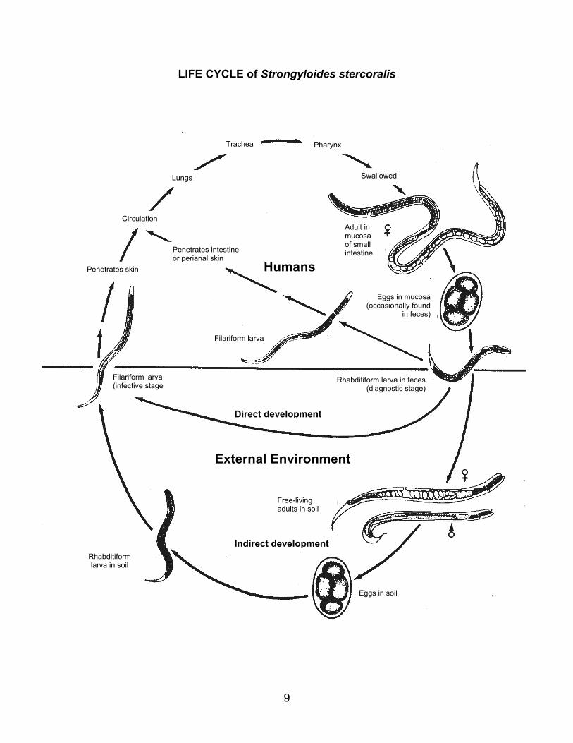
LIFE CYCLE of Strongyloides stercoralis
Rhabditiform larva in feces(diagnostic stage)
Eggs in mucosa(occasionally found
in feces)
Filariform larva (infective stage
Adult in mucosa of small intestine
Free-living adults in soil
Filariform larva
Penetrates intestine or perianal skin
Rhabditiform larva in soil
Swallowed
PharynxTrachea
Lungs
Circulation
Penetrates skin
External Environment
Humans
Indirect development
Direct development
Eggs in soil
9

III. Cestodes
With one exception, Hymenolepis nana, the life cycles of the human cestodes involve two or more hosts: a definitive host in which the adult parasite lives and one or two intermediate hosts in which larval development occurs. Table 2 gives the usual developmental times in both intermediate and definitive hosts.
As with the diagnostic stages of nematodes, the proglottids and eggs of the cestodes have been drawn to scale, with relation to each other, but no attempt has been made to correlate the size of the adult worms. The eggs of the cestodes, however, are not proportional to those of the nematodes or trematodes.
The Taenia species utilize vertebrates as intermediate hosts, cattle for T. saginata and swine for T. solium. Larval development to the infective cysticercus requires about 2 months and man acquires the infection by ingestion of these cysticerci in improperly cooked beef or pork. Eggs are liberated from the gravid proglottids only when they are broken or ruptured and, in general, it is the proglottids which break off from the strobila that are found in or on the fecal specimen. The eggs of the two species, however, are identical and species differentiation is based on the number of uterine branches in the proglottid (or on the appearance of the scolex, if it is recovered).
In addition to being the definitive host for these two species, man may serve as the intermediate host for T. solium. Human infections with Cysticercus cellulosae, the larval stage of the latter, result from a transfer of eggs from anus to mouth or from the ingestion of eggs or proglottids in contaminated food or water. Occasionally, infections may occur when gravid proglottids are swept by reverse peristalsis into the stomach where they are digested and the eggs liberated.
Table 2 USUAL TIME FOR COMPLETION OF LIFE CYCLES
Cestode Definitive Host
Intermediate Host(s)
T. saginata
T. solium
D. latum
H. nana
H. diminuta
D. caninum
8-10 weeks
8-10 weeks
3 weeks
4 weeks
3 weeks
3 weeks
9-10 weeks
9-10 weeks
4-8 weeks
……………
2 weeks
3-4 weeks
10

Diphyllobothrium latum also reaches its final infective stage in a vertebrate host but differs from other cestodes in having two intermediate hosts in its life cycle: first, an invertebrate, copepod, (Cyclops and Diaptomus species), and second, certain species of fresh water fish. In North America, the fish usually involved are pike, burbot, and carp. Unlike Taenia species, the eggs of D. latum are liberated from the gravid proglottids through a special uterine pore and are found in feces. Proglottids are found less frequently. The eggs are unembryonated when passed, in contrast to the embryonated eggs of other human cestode species, and require several days in water to embryonate and hatch. D. latum is the only human cestode which has a free-living stage since the eggs hatch and the larval form (the coracidium) is free-swimming before being ingested by a copepod. This cestode is also unique in that the plerocercoid larva can transfer from one fish to another, if the first fish host is ingested by a larger one. Hymenolepis nana, the most common cestode parasite of man, has a direct cycle similar to that of Enterobius vermicularis. An intermediate host is not necessary, although the parasite can utilize an arthropod for larval development under certain conditions. Ordinarily, a direct anus-to-mouth transmission occurs and, in essence, man serves as both intermediate and definitive hosts. Larval development occurs in the villi of the upper part of the small intestine and in this way, the species has combined the intermediate and definitive hosts within a single animal. The host-specificity of the cestodes is more or less limited depending on species. The adult Taenia worms occur only in man, while D. latum adults have been reported from bears and dogs as well and H. nana from certain rodents. Man is an accidental host of two species of cestodes, H. diminuta from rodents and D. caninum from dogs and cats. The role of accidental host is indicated by the parenthesis around HUMAN on the charts. The arthropods commonly involved as hosts for these two species are as follows:
H. diminuta -Tribolium spp. -"meal beetles"
Tenebrio spp. -"meal beetles"
D. caninum -Ctenocephalides canis, C. felis,
and other species of fleas.
Trichodectus canis, dog louse
11

LIFE CYCLE of Taenia saginata
Oncosphere hatches penetrates intestinal wal
Cysticercus in muscle (infective stage)
Circulation
Ingested
Adult in small intestine
Scolex attaches to intestine
Human
Embryonated eggs or proglottids ingested l
in feces or environment
Gravid proglottid
(Diagnostic stages)
Cattle
12
Egg in feces

LIFE CYCLE of Taenia solium
O cosphere hatches p netrates intestinal wall
Cysticercus in lungs, brain, eye, connective tissue
irculation
Gravid proglottid in
Oncosphere hatchpenetrates intestin
Circulation
Cysticercus in muscle (infective stage)
Adult in small intestineScolex attaches to intestine
Human
Embryonated eggs or proglottids occasionally ingested
A to-infection
i feces
Egg
feces or environment
Embryonated eggs or proglottids ingested
es al wall
(Diagnostic stages)
Swine
13
n
u
C
ne

LIFE CYCLE of Diphyllobothrium latum
Cringfre
Human
Adult in small intestine
gested
Scolex attaches to intestine
In
(diagnostic stage)
Unembryonated eggin feces
Ingested by crustacean
Procercoid in body cavity of crustacean
ustacean ested by sh water fish
P cercoid in muscle of fish (infective stage)
Crustacean Fish
ro
leCoracidium hatches from egg
Embryonatedegg in water
14

LIFE CYCLE of Hymenolepis nana
C ticercoid emerges from villus
Adult in smallintestine
Scolex attaches to intestine
ys
gested
Oncosphere hatches Cysticercoid develops in villus
Human
Gravid proglottidsdisintegrate
In
Embryonat(di
Embryonated egg (infective stage)
External Environment
15
ed egg in fecesagnostic stage)

LIFE CYCLE of Hymenolepis diminuta
Embryonat(di
(infective stage)
Cysticercoid in body cavity
Oncosphere hatches penetrates intestinal wall
Insects
Rat-Mouse(Human) Ingested
Gravid proglottids disintegrate
Adult in small intestine
Scolex attaches to intestine
16
ed egg in fecesagnostic stage)
Ingested

LIFE CYCLE of Dipylidium caninum
Egg packet
Dog and Cat (Human)
ngested
Adult in smallintestine
Scolex attaches to intestine
I
Oncosphere hatches penetrates intestinal wall
Embryonated eggingested
n feces or on perianal hairs
(diagnostic stage)
Gravidproglottid
Insects
Cysticercoid in body cavity (infective stage)
17
i

IV. Trematodes The trematode life histories, in general, are more complex than those of the nematodes or cestodes and are somewhat more restricted in geographical distribution than the other helminths because of their rigid host specificities with regard to larval development. As previously stated for nematodes and cestodes, the eggs of the various trematodes have been drawn to scale in relation to each other insofar as possible. Because of its very small size as compared to the other species, the egg of Clonorchis sinensis has been made larger than the proportionate scale drawing would have been. The trematode eggs are not in proportion to those of the cestodes or nematodes. No attempt has been made to correlate the sizes of the adults due to the great variations. The life histories of the trematodes are similar in that the first intermediate host is a species of snail and development to the cercarial stage occurs in this first host. Some of the most common snail hosts for each trematode are listed in Table 3. The developmental periods in the snail vary greatly with the trematode species ranging from approximately 3 to 13 weeks under optimum conditions. Unlike the nematodes and cestodes included here, an increase in progeny takes place in the intermediate host so that the number of cercariae released from the snail far exceeds the number of miracidia entering. The manner of entrance into the snail host varies with the species. Certain ones (Paragonimus, Fasciola, and Fasciolopsis) are passed unembryonated and like D. latum (cestode), must undergo embryonation in water prior to infection of the snail. Some species (schistosomes and Clonorchis) are embryonated when passed. The miracidium gains entrance into the snail either by being ingested within the egg (Clonorchis) or by penetration (after hatching) into the snail tissue (schistosomes, Paragonimus, Fasciola, Fasciolopsis). Within the snail, the trematodes other than schistosomes have a larval stage called a redia in addition to the usual sporocyst. The rediae differ from sporocysts in having an oral sucker, a pharynx, and a primitive gut. One or more generations of either sporocysts or rediae may occur depending on species. Only in the schistosomes, do the escaped cercariae penetrate directly into the definitive host. Other species enter or adhere to a second intermediate host in or on which the cercariae lose the tail, spines, and lytic glands and encyst (metacercariae). The definitive host becomes infected by ingesting these encysted forms. The free-swimming cercariae, such as those of the schistosomes, usually live only 1 to 3 or 4 days, but the encysted metacercariae are more resistant and survive for much longer periods, several weeks or months or even longer.
18

The patterns of the three species of schistosomes are similar and have been represented by a simple chart. The specific locations of the adults in man have been indicated, as well as the usual body material (urine or feces) in which the specific eggs are normally found. By means of broken line arrows, the chart indicates that S. haematobium eggs may occasionally be found in feces although usually passed in urine, and that S. japonicum eggs may sometimes be found in urine as well as feces. The schistosomes may occur naturally in mammals other than man: S. mansoni, in rodents and primates; S. japonicum in domestic animals such as dogs, cats, ruminants, hogs, equines, as well as rodents, S. haematobium in monkeys and rodents, but only rarely. Paragonimus occurs in cats, dogs, hogs, fur-bearing carnivores and rodents as well as man. Clonorchis sinensis is a parasite of man and other fish-eating mammals such as dogs and cats. In addition to man, F. buski occurs in hogs and occasionally, dogs. Fasciola hepatica is primarily a parasite of sheep and cattle and other herbivores. Man is only an occasional host of this species and is indicated as such by the parenthesis around HUMAN on the chart. The following table gives some of the common snail hosts and the developmental times required to complete the life cycle in both intermediate host and man.
Table 3
USUAL TIME: FOR COMPLETION OF LIFE CYCLE UNDER FAVORABLE CONDITIONS
Developmental Time Trematodes Definitive Host (man)
Intermediate Host (snail)*
S. mansoni
S. japonicum
S. haematobium
P. westermani
C. sinensis
F. hepatica
F. buski
5-7 weeks
4-5 weeks
4-8 weeks
5-6 weeks
4 weeks
12 weeks
4 weeks
4-5 weeks (Biomphalaria, Australorbis)
4-5 weeks (Oncomelania)
4-5 weeks (Bulinus, Physopsis)
13 weeks (Pomatiopsis, Hua, Thiara)
3-4 weeks (Alocinma, Bulinus, Hua)
5-8 weeks (Lymnoea)
4-7 weeks (Segmentina, Hippeutis)
*The genera of snails listed in parenthesis represents only a few of the common hosts for the trematodes and is by no means a complete listing.
19

LIFE CYCLE of Schistosomes
P n
Sporocyst in snail(2 generations)
Carcaria free – swimming (infective stage)
P netrates skin
Cercaria (tail lost during penetrat
Circulation
M ure intrahepatic po al blood
at
etrat
S
H
ion)
inrt
Adults in blood vessels S. mansoni: lower intestine S. japonicum: intestine S. haematobium: bladder
e
es into snail tissue
Miracidium hatches
S.m.
nails
in rine feces
uman
20
in
S.j.
u
S h.
.e

LIFE CYCLE of Paragonimus westermani
Unembryonated egg in sputum
Redia in snail tissue Miracidium hatches penetrates snail
Sporocyst in snail tissue
Egg embryonates in waterCercaria in crustacean
(in feces if swallowed)
Metacercaria in crustacean (infective stage)
(d iagnostic stage)
Ingested
Adult in cysticcavities in lungs(and other sites)
Pleural cavity
Penetrates diaphragm Abdominal cavity
Penetrates intestinal wall
Excysts in stomach
Snails Crustacea
Human
21

LIFE CYCLE of Clonorchis sinensis
Redia in snail tissue
Cercaria free swimming-
P etrates under scales of fresh water fish
Metacercaria in fresh water fish(infective stage)
gested
Excysts in duodenum
igrates to ile ducts
Mb
Human
Adult in bile duct
In
Ingested by snail
(diagnostic stage)
Embryonated egg in feces
en
Sporocyst in snail tissue
Miracidium hatches
Snails Fish
22

LIFE CYCLE of Fasciola hepatica
Spo
Redia in snail tissue
Cercaria free-swimming
W
Metacercaria on water plant (infective stage)
In ested
H
Excysts in duodenum
Pe trates intestinal wall
dominal cavity
Ab
enetrates liver
nerocyst in snail tissue
Egg embr
Snails ater Plants
Un
erbivores(Human)
Ad
23
P
ult in bile duct
g
Miracidium hatches penetrates snail
yonates in water
(diagnostic stage)
embryonated egg in feces

LIFE CYCLE of Fasciolopsis buski
gested
Human
Adult in small intestineExcysts in duodenum
Attaches to mucosa of
small intestine
In
(diagnostic stage)
Unembryonated egg in feces
Redia in snail tissueMiracidium hatches penetrates snail
Egg embryonates in water
Sporocyst in snail tissue
Cercaria free-swimming
Metacercaria on water plant (infective stage)
Snails Water Plants
24


![Prevalence of intestinal parasitic infections and ... · intestinal parasitic infections caused by helminths and intestinal protozoa [1, 11–15]. In Burkina Faso, where polyparasitism](https://static.fdocuments.net/doc/165x107/5ecdb4a171fb394e4f7767a3/prevalence-of-intestinal-parasitic-infections-and-intestinal-parasitic-infections.jpg)

![Effects of Hygiene and Defecation Behavior on Helminths ... · [23]. To our knowledge, the effect of CLTS on re-infection patterns with helminths and intestinal protozoa infections](https://static.fdocuments.net/doc/165x107/5ecdb4b071fb394e4f7767d0/effects-of-hygiene-and-defecation-behavior-on-helminths-23-to-our-knowledge.jpg)


