
The Eardrum Made Simple Dr. Ramesh Mehay Programme Director, Bradford VTS.

Common Eye Emergencies
Namir Kafil‐HussainC l O h h l i SConsultant Ophthalmic Surgeon

First Stop in The Journey of Ophthalmic patients First Stop in The Journey of Ophthalmic patients ‐‐ A GP Survey; A GP Survey; 150 practices 150 practices ShieffieldShieffield area 2008area 2008
•• Task Task •• Yes response %Yes response %
•• Visual acuity testing Visual acuity testing
•• Use of pinhole Use of pinhole •• 8585
•• 1010•• Confident with your Confident with your findings findings
•• 1010
•• 6161•• Able to tell if disc is normal Able to tell if disc is normal
or not by or not by ophthamoscopyophthamoscopy
Abl t t ll if l iAbl t t ll if l i
•• 7676
•• 3030•• Able to tell if macula is Able to tell if macula is normal or not by normal or not by ophthamoscopyophthamoscopy
•• 3030
•• 1313ophthamoscopyophthamoscopy
•• Do you dilate the pupils? Do you dilate the pupils?

First Stop in The Journey of Ophthalmic patients First Stop in The Journey of Ophthalmic patients ‐‐ A GP Survey; A GP Survey; 150 practices 150 practices ShieffieldShieffield area 2008area 2008
•• Eye condition treatedEye condition treated
•• Acute red eye with Acute red eye with di hdi h
•• Yes response %Yes response %
•• 9898dischargedischarge
•• BlepharitisBlepharitis
DD
•• 9595
•• 9494•• Dry eyeDry eye
•• Itchy eyeItchy eye
•• 9494
•• 9797•• Recurrent Recurrent iritisiritis
•• Painful red eyePainful red eye•• 4343
•• 4040•• Minor surgical proceduresMinor surgical procedures •• 5656

The Red EyeThe Red Eyeyy
•• About 80 % of the patients referred to our casualty present with a red About 80 % of the patients referred to our casualty present with a red eye A careful clinicaleye A careful clinical assessment usually produced a correct diagnosisassessment usually produced a correct diagnosiseye. A careful clinicaleye. A careful clinical assessment usually produced a correct diagnosis.assessment usually produced a correct diagnosis.
•• HistoryHistory•• Use of contact lens Use of contact lens (consider corneal ulcer in contact(consider corneal ulcer in contact lens users with lens users with
painful eye)painful eye)painful eye)painful eye)•• Sticky dischargeSticky discharge (suggest(suggest infective conjunctivitis)infective conjunctivitis)•• Past history of Past history of iritisiritis (consider recurrence)(consider recurrence)
P f it hiP f it hi ( ll i j ti iti )( ll i j ti iti )•• Presence of itching Presence of itching (allergic conjunctivitis)(allergic conjunctivitis)•• ExaminationExamination
•• Assessment of both eyes with Assessment of both eyes with SnellenSnellen chart chart (reduced vision needs (reduced vision needs t f l )t f l )urgent referrals)urgent referrals)
•• Examine the anterior segment Examine the anterior segment with a bright torch and note: with a bright torch and note: •• injection of the conjunctiva injection of the conjunctiva (conjunctivitis)(conjunctivitis)•• cornea for opacitycornea for opacity (ulcer or acute glaucoma)(ulcer or acute glaucoma)•• pupil reaction to lightpupil reaction to light (fixed pupil is seen in(fixed pupil is seen in acute glaucoma and acute glaucoma and
iritisiritis))••••

The Red EyeThe Red Eyeyy
•• Patients with pain +/Patients with pain +/‐‐ blurring of vision is likely to blurring of vision is likely to have a sighthave a sight‐‐threatening conditions.threatening conditions. The most The most importantimportant differential diagnosis are:differential diagnosis are:
•• Acute glaucomaAcute glaucoma•• Acute glaucomaAcute glaucoma•• Corneal infectionCorneal infection•• IritisIritisIritisIritis•• Patient without pain are likely to have a selfPatient without pain are likely to have a self‐‐limiting limiting conditions, the most common are:conditions, the most common are:
•• ConjunctivitisConjunctivitis•• EpiscleritisEpiscleritis
b l h hb l h h•• Subconjunctival haemorrhageSubconjunctival haemorrhage
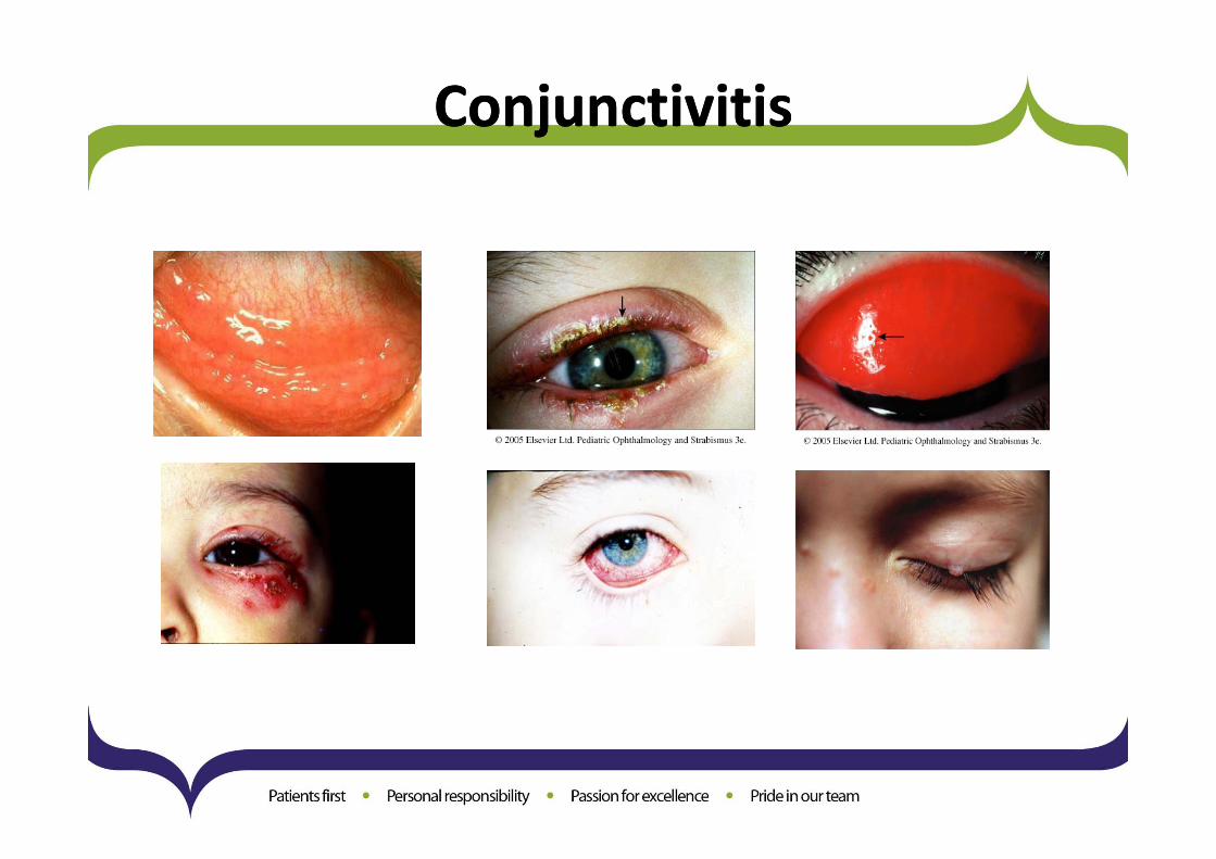
ConjunctivitisConjunctivitisjj

ConjunctivitisConjunctivitisjj
•• Inflammation of the conjunctiva is usually caused by either Inflammation of the conjunctiva is usually caused by either infection or allergyinfection or allergy. The eye is . The eye is dd d f bl b i id f bl b i iredred and uncomfortable but pain is not common.and uncomfortable but pain is not common.
•• Presentation: Presentation: –– Infective conjunctivitis Infective conjunctivitis usually present with discharging or sticky eyes. usually present with discharging or sticky eyes.
There may be aThere may be a history of contacthistory of contact with people with red eyeswith people with red eyes–– There may be a There may be a history of contact history of contact with people with red eyes. with people with red eyes.
–– Allergic conjunctivitis Allergic conjunctivitis is commonly seen in patients with is commonly seen in patients with atopyatopy or hay fever. Itchy redor hay fever. Itchy redeye is a prominent featureeye is a prominent feature
•• Examination: Examination: –– The visual acuity is usually normalThe visual acuity is usually normal
–– One or both eyesOne or both eyes may be affected and the eyelids may be swollen may be affected and the eyelids may be swollen
–– The The conjunctiva is oedematous conjunctiva is oedematous and there are visible changes on the tarsal conjunctiva and there are visible changes on the tarsal conjunctiva
•• Treatment: Treatment: –– In the general practice, it is difficult to differentiate between bacterial from viral conjunctivitis. However, it is In the general practice, it is difficult to differentiate between bacterial from viral conjunctivitis. However, it is
acceptable to treat all infective conjunctivitis with acceptable to treat all infective conjunctivitis with topical antibiotics such astopical antibiotics such as chloramphenicolchloramphenicol as it can prevent as it can prevent secondary infection in viral conjunctivitis. The conjunctivitissecondary infection in viral conjunctivitis. The conjunctivitis usually takes about one or two weeks to settle. usually takes about one or two weeks to settle.
–– Patient with Patient with allergic conjunctivitis allergic conjunctivitis will benefit from topical sodium will benefit from topical sodium cromoglycatecromoglycate such as such as opticromopticrom..It is important to determine the cause as the allergen (for example eye drops or cosmetic) may be eliminated. It is important to determine the cause as the allergen (for example eye drops or cosmetic) may be eliminated.
•• Refer the patient to the casualty only if the Refer the patient to the casualty only if the conjunctivitis fails to respond to treatment conjunctivitis fails to respond to treatment

IritisIritis•• Seen mainly in young people. Occasionally associated with systemic conditions such as Seen mainly in young people. Occasionally associated with systemic conditions such as
ankylosingankylosing spondylitisspondylitis andand sarcoidosissarcoidosisankylosingankylosing spondylitisspondylitis and and sarcoidosissarcoidosis. .
•• Presentation: Presentation:
–– Painful red eye Painful red eye
Photophobia with reduced visionPhotophobia with reduced vision–– Photophobia with reduced vision Photophobia with reduced vision
–– May have been treated for resistant conjunctivitis May have been treated for resistant conjunctivitis
•• Examination:Examination:
Visual acuity is reducedVisual acuity is reduced to varying degreeto varying degree–– Visual acuity is reduced Visual acuity is reduced to varying degree to varying degree
–– Redness mainly around the cornea (Redness mainly around the cornea (ciliaryciliary injectioninjection) )
–– PupilPupil is usually constricted or irregular reacting poorly to light. is usually constricted or irregular reacting poorly to light.
I l f hit ll (I l f hit ll (k titik titi i it ti it t b b hi d thb b hi d th–– In severe cases, clumps of white cells (In severe cases, clumps of white cells (keratitickeratitic precipitates precipitates may be seen behind the may be seen behind the cornea) cornea)
•• Management: Refer the patient Management: Refer the patient within 24 hourswithin 24 hours. .
SlitSlit lamplamp examination by ophthalmologists to confirm the diagnosisexamination by ophthalmologists to confirm the diagnosis–– SlitSlit‐‐lamplamp examination by ophthalmologists to confirm the diagnosis. examination by ophthalmologists to confirm the diagnosis.
–– Treatment is with Treatment is with intensive topical steroid intensive topical steroid to reduce inflammation and to reduce inflammation and mydriaticmydriatic to to dilate the pupil so that the iris does not stick to the cornea causing problem with dilate the pupil so that the iris does not stick to the cornea causing problem with glaucoma.glaucoma.glaucoma. glaucoma.
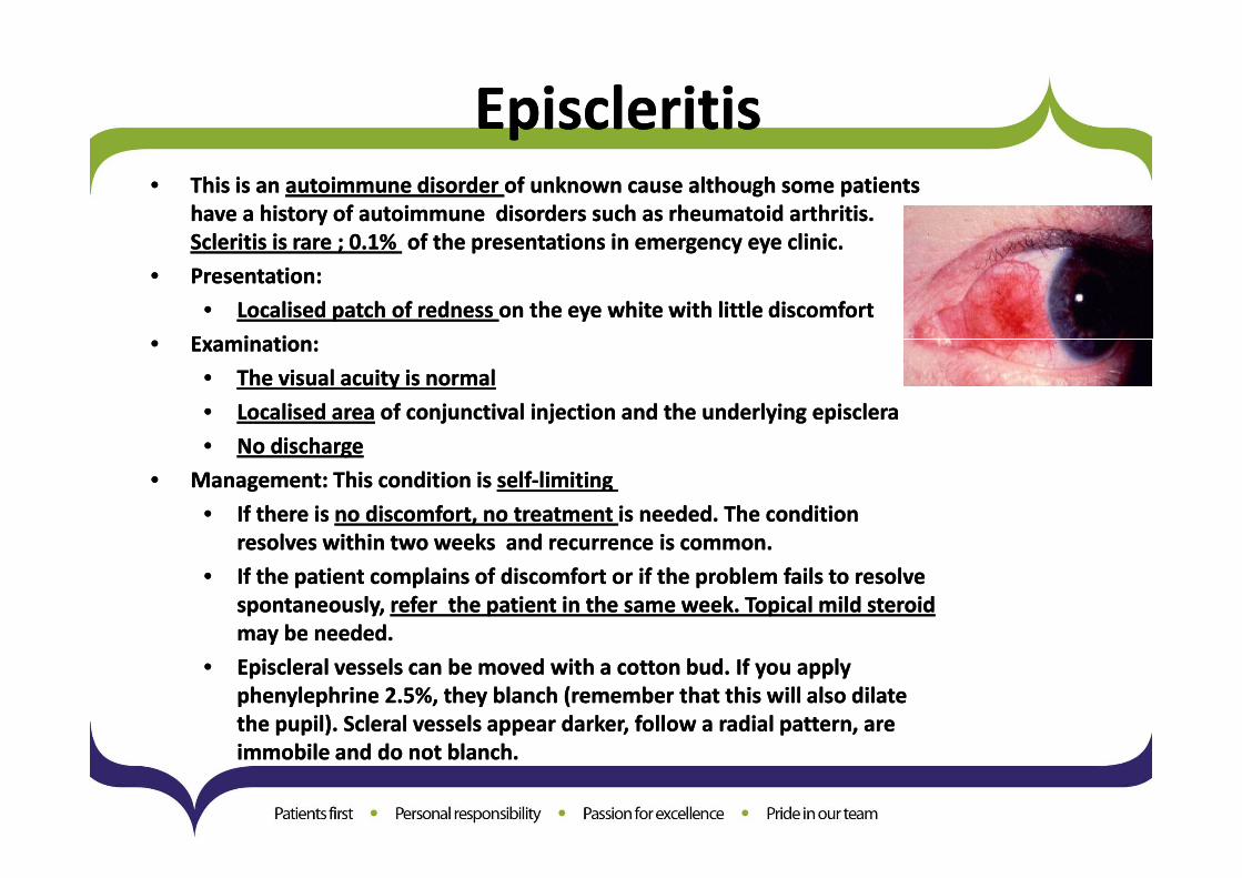
EpiscleritisEpiscleritispp•• This is an This is an autoimmune disorder autoimmune disorder of unknown cause although some patients of unknown cause although some patients
have a history of autoimmune disorders such as rheumatoid arthritis. have a history of autoimmune disorders such as rheumatoid arthritis. ScleritisScleritis is rare ; 0 1%is rare ; 0 1% of the presentations in emergency eye clinicof the presentations in emergency eye clinicScleritisScleritis is rare ; 0.1% is rare ; 0.1% of the presentations in emergency eye clinic.of the presentations in emergency eye clinic.
•• Presentation: Presentation:
•• Localised patch of redness Localised patch of redness on the eye white with little discomfort on the eye white with little discomfort
E i tiE i ti•• Examination: Examination:
•• The visual acuity is normal The visual acuity is normal
•• Localised areaLocalised area of of conjunctivalconjunctival injection and the underlying injection and the underlying episcleraepisclera
N di hN di h•• No dischargeNo discharge
•• Management: This condition is Management: This condition is selfself‐‐limitinglimiting
•• If there is If there is no discomfort, no treatment no discomfort, no treatment is needed. The condition is needed. The condition resolves within two weeksresolves within two weeks and recurrence is commonand recurrence is commonresolves within two weeksresolves within two weeks and recurrence is common. and recurrence is common.
•• If the patient complains of discomfort or if the problem fails to resolve If the patient complains of discomfort or if the problem fails to resolve spontaneously, spontaneously, referrefer the patient in the same week. Topical mild steroid the patient in the same week. Topical mild steroid may be needed.may be needed.may be needed.may be needed.
•• EpiscleralEpiscleral vessels can be moved with a cotton bud. If you apply vessels can be moved with a cotton bud. If you apply phenylephrinephenylephrine 2.5%, they blanch (remember that this will also dilate 2.5%, they blanch (remember that this will also dilate the pupil). the pupil). ScleralScleral vessels appear darker, follow a radial pattern, are vessels appear darker, follow a radial pattern, are immobile and do not blanch.immobile and do not blanch.

Acute glaucomaAcute glaucomagg
•• Rare cause of painful red eye but early diagnosis important to prevent severe Rare cause of painful red eye but early diagnosis important to prevent severe visual loss. visual loss. About 1 in 1,000 people get AACG. It is more likely in people over the age of 40 years, and most often happens at around age 60 to 70 years.
•• Presentation: Presentation: •• SeverelySeverely painful red eye.painful red eye.
•• Haloes Haloes around light common.around light common.
•• Patients usually Patients usually over 50 over 50 years old.years old.
•• Nausea and vomiting Nausea and vomiting common common
•• Examination: Examination: •• Reduced visual acuityReduced visual acuity..Reduced visual acuityReduced visual acuity. .
•• Hazy cornea Hazy cornea and the iris is not clearly visible.and the iris is not clearly visible.
•• PupilPupil is fixed or semiis fixed or semi‐‐dilated, dilated, unreactiveunreactive to light to light
•• Management:Management:•• Management: Management: •• Urgent referrals Urgent referrals i.e. as soon as possible and not the next day. i.e. as soon as possible and not the next day.
•• Patient is usually admitted and given Patient is usually admitted and given acetazolamideacetazolamide IV to lower pressure.IV to lower pressure.TopicalTopical pilocarpinepilocarpine and steroid (to reduce inflammation) are also givenand steroid (to reduce inflammation) are also givenTopical Topical pilocarpinepilocarpine and steroid (to reduce inflammation) are also given.and steroid (to reduce inflammation) are also given.

Corneal infectionsCorneal infections•• This is a potentially sight threatening condition. Avoid using steroid if corneal infection can This is a potentially sight threatening condition. Avoid using steroid if corneal infection can
not be excluded as steroid can worsen the infection. not be excluded as steroid can worsen the infection.
•• Presentation: Presentation:
–– Painful red eye Painful red eye
–– Photophobia Photophobia
–– There may be a history of There may be a history of
–– contact lens use or previous herpes keratitiscontact lens use or previous herpes keratitis. .
•• Examination: Examination:
–– The visual acuity is reduced The visual acuity is reduced
–– FluoresceinFluorescein dye dye reveals corneal defect reveals corneal defect
–– In severe bacterial infection, there may be In severe bacterial infection, there may be hypopyonhypopyon
–– (pus in the anterior chamber) (pus in the anterior chamber)
•• Management: Refers within 24 hoursManagement: Refers within 24 hours
–– In In herpes keratitisherpes keratitis, topical acyclovir 3% 5X a day for one week . About 1, topical acyclovir 3% 5X a day for one week . About 1‐‐2 people in 2 people in pp , p y y, p y y p pp p1000 will develop at least one episode of active herpes simplex eye infection at some 1000 will develop at least one episode of active herpes simplex eye infection at some stage in their life. stage in their life.
–– In In bacterial corneal ulcerbacterial corneal ulcer, the patient may be admitted for intensive antibiotic , the patient may be admitted for intensive antibiotic treatmenttreatment if severe or treated as an outif severe or treated as an out‐‐patient if mild.patient if mild.

Subconjunctival haemorrhageSubconjunctival haemorrhagej gj g
•• Presentation: Presentation:
•• May be related to trauma May be related to trauma but the majority occurs but the majority occurs spontaneously. spontaneously.
•• Some may be precipitated by severe prolonged Some may be precipitated by severe prolonged coughingcoughing. .
•• Redness may be Redness may be limited limited to one part of the eye or the whole eye. to one part of the eye or the whole eye. yy p y yp y y
•• Examination: Examination:
•• The redness looks like The redness looks like blood blood under the conjunctiva under the conjunctiva
•• The eye is The eye is quiet quiet yy qq
•• Normal visual acuityNormal visual acuity
•• Management: Management:
•• The condition looks alarming butThe condition looks alarming but resolves within two weeksresolves within two weeks..The condition looks alarming but The condition looks alarming but resolves within two weeksresolves within two weeks. .
•• ReassuranceReassurance isis all that is needed. all that is needed.
•• Check the Check the blood pressure blood pressure in elderly patientin elderly patient
•• Refer the patient only if theRefer the patient only if the subconjunctivalsubconjunctival haemorrhage ishaemorrhage is traumatictraumaticRefer the patient only if the Refer the patient only if the subconjunctivalsubconjunctival haemorrhage is haemorrhage is traumatictraumatic
•• Figure 1. This patient presented with a painless red eye caused by Figure 1. This patient presented with a painless red eye caused by subconjunctivalsubconjunctivalhaemorrhage.haemorrhage. Note that the eye is quiet and the redness of the conjunctiva is uniform.Note that the eye is quiet and the redness of the conjunctiva is uniform.

Sudden Painless Visual LossSudden Painless Visual Loss
•• History: History:
•• Transient visual loss (Transient visual loss ( like alike a curtain coming down, this is suggestive of curtain coming down, this is suggestive of
•• amourosisamourosis fugaxfugax
•• Visual loss or field loss preceded by Visual loss or field loss preceded by sudden onset floaters and flashingsudden onset floaters and flashinglight (light (photopsiaphotopsia), this is suggestive of ), this is suggestive of
•• retinal detachment retinal detachment
•• History of History of poorly controlled diabetespoorly controlled diabetes and laser treatment to theand laser treatment to the retina, this is suggestive of retina, this is suggestive of
•• vitreous haemorrhage vitreous haemorrhage
•• Headache +/Headache +/‐‐ jaw jaw claudicationclaudication (pain(pain in the jaw on eating) in the elderlyin the jaw on eating) in the elderly , suggestive of, suggestive of
•• Temporal Temporal arteritisarteritis
•• Pain on eye movement Pain on eye movement in young patients , suggestive ofin young patients , suggestive of
•• optic neuritisoptic neuritis

Sudden Painless Visual LossSudden Painless Visual Loss
T i t L f i i ( i t )T i t L f i i ( i t )•• Transient Loss of vision (minutes)Transient Loss of vision (minutes)•• UnilateralUnilateral
•• AmaurosisAmaurosis fugaxfugax•• Ischemic Optic NeuropathyIschemic Optic Neuropathy
•• BilateralBilateral•• VertebrobasilarVertebrobasilar insufficiencyinsufficiencyVertebrobasilarVertebrobasilar insufficiencyinsufficiency
•• Unilateral or BilateralUnilateral or Bilateral•• MigraineMigraine
•• Sudden Painless Loss of VisionSudden Painless Loss of Vision•• Sudden Painless Loss of VisionSudden Painless Loss of Vision•• Retinal artery or vein occlusionRetinal artery or vein occlusion•• Vitreous HaemorrhageVitreous Haemorrhagegg•• Retinal detachmentRetinal detachment•• Optic neuritis +/Optic neuritis +/‐‐ painpain

Central or branch retinal artery occlusion Central or branch retinal artery occlusion •• Occlusion of the retinal artery may be caused by arteriosclerotic changes, embolusOcclusion of the retinal artery may be caused by arteriosclerotic changes, embolus (from heart or (from heart or
carotid artery) or inflammation (rare) . It has an estimated incidence of 0.85/100,000/year.carotid artery) or inflammation (rare) . It has an estimated incidence of 0.85/100,000/year.
•• History: History:
•• Sudden painless visual lossSudden painless visual loss which may be complete (due to central retinal arterywhich may be complete (due to central retinal artery occlusion) or occlusion) or partial (due to branch retinal artery occlusion) partial (due to branch retinal artery occlusion)
•• Patient usually have a history of Patient usually have a history of hypertension or heart disease hypertension or heart disease
E i tiE i ti•• Examination: Examination:
•• The visual acuity The visual acuity is reduced in CRAO but may be normal in BRCO.is reduced in CRAO but may be normal in BRCO.
•• Relative afferent pupillary defect Relative afferent pupillary defect is present in central retinal artery occlusion is present in central retinal artery occlusion
•• The retinal arteriesThe retinal arteries are narrow or collapsed.are narrow or collapsed.The retinal arteries The retinal arteries are narrow or collapsed.are narrow or collapsed.
•• In CRAO, the fovea shows a In CRAO, the fovea shows a cherrycherry‐‐red spot red spot against theagainst the white white infarctedinfarcted retina.retina.
•• In BRAO In BRAO the white the white infarctedinfarcted retina corresponds to theretina corresponds to the occluded retina.occluded retina.
•• Emboli Emboli may be seen in the arteries if the cause is emboli may be seen in the arteries if the cause is emboli
•• Management: Management:
•• Immediate referral Immediate referral if the visual loss is less than 6 hours if the visual loss is less than 6 hours
•• Treatment involves the use of Treatment involves the use of intravenous intravenous acetazolamideacetazolamide and globe massage and globe massage toto lower the lower the intraocular pressure and hopefully reintraocular pressure and hopefully re‐‐establish the arterial flowestablish the arterial flowintraocular pressure and hopefully reintraocular pressure and hopefully re‐‐establish the arterial flow. establish the arterial flow.
•• Further management aim to uncover any Further management aim to uncover any underlying diseases underlying diseases such as hypertension, cardiac or such as hypertension, cardiac or carotid thrombus. An ESR is usually performed in the absence of obviouscarotid thrombus. An ESR is usually performed in the absence of obvious embolus to exclude embolus to exclude arteriticarteritic causes. causes.
l dl d d d d h k fd d d h k f•• Long term low dose aspirin Long term low dose aspirin is advised to reduce the risk of occurrence. is advised to reduce the risk of occurrence.

Central or branch retinal vein occlusion Central or branch retinal vein occlusion
•• Retinal vein occlusion is a common vascular disorder caused by impaired venous blood Retinal vein occlusion is a common vascular disorder caused by impaired venous blood flfl I i d l di b lli l f i i d i i 2I i d l di b lli l f i i d i i 2flow.flow. It is second only to diabetes mellitus as a vascular cause of impaired vision. 2 per It is second only to diabetes mellitus as a vascular cause of impaired vision. 2 per 1,000 in those >40 years and 5.4 cases per 1,000 aged >64 years. There is an equal sex 1,000 in those >40 years and 5.4 cases per 1,000 aged >64 years. There is an equal sex distribution.distribution.
•• Presentation:Presentation:Presentation: Presentation:
•• Sudden painless blurred vision Sudden painless blurred vision
•• ExaminationExamination•• Examination Examination
•• The The visual acuity is reduced +/visual acuity is reduced +/‐‐
•• Relative afferent Relative afferent pupillarypupillary defect +/defect +/‐‐
•• OphthalmoscopyOphthalmoscopy reveals extensivereveals extensive retinal haemorrhageretinal haemorrhage•• OphthalmoscopyOphthalmoscopy reveals extensive reveals extensive retinal haemorrhageretinal haemorrhage
•• Management: Refer within one week.Management: Refer within one week.
•• Although there is no immediate treatment that can restore the vision, it is important Although there is no immediate treatment that can restore the vision, it is important toto examine the patient forexamine the patient for hypertension and glaucomahypertension and glaucoma A blood test is usually performedA blood test is usually performedtoto examine the patient for examine the patient for hypertension and glaucomahypertension and glaucoma. A blood test is usually performed . A blood test is usually performed forfor full blood count, ESR and in young patients autofull blood count, ESR and in young patients auto‐‐immune screening. immune screening.
•• FollowFollow‐‐up up in the clinic is arranged so that those at risk of neovascular glaucoma may in the clinic is arranged so that those at risk of neovascular glaucoma may bebe treated with laser pantreated with laser pan‐‐photocoagulationphotocoagulationpp p gp g

Retinal detachment Retinal detachment •• Incidence is about 1 in 10,000 with a prevalence of about 0.3% of the general population Incidence is about 1 in 10,000 with a prevalence of about 0.3% of the general population
and a lifetime risk of 3% by the age of 85and a lifetime risk of 3% by the age of 85and a lifetime risk of 3% by the age of 85.and a lifetime risk of 3% by the age of 85.
•• History: History:
A recent history ofA recent history of floaters and flashesfloaters and flashes–– A recent history of A recent history of floaters and flashesfloaters and flashes
–– Curtain Curtain coming across the visioncoming across the vision
•• Examination: Examination:
Visual acuity variableVisual acuity variable depending if the macula is involveddepending if the macula is involved–– Visual acuity variable Visual acuity variable depending if the macula is involved. depending if the macula is involved.
–– Visual field defectVisual field defect
–– Ophthalmic examination in a dilated pupil shows Ophthalmic examination in a dilated pupil shows greyish retina, hole and teargreyish retina, hole and tear may be may be seenseenseen.seen.
•• Management: Management:
•• Refer the patient the same dayRefer the patient the same day
•• Patients will require surgical management which consists of sealing the retinalPatients will require surgical management which consists of sealing the retinal•• Patients will require surgical management which consists of sealing the retinal Patients will require surgical management which consists of sealing the retinal breaksbreaks (using (using cryotherapycryotherapy or laser) and relieving the vitreous traction (vitrectomy).or laser) and relieving the vitreous traction (vitrectomy).

IschaemicIschaemic optic neuropathyoptic neuropathyp p yp p y
•• In In ischaemicischaemic optic neuropathy, there is optic neuropathy, there is occlusion of the small arteries around the optic discocclusion of the small arteries around the optic disc.. It is important It is important to differentiate to differentiate arteriticarteritic optic neuropathy from optic neuropathy from nonnon‐‐arteriticarteritic optic neuropathy.optic neuropathy. ArteriticArteritic optic neuropathy is optic neuropathy is caused by giant cell caused by giant cell arteritisarteritis and prompt treatment with systemicand prompt treatment with systemic steroid can prevent involvement of the steroid can prevent involvement of the contralateralcontralateral eye.eye. The incidence of giant cell The incidence of giant cell arteritisarteritis increases progressively after 50 years of age and is in increases progressively after 50 years of age and is in the region of 20 per 100,000 people older than 50 years.the region of 20 per 100,000 people older than 50 years.
•• Presentation: Presentation:
•• Sudden visual loss Sudden visual loss persistent headache or persistent headache or jaw jaw claudicationclaudication suggest GCA.suggest GCA.
•• Examination: Examination:
ThTh i l l i ll f di l l i ll f d 6/606/60•• The The visual loss is usually profound visual loss is usually profound 6/60 or6/60 or
•• Afferent pupillary defect Afferent pupillary defect
•• FundalFundal examination reveals examination reveals swollen optic disc swollen optic disc caused by occlusion of the arteries aroundcaused by occlusion of the arteries around the optic disc the optic disc
•• In giant cell In giant cell arteritisarteritis there is there is tendernesstenderness over the affected artery (usually the temporal artery)over the affected artery (usually the temporal artery)In giant cellIn giant cell arteritisarteritis there isthere is tendernesstenderness over the affected artery (usually the temporal artery)over the affected artery (usually the temporal artery)and the artery is usually not palpable.and the artery is usually not palpable.
•• Management: Management:
•• Refer immediatelyRefer immediately any patient with sudden visual loss and swollen disc for exclusion ofany patient with sudden visual loss and swollen disc for exclusion of giant cell giant cell arteritisarteritis. .
•• ESR and the C reactive ESR and the C reactive protein are usually raised in giant cell protein are usually raised in giant cell arteritisarteritis but nonbut non‐‐specific.specific. A definite diagnosis is A definite diagnosis is by by temporal artery biopsy temporal artery biopsy for the typical for the typical granulomatousgranulomatous changeschanges in the arterial wall. However, systemic in the arterial wall. However, systemic steroid is usually given while this is arranged. steroid is usually given while this is arranged.

Optic neuritis Optic neuritis pp•• This condition typically affects patients in the This condition typically affects patients in the 20 20 ‐‐ 45 45 age group. Occurs in 1 to 5 age group. Occurs in 1 to 5
individuals/100 000/yearindividuals/100 000/yearindividuals/100 000/year.individuals/100 000/year.
•• Presentation: Presentation: –– Impaired visionImpaired vision
C t l fi ld d f tC t l fi ld d f t–– Central field defect Central field defect
•• Examination: Examination: –– Visual acuityVisual acuity may be as poor as perception of light may be as poor as perception of light
C t lC t l tt i t i li t i l–– Central Central scotomascotoma is typical is typical
–– Impaired colourImpaired colour discrimination (best demonstrated with red object, the affecteddiscrimination (best demonstrated with red object, the affected eye will see the eye will see the red object less bright than the unaffected eye) red object less bright than the unaffected eye)
–– Relative afferent pupillary defect Relative afferent pupillary defect of the affected eye of the affected eye Pain on eye movement Pain on eye movement especially on adduction. especially on adduction.
–– FundalFundal examination is normal examination is normal as most cases have retrobulbar neuritisas most cases have retrobulbar neuritis..
•• Management: Management: –– Refer the patient within 24 hours Refer the patient within 24 hours for confirmation of the diagnosis for confirmation of the diagnosis
–– Normal or nearNormal or near‐‐normal vision usually returns normal vision usually returns within 6 weeks within 6 weeks
–– As treatment does not affect the outcome, unilateral optic neuritis is not treated byAs treatment does not affect the outcome, unilateral optic neuritis is not treated by most most ophthalmologists. However, followophthalmologists. However, follow‐‐up is important asup is important as radiological investigationradiological investigation may be needed tomay be needed toophthalmologists. However, followophthalmologists. However, follow up is important as up is important as radiological investigationradiological investigation may be needed to may be needed to exclude compressive lesion in cases where spontaneous recoveryexclude compressive lesion in cases where spontaneous recovery fails to recur. fails to recur.

The swollen eyelidsThe swollen eyelidsyy
•• The The most common causes ofmost common causes of swollen eyelids swollen eyelids are:are:•• AllergyAllergy
•• ChalazionChalazion (Meibomian cyst)(Meibomian cyst)•• ChalazionChalazion (Meibomian cyst)(Meibomian cyst)
•• PrePre‐‐septalseptal or Orbital Cellulitisor Orbital Cellulitis
•• Herpes ZosterHerpes Zoster
•• Acute Acute dacryocystitisdacryocystitisy yy y
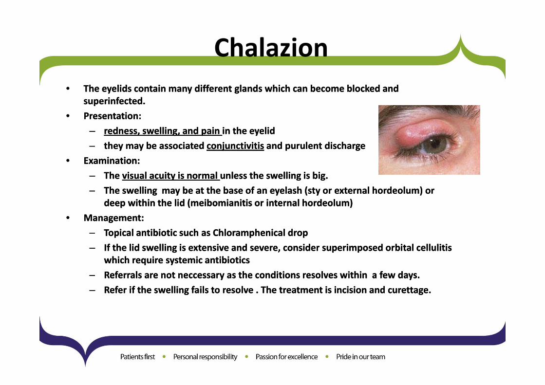
Chalazion•• The eyelids contain many different glands which can become blocked and The eyelids contain many different glands which can become blocked and
superinfectedsuperinfected..pp
•• Presentation:Presentation:
–– redness, swelling, and pain redness, swelling, and pain in the eyelidin the eyelid
–– they may be associatedthey may be associated conjunctivitisconjunctivitis and purulent dischargeand purulent dischargethey may be associated they may be associated conjunctivitisconjunctivitis and purulent discharge and purulent discharge
•• Examination: Examination:
–– The The visual acuity is normal visual acuity is normal unless the swelling is big. unless the swelling is big.
–– The swellingThe swelling may be at the base of an eyelash (sty or externalmay be at the base of an eyelash (sty or external hordeolumhordeolum) or) orThe swellingThe swelling may be at the base of an eyelash (sty or external may be at the base of an eyelash (sty or external hordeolumhordeolum) or ) or deep within the lid (deep within the lid (meibomianitismeibomianitis or internal or internal hordeolumhordeolum) )
•• Management: Management:
–– Topical antibiotic such as Topical antibiotic such as ChloramphenicalChloramphenical dropdroppp pp pp
–– If the lid swelling is extensive and severe, consider superimposed orbital cellulitis If the lid swelling is extensive and severe, consider superimposed orbital cellulitis which require systemic antibiotics which require systemic antibiotics
–– Referrals are not Referrals are not neccessaryneccessary as the conditions resolves withinas the conditions resolves within a few days. a few days. yy yy
–– Refer if the swelling fails to resolve . The treatment is incision and curettage. Refer if the swelling fails to resolve . The treatment is incision and curettage.

Orbital CellulitisOrbital Cellulitis
•• This is a potentially sightThis is a potentially sight‐‐threatening condition and the patient should be referred to the paediatricians or threatening condition and the patient should be referred to the paediatricians or ophthalmologists for further management. Sight loss may result from central retinal artery occlusion or ophthalmologists for further management. Sight loss may result from central retinal artery occlusion or optic nerve inflammation.optic nerve inflammation.In adults the most common infection are In adults the most common infection are Staph Staph aureusaureus, , StreptStrept pyogenespyogenes oror StreptStrept penumoniaepenumoniae. In children . In children , it is often secondary to infection in the adjacent sinuses and, it is often secondary to infection in the adjacent sinuses and HaemophiliusHaemophilius is an important pathogen. is an important pathogen.
•• Presentation: Presentation:
–– Severe pain Severe pain
–– Tense and red orbit with lid closure Tense and red orbit with lid closure
P iP i–– PyrexiaPyrexia
•• Examination Examination
–– Intense Intense swelling of the lids swelling of the lids
–– ProptosisProptosisProptosisProptosis
–– Congestion Congestion of the of the conjunctivalconjunctival and and episcleralepiscleral vessels vessels
–– ChemosisChemosis (swollen conjunctiva) (swollen conjunctiva)
–– Double visionDouble vision
•• Treatment: Treatment:
•• Refer to the ophthalmologist within 24 hoursRefer to the ophthalmologist within 24 hours. .
•• Treatment require systemic antibiotics and analgesia. Treatment require systemic antibiotics and analgesia.

Herpes zoster Herpes zoster ophthalmicusophthalmicus
•• This is caused by This is caused by reactivation of herpes zoster virusreactivation of herpes zoster virus in patient who previously had chickenpox. The eye is in patient who previously had chickenpox. The eye is affected in affected in 50% 50% of zoster of zoster ophthalmicusophthalmicus and is increased in patients with and is increased in patients with involvement of the involvement of the nasociliarynasociliarynerve (rash at the tip of the nose).nerve (rash at the tip of the nose).
•• Presentation: Presentation:
–– pain in the distribution of the ophthalmic nervepain in the distribution of the ophthalmic nerve followed in a few days with vesicular eruptionfollowed in a few days with vesicular eruptionpain in the distribution of the ophthalmic nervepain in the distribution of the ophthalmic nerve followed in a few days with vesicular eruptionfollowed in a few days with vesicular eruption
•• Examination Examination
–– Vesicular rash Vesicular rash affecting the scalps and lids affecting the scalps and lids
–– Vision may be reduced Vision may be reduced with ocular involvement (keratitis and anterior with ocular involvement (keratitis and anterior uveitisuveitis) )
–– Swollen lids Swollen lids may make eye examination difficult may make eye examination difficult
–– Ocular injections Ocular injections
–– Discharge from conjunctivitis Discharge from conjunctivitis
M tM t•• Management: Management:
–– Oral acyclovir Oral acyclovir is useful in speeding up the resolution of the rash is useful in speeding up the resolution of the rash
–– Analgesia should be given as the condition is very painful Analgesia should be given as the condition is very painful
–– Conjunctivitis is common and does not require treatment Conjunctivitis is common and does not require treatment j qj q
•• Refer to the ophthalmologists withinRefer to the ophthalmologists within 24 hours 24 hours from seeing for exclusion of ocular involvement such as from seeing for exclusion of ocular involvement such as iritisiritis and keratitis. and keratitis.

Acute Acute dacryocystitisdacryocystitisy yy y•• This is caused by inflammation of the This is caused by inflammation of the lacrimallacrimal sac. It is often associated with obstruction of sac. It is often associated with obstruction of
thethe nasolacrimalnasolacrimal duct with watering of the eye Infection are often due to streptococcusduct with watering of the eye Infection are often due to streptococcusthethe nasolacrimalnasolacrimal duct with watering of the eye. Infection are often due to streptococcus duct with watering of the eye. Infection are often due to streptococcus andand staphylococcus. staphylococcus.
•• Presentation: Presentation:
–– Painful swellingPainful swelling at the nasal side of the lower lid.at the nasal side of the lower lid.Painful swelling Painful swelling at the nasal side of the lower lid.at the nasal side of the lower lid.
•• Examination: Examination:
–– Visual acuity is normalVisual acuity is normal. .
–– The swellingThe swelling is tense andis tense and tendertenderThe swelling The swelling is tense andis tense and tendertender
–– In severe cases, the whole of the lower lid may be swollen due to superimposed In severe cases, the whole of the lower lid may be swollen due to superimposed cellulitis cellulitis
•• Management:Management:Management: Management:
–– Refer the patient to the ophthalmologists within 24 hoursRefer the patient to the ophthalmologists within 24 hours. .
–– High dose systemic antibiotic is required either orally or by intravenous. High dose systemic antibiotic is required either orally or by intravenous.
–– Incision of the swelling should be avoided as this can cause fistula formationIncision of the swelling should be avoided as this can cause fistula formationIncision of the swelling should be avoided as this can cause fistula formation Incision of the swelling should be avoided as this can cause fistula formation
–– Most patient will require Most patient will require dacryocystorhninostomydacryocystorhninostomy (an artificial passage is (an artificial passage is createdcreated between the between the lacrimallacrimal sac and the nasal cavity to bypass the blockage)sac and the nasal cavity to bypass the blockage) when when thethe acute episode settle.acute episode settle.

Chemical burnChemical burn
•• Exposure of the eye to any chemical can cause significant damage to the anterior segment. Exposure of the eye to any chemical can cause significant damage to the anterior segment. I d i l i i lk li id i ll d i h Alk liI d i l i i lk li id i ll d i h Alk liIndustrial agents containing alkali or acid are especially devastating to the eye. Alkalis Industrial agents containing alkali or acid are especially devastating to the eye. Alkalis areare more dangerous than acids. Chemical burn is one condition where immediate treatment more dangerous than acids. Chemical burn is one condition where immediate treatment should precede examination as the amount of damage is related to the duration of the should precede examination as the amount of damage is related to the duration of the exposure. exposure. pp
•• Management: Management:
•• Wash the eye with copious amounts of waterWash the eye with copious amounts of water with the eye open.with the eye open.Wash the eye with copious amounts of water Wash the eye with copious amounts of water with the eye open. with the eye open.
•• If the patient had severe If the patient had severe blepharospasmblepharospasm. . InstillInstill topical anaesthesia topical anaesthesia
•• If the burn is caused by household detergent and there was minimal discomfort thenIf the burn is caused by household detergent and there was minimal discomfort then referral referral is not necessary is not necessary yy
•• If the burn is caused by industrial agents or any unknown agents, refer the patient to the eye If the burn is caused by industrial agents or any unknown agents, refer the patient to the eye casualty immediately casualty immediately
•• In the casualty, the pH of the eye is measured (normal pH is around 8). If the pH is tooIn the casualty, the pH of the eye is measured (normal pH is around 8). If the pH is too high or high or low further irrigation is performed. low further irrigation is performed.
•• The severity is assessed by the degree of The severity is assessed by the degree of corneal opacities and corneal opacities and limballimbal ischaemiaischaemia (whiteness (whiteness around the cornea) around the cornea)

Ocular TraumaOcular Trauma
•• OcularOcular traumatrauma isis aa commoncommon casualtycasualty referralreferral.. TheyThey cancan resultresultfromfrom fight,fight, fall,fall, foreignforeign bodybody atat workwork oror roadroad traffictraffic accidentaccident..ItIt isis importantimportant forfor thethe referringreferring doctordoctor toto differentiatedifferentiate bluntbluntocularocular traumatrauma fromfrom perforatingperforating ocularocular injuriesinjuries TheThe latterlatterocularocular traumatrauma fromfrom perforatingperforating ocularocular injuriesinjuries.. TheThe latterlattermaymay leaveleave thethe eyeeye withwith anan openopen woundwound whichwhich cancan leadleadrapidlyrapidly toto sightsight‐‐threateningthreatening infectioninfection ifif notnot referredreferred earlyearly..rapidlyrapidly toto sightsight threateningthreatening infectioninfection ifif notnot referredreferred earlyearly..
•• OcularOcular traumatrauma oftenoften hashas medicolegalmedicolegal implicationimplication,, itit isisimportantimportant forfor thethe attendingattending physicianphysician toto keepkeep aa goodgood recordrecordpp gg p yp y pp ggincludingincluding thethe presentingpresenting visualvisual acuityacuity..
•• BluntBlunt traumatrauma
•• OpenOpen eyeeye traumatrauma
•• OcularOcular foreignforeign bodybody

Blunt trauma Blunt trauma •• This usually results from fist, sport injury (tennis or squash ball injury). This usually results from fist, sport injury (tennis or squash ball injury).
•• Presentation: Presentation:
–– Black eye Black eye is common due to skin is common due to skin ecchymosisecchymosis
–– Painful eye Painful eye results from corneal abrasion and rarely raised intraocular pressure results from corneal abrasion and rarely raised intraocular pressure
–– Reduced vision Reduced vision from from hyphemahyphema or retina contusion or retina contusion ypyp
–– Double vision Double vision may occur due to blowmay occur due to blow‐‐out fracture or out fracture or introrbitalintrorbital haemorrhage haemorrhage
•• Examination: Examination:
–– Corneal abrasion Corneal abrasion is best seen by instillation of is best seen by instillation of fluoresceinfluorescein dye and examinedye and examineCo ea ab as oCo ea ab as o s best see by st at o os best see by st at o o uo esceuo esce dye a d e a edye a d e a ewith a blue light with a blue light
–– HyphemaHyphema may show up as blood level in the anterior chamber may show up as blood level in the anterior chamber
–– The pupil may be dilated due to The pupil may be dilated due to traumatic traumatic mydriasismydriasisp p yp p y yy
–– Posterior segment Posterior segment examination with direct ophthalmoscope is usually difficultexamination with direct ophthalmoscope is usually difficult due to due to swollen lid, abrasion or swollen lid, abrasion or hyphaemahyphaema..
•• Management: Refer the patient within 24 hours after seeing to exclude any serious Management: Refer the patient within 24 hours after seeing to exclude any serious ocularocular injury which may include: injury which may include:
•• HyphaemaHyphaema, cataract, retinal oedema, retinal haemorrhage, globe perforation , cataract, retinal oedema, retinal haemorrhage, globe perforation (rare) (rare)
•• blow out fracture. blow out fracture.

Blunt trauma Blunt trauma •• Figure 1 Figure 1
Picture showing potential site of Picture showing potential site of g pg phaemorrhage in blunt trauma. haemorrhage in blunt trauma.
•• Figure 2 Figure 2 This patient suffers a traumatic corneal This patient suffers a traumatic corneal abrasion Note theabrasion Note the fluoresceinfluorescein stainedstainedabrasion. Note the abrasion. Note the fluoresceinfluorescein stained stained area of abrasion (appears as green). area of abrasion (appears as green).
•• Figure 3 Figure 3 An eye with An eye with hyphaemahyphaema (note the blood (note the blood clot in the anterior chamber)clot in the anterior chamber)clot in the anterior chamber). clot in the anterior chamber).
•• Figure 4. Figure 4. A child with a right A child with a right iridodialysisiridodialysis(avulsion of the iris root) from blunt (avulsion of the iris root) from blunt trauma. trauma.
•• Figure 5. Figure 5. This young man was assaulted two This young man was assaulted two weeks earlier and sustained a leftweeks earlier and sustained a leftblack eye. He complained of double black eye. He complained of double vision on upgaze when the swellingvision on upgaze when the swellingresolved. The picture shows restricted resolved. The picture shows restricted left upgaze caused by orbital floor left upgaze caused by orbital floor fracture.fracture.

Open eye trauma Open eye trauma p yp y
d f ld f l b f h k fb f h k f•• Penetrating eye injury requires Penetrating eye injury requires immediate referral immediate referral because of the risk of because of the risk of ocular infection. ocular infection.
•• Presentation: Presentation:
–– Most commonly seen in children at play with sharp object Most commonly seen in children at play with sharp object
–– Shattered windscreen in road traffic accidents Shattered windscreen in road traffic accidents
–– High velocity missiles at work place High velocity missiles at work place
•• Examination: Visual acuity is reduced due to cornea distortion or blood Examination: Visual acuity is reduced due to cornea distortion or blood
•• Most injuries involves the cornea or at theMost injuries involves the cornea or at the corneocorneo scleralscleral junctionsjunctions•• Most injuries involves the cornea or at the Most injuries involves the cornea or at the corneocorneo‐‐scleralscleral junctions. junctions. Therefore displacementTherefore displacement of the iris or pupil should alert the possibility of of the iris or pupil should alert the possibility of open eye injuryopen eye injury..
•• Management: Refer the patient immediately to the eye casualtyManagement: Refer the patient immediately to the eye casualty

Open eye trauma Open eye trauma p yp y•• Figure 1.This patient sustained a left perforating eye Figure 1.This patient sustained a left perforating eye
injury when his friend threw him ainjury when his friend threw him a sharp pencil at sharp pencil at j yj y p pp pschool. The visual acuity was hand movement. Note school. The visual acuity was hand movement. Note the displacement of the iris and pupil towards 8 the displacement of the iris and pupil towards 8 O'clock where the perforationO'clock where the perforation occurs at the occurs at the
l ll l j ti H d itt d f dj ti H d itt d f dcorneoslceralcorneoslceral junction. He was admitted for wound junction. He was admitted for wound repair andrepair and was given antibiotic cover. The eventual was given antibiotic cover. The eventual visual acuity was 6/12 with glasses. visual acuity was 6/12 with glasses.
•• Figure 2.Figure 2.Figure 2. Figure 2. Another patient with a penetrating injury.Another patient with a penetrating injury.Note the iris prolapse and the "tear drop" shape iris. Note the iris prolapse and the "tear drop" shape iris. It isIt is important to exclude the presence of intraocular important to exclude the presence of intraocular foreign body. foreign body.
•• Figure 3. Figure 3. This man sustained a right corneal laceration in a This man sustained a right corneal laceration in a road traffic accident from a broken windscreen Theroad traffic accident from a broken windscreen Theroad traffic accident from a broken windscreen. The road traffic accident from a broken windscreen. The picture shows thepicture shows the cornea immediately following cornea immediately following primary repair . primary repair .

Ocular foreign body Ocular foreign body g yg y•• Perforating eye injuries from foreign body are uncommon. More commonly the foreign Perforating eye injuries from foreign body are uncommon. More commonly the foreign
bodies are found in thebodies are found in the subtarsalsubtarsal area and cornea where there can be easily removed. area and cornea where there can be easily removed.
ii•• Presentation: Presentation:
–– painpain
–– red eye and red eye and
–– watery eyewatery eye
•• Examination: visual acuity is important, in the presence of severe pain and Examination: visual acuity is important, in the presence of severe pain and blepharospasmblepharospasmvisual acuity is checked after instillation of topical anaesthesia. Intraocular foreign body visual acuity is checked after instillation of topical anaesthesia. Intraocular foreign body can cause drop in visual acuity through cataract or vitreous haemorrhagecan cause drop in visual acuity through cataract or vitreous haemorrhagecan cause drop in visual acuity through cataract or vitreous haemorrhage can cause drop in visual acuity through cataract or vitreous haemorrhage
•• note any distortion of the pupil or iris which may be caused by a perforating injury note any distortion of the pupil or iris which may be caused by a perforating injury
•• eversioneversion of the upper lid is essential as foreign body may be lodged in the of the upper lid is essential as foreign body may be lodged in the subtarsalsubtarsal area area causing corneal abrasioncausing corneal abrasioncausing corneal abrasion causing corneal abrasion
•• Management: Management: subtarsalsubtarsal or corneal foreign bodies can easily be removed with a cotton bud or corneal foreign bodies can easily be removed with a cotton bud following instillation of topical anaesthesia. following instillation of topical anaesthesia.
•• refer patient within 24 hours if the corneal foreign body cannot be easily or completelyrefer patient within 24 hours if the corneal foreign body cannot be easily or completelyrefer patient within 24 hours if the corneal foreign body cannot be easily or completely refer patient within 24 hours if the corneal foreign body cannot be easily or completely removed. removed.
•• any patient with suspected intraocular foreign body should be referred immediately. any patient with suspected intraocular foreign body should be referred immediately. History suggestive of intraocular foreign body include the use of handHistory suggestive of intraocular foreign body include the use of hand‐‐hammer on metal or hammer on metal or accidents with industrial power tool accidents with industrial power tool

Ocular foreign body Ocular foreign body g yg y•• Figure 1. Figure 1.
Metal corneal foreign body This can beMetal corneal foreign body This can beMetal corneal foreign body. This can be Metal corneal foreign body. This can be easily removed with a cotton bud easily removed with a cotton bud afterafter application of topical anaesthesia. application of topical anaesthesia.
Fi 2Fi 2•• Figure 2 Figure 2 A painful eye caused by a A painful eye caused by a subtarsalsubtarsal foreign foreign body. body. EversionEversion of the upper lid reveals the of the upper lid reveals the foreign body which may otherwise beforeign body which may otherwise beforeign body which may otherwise be foreign body which may otherwise be missed. missed.
•• Figure 3 Figure 3 Thi ld t i d t ti i jThi ld t i d t ti i jThis welder sustained a penetrating injury This welder sustained a penetrating injury at work. The picture shows a pieceat work. The picture shows a piece of iron of iron foreign body embedded in the vitreous. foreign body embedded in the vitreous. This was removed within 24 hours by theThis was removed within 24 hours by theThis was removed within 24 hours by the This was removed within 24 hours by the vitreoretinalvitreoretinal surgeon. Intraocular iron is surgeon. Intraocular iron is toxic to the eye tissue and should be toxic to the eye tissue and should be removedremovedremoved.removed.

Pain following cataract operation Pain following cataract operation g pg p
•• Although uncommon (1:1000 cataract cases) infective endophthalmitis should be Although uncommon (1:1000 cataract cases) infective endophthalmitis should be suspected in any postsuspected in any post‐‐operative patients presenting with pain +/operative patients presenting with pain +/‐‐ reduced vision. This reduced vision. This occurs most commonly within the first postoccurs most commonly within the first post‐‐operative week. All postoperative week. All post‐‐cataract patients cataract patients in our department are advised to contact us directly should pain or reduced vision in our department are advised to contact us directly should pain or reduced vision occursoccursoccurs.occurs.
•• Presentation: Presentation:
•• Painful red eyePainful red eye ( usually within the first week) ( usually within the first week)
•• Reduced visionReduced vision•• Reduced vision. Reduced vision.
•• Examination: Examination:
•• Visual acuity is reduced. Visual acuity is reduced.
I j ti f th j ti /I j ti f th j ti / ll lidll lid•• Injection of the conjunctiva +/Injection of the conjunctiva +/‐‐ swollen lids swollen lids
•• Hypopyon (pus in the anterior chamber )Hypopyon (pus in the anterior chamber )
•• Management: Management:
f h i iblf h i ibl f l i f d h h l i i ff l i f d h h l i i f•• Refers the patient as soon as possible Refers the patient as soon as possible for exclusion of endophthalmitis . If for exclusion of endophthalmitis . If endophthalmitis were present or suspected, the patient is admitted andendophthalmitis were present or suspected, the patient is admitted and treated with treated with vitreous tap for culture and sensitivity and vitreous tap for culture and sensitivity and intravitrealintravitreal antibiotics injection.antibiotics injection.

Double visionDouble vision•• It is important to differentiate It is important to differentiate binocular double vision from monocular visionbinocular double vision from monocular vision. Binocular double vision . Binocular double vision
disappears when one eye is covered and is usually caused by an imbalance of the extraocular muscles. disappears when one eye is covered and is usually caused by an imbalance of the extraocular muscles. pp y y ypp y y yThere may be associated systemic diseases such as hypertension, diabetes mellitus or intracranial There may be associated systemic diseases such as hypertension, diabetes mellitus or intracranial lesions. lesions.
•• Presentation: Presentation:
•• Double visionDouble vision•• Double visionDouble vision
•• Some may present with blurred vision or headache Some may present with blurred vision or headache
•• Examination: Examination:
•• Determine if the double vision is binocular by getting the patient to cover one eye and observe if the Determine if the double vision is binocular by getting the patient to cover one eye and observe if the y g g p yy g g p ydouble vision resolves double vision resolves
•• Determine if the double vision is vertical or horizontal, an imbalance of horizontal muscles causes Determine if the double vision is vertical or horizontal, an imbalance of horizontal muscles causes horizontal double vision whereas an imbalance of vertical muscles causes vertical double vision. horizontal double vision whereas an imbalance of vertical muscles causes vertical double vision.
•• Examine the ocular movement for anyExamine the ocular movement for any underactionunderaction of the extraocular musclesof the extraocular muscles•• Examine the ocular movement for any Examine the ocular movement for any underactionunderaction of the extraocular muscles. of the extraocular muscles.
•• Look for associated signs especially the presence of Look for associated signs especially the presence of ptosisptosis and dilated pupil (third nerve palsy) and dilated pupil (third nerve palsy)
•• Management: Management:
•• If the If the double vision was binocular refers the patient within 24 hoursdouble vision was binocular refers the patient within 24 hours. The patient will be evaluated by . The patient will be evaluated by pp p yp ythe orthoptic department and may be prescribed prism to fuse the images. the orthoptic department and may be prescribed prism to fuse the images.
•• If the double vision was monocular, advise the patient to consult an optician as the problem may be If the double vision was monocular, advise the patient to consult an optician as the problem may be refractive. If the double vision can not be abolished with glasses refers the patientrefractive. If the double vision can not be abolished with glasses refers the patient to the clinic. to the clinic.

Unequal pupil size/Unequal pupil size/anisocoriaanisocoriaq p pq p p•• Although an uncommon presentation to the eye casualty, causes of unequal pupil size ranges from Although an uncommon presentation to the eye casualty, causes of unequal pupil size ranges from
benign to lifebenign to life‐‐threatening conditions. The history and physical examination are important to determine threatening conditions. The history and physical examination are important to determine which patients needs urgent referrals or routine clinical appointment. which patients needs urgent referrals or routine clinical appointment. The main task of the attending The main task of the attending p g ppp g pp ggdoctor is to exclude third nerve palsy which requires immediate referrals as the patient may harbour a doctor is to exclude third nerve palsy which requires immediate referrals as the patient may harbour a cerebral aneurysm. cerebral aneurysm.
•• Presentation: Presentation:
•• Patients may present with unequal pupils as an incidental finding whichPatients may present with unequal pupils as an incidental finding which realtivedrealtived or friends commentor friends comment•• Patients may present with unequal pupils as an incidental finding which Patients may present with unequal pupils as an incidental finding which realtivedrealtived or friends comment or friends comment on.on.
•• More seriously are patients who present with More seriously are patients who present with sudden onset unequal pupils associated with headache sudden onset unequal pupils associated with headache +/+/‐‐ double vision double vision
•• Examination: Examination:
•• Determine which is the abnormal pupil, as a rule of thumb, if the abnormal pupil is small immediate Determine which is the abnormal pupil, as a rule of thumb, if the abnormal pupil is small immediate referral to the casualty is not needed.referral to the casualty is not needed. If the abnormal pupil is the small one, the If the abnormal pupil is the small one, the anisocoriaanisocoria tends to tends to increase in the dark; on the other hand, if the abnormal pupil is the large one, the increase in the dark; on the other hand, if the abnormal pupil is the large one, the anisocoriaanisocoria increases increases ; , p p g ,; , p p g ,in bright light. A large pupil that does not respond or responds sluggishly to light is the abnormal pupil. in bright light. A large pupil that does not respond or responds sluggishly to light is the abnormal pupil.
•• Look for associated signs such Look for associated signs such as as ptosisptosis (third nerve palsy) (third nerve palsy) and abnormal eye movement (third nerve and abnormal eye movement (third nerve palsy). palsy).
•• Management:Management:•• Management: Management:
•• A dilated pupil with A dilated pupil with ptosisptosis and/or poor eye movement should be assumed to be caused by third nerve and/or poor eye movement should be assumed to be caused by third nerve palsy until proven otherwise. palsy until proven otherwise.
•• Also refer unequal pupils associated with head injury to the eye casualty within 24 hours Also refer unequal pupils associated with head injury to the eye casualty within 24 hours
•• Unequal pupils without other associated signs can be referred to the clinic Unequal pupils without other associated signs can be referred to the clinic

Unequal pupil size/Unequal pupil size/anisocoriaanisocoriaq p pq p p
Fi 1Fi 1•• Figure 1.Figure 1.This 70 yearThis 70 year‐‐old man presented with a sudden onset old man presented with a sudden onset left dilated pupil with left dilated pupil with ptosisptosis andand ocular palsy. He also ocular palsy. He also suffered from headache. He was seen at the eye suffered from headache. He was seen at the eye yycasualty and was referred to the neurosurgeon. MRI casualty and was referred to the neurosurgeon. MRI scan with contrast revealed a posterior scan with contrast revealed a posterior communicatingcommunicating artery aneurysm which was clipped artery aneurysm which was clipped
i lli llsurgically. surgically.
•• Figure 2 Figure 2 This man had unequal pupils with the left pupil being This man had unequal pupils with the left pupil being smaller Note the partialsmaller Note the partial ptosisptosis The patient had a leftThe patient had a leftsmaller. Note the partial smaller. Note the partial ptosisptosis.. The patient had a left The patient had a left Horner's syndrome. He was referred to the clinic for Horner's syndrome. He was referred to the clinic for furtherfurther evaluation. In Horner's syndrome, it is evaluation. In Horner's syndrome, it is important to exclude pathological process such asimportant to exclude pathological process such astumour in the pulmonary apex. Once this is excluded, tumour in the pulmonary apex. Once this is excluded, most cases are benign.most cases are benign.

Guidelines for ocular referralsGuidelines for ocular referrals
di f ldi f l•• Immediate referralsImmediate referrals
•• Red eyeRed eye
–– Acute glaucomaAcute glaucoma
–– Painful red eye after cataract surgery Painful red eye after cataract surgery
•• TraumaTrauma
Chemical burnChemical burn–– Chemical burnChemical burn
–– Corneal lacerationCorneal laceration
–– Globe perforationGlobe perforation
•• Sudden visual lossSudden visual loss
–– Temporal Temporal arteritisarteritis
–– Retinal artery occlusionRetinal artery occlusionRetinal artery occlusionRetinal artery occlusion
•• Painful third nerve palsy with dilated pupilPainful third nerve palsy with dilated pupil

Guidelines for ocular referralsGuidelines for ocular referrals•• Same day ( within 24 hours)Same day ( within 24 hours)
•• Red eyeRed eye•• Red eyeRed eye
–– IritisIritis
–– Corneal infectionCorneal infection
•• TraumaTrauma
–– Blunt traumaBlunt trauma
–– Corneal abrasionCorneal abrasionCorneal abrasionCorneal abrasion
–– Foreign bodyForeign body
•• Swollen lidsSwollen lids
i h i li h i l–– Herpes zoster with eye involvementHerpes zoster with eye involvement
–– Orbital cellulitisOrbital cellulitis
•• Sudden visual lossSudden visual loss
–– Vitreous haemorrhageVitreous haemorrhage
–– Retinal detachmentRetinal detachment

Guidelines for ocular referralsGuidelines for ocular referrals
•• Same weekSame week–– Persistent conjunctivitisPersistent conjunctivitisjj
–– EpiscleritisEpiscleritis
Facial nerve palsyFacial nerve palsy–– Facial nerve palsyFacial nerve palsy