Common Diagnostic procedures in pediatrics
54
Common Diagnostic procedures in pediatrics Prepared by : Maha Hmeidan Nahal
-
Upload
illiana-aguilar -
Category
Documents
-
view
41 -
download
2
description
Common Diagnostic procedures in pediatrics. Prepared by : Maha Hmeidan Nahal. Common Diagnostic procedures in pediatrics. Lumbar puncture Arterial Blood Gases. Lumbar Puncture. Lumbar Puncture. - PowerPoint PPT Presentation
Transcript of Common Diagnostic procedures in pediatrics
Common Diagnostic procedures in pediatrics
Prepared by :Maha Hmeidan Nahal

Common Diagnostic procedures in
pediatricsLumbar puncture
Arterial Blood Gases
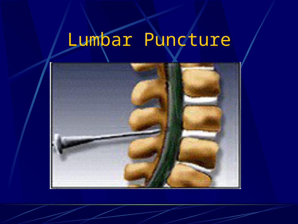
Lumbar Puncture
Lumbar Puncture
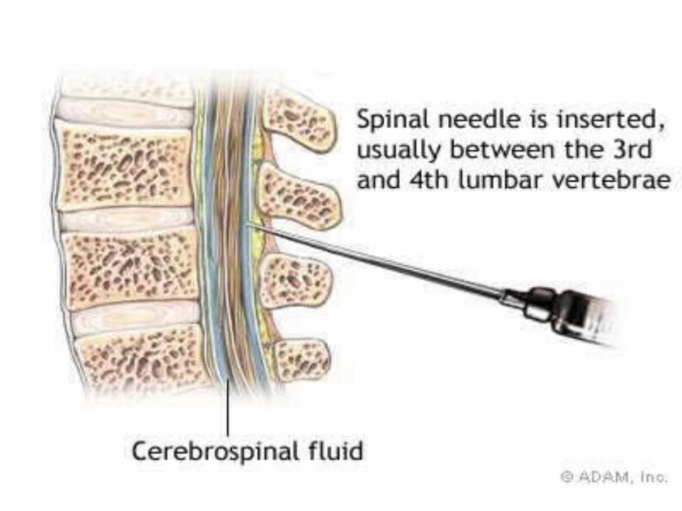
Lumbar Puncture - involves withdrawing cerebrospinal fluid by the insertion of a hollow needle into the lumbar subarachnoid space”.
Cerebral Spinal Fluid – Clear, lymph-like fluid that fills the entire subarachnoid space and surrounds and protects the brain.
Indications for Lumbar Puncture
Primary indication for emergent spinal tap is possibility of CNS infection
The second indication for an emergent spinal puncture is a suspected spontaneous subarachnoid hemorrhage.
Subarachnoid Hemorrhage
Diagnosis usually made by CT scan or by blood in CSF.
Initial presentation: CT 92-98% accurate
Later than 24 hr presentation: 76% accurate
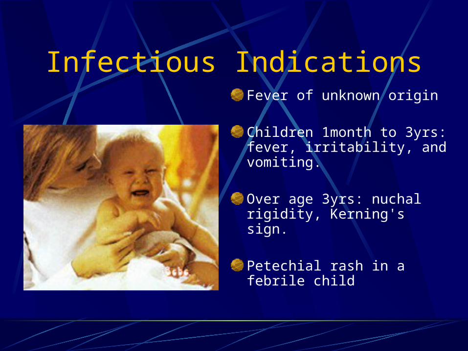
Infectious IndicationsFever of unknown origin
Children 1month to 3yrs: fever, irritability, and vomiting.
Over age 3yrs: nuchal rigidity, Kerning's sign.
Petechial rash in a febrile child

Contraindications for LP
presence of infection in the tissues near the puncture site.
Increased ICP--The presence of papilledema, retinal hemorrhage.
A sudden drop in intraspinal pressure by rapid release of cerebrospinal fluid (CSF) may cause fatal
herniation. Bleeding diathesis: A platelet count is desirable before LP.

EquipmentSpinal needleThree-way stopcockManometer3 specimen tubesLocal anesthesiaBetadine Plaster dressingSterile towel
Equipment required
Three sterile specimen bottles: should be labeled 1, 2 and 3. The first specimen, which may be bloodstained due to needle trauma, should go into the first bottle. This will assist the laboratory to differentiate between blood due to procedure trauma and that due to Subarachnoid hemorrhage.
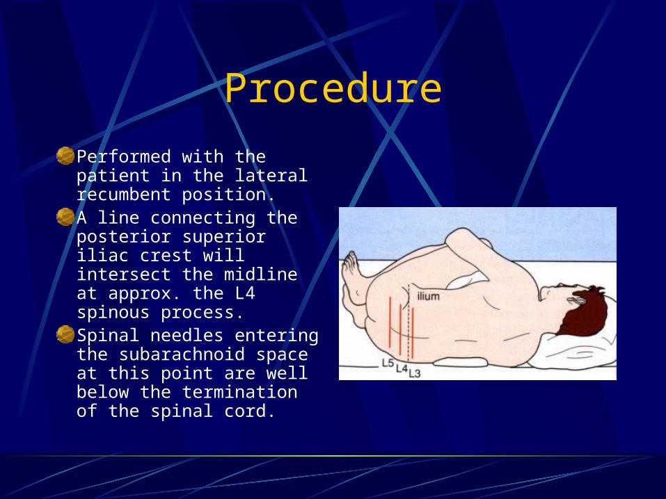
Procedure
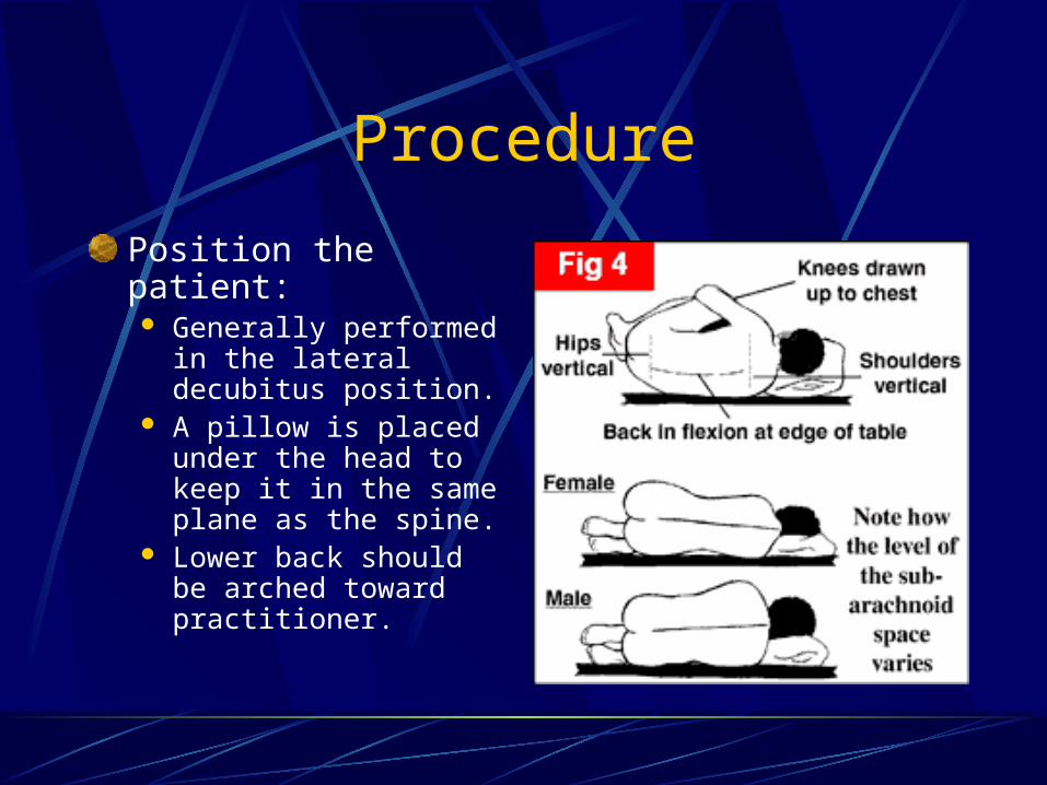
Performed with the patient in the lateral recumbent position.A line connecting the posterior superior iliac crest will intersect the midline at approx. the L4 spinous process.Spinal needles entering the subarachnoid space at this point are well below the termination of the spinal cord.
Procedure
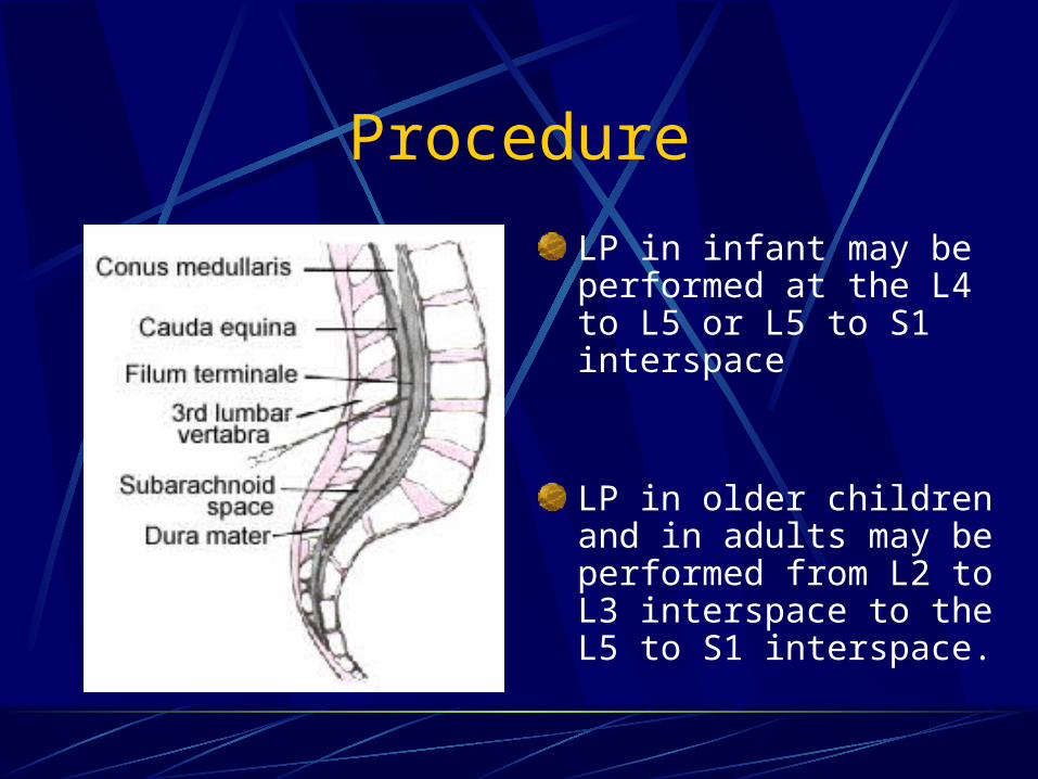
LP in infant may be performed at the L4 to L5 or L5 to S1 interspace
LP in older children and in adults may be performed from L2 to L3 interspace to the L5 to S1 interspace.
Procedure
Position the patient: Generally performed in
the lateral decubitus position.
A pillow is placed under the head to keep it in the same plane as the spine.
Lower back should be arched toward practitioner.
Procedure
Almost all patients are afraid of an LP. Explaining the procedure in advance and discussing each step aids in reducing anxiety.
Inquire about allergies to anesthetics.
Informed consent.
Procedure
Sterile gloves MUST be used.Wash back with antiseptic solution.Sterile towel under hips.The skin and deeper subcutaneous tissue are infiltrated with local anesthetic.Warn patient of transient discomfort of anesthetic.
Procedure
The patient should be told to report any pain and should be informed that he or she will feel some pressure.
The needle is placed into the skin in the midline parallel to the bed.
The needle is held with both thumbs and index fingers.
Procedure
The ligaments offer resistance to the needle, and a “pop” is often felt as they are penetrated.
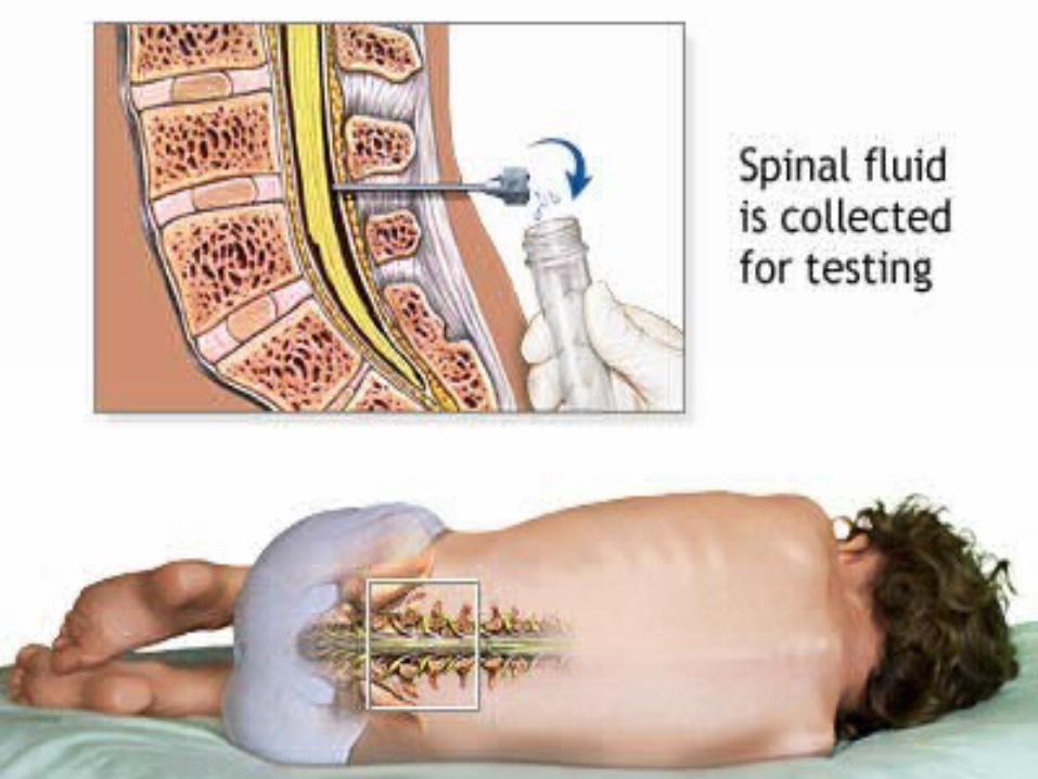
Clear fluid will flow from the needle when the subarachnoid space has been penetrated.
Procedure
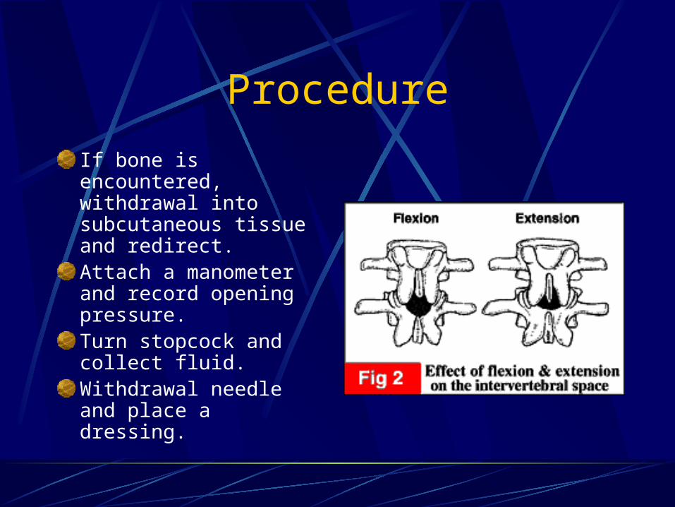
If bone is encountered, withdrawal into subcutaneous tissue and redirect.Attach a manometer and record opening pressure.Turn stopcock and collect fluid.Withdrawal needle and place a dressing.
Procedure
Tube 1 is used for determining protein and glucose
Tube 2 is used for microbiologic and cytologic studies
Tube 3 is for cell counts and serologic tests for syphilis
The Traumatic Tap
It should not be difficult to distinguish between subarachnoid hemorrhage and a traumatic tap.
In traumatic taps, the fluid generally clears between 1st and 3rd tubes.

Interpretation
Appearance If CSF is not crystal clear, a pathologic
condition of the CNS should be suspectedCompare fluid to waterFluid may be clear with as many as 400
RBCs/mm3 and 200 WBCs/mm3

Interpretation
Cells WBC counts over 5 cells/mm3 should be taken to
indicate the presence of pathologic condition
Neutrophilic pleocytosis (increase in number) is commonly associated with bacterial infections or early stages of viral infections, tuberculosis, or meningitis.
Interpretation
Cells
Eosinophils are most commonly represent a parasite infestation.
Eosinophils have also been reported in cases of subarachnoid hemorrhage, lymphoma, Hodgkin’s disease, brucellosis, fungal meningitis, mycoplasma pneumonia infection, measles, and many other infectious disease.
Interpretation
CellsNormal CSF RBCs are less than 10/mm3.Counts that are otherwise unexplained
may be due to a traumatic tap.Herpes simplex virus encephalitis may
elevate the CSF RBC count in many patients.
Interpretation
GlucoseLow CSF glucose concentration indicates
increased glucose use in the brain and the spinal cord.
The normal range of CSF glucose is between 50 and 80 mg/dl
60-70% of serum glucose concentrationOnly low concentrations of glucose are
significance

Interpretation
Low CSF Glucose SyndromesBacterial meningitis Syphilis
Tuberculous meningitis Chemical meningitis
Fungal meningitis Subarachnoid meningitis
Sarcoidosis Mumps meningitis
Meningeal carcinomatosis
Herpes simplex encephalitis
Amebic meningitis Hypoglycemia
Interpretation
Protein Increase in CSF total protein levels are a
nonspecific abnormality associated with many disease states.
Levels > 500mg/dl are uncommon and are seen mainly in meningitis, in subarachnoid bleeding, and with spinal tumors.

CSF Analysis with Infections
Bacterial Infections While the culture is pending, one may suspect a
bacterial infection in the presence of an elevated opening pressure and a marked pleocytosis ranging between 500 and 20,000 WBCs/mm3.
The differential count is usually chiefly neutrophils. A count above 1000 cells/mm3 seldom occurs in
viral infections.

CSF Analysis with Infections
Bacterial InfectionsCSF glucose levels less than 40 mg/dl or
less than 50% of a simultaneous blood glucose level should raise the question of bacterial meningitis.
The CSF protein content in bacterial meningitis ranges from 500 to 1500 mg/dl.

CSF Analysis with Infections
Viral StudiesThe organisms most commonly isolated in
viral meningitis are enteroviruses and mumps.
Enteroviruses: summer and fallMumps: winter and spring

CSF Analysis with Infections
Viral StudiesWBC count in viral meningitis and
encephalitis usually: 10 to 1000 cells/mm3.The differential count is predominantly
lymphocytic and mononuclear in type.Protein levels are usually mildly elevated
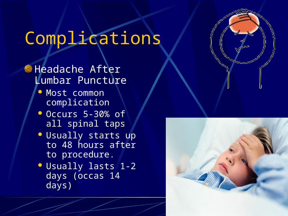
Complications
Headache After Lumbar Puncture Most common
complication Occurs 5-30% of all
spinal taps Usually starts up to
48 hours after to procedure.
Usually lasts 1-2 days (occas 14 days)
Complications
Headache After Lumbar PunctureUsually begins within minutes after arising
and resolves with recumbent position.Pain is mild to incapacitating and is usually
cervical and sub-occipital, but may involve the shoulders and the entire cranium.
Caused by leaking of fluid through dural puncture site.
Complications
Headache After Lumbar Puncture Incidence is higher in younger patients and
females, and those with headache history.Treatment: barbiturates, fluids (500mg in 2
ml NS IV push) more common 500mg in 2 L over 1 hr.
Blood patch by anesthesia if no improvement.
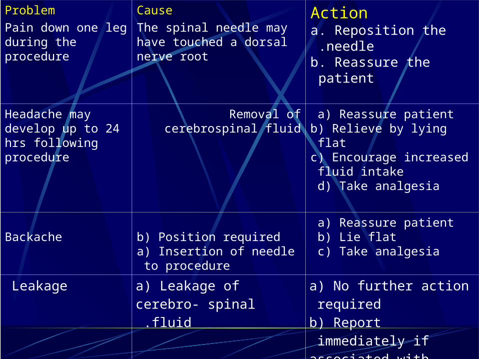
Problem
Pain down one leg during the procedure
Cause
The spinal needle may have touched a dorsal nerve root
Actiona. Reposition the needle .b. Reassure the patient
Headache may develop up to 24 hrs following procedure
Removal of cerebrospinal fluid a) Reassure patient b) Relieve by lying flat c) Encourage increased fluid intake d) Take analgesia
Backache b) Position required a) Insertion of needle to procedure
a) Reassure patient b) Lie flat c) Take analgesia
Leakage a) Leakage of cerebro- spinal fluid .
a) No further action required b) Report immediately if associated with other symptoms
Deterioration in neurological status
Presence of space occupying lesion in the brain not appreciated
Need medical assistance immediately .
Recommendations for nurses
1- prepare all equipments before starting the procedure.2- explain procedure to family , why , how the procedure done.3- keep child in sterile field as possible.4- sending all samples to lab after procedure immediately.5- explain to family how to care of child after procedure to decrease potential problems.

Arterial Blood GasesArterial blood gases: are measured to assess a child or a client’s oxygenation, ventilation, and acid-base balance. The blood sample is easily, although often painfully, obtained from an artery and is analyzed for:-arterial blood pH- partial pressure of oxygen (PaO2)- partial pressure of carbon dioxide (PaCO2)-arterial oxygen saturation (SaO2).- Rate and depth of respirations can affect the results of an ABG sample.
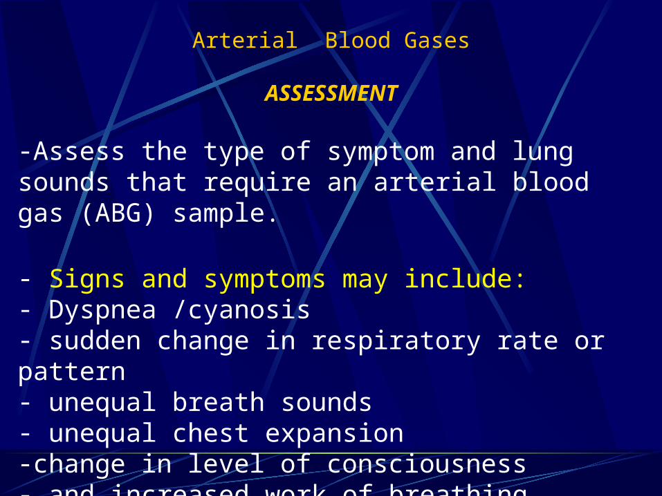
Arterial Blood Gases
ASSESSMENT
-Assess the type of symptom and lung sounds that require an arterial blood gas (ABG) sample.
- Signs and symptoms may include:- Dyspnea /cyanosis- sudden change in respiratory rate or pattern- unequal breath sounds- unequal chest expansion -change in level of consciousness- and increased work of breathing.
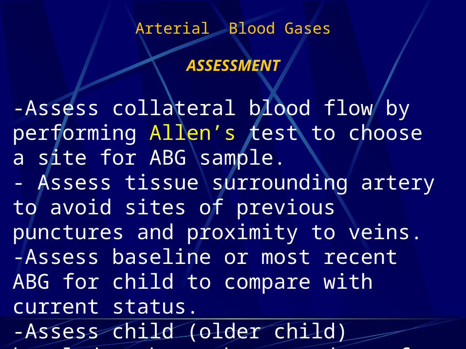
Arterial Blood Gases
ASSESSMENT
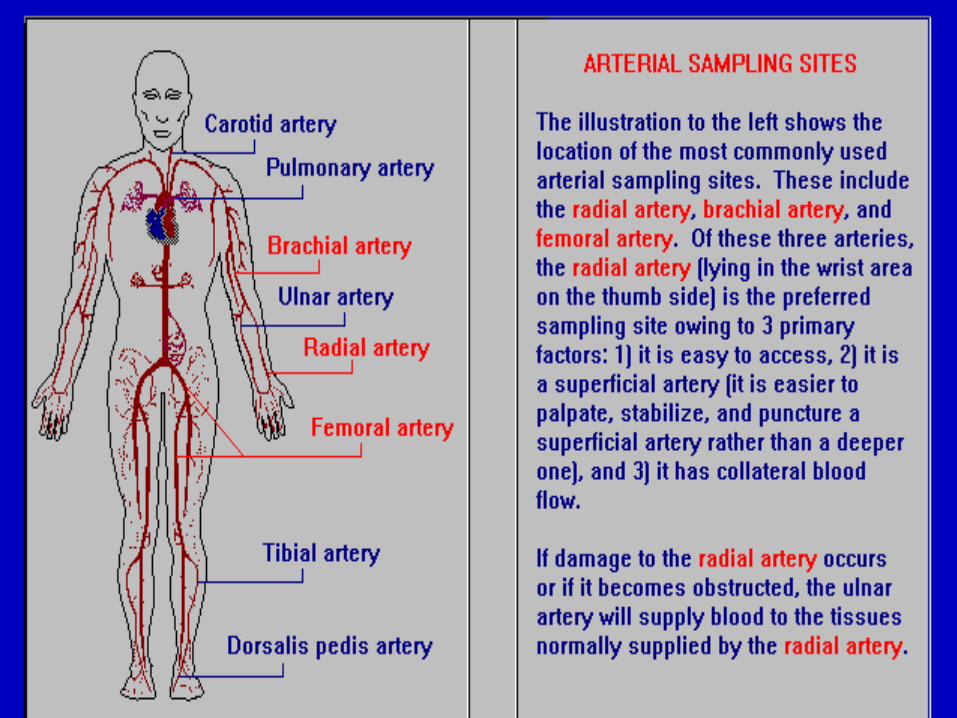
-Assess collateral blood flow by performing Allen’s test to choose a site for ABG sample.- Assess tissue surrounding artery to avoid sites of previous punctures and proximity to veins.-Assess baseline or most recent ABG for child to compare with current status.-Assess child (older child) knowledge about the procedure of obtaining an ABG sample to ensure cooperation and reduce anxiety.
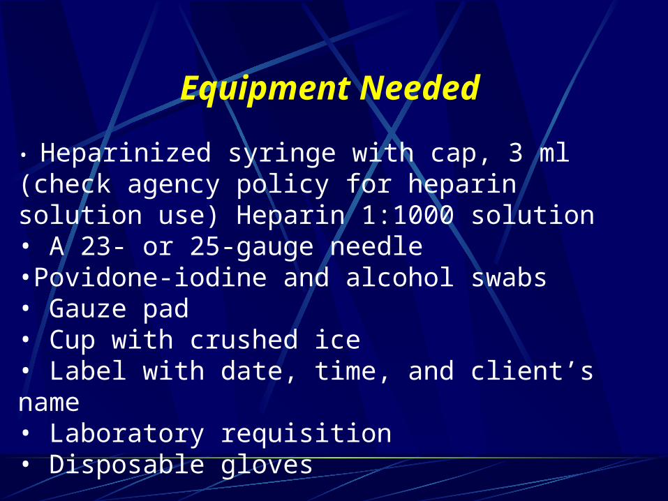
Equipment Needed
• Heparinized syringe with cap, 3 ml (check agency policy for heparin solution use) Heparin 1:1000 solution• A 23- or 25-gauge needle•Povidone-iodine and alcohol swabs• Gauze pad• Cup with crushed ice• Label with date, time, and client’s name• Laboratory requisition• Disposable gloves
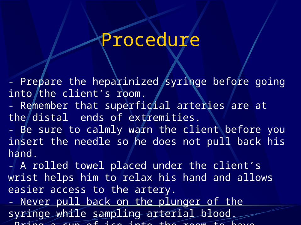
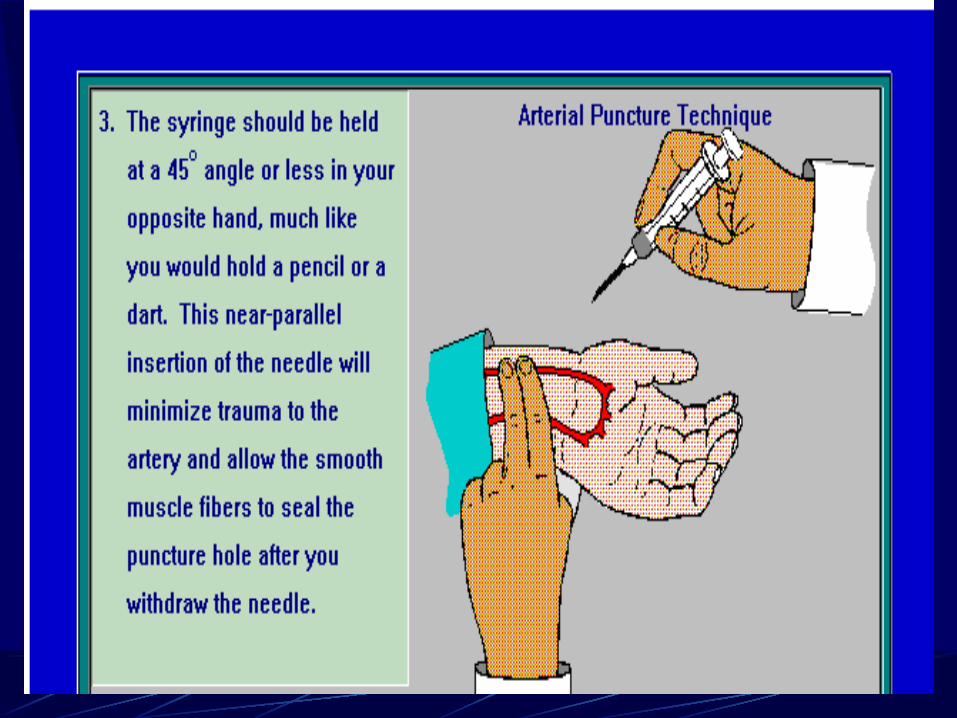
Procedure - Prepare the heparinized syringe before going into the client’s room.- Remember that superficial arteries are at the distal ends of extremities.- Be sure to calmly warn the client before you insert the needle so he does not pull back his hand.- A rolled towel placed under the client’s wrist helps him to relax his hand and allows easier access to the artery.- Never pull back on the plunger of the syringe while sampling arterial blood.-Bring a cup of ice into the room to have available to transport the sample.
Procedure
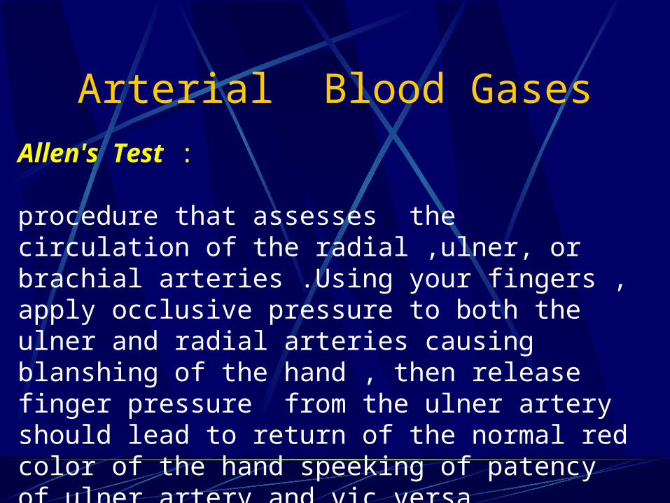
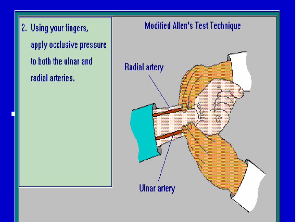
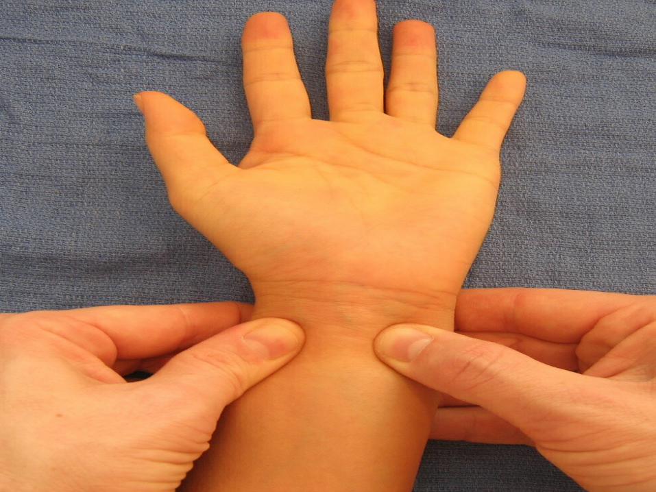
Arterial Blood GasesAllen's Test :
procedure that assesses the circulation of the radial ,ulner, or brachial arteries .Using your fingers , apply occlusive pressure to both the ulner and radial arteries causing blanshing of the hand , then release finger pressure from the ulner artery should lead to return of the normal red color of the hand speeking of patency of ulner artery and vic versa .
Blood Gas Sampling Errors
- Air or air bubbles left in the blood gas sample.-Delay in icing or analyzing the blood gas sample.- Excess heparin left in the blood gas syringe.- Obtaining a venous sample or a venous admixture sample.
- Alterations in temperature
COMMON ERRORS.
Prevention:- Do not pull back on the plunger of the syringe while obtaining arterial blood. - Be sure the needle is attached securely to the syringe before inserting the needle into the artery. - If a sufficient amount of blood has been obtained, remove the needle and expel the air bubbles from the syringe. - If not, remove the needle, apply pressure to the site, wait 5 minutes, and obtain the sample at another site with a new needle and syringe.
Arterial Blood Gases. The date and time of the ABG sampling should be recorded in the narrative notes.• Also record the reason for the test, the results of the Allen’s test, the client’s response to the blood sampling, and any unusual observations.• Note the route and amount of oxygen the client is receiving and any respiratory assessment• Record the condition of the puncture site prior to the blood draw and after the blood draw.• Be sure to note the follow-up check on the condition of the site.

Any question

Thanks for Attention