Commensal Protection of Staphylococcus aureus against ... · Eric F. Kong,a,b,c Christina Tsui,c...
12
Commensal Protection of Staphylococcus aureus against Antimicrobials by Candida albicans Biofilm Matrix Eric F. Kong, a,b,c Christina Tsui, c Sona Kucharíková, d,e David Andes, f Patrick Van Dijck, d,e Mary Ann Jabra-Rizk b,c Graduate Program in Life Sciences, Molecular Microbiology and Immunology Program, University of Maryland, Baltimore, Maryland, USA a ; Department of Microbiology and Immunology, School of Medicine, University of Maryland, Baltimore, Maryland, USA b ; Department of Oncology and Diagnostic Sciences, Dental School, University of Maryland, Baltimore, Maryland, USA c ; Laboratory of Molecular Cell Biology, KU Leuven, Leuven-Heverlee, Flanders, Belgium d ; Department of Molecular Microbiology, VIB, Leuven-Heverlee, Flanders, Belgium e ; Department of Medicine, Microbiology and Immunology, University of Wisconsin, Madison, Wisconsin, USA f E.F.K. and C.T. contributed equally to the study. ABSTRACT Biofilm-associated polymicrobial infections, particularly those involving fungi and bacteria, are responsible for sig- nificant morbidity and mortality and tend to be challenging to treat. Candida albicans and Staphylococcus aureus specifically are considered leading opportunistic fungal and bacterial pathogens, respectively, mainly due to their ability to form biofilms on catheters and indwelling medical devices. However, the impact of mixed-species biofilm growth on therapy remains largely un- derstudied. In this study, we investigated the influence of C. albicans secreted cell wall polysaccharides on the response of S. au- reus to antibacterial agents in biofilm. Results demonstrated significantly enhanced tolerance for S. aureus to drugs in the pres- ence of C. albicans or its secreted cell wall polysaccharide material. Fluorescence confocal time-lapse microscopy revealed impairment of drug diffusion through the mixed biofilm matrix. Using C. albicans mutant strains with modulated cell wall poly- saccharide expression, exogenous supplementation, and enzymatic degradation, the C. albicans-secreted -1,3-glucan cell wall component was identified as the key matrix constituent providing the bacteria with enhanced drug tolerance. Further, antibody labeling demonstrated rapid coating of the bacteria by the C. albicans matrix material. Importantly, via its effect on the fungal biofilm matrix, the antifungal caspofungin sensitized the bacteria to the drugs. Understanding such symbiotic interactions with clinical relevance between microbial species in biofilms will greatly aid in overcoming the limitations of current therapies and in defining potential new targets for treating polymicrobial infections. IMPORTANCE The fungus Candida albicans and the bacterium Staphylococcus aureus are important microbial pathogens re- sponsible for the majority of infections in hospitalized patients and are often coisolated from a host. In this study, we demon- strated that when grown together, the fungus provides the bacterium with enhanced tolerance to antimicrobial drugs. This pro- cess was mediated by polysaccharides secreted by the fungal cell into the environment. The biofilm matrix formed by these polysaccharides prevented penetration by the drugs and provided the bacteria with protection. Importantly, we show that by inhibiting the production of the fungal polysaccharides, a specific antifungal agent indirectly sensitized the bacteria to antimi- crobials. Understanding the therapeutic implications of the interactions between these two diverse microbial species will aid in overcoming the limitations of current therapies and in defining new targets for treating complex polymicrobial infections. Received 27 July 2016 Accepted 12 September 2016 Published 11 October 2016 Citation Kong EF, Tsui C, Kucharíková S, Andes D, Van Dijck P, Jabra-Rizk MA. 2016. Commensal protection of Staphylococcus aureus against antimicrobials by Candida albicans biofilm matrix. mBio 7(5):e01365-16. doi:10.1128/mBio.01365-16. Editor Joseph Heitman, Duke University Copyright © 2016 Kong et al. This is an open-access article distributed under the terms of the Creative Commons Attribution 4.0 International license. Address correspondence to Mary Ann Jabra-Rizk, [email protected]. P olymicrobial infections caused by a combination of micro- organisms are responsible for significant mortality and morbidity, particularly those associated with biofilms formed on indwelling medical devices (1–3). Biofilms are structured three-dimensional communities of surface-associated micro- bial populations embedded in a matrix of extracellular polysac- charides, proposed to provide a structural scaffold and protec- tion for biofilm cells (4–6). Therefore, in a biofilm, microbes are afforded a stable environment and can tolerate high concentra- tions of antimicrobials. The impact of these biofilms on public health is dramatic, as cells released from biofilms can migrate into the bloodstream and cause systemic infections with high mortality (7). Importantly, the increase in drug resistance has provided a strong impetus to understand the mechanisms of the enhanced tolerance of biofilm-associated infections to antimicrobial ther- apy and particularly polymicrobial infections. Although mixed fungal-bacterial infections tend to be the most complex and chal- lenging to treat, the impact of these interactions on therapy re- mains largely understudied. Among the fungal species, Candida albicans is the most com- mon human pathogen, causing diseases ranging from superficial mucosal to life-threatening systemic infections (8–10). The ability of C. albicans to transition from commensal to pathogen is pri- marily the result of its aptitude for morphologically switching be- tween yeast and hyphal forms (9, 11). In fact, the majority of C. albicans infections are associated with its ability to form bio- RESEARCH ARTICLE crossmark September/October 2016 Volume 7 Issue 5 e01365-16 ® mbio.asm.org 1 on February 23, 2019 by guest http://mbio.asm.org/ Downloaded from
Transcript of Commensal Protection of Staphylococcus aureus against ... · Eric F. Kong,a,b,c Christina Tsui,c...
Commensal Protection of Staphylococcus aureus againstAntimicrobials by Candida albicans Biofilm Matrix
Eric F. Kong,a,b,c Christina Tsui,c Sona Kucharíková,d,e David Andes,f Patrick Van Dijck,d,e Mary Ann Jabra-Rizkb,c
Graduate Program in Life Sciences, Molecular Microbiology and Immunology Program, University of Maryland, Baltimore, Maryland, USAa; Department of Microbiologyand Immunology, School of Medicine, University of Maryland, Baltimore, Maryland, USAb; Department of Oncology and Diagnostic Sciences, Dental School, University ofMaryland, Baltimore, Maryland, USAc; Laboratory of Molecular Cell Biology, KU Leuven, Leuven-Heverlee, Flanders, Belgiumd; Department of Molecular Microbiology, VIB,Leuven-Heverlee, Flanders, Belgiume; Department of Medicine, Microbiology and Immunology, University of Wisconsin, Madison, Wisconsin, USAf
E.F.K. and C.T. contributed equally to the study.
ABSTRACT Biofilm-associated polymicrobial infections, particularly those involving fungi and bacteria, are responsible for sig-nificant morbidity and mortality and tend to be challenging to treat. Candida albicans and Staphylococcus aureus specifically areconsidered leading opportunistic fungal and bacterial pathogens, respectively, mainly due to their ability to form biofilms oncatheters and indwelling medical devices. However, the impact of mixed-species biofilm growth on therapy remains largely un-derstudied. In this study, we investigated the influence of C. albicans secreted cell wall polysaccharides on the response of S. au-reus to antibacterial agents in biofilm. Results demonstrated significantly enhanced tolerance for S. aureus to drugs in the pres-ence of C. albicans or its secreted cell wall polysaccharide material. Fluorescence confocal time-lapse microscopy revealedimpairment of drug diffusion through the mixed biofilm matrix. Using C. albicans mutant strains with modulated cell wall poly-saccharide expression, exogenous supplementation, and enzymatic degradation, the C. albicans-secreted �-1,3-glucan cell wallcomponent was identified as the key matrix constituent providing the bacteria with enhanced drug tolerance. Further, antibodylabeling demonstrated rapid coating of the bacteria by the C. albicans matrix material. Importantly, via its effect on the fungalbiofilm matrix, the antifungal caspofungin sensitized the bacteria to the drugs. Understanding such symbiotic interactions withclinical relevance between microbial species in biofilms will greatly aid in overcoming the limitations of current therapies and indefining potential new targets for treating polymicrobial infections.
IMPORTANCE The fungus Candida albicans and the bacterium Staphylococcus aureus are important microbial pathogens re-sponsible for the majority of infections in hospitalized patients and are often coisolated from a host. In this study, we demon-strated that when grown together, the fungus provides the bacterium with enhanced tolerance to antimicrobial drugs. This pro-cess was mediated by polysaccharides secreted by the fungal cell into the environment. The biofilm matrix formed by thesepolysaccharides prevented penetration by the drugs and provided the bacteria with protection. Importantly, we show that byinhibiting the production of the fungal polysaccharides, a specific antifungal agent indirectly sensitized the bacteria to antimi-crobials. Understanding the therapeutic implications of the interactions between these two diverse microbial species will aid inovercoming the limitations of current therapies and in defining new targets for treating complex polymicrobial infections.
Received 27 July 2016 Accepted 12 September 2016 Published 11 October 2016
Citation Kong EF, Tsui C, Kucharíková S, Andes D, Van Dijck P, Jabra-Rizk MA. 2016. Commensal protection of Staphylococcus aureus against antimicrobials by Candida albicansbiofilm matrix. mBio 7(5):e01365-16. doi:10.1128/mBio.01365-16.
Editor Joseph Heitman, Duke University
Copyright © 2016 Kong et al. This is an open-access article distributed under the terms of the Creative Commons Attribution 4.0 International license.
Address correspondence to Mary Ann Jabra-Rizk, [email protected].
Polymicrobial infections caused by a combination of micro-organisms are responsible for significant mortality and
morbidity, particularly those associated with biofilms formedon indwelling medical devices (1–3). Biofilms are structuredthree-dimensional communities of surface-associated micro-bial populations embedded in a matrix of extracellular polysac-charides, proposed to provide a structural scaffold and protec-tion for biofilm cells (4–6). Therefore, in a biofilm, microbes areafforded a stable environment and can tolerate high concentra-tions of antimicrobials. The impact of these biofilms on publichealth is dramatic, as cells released from biofilms can migrate intothe bloodstream and cause systemic infections with high mortality(7). Importantly, the increase in drug resistance has provided a
strong impetus to understand the mechanisms of the enhancedtolerance of biofilm-associated infections to antimicrobial ther-apy and particularly polymicrobial infections. Although mixedfungal-bacterial infections tend to be the most complex and chal-lenging to treat, the impact of these interactions on therapy re-mains largely understudied.
Among the fungal species, Candida albicans is the most com-mon human pathogen, causing diseases ranging from superficialmucosal to life-threatening systemic infections (8–10). The abilityof C. albicans to transition from commensal to pathogen is pri-marily the result of its aptitude for morphologically switching be-tween yeast and hyphal forms (9, 11). In fact, the majority ofC. albicans infections are associated with its ability to form bio-
RESEARCH ARTICLE
crossmark
September/October 2016 Volume 7 Issue 5 e01365-16 ® mbio.asm.org 1
on February 23, 2019 by guest
http://mbio.asm
.org/D
ownloaded from

films, where adhesion of yeast cells to the substrate is followed byproliferation and hypha formation, resulting in a network of cellsembedded in a matrix (7, 12, 13). Candida albicans biofilm matrixis complex, with major polysaccharide constituents being�-mannan, �-1,6-glucan, and �-1,3-glucan (14, 15). Although arelatively minor component, �-1,3-glucan is considered the crit-ical matrix polysaccharide, as extracellular glucan has been linkedto biofilm resistance to antifungals (16, 17). In fact, previous stud-ies have shown elevated �-1,3-glucan levels to be characteristic ofbiofilm cells both in the fungal cell walls and as a secreted form. Ofmore significance, the increase in �-1,3-glucan secretion by bio-film cells was shown in vivo in animal models of catheter infectionand disseminated candidiasis (12). Glucan synthase Fks1p is re-sponsible for the synthesis of cell wall �-1,3-glucan during biofilmgrowth, and FKS1 disruption was shown to reduce manufactureand deposition of �-1,3-glucan in the biofilm matrix (18). Impor-tantly, using strains with modulated FKS1 expression, a study byNett et al. (18) demonstrated that reduction in expression ren-dered biofilms more susceptible to various antifungals, whereasoverexpression resulted in increased resistance.
In various niches in the host, C. albicans coexists with variousbacterial species, including Staphylococcus aureus (2, 19–21). Al-though S. aureus primarily exists as a commensal organism, thisbacterial pathogen is implicated in a variety of diseases rangingfrom minor skin infections to more serious invasive diseases andspecifically device-associated biofilm infections (22–24). With theemergence of methicillin-resistant S. aureus (MRSA), this ubiqui-tous pathogen is becoming an even greater therapeutic challenge(25, 26). S. aureus is a poor former of biofilms; however, togetherwith C. albicans, this species forms a substantial biofilm where thefungus creates a scaffold for the bacteria (27–29). Our previous invitro studies have demonstrated that S. aureus exhibits high affin-ity to the C. albicans hyphal form, as these species coadhere andinteract synergistically in a biofilm. Further, our studies identifiedthe C. albicans hypha-specific adhesin Als3p to be involved in thecoadherence process (27).
The growing use of implanted medical devices is another rea-son why the incidence of Candida and staphylococcal infectionshas steadily increased, since the majority of these infections areemerging from biofilms formed on medical implants (10, 13, 23,30, 31). In fact, a recent analysis of cases of endocarditis associatedwith an implanted device found ~25% of the infections to bepolymicrobial, and in another study, 27% of nosocomial C. albi-cans bloodstream infections were estimated to be polymicrobial(32, 33). Importantly, S. aureus was found to be the third mostcommonly coisolated species with C. albicans (33). Although nu-merous studies have reported the coisolation of C. albicans andS. aureus from a multitude of diseases such as periodontitis, den-ture stomatitis, cystic fibrosis, keratitis, ventilator-associatedpneumonia, and urinary tract catheter and burn wound infec-tions, the clinical significance of their interaction in a host remainslargely understudied, likely due to lack of suitable animal models(34–38). However, using a mouse model of oral infection, we re-cently demonstrated that upon onset of oral candidiasis (thrush),mice cocolonized with C. albicans and S. aureus suffered systemicbacterial infection with high morbidity and mortality (39).
The inherent characteristics of biofilms are multifactorial butare largely due to the extracellular matrix encasing the biofilmcells, which prevents drugs and other stresses from penetrating thebiofilm. Therefore, S. aureus and C. albicans interactions in mixed
biofilm infections may also impact response to antimicrobial ther-apy (39). Although enhanced in vitro tolerance to antimicrobialsin C. albicans and staphylococcus mixed biofilms has been re-ported, the mechanism and specific factors behind these observa-tions remain undefined (40, 41). To that end, in this study, exper-iments were designed to demonstrate the impact of C. albicansbiofilm matrix and secreted components on the susceptibility ofS. aureus to antibacterial drugs in biofilm, focusing on vancomy-cin, the drug of choice for treatment of MRSA infections. Theoverall goal of this study is to provide crucial insights into theenhanced tolerance of biofilm-associated polymicrobial infec-tions to antimicrobial therapy.
RESULTSComparative assessment of the susceptibility of S. aureus tovancomycin in single and mixed biofilms with C. albicans. Theimpact of mixed-species biofilm growth on S. aureus susceptibilityto vancomycin was assessed following 24-h treatment of pre-formed single and mixed biofilms. Based on CFU, results demon-strated a significant increase in S. aureus survival in mixed biofilmsfollowing vancomycin treatment compared to survival in singlebiofilm (Fig. 1A). Analysis of biofilms using crystal violet stainingindicated a significant increase in biofilm biomass in mixed bio-films relative to S. aureus biofilm (Fig. 1B). These results werecorroborated by scanning electron microscopy (SEM) analysiswhere images of mixed biofilms revealed a thick matrix, withS. aureus adhering to and forming aggregates around the C. albi-cans hyphae (Fig. 1C and D).
Exogenous supplementation of S. aureus biofilms with C. al-bicans biofilm matrix material enhances S. aureus tolerance tovancomycin. To isolate the role of C. albicans biofilm matrix inthe enhanced tolerance of S. aureus to vancomycin in mixed bio-films, purified matrix material recovered from C. albicans biofilmswas incorporated in susceptibility testing. Assessment of S. aureusviability by the MTS [3-(4,5-dimethylthiazol-2-yl)-5-(3-carboxy-methoxyphenyl)-2-(4-sulfophenyl)-2H-tetrazolium] metabolicassay and CFU recovery demonstrated a significant increase inS. aureus survival in single biofilms formed in the presence ofmatrix material (Fig. 2). In order to identify the contribution ofspecific matrix components, similar experiments were also per-formed where S. aureus biofilms were allowed to form in the pres-ence of glucan or mannan. Results from these experiments dem-onstrated a comparable increase in survival with vancomycin.
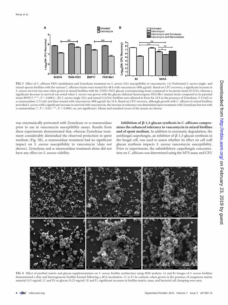
Differential C. albicans �-1,3-glucan but not �-mannan ex-pression modulates vancomycin tolerance in mixed biofilms.The key role for the �-1,3-glucan matrix component was demon-strated using C. albicans strains with modulated �-1,3-glucan ex-pression in mixed biofilms treated with vancomycin. Comparedto their respective reference strains, S. aureus recovery followingvancomycin treatment was consistently higher in the presence ofthe TDH3-FKS1 glucan-overexpressing strain but lower whengrown with the FKS1/fks1� glucan-deficient heterozygous mutantstrain (Fig. 3A). No impact on S. aureus vancomycin susceptibilitywas seen in mixed biofilms with the mannosylation mutants or thezap1�/zap1� mutant lacking the zinc response transcription fac-tor Zap1 (a negative regulator of �-1,3-glucan) (data not shown).
�-1,3-Glucanase but not �-mannosidase treatment of mixedbiofilms abrogates the enhanced tolerance to vancomycin. Todemonstrate the importance of the C. albicans matrix polysaccha-rides, experiments were also performed where single- and mixed-
Kong et al.
2 ® mbio.asm.org September/October 2016 Volume 7 Issue 5 e01365-16
on February 23, 2019 by guest
http://mbio.asm
.org/D
ownloaded from

species biofilms were treated with vancomycin in the presence ofthe glucan-degrading enzyme Zymolyase or the mannan-degrading enzyme �-mannosidase. Based on CFU recovery, theincrease in S. aureus vancomycin tolerance in mixed biofilms was
significantly diminished upon Zymolyase treatment, to a levelcomparable to that for S. aureus single-species biofilm treated withvancomycin. However, no significant differences were noted withthe �-mannosidase treatment (Fig. 3B). Since Zymolyase isknown to have contaminating protease activity, enzymatic diges-tion experiments were also performed using proteinase K; resultsfrom these experiments demonstrated no effect for proteinase Kon S. aureus response to vancomycin (data not shown).
SEM of S. aureus biofilm architecture supplemented withC. albicans biofilm matrix material and exogenous �-1,3-glucan. To visualize the structure of the S. aureus biofilm supple-mented with C. albicans matrix material, S. aureus biofilms wereallowed to form for 24 h in the absence and presence of purifiedmatrix material or glucan, and the formed biofilms were compar-atively examined by SEM analysis. Images revealed that wherenonsupplemented S. aureus biofilms appeared thin and heteroge-neous (Fig. 4A and B), growth in the presence of matrix material(Fig. 4C and D) or �-1,3-glucan (Fig. 4E and F) resulted in signif-icantly increased biofilm mass with considerable aggregation ofbacterial cells.
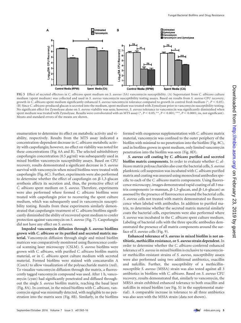
C. albicans spent culture medium provides S. aureus withenhanced tolerance to vancomycin in single-species biofilm.Since C. albicans cell wall polysaccharides are secreted, experi-ments were performed where C. albicans spent biofilm culturemedium was recovered and used in vancomycin susceptibilitytesting of S. aureus single biofilms. Results from these experimentsdemonstrated that C. albicans cell-free spent medium providedS. aureus with significantly enhanced tolerance to vancomycin inthe absence of C. albicans in the biofilm (Fig. 5A). To identify thesecreted component conferring the tolerance, the spent medium
FIG 1 Assessment of relative biomass and vancomycin susceptibility in single S. aureus (SA) and mixed S. aureus and C. albicans (SA/CA) biofilms. (A)Preformed (24-h) S. aureus single and mixed biofilms were treated with vancomycin (800 �g/ml) for an additional 24 h. CFU recovery of S. aureus from bothbiofilms showed a significant increase in S. aureus recovery from mixed biofilms following vancomycin treatment (***, P � 0.001). (B) To assess biomass, 24-hsingle- and mixed-species biofilms were treated with crystal violet. Based on absorbance (595 nm), results demonstrated significantly higher biomass for mixedbiofilms than for S. aureus biofilm (**, P � 0.01; ***, P � 0.001). Means and standard errors of the means are shown. (C and D) These results were corroboratedby SEM analysis where images demonstrated adherence to and clumping of S. aureus around C. albicans hyphae, forming thick biofilm aggregates.
FIG 2 Effect of C. albicans matrix polysaccharides on S. aureus (SA) suscep-tibility to vancomycin. S. aureus single biofilms were allowed to form in thepresence of purified C. albicans (CA) biofilm matrix material (0.02 mg/liter),glucan (1 mg/ml), or mannan (1 mg/ml). S. aureus dual-species biofilms weregrown with C. albicans (SA/CA). Preformed biofilms were treated with vanco-mycin (800 �g/ml) for 24 h. S. aureus CFU recovery from biofilms demon-strated significant increase in S. aureus tolerance to vancomycin in the pres-ence of matrix material, glucan, and mannan. Susceptibility to vancomycinwas assessed by CFU counts and corroborated by an MTS assay (**, P � 0.01;***, P � 0.001). Means and standard errors of the means are shown.
Fungal-Bacterial Biofilms and Drug Resistance
September/October 2016 Volume 7 Issue 5 e01365-16 ® mbio.asm.org 3
on February 23, 2019 by guest
http://mbio.asm
.org/D
ownloaded from
was enzymatically pretreated with Zymolyase or �-mannosidaseprior to use in vancomycin susceptibility assays. Results fromthese experiments demonstrated that, whereas Zymolyase treat-ment considerably diminished the observed protection in spentmedium (Fig. 5B), �-mannosidase treatment had no significantimpact on S. aureus susceptibility to vancomycin (data notshown). Zymolyase and �-mannosidase treatment alone did nothave any effect on S. aureus viability.
Inhibition of �-1,3-glucan synthesis in C. albicans compro-mises the enhanced tolerance to vancomycin in mixed biofilmsand of spent medium. In addition to enzymatic degradation, theantifungal caspofungin, an inhibitor of �-1,3-glucan synthesis inthe fungal cell, was used to assess whether its effect on cell wallglucan synthesis impacts S. aureus vancomycin susceptibility.Prior to experiments, the subinhibitory caspofungin concentra-tion on C. albicans was determined using the MTS assay and CFU
FIG 3 Effect of C. albicans FKS1 modulation and Zymolyase treatment on S. aureus (SA) susceptibility to vancomycin. (A) Preformed S. aureus single- andmixed-species biofilms with the various C. albicans strains were treated for 48 h with vancomycin (800 �g/ml). Based on CFU recovery, a significant increase inS. aureus survival was seen when grown in mixed biofilms with the TDH3-FKS1 glucan-overexpressing strain compared to its parent strain SC5314, whereas asignificant decrease in survival was noted when S. aureus was grown with the glucan-deficient heterozygous FKS1/fks1 mutant strain compared to its parentalstrain BWP17 (***, P � 0.0001). (B) S. aureus single (SA) and mixed (CA/SA) biofilms were allowed to form for 24 h in the presence of Zymolyase (5 U/ml) or�-mannosidase (2 U/ml) and then treated with vancomycin (800 �g/ml) for 24 h. Based on CFU recovery, although growth with C. albicans in mixed biofilmsprovided S. aureus with a significant increase in survival with vancomycin, the increase in tolerance was diminished upon treatment with Zymolyase but not with�-mannosidase (*, P � 0.05; ***, P � 0.0001; ns, not significant). Means and standard errors of the means are shown.
FIG 4 Effect of purified matrix and glucan supplementation on S. aureus biofilm architecture using SEM analysis. (A and B) Images of S. aureus biofilmsdemonstrated a thin and heterogeneous biofilm formed following a 48-h incubation. (C to F) In contrast, when grown in the presence of exogenous matrixmaterial (0.5 mg/ml) (C and D) or glucan (0.25 mg/ml) (E and F), significant increases in biofilm matrix, mass, and bacterial cell clumping were seen.
Kong et al.
4 ® mbio.asm.org September/October 2016 Volume 7 Issue 5 e01365-16
on February 23, 2019 by guest
http://mbio.asm
.org/D
ownloaded from
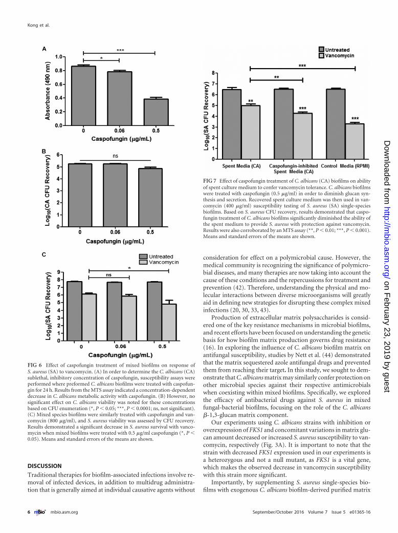
enumeration to determine its effect on metabolic activity and vi-ability, respectively. Results from the MTS assay indicated aconcentration-dependent decrease in C. albicans metabolic activ-ity with caspofungin; however, no effect on viability was noted forthese concentrations (Fig. 6A and B). The selected subinhibitorycaspofungin concentration (0.5 �g/ml) was subsequently used inmixed biofilm vancomycin susceptibility assays. Based on CFUrecovery, results demonstrated a significant decrease in S. aureussurvival with vancomycin when mixed biofilms were treated withcaspofungin (Fig. 6C). Further, experiments were also performedto determine whether the effect of caspofungin on �-1,3-glucansynthesis affects its secretion and, thus, the protective effect ofC. albicans spent medium on S. aureus. Therefore, experimentswere also performed where formed C. albicans biofilms weretreated with caspofungin prior to recovering the spent culturemedium, which was subsequently used in vancomycin suscepti-bility testing. Results from these experiments similarly demon-strated that caspofungin treatment of C. albicans biofilms signifi-cantly diminished the ability of recovered spent medium to conferprotection against vancomycin on S. aureus (Fig. 7). Caspofungindid not have any effect on S. aureus.
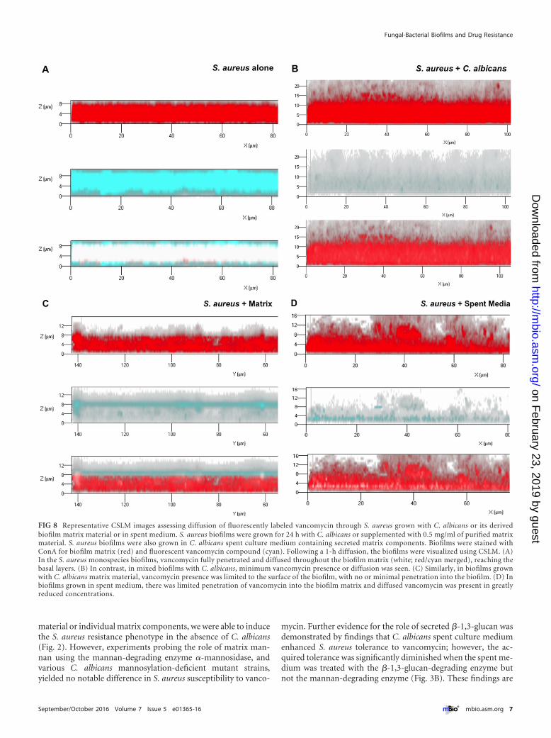
Impeded vancomycin diffusion through S. aureus biofilmsgrown with C. albicans or its purified and secreted matrix ma-terial. Vancomycin diffusion through single and mixed biofilmmatrices was comparatively monitored using fluorescence confo-cal scanning laser microscopy (CSLM). S. aureus biofilms weregrown with C. albicans, with purified C. albicans biofilm matrixmaterial, or in C. albicans spent culture medium with secretedmaterial. Formed biofilms were stained with concanavalin A(ConA) to allow visualization of the polysaccharide matrix (red).To visualize vancomycin diffusion through the matrix, a fluores-cently tagged vancomycin compound was used. After 1 h, vanco-mycin (cyan) had significantly penetrated and diffused through-out the single S. aureus biofilm matrix, reaching the basal layer(Fig. 8A). In contrast, in the mixed biofilms with C. albicans, van-comycin signal was minimally detected, with negligible to no pen-etration into the matrix seen (Fig. 8B). Similarly, in the biofilms
formed with exogenous supplementation with C. albicans matrixmaterial, vancomycin was confined to the outer periphery of thebiofilm with minimal to no penetration into the biofilm (Fig. 8C),and in biofilms grown in spent medium, only limited vancomycinpenetration into the biofilm was seen (Fig. 8D).
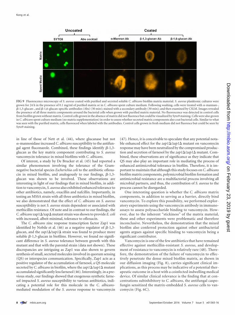
S. aureus cell coating by C. albicans purified and secretedbiofilm matrix components. In order to evaluate whether C. al-bicans matrix polysaccharides also coat the bacterial cells, S. aureusplanktonic cell suspension was incubated with C. albicans purifiedmatrix and coating was assessed using monoclonal antibodies spe-cific to each of the C. albicans matrix components. Using fluores-cence microscopy, images demonstrated rapid coating of all 3 ma-trix components (�-mannan, �-1,3-glucan, and �-1,6-glucan) asassessed by the presence of fluorescence around the bacterial cells.S. aureus cells not treated with matrix demonstrated no fluores-cence when labeled with antibodies. In addition to purified ma-trix, to determine whether the secreted matrix material similarlycoats the bacterial cells, experiments were also performed whereS. aureus was incubated in the C. albicans spent culture medium.Labeling of bacterial cells with the three specific antibodies dem-onstrated the presence of all matrix components around the sur-face of S. aureus cells (Fig. 9).
Enhanced tolerance of S. aureus in mixed biofilm is not an-tibiotic, methicillin resistance, or S. aureus strain dependent. Inorder to determine whether the C. albicans-conferred enhancedtolerance of S. aureus in mixed biofilm is exclusive to vancomycin-or methicillin-resistant strains of S. aureus, susceptibility assayswere also performed using two additional antibiotics, oxacillinand nafcillin. Further, the susceptibility of a methicillin-susceptible S. aureus (MSSA) strain was also tested against all 3antibiotics in biofilms with C. albicans. Based on S. aureus CFUrecovery, results demonstrated that, similarly to vancomycin, theMRSA strain exhibited enhanced tolerance to both oxacillin andnafcillin in mixed biofilm (see Fig. S1 in the supplemental mate-rial). A comparable increase in tolerance to all three antibioticswas also seen with the MSSA strain (data not shown).
FIG 5 Effect of secreted effectors in C. albicans spent medium on S. aureus (SA) vancomycin susceptibility. (A) Supernatant from C. albicans culturemedium (spent medium) was collected and used in S. aureus vancomycin susceptibility testing assays. Based on results from S. aureus CFU recovery,growth in C. albicans spent medium significantly enhanced S. aureus vancomycin tolerance compared to growth in control fresh medium (*, P � 0.05).(B) Since C. albicans-produced glucan is secreted into the medium, spent medium was treated with Zymolyase prior to vancomycin susceptibility testing.No significant effect for Zymolyase alone on S. aureus viability was seen; however, S. aureus tolerance to vancomycin was significantly diminished whenspent medium was treated with Zymolyase. Results were corroborated with an MTS assay (*, P � 0.05; **, P � 0.001; ***, P � 0.0001; ns, not significant).Means and standard errors of the means are shown.
Fungal-Bacterial Biofilms and Drug Resistance
September/October 2016 Volume 7 Issue 5 e01365-16 ® mbio.asm.org 5
on February 23, 2019 by guest
http://mbio.asm
.org/D
ownloaded from
DISCUSSION
Traditional therapies for biofilm-associated infections involve re-moval of infected devices, in addition to multidrug administra-tion that is generally aimed at individual causative agents without
consideration for effect on a polymicrobial cause. However, themedical community is recognizing the significance of polymicro-bial diseases, and many therapies are now taking into account thecause of these conditions and the repercussions for treatment andprevention (42). Therefore, understanding the physical and mo-lecular interactions between diverse microorganisms will greatlyaid in defining new strategies for disrupting these complex mixedinfections (20, 30, 33, 43).
Production of extracellular matrix polysaccharides is consid-ered one of the key resistance mechanisms in microbial biofilms,and recent efforts have been focused on understanding the geneticbasis for how biofilm matrix production governs drug resistance(16). In exploring the influence of C. albicans biofilm matrix onantifungal susceptibility, studies by Nett et al. (44) demonstratedthat the matrix sequestered azole antifungal drugs and preventedthem from reaching their target. In this study, we sought to dem-onstrate that C. albicans matrix may similarly confer protection onother microbial species against their respective antimicrobialswhen coexisting within mixed biofilms. Specifically, we exploredthe efficacy of antibacterial drugs against S. aureus in mixedfungal-bacterial biofilms, focusing on the role of the C. albicans�-1,3-glucan matrix component.
Our experiments using C. albicans strains with inhibition oroverexpression of FKS1 and concomitant variations in matrix glu-can amount decreased or increased S. aureus susceptibility to van-comycin, respectively (Fig. 3A). It is important to note that thestrain with decreased FKS1 expression used in our experiments isa heterozygous and not a null mutant, as FKS1 is a vital gene,which makes the observed decrease in vancomycin susceptibilitywith this strain more significant.
Importantly, by supplementing S. aureus single-species bio-films with exogenous C. albicans biofilm-derived purified matrix
FIG 6 Effect of caspofungin treatment of mixed biofilms on response ofS. aureus (SA) to vancomycin. (A) In order to determine the C. albicans (CA)sublethal, inhibitory concentration of caspofungin, susceptibility assays wereperformed where preformed C. albicans biofilms were treated with caspofun-gin for 24 h. Results from the MTS assay indicated a concentration-dependentdecrease in C. albicans metabolic activity with caspofungin. (B) However, nosignificant effect on C. albicans viability was noted for these concentrationsbased on CFU enumeration (*, P � 0.05; ***, P � 0.0001; ns, not significant).(C) Mixed species biofilms were similarly treated with caspofungin and van-comycin (800 �g/ml), and S. aureus viability was assessed by CFU recovery.Results demonstrated a significant decrease in S. aureus survival with vanco-mycin when mixed biofilms were treated with 0.5 �g/ml caspofungin (*, P �0.05). Means and standard errors of the means are shown.
FIG 7 Effect of caspofungin treatment of C. albicans (CA) biofilms on abilityof spent culture medium to confer vancomycin tolerance. C. albicans biofilmswere treated with caspofungin (0.5 �g/ml) in order to diminish glucan syn-thesis and secretion. Recovered spent culture medium was then used in van-comycin (400 �g/ml) susceptibility testing of S. aureus (SA) single-speciesbiofilms. Based on S. aureus CFU recovery, results demonstrated that caspo-fungin treatment of C. albicans biofilms significantly diminished the ability ofthe spent medium to provide S. aureus with protection against vancomycin.Results were also corroborated by an MTS assay (**, P � 0.01; ***, P � 0.001).Means and standard errors of the means are shown.
Kong et al.
6 ® mbio.asm.org September/October 2016 Volume 7 Issue 5 e01365-16
on February 23, 2019 by guest
http://mbio.asm
.org/D
ownloaded from
material or individual matrix components, we were able to inducethe S. aureus resistance phenotype in the absence of C. albicans(Fig. 2). However, experiments probing the role of matrix man-nan using the mannan-degrading enzyme �-mannosidase, andvarious C. albicans mannosylation-deficient mutant strains,yielded no notable difference in S. aureus susceptibility to vanco-
mycin. Further evidence for the role of secreted �-1,3-glucan wasdemonstrated by findings that C. albicans spent culture mediumenhanced S. aureus tolerance to vancomycin; however, the ac-quired tolerance was significantly diminished when the spent me-dium was treated with the �-1,3-glucan-degrading enzyme butnot the mannan-degrading enzyme (Fig. 3B). These findings are
FIG 8 Representative CSLM images assessing diffusion of fluorescently labeled vancomycin through S. aureus grown with C. albicans or its derivedbiofilm matrix material or in spent medium. S. aureus biofilms were grown for 24 h with C. albicans or supplemented with 0.5 mg/ml of purified matrixmaterial. S. aureus biofilms were also grown in C. albicans spent culture medium containing secreted matrix components. Biofilms were stained withConA for biofilm matrix (red) and fluorescent vancomycin compound (cyan). Following a 1-h diffusion, the biofilms were visualized using CSLM. (A)In the S. aureus monospecies biofilms, vancomycin fully penetrated and diffused throughout the biofilm matrix (white; red/cyan merged), reaching thebasal layers. (B) In contrast, in mixed biofilms with C. albicans, minimum vancomycin presence or diffusion was seen. (C) Similarly, in biofilms grownwith C. albicans matrix material, vancomycin presence was limited to the surface of the biofilm, with no or minimal penetration into the biofilm. (D) Inbiofilms grown in spent medium, there was limited penetration of vancomycin into the biofilm matrix and diffused vancomycin was present in greatlyreduced concentrations.
Fungal-Bacterial Biofilms and Drug Resistance
September/October 2016 Volume 7 Issue 5 e01365-16 ® mbio.asm.org 7
on February 23, 2019 by guest
http://mbio.asm
.org/D
ownloaded from
in line of those of Nett et al. (44), where glucanase but not�-mannosidase increased C. albicans susceptibility to the antifun-gal agent fluconazole. Combined, these findings identify �-1,3-glucan as the key matrix component contributing to S. aureusvancomycin tolerance in mixed biofilms with C. albicans.
Of interest, a study by De Brucker et al. (45) had reported asimilar phenomenon involving the tolerance of the Gram-negative bacterial species Escherichia coli to the antibiotic ofloxa-cin in mixed biofilm, and analogously to our findings, �-1,3-glucan was shown to be involved. These observations areinteresting in light of our findings that in mixed biofilm, in addi-tion to vancomycin, S. aureus also exhibited enhanced tolerance toother antibiotics, namely, oxacillin and nafcillin. Importantly, intesting an MSSA strain with all three antibiotics in mixed biofilm,we also demonstrated that the effect of C. albicans on S. aureussusceptibility is not S. aureus strain dependent or associated withmethicillin resistance. Of note and in contrast to our findings, theC. albicans zap1�/zap� mutant strain was shown to provide E. coliwith increased, albeit minimal, tolerance to ofloxacin.
The C. albicans zinc response transcription factor Zap1 wasidentified by Nobile et al. (46) as a negative regulator of �-1,3-glucan, and the zap1�/zap1� strain was found to produce moresoluble �-1,3-glucan in biofilms. However, we found no signifi-cant difference in S. aureus tolerance between growth with thismutant and that with the parental strain (data not shown). Thesediscrepancies are intriguing as Zap1 was also shown to governsynthesis of small, secreted molecules involved in quorum sensing(QS) or interspecies communication. Specifically, Zap1 acts as apositive regulator of the accumulation of farnesol, a QS moleculesecreted by C. albicans in biofilm, where the zap1�/zap1� mutantaccumulated significantly less farnesol (46). Interestingly, in a pre-vious study, our findings showed that exogenous synthetic farne-sol impacted S. aureus susceptibility to various antibiotics, indi-cating a potential role for this molecule in the C. albicans-mediated modulation of the S. aureus response to vancomycin
(47). Hence, it is conceivable to speculate that any potential nota-ble enhanced effect for the zap1�/zap1� mutant on vancomycinresponse may have been neutralized by the compromised produc-tion and secretion of farnesol by the zap1�/zap1� mutant. Com-bined, these observations are of significance as they indicate thatQS may also play an important role in mediating the process ofenhanced antimicrobial tolerance in biofilm. Therefore, it is im-portant to maintain that although this study focuses on C. albicansbiofilm matrix components, polymicrobial biofilm formation andantimicrobial resistance are a multifactorial process involving allmicrobial partners, and thus, the contribution of S. aureus to theprocess cannot be disregarded.
One interesting question is whether the C. albicans matrixcomponents, in addition to serving as a barrier, directly bindvancomycin. To explore this possibility, we performed explor-atory experiments using the vancomycin antibody in immuno-assays to assess polysaccharide binding to vancomycin. How-ever, due to the inherent “stickiness” of the matrix material,these and other experiments were problematic and thereforeinconclusive. Nevertheless, the demonstration that the mixedbiofilm also conferred protection against other antibacterialagents argues against specific binding to vancomycin being asignificant contributor.
Vancomycin is one of the few antibiotics that have remainedeffective against methicillin-resistant S. aureus, and develop-ment of resistance to vancomycin is relatively rare (48). There-fore, the demonstration of the failure of vancomycin to effec-tively penetrate the dense mixed biofilm matrix, as shown inour diffusion imaging (Fig. 8), carries significant clinical im-plications, as this process may be indicative of a potential ther-apeutic outcome in a host with a coinfected indwelling medicaldevice. Of similar clinical relevance is the finding that at con-centrations subinhibitory to C. albicans, the antifungal caspo-fungin sensitized the matrix-embedded S. aureus cells to van-comycin (Fig. 6C).
FIG 9 Fluorescence microscopy of S. aureus coated with purified and secreted soluble C. albicans biofilm matrix material. S. aureus planktonic cultures weregrown for 24 h in the presence of 0.1 mg/ml of purified matrix or in C. albicans spent culture medium. Following washing, cells were treated with �-mannan-,�-1,3-glucan-, and �-1,6-glucan-specific antibodies (Abs) (30 min); stained with a secondary antibody (30 min); and then examined by CSLM. Images revealedthe presence of all three matrix components around the bacterial cells when grown with purified matrix material. No fluorescence was detected in control cellsfrom biofilm grown without matrix. Control cells grown in the absence of matrix did not fluoresce but could be visualized by Syto9 staining. Cells were also grownin C. albicans spent culture medium (no matrix supplementation) in order to assess whether secreted matrix components also coat bacterial cells. Similar to whatwas seen with the purified matrix, cells fluoresced when labeled with the antibodies. Control cells grown in fresh medium did not fluoresce but could be seen bySyto9 staining.
Kong et al.
8 ® mbio.asm.org September/October 2016 Volume 7 Issue 5 e01365-16
on February 23, 2019 by guest
http://mbio.asm
.org/D
ownloaded from
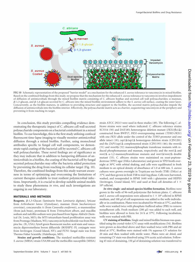
In conclusion, this study provides compelling evidence dem-onstrating the therapeutic impact of C. albicans cell wall secretedpolysaccharide components on a bacterial coinhabitant in a mixedbiofilm. To our knowledge, this is the first study utilizing confocalfluorescent time-lapse imaging to visually monitor antimicrobialdiffusion through a mixed biofilm. Further, using monoclonalantibodies specific to fungal cell wall components, we demon-strate rapid coating of the bacterial cell by secreted C. albicans cellwall polysaccharides. These novel findings are of significance asthey may indicate that in addition to hampering diffusion of an-timicrobials in a biofilm, the coating of the bacterial cell by fungalsecreted polysaccharides may offer the bacteria added protectionby preventing the drug from reaching its cellular target (Fig. 10).Therefore, the combined findings from this study warrant aware-ness in terms of optimizing and overcoming the limitations ofcurrent therapies available to treat resilient polymicrobial infec-tions. Importantly, it is crucial to develop suitable animal modelsto study these phenomena in vivo, and such investigations areongoing in our laboratory.
MATERIALS AND METHODSReagents. �-1,3-Glucan (laminarin from Laminaria digitata), lyticasefrom Arthrobacter luteus (Zymolyase), mannan (from Saccharomycescerevisiae), concanavalin A (from Canavalia ensiformis), �-mannosidase(from jack bean), proteinase K from Tritirachium album, and oxacillinsodium and nafcillin sodium were purchased from Sigma-Aldrich Chem-ical (St. Louis, MO); the MTS tetrazolium-based proliferation assay wasfrom Promega (Madison, WI); vancomycin hydrochloride was from Hos-pira Inc. (IL, USA); Syto9 green fluorescent nucleic acid stain and vanco-mycin dipyrromethene boron difluoride (BODIPY) FL conjugate werefrom Invitrogen (Grand Island, NY); and FUN1 fungal stain was fromThermo Fisher Scientific (Halethorpe, MD).
Strains and growth conditions. The standard methicillin-resistantS. aureus (MRSA) strain USA300 and the methicillin-susceptible (MSSA)
strain ATCC 29213 were used in these studies (49). The following C. al-bicans strains were used where indicated: C. albicans reference strainsSC5314 (50) and DAY185; heterozygous deletion mutant (FKS1/fks1�)constructed from BWP17, FKS1-overexpressing mutant (TDH3-FKS1)with one FKS1 allele under the control of the TDH3 promoter and oneallele intact (18); zap1�/zap1� homozygous deletion strain (CJN1201)and the ZAP1/zap1� complemented strain (CJN1193) (46); the mnn4�(51) and mnn9� (52) mannosylphosphate transferase mutants with re-duced phosphomannan and mannan, respectively; and the mnt1� andmnt2� �-1,2-mannosyltransferase mutants and mnt1�/mnt2� doublemutant (53). C. albicans strains were maintained on yeast-peptone-dextrose (YPD) agar (Difco Laboratories) and grown in YPD broth over-night at 30°C with orbital shaking, and cells were equilibrated in freshmedium to an optical density of absorbance of 1.0 at 600 nm. S. aureuscultures were grown overnight in Trypticase soy broth (TSB) (Difco) at37°C and then grown in fresh TSB to mid-log phase. Cells were harvested,washed, and resuspended in RPMI 1640 with L-glutamine and HEPES(Invitrogen, Grand Island, NY) and used at final cell densities of 1 �106 cells/ml.
In vitro single- and mixed-species biofilm formation. Biofilms weregrown in the wells of 96-well polystyrene flat-bottom plates. C. albicansand S. aureus cell suspensions were adjusted to 1 � 106 cells/ml in RPMImedium, and 100 �l of cell suspensions was added to the wells individu-ally or in combination. Plates were incubated for 90 min at 37°C, and thenwells were washed twice with phosphate-buffered saline (PBS) to removenonadherent cells. Fresh medium (200 �l) was added to each well, andbiofilms were allowed to form for 24 h at 37°C. Following incubation,wells were washed with PBS.
CV staining of biofilms. Single and mixed biofilm biomass was quan-tified using the crystal violet (CV) assay with modifications (54). Biofilmswere grown as described above and then washed twice with PBS and airdried at 37°C. Biofilms were stained with 1% aqueous CV solution for20 min and then washed with sterile water. Plates were air dried, andremaining CV stain was dissolved using 33% acetic acid solution. Follow-ing 45 min of destaining, 150 �l of destaining solution was transferred to
FIG 10 Schematic representation of the proposed “barrier model” as a mechanism for the enhanced S. aureus tolerance to vancomycin in mixed biofilm.Based on the combined findings from this study, we propose that the mechanism for the enhanced S. aureus tolerance to vancomycin involves impedimentof diffusion of antimicrobials through the mixed biofilm matrix consisting of C. albicans hyphae and secreted cell wall polysaccharides; �-mannan,�-1,3-glucan, and �-1,6-glucan secreted by C. albicans into the mixed biofilm environment adhere to the S. aureus cell surface, coating the outer layer.Concurrently, as the biofilm matures, in addition to providing structure and support to the biofilm, the secreted matrix polysaccharides impede thediffusion of antimicrobials into the biofilm interior. Effectively, the polysaccharide matrix acts as a barrier, sequestering vancomycin at the periphery andpreventing it from reaching its target.
Fungal-Bacterial Biofilms and Drug Resistance
September/October 2016 Volume 7 Issue 5 e01365-16 ® mbio.asm.org 9
on February 23, 2019 by guest
http://mbio.asm
.org/D
ownloaded from

a new well, and the amount of CV stain was measured with a microtiterplate reader at 595 nm (Titertek; Multiskan MCC1340).
Biofilm vancomycin susceptibility testing. The impact of C. albicanson the response of S. aureus to vancomycin was assessed in biofilms grownas described above. Mixed biofilms were grown using S. aureus and each ofthe C. albicans mutant strains for 24 h. Following washing, the wells weresupplemented with fresh medium, vancomycin hydrochloride at a finalconcentration of 800 �g/ml (predetermined based on activity againstS. aureus single and mixed biofilms) (see Fig. S2 in the supplementalmaterial) was added, and plates were incubated for an additional 24 h at37°C. Biofilms were then washed twice with PBS, and 100 �l of PBS wasadded; cells from the biofilms were recovered by sonication followed byvigorous vortexing and pipetting. Cell suspensions were diluted andplated on C. albicans and S. aureus-specific chromogenic medium(CHROMagar; DRG International, Inc.) for CFU count. Drug-free wellswere included as controls. In addition, in order to assess the effect ofmixed biofilm growth on S. aureus susceptibility to other antibiotics, ex-periments were also performed using two additional antibiotics, oxacillin(0 to 480 �g/ml) and nafcillin (0 to 160 �g/ml). Further, susceptibilitytesting using all 3 antibiotics was performed using an alternate strain ofS. aureus that is susceptible to methicillin (MSSA).
MTS assay. Viability was also assessed using the MTS metabolic assayaccording to the manufacturer’s directions. Following washing with PBS,100 �l of PBS was added to the wells followed by 20 �l of MTS reagent andplates were incubated at 37°C until color fully developed. Following colordevelopment, colorimetric change at 490 nm (A490) was measured with amicrotiter plate reader. On each occasion, reactions were performed intriplicate.
SEM of single- and mixed-species biofilms. For SEM analysis, S. au-reus was grown in single species biofilm with and without exogenoussupplementation of purified matrix material (0.5 mg/ml) or glucan(0.25 mg/ml) or with C. albicans in mixed biofilms, on coverslips for 48 h.Coverslips were washed twice with PBS and then fixed in 2% paraformal-dehyde, 2.5% glutaraldehyde in phosphate buffer, pH 7.4, for 1 h at roomtemperature and then at 4°C overnight. Following initial fixation, speci-mens were washed in three changes of 0.1 M PBS for a total of 30 min,postfixed with 1% osmium tetroxide in PBS for 1 h, and washed again inthree changes of buffer. Dehydration of specimens was done using a seriesof graded ethyl alcohol, 30%, 50%, 70%, 90%, and 100% for 10 min each,and two more changes of 100% ethyl alcohol. Specimens were then chem-ically dried by immersing them sequentially in 2 parts 100% ethylalcohol-1 part hexamethyldisilazane (HMDS) (Electron Microscopy Sci-ences, Fort Washington, PA) for 10 min, 1 part 100% ethyl alcohol-1 partHDMS for 10 min, 1 part 100% ethyl alcohol-2 parts HDMS for 10 min,and then 2 changes for 10 min each with 100% HDMS. Specimens were airdried in a hood overnight, mounted on SEM pin mounts, and sputtercoated with 10 to 20 nm of platinum-palladium in a sputter coater (EMS150T ES). SEM images were captured using a Quanta 200 scanning elec-tron microscope (FEI Co., Hillsboro, OR).
Effect of C. albicans purified biofilm-derived matrix material and�-1,3-glucan and mannan exogenous supplementation on S. aureussusceptibility to vancomycin in single-species biofilm. C. albicans ma-trix polysaccharides were extracted from C. albicans biofilms and purifiedas previously described (55). The recovered matrix material (0.25 mg/ml)was used to supplement S. aureus biofilms, which were allowed to form for24 h as described above. S. aureus biofilms were also formed in the pres-ence of exogenous �-1,3-glucan (1 mg/ml) or mannan (1 mg/ml) as pre-viously performed (44). Following incubation, biofilms were washed andsupplemented with fresh RPMI medium and vancomycin (800 �g/ml),and plates were incubated for an additional 24 h. Following washing,S. aureus viability was assessed using the MTS assay and confirmed byCFU recovery for viability.
Impact of C. albicans �-1,3-glucan and mannan enzymatic degra-dation on S. aureus susceptibility to vancomycin in mixed biofilms.Prior to performing experiments, susceptibility of C. albicans to Zy-
molyase and �-mannosidase was tested to determine the subinhibitoryconcentrations to be used in mixed biofilm susceptibility testing. Mixed-species biofilms were grown alone or in the presence of glucanase (Zy-molyase) (5 U/ml) or �-mannosidase (2 U/ml). Vancomycin (800 �g/ml)was then added alone or in combination with the enzymes, and plateswere incubated for an additional 24 h at 37°C. Following incubation, wellswere washed three times with PBS and biofilm cells were recovered bysonication and plated for CFU counts. In order to demonstrate lack ofcontribution of contaminating protease activity in Zymolyase, experi-ments were also performed using enzymatic digestion with proteinase K.
Impact of inhibition of C. albicans in �-1,3-glucan synthesis onS. aureus susceptibility to vancomycin in mixed biofilms. Mixed 24-hbiofilms were treated with the �-1,3-glucan synthase inhibitor caspofun-gin at final concentrations of 0, 0.06, and 0.5 �g/ml with and withoutvancomycin (800 �g/ml), and plates were incubated for an additional24 h. Following washing, biofilms were sonicated and S. aureus viabilitywas assessed based on CFU counts. Prior to experiments, susceptibility ofC. albicans to caspofungin in single species was assessed in order to deter-mine the subinhibitory caspofungin concentration to be used in mixedbiofilms. Viability was evaluated using the MTS assay to determinechanges in metabolic activity and CFU enumeration for viability.
Effect of C. albicans soluble secreted matrix components on S. au-reus susceptibility to vancomycin. In order investigate the role of C. al-bicans secreted matrix components on vancomycin susceptibility, cell-free C. albicans biofilm spent culture medium was recovered. Briefly,C. albicans (1 � 106 cells/ml) biofilms were grown in 10 ml of RPMImedium in canted-neck flasks and incubated at 37°C for 48 h. Followingincubation, spent culture medium was recovered and filter sterilizedthrough a 0.22-�m pore. The filtered medium was then supplemented 1:1with fresh RPMI medium (prewarmed to 37°C) and used to grow S. au-reus single-species biofilms. Following 24-h incubation, biofilms werewashed and then supplemented with fresh medium and vancomycin(400 �g/ml) (predetermined to be subinhibitory in S. aureus single-species biofilm) (see Fig. S2 in the supplemental material) and incubatedfor an additional 24 h. S. aureus viability was assessed using the MTS assayand based on CFU recovery. To identify �-1,3-glucan as the key secretedcomponent in C. albicans culture medium, experiments were also per-formed where S. aureus biofilms were cultured in the spent medium (asdescribed above) with the addition of 5 U/ml of the glucan-degradingenzyme glucanase (Zymolyase) for 24 h at 37°C. Following incubation,vancomycin susceptibility testing was performed. Spent medium withoutglucanase was included as a control. In addition, C. albicans biofilms weregrown for 24 h and then treated with the �-1,3-glucan synthesis inhibitorcaspofungin (0.5 �g/ml) (subinhibitory dose for C. albicans) for an addi-tional 24 h. Spent medium was then collected and used in vancomycinsusceptibility testing of S. aureus single-species biofilms as describedabove.
Confocal scanning laser microscopy analysis of vancomycin diffu-sion through single and mixed biofilms. To visualize the process of van-comycin diffusion through single and mixed biofilms, confocal scanninglaser microscopy was performed as based on a previously describedmethod for S. aureus with modifications (48). In addition to monitoringvancomycin diffusion through mixed biofilms, experiments were alsoperformed where S. aureus single biofilms were formed in the presence ofexogenous supplementation of purified C. albicans biofilm matrix to iso-late the role of the matrix. Further, as matrix components are secretedduring C. albicans growth, biofilm spent culture medium was used togrow S. aureus biofilms. For these experiments, biofilms were grown onglass coverslip-bottom dishes (MatTek Co., Ashland, MA) in RPMI me-dium at 37°C for 24 h. Following incubation, biofilms were gently washedthree times with PBS and stained with concanavalin A (ConA) for poly-saccharide biofilm matrix (100 �g/ml) (red; 488/545). In order to visual-ize the diffusion of vancomycin, the fluorescent BODIPY FL conjugate ofvancomycin (cyan; 488/650) was added to the biofilms to a final concen-tration of 1 �g/ml. Biofilms were incubated for 1 h and then washed three
Kong et al.
10 ® mbio.asm.org September/October 2016 Volume 7 Issue 5 e01365-16
on February 23, 2019 by guest
http://mbio.asm
.org/D
ownloaded from
times with PBS to remove nonpenetrated vancomycin and observed usinga 63� oil immersion objective and a Zeiss 710 confocal microscope. Im-ages were obtained by LSM 5 Image Browser software at a resolution of512 by 512 pixels, with an average of 8 images per line. To evaluate thestructure and size of the biofilms, a series of images at �1-�m intervals inthe z axis were acquired for the full depth of the biofilm. At least threerandom fields were visualized for each biofilm, and representative imagesare presented.
Fluorescence microscopy analysis of S. aureus cell coating with pu-rified and secreted soluble C. albicans biofilm matrix material. In orderto examine whether, in addition to their role in biofilms, purified andsecreted matrix components also coat the bacterial cell, S. aureus cells (2 �107 cells/ml) were incubated with 0.1 mg/ml of the purified matrixmaterial for 30 min at 37°C. S. aureus cells with no matrix treatmentwere included as controls. In addition to purified matrix, experimentswere also performed where S. aureus was exposed to C. albicans spentculture medium in order to determine whether soluble matrix com-ponents secreted by C. albicans during biofilm growth similarly coatthe bacterial cells. In these experiments, 2 � 107 cells/ml of S. aureuswere incubated with 1 ml of C. albicans spent medium for 30 min at37°C. S. aureus cells incubated in fresh medium were included as acontrol. Following three washes with PBS, cells were treated withmonoclonal antibodies specific to �-mannan (1/100 dilution), �-1,3-glucans (1/6,000 dilution), and �-1,6-glucans (1/4,000 dilution) (pro-duced as previously described) (14) for 1 h at 37°C. Following incuba-tion with primary antibodies, cells were washed three times with PBSand subsequently incubated with a goat anti-mouse IgG Alexa Fluor488 (orange; 495/519) secondary antibody (1/100 dilution) (ThermoFisher Scientific) for 30 min at 37°C. Cells were then washed, pelleted,and resuspended in 50 �l PBS. One drop of cell suspension was placedon a slide and covered with a coverslip, and fluorescence was assessedby CSLM. Non-matrix-coated S. aureus cells similarly treated with theantibodies were used as a control. However, as control cells with nomatrix did not react with antibodies and therefore were not visible,they were stained with Syto9 nucleic acid stain (10 �M) (green; 488/505) in order to be visualized. Cells were observed using a 63� oilimmersion objective and a Zeiss 710 confocal microscope. Imageswere obtained by LSM 5 Image Browser software at a resolution of 512by 512 pixels, with an average of 8 images per line. At least threerandom fields were visualized for each sample, and representative im-ages are presented.
Data analysis. All experiments were performed on at least 3 separateoccasions and in triplicate where applicable, and averages were used topresent data. All statistical analysis was performed using GraphPad Prism5.0 software. The Kruskal-Wallis one-way analysis of variance test wasused to compare differences between multiple groups, and Dunn’smultiple-comparison test was used to determine whether the differencebetween two samples was statistically significant. Student’s unpaired t testwas used to compare differences between two samples. P values of �0.05were considered to be significant.
SUPPLEMENTAL MATERIALSupplemental material for this article may be found at http://mbio.asm.org/lookup/suppl/doi:10.1128/mBio.01365-16/-/DCSupplemental.
Figure S1, TIF file, 0.8 MB.Figure S2, TIF file, 0.5 MB.
ACKNOWLEDGMENTS
We thank Hiram Sanchez for his contribution and Vincent Bruno andAaron Mitchell for providing us with C. albicans strains.
This work was supported by NIH grant DE14424 to Mary Ann Jabra-Rizk; the Interuniversity Attraction Poles Programme initiated by the Bel-gian Science Policy Office to Patrick Van Dijck and Mary Ann Jabra-Rizk;the Flemish Science Foundation (FWO), WO.026.11N, to Patrick VanDijck and Mary Ann Jabra-Rizk; and the FWO postdoctoral fellowships
awarded to Sona Kucharíková. The funders had no role in study design,data collection and interpretation, or the decision to submit the work forpublication.
FUNDING INFORMATIONThis work, including the efforts of Mary Ann Jabra-Rizk, was funded byNIH (DE14424). This work, including the efforts of Patrick Van Dijck andMary Ann Jabra-Rizk, was funded by Flemish Science Foundation(WO.026.11N).
REFERENCES1. Brogden KA, Guthmiller JM, Taylor CE. 2005. Human polymicrobial
infections. Lancet 365:253–255. http://dx.doi.org/10.1016/S0140-6736(05)17745-9.
2. Peters BM, Jabra-Rizk MA, O’May GA, Costerton JW, Shirtliff ME.2012. Polymicrobial interactions in biofilms: impact on pathogenesis andhuman disease. Clin Microbiol Rev 25:193–213. http://dx.doi.org/10.1128/CMR.00013-11.
3. Jabra-Rizk MA. 2011. Pathogenesis of polymicrobial biofilms. Open My-col J 5:39 – 43. http://dx.doi.org/10.2174/1874437001105010039.
4. O’Toole G, Kaplan HB, Kolter R. 2000. Biofilm formation as microbialdevelopment. Annu Rev Microbiol 54:49 –79. http://dx.doi.org/10.1146/annurev.micro.54.1.49.
5. Lewis K. 2001. Riddle of biofilm resistance. Antimicrob Agents Che-mother 45:999 –1007. http://dx.doi.org/10.1128/AAC.45.4.999-1007.2001.
6. Ghannoum M, Roilides E, Katragkou A, Petraitis V, Walsh TJ. 2015.The role of echinocandins in Candida biofilm–related vascular catheterinfections: in vitro and in vivo model systems. Clin Infect Dis 61(Suppl6):S618 –S621. http://dx.doi.org/10.1093/cid/civ815.
7. Finkel JS, Mitchell AP. 2011. Genetic control of Candida albicans biofilmdevelopment. Nat Rev Microbiol 9:109 –118. http://dx.doi.org/10.1038/nrmicro2475.
8. Ganguly S, Mitchell AP. 2011. Mucosal biofilms of Candida albicans.Curr Opin Microbiol 14:380 –385. http://dx.doi.org/10.1016/j.mib.2011.06.001.
9. Calderone R (ed). 2012. Candida and candidiasis, 2nd ed. ASM Press,Washington, DC.
10. Pfaller MA, Diekema DJ. 2007. Epidemiology of invasive candidiasis: apersistent public health problem. Clin Microbiol Rev 20:133–163. http://dx.doi.org/10.1128/CMR.00029-06.
11. Chauvel M, Nesseir A, Cabral V, Znaidi S, Goyard S, Bachellier-Bassi S,Firon A, Legrand M, Diogo D, Rossignol T, Naulleau C, d’Enfert C.2012. A versatile overexpression strategy in the pathogenic yeast Candidaalbicans: identification of regulators of morphogenesis and fitness. PLoSOne 7:e45912. http://dx.doi.org/10.1371/journal.pone.0045912.
12. Nett J, Andes D. 2006. Candida albicans biofilm development, modelinga host-pathogen interaction. Curr Opin Microbiol 9:340 –345. http://dx.doi.org/10.1016/j.mib.2006.06.007.
13. Tournu H, Van Dijck P. 2012. Candida biofilms and the host: models andnew concepts for eradication. Int J Microbiol 2012: http://dx.doi.org/10.1155/2012/845352.
14. Mitchell KF, Zarnowski R, Sanchez H, Edward JA, Reinicke EL, Nett JE,Mitchell AP, Andes DR. 2015. Community participation in biofilm ma-trix assembly and function. Proc Natl Acad Sci U S A 112:4092– 4097.http://dx.doi.org/10.1073/pnas.1421437112.
15. Hall R, Gow NA. 2013. Mannosylation in Candida albicans: role in cellwall function and immune recognition. Mol Microbiol 90:1147–1161.http://dx.doi.org/10.1111/mmi.12426.
16. Taff HT, Nett JE, Zarnowski R, Ross KM, Sanchez H, Cain MT,Hamaker J, Mitchell AP, Andes DR. 2012. A Candida biofilm-inducedpathway for matrix glucan delivery: implications for drug resistance. PLoSPathog 8:e1002848. http://dx.doi.org/10.1371/journal.ppat.1002848.
17. Taff HT, Mitchell KF, Edward JA, Andes DR. 2013. Mechanisms ofCandida biofilm drug resistance. Future Microbiol 8:1325–1337. http://dx.doi.org/10.2217/fmb.13.101.
18. Nett JE, Crawford K, Marchillo K, Andes DR. 2010. Role of Fks1p andmatrix glucan in Candida albicans biofilm resistance to an echinocandin,pyrimidine, and polyene. Antimicrob Agents Chemother 54:3505–3508.http://dx.doi.org/10.1128/AAC.00227-10.
19. Jenkinson HF, Barbour ME, Jagger DC, Miles M, Bamford CM, NobbsAH, Dutton LC, Silverman RJ, McNally L, Vickerman MM, Gill S. 2008.
Fungal-Bacterial Biofilms and Drug Resistance
September/October 2016 Volume 7 Issue 5 e01365-16 ® mbio.asm.org 11
on February 23, 2019 by guest
http://mbio.asm
.org/D
ownloaded from

Candida albicans-bacteria interactions in biofilms and disease, p 1– 6. Uni-versity of Bristol Dental School, Bristol, United Kingdom.
20. Shirtliff ME, Peters BM, Jabra-Rizk MA. 2009. Cross-kingdominteractions: Candida albicans and bacteria. FEMS Microbiol Lett 299:1– 8. http://dx.doi.org/10.1111/j.1574-6968.2009.01668.x.
21. Morales DK, Hogan DA. 2010. Candida albicans interactions with bacte-ria in the context of human health and disease. PLoS Pathog 6:e1000886.http://dx.doi.org/10.1371/journal.ppat.1000886.
22. Rehm SJ. 2008. Staphylococcus aureus: the new adventures of a legendarypathogen. Cleve Clin J Med 75:177–192. http://dx.doi.org/10.3949/ccjm.75.3.177.
23. Otto M. 2013. Staphylococcal infections: mechanisms of biofilm matura-tion and detachment as critical determinants of pathogenicity. Annu RevMed 64:175–188. http://dx.doi.org/10.1146/annurev-med-042711-140023.
24. McGavin MJ, Heinrichs DE. 2012. The staphylococci and staphylococcalpathogenesis. Front Cell Infect Microbiol 2: http://dx.doi.org/10.3389/fcimb.2012.00066.
25. Sakoulas G, Moellering RC. 2008. Increasing antibiotic resistance amongmethicillin-resistant Staphylococcus aureus strains. Clin Infect Dis46(Suppl 5):S360 –S367. http://dx.doi.org/10.1086/533592.
26. Gordon RJ, Lowy FD. 2008. Pathogenesis of methicillin-resistant Staph-ylococcus aureus infection. Clin Infect Dis 46(Suppl 5):S350 –S359. http://dx.doi.org/10.1086/533591.
27. Peters B, Ovchinnikova E, Schlecht L, Hoyer L, Busscher H, van derMei H, Krom B, Jabra-Rizk MA, Shirtliff M. 2012. Staphylococcus aureusadherence to Candida albicans hyphae is mediated by the hyphal adhesinAls3p. Microbiology 158:2975–2986. http://dx.doi.org/10.1099/mic.0.062109-0.
28. Peters BM, Jabra-Rizk MA, Scheper MA, Leid JG, Costerton JW,Shirtliff ME. 2010. Microbial interactions and differential protein expres-sion in Staphylococcus aureus and Candida albicans dual-species biofilms.FEMS Immunol Med Microbiol 59:493–503. http://dx.doi.org/10.1111/j.1574-695X.2010.00710.x.
29. Harriott MM, Noverr MC. 2009. Candida albicans and Staphylococcusaureus form polymicrobial biofilms: effects on antimicrobial resistance.Antimicrob Agents Chemother 53:3914 –3922. http://dx.doi.org/10.1128/AAC.00657-09.
30. Harriott MM, Noverr MC. 2011. Importance of Candida-bacterial poly-microbial biofilms in disease. Trends Microbiol 19:557–563. http://dx.doi.org/10.1016/j.tim.2011.07.004.
31. Lazzell AL, Chaturvedi AK, Pierce CG, Prasad D, Uppuluri P, Lopez-Ribot JL. 2009. Treatment and prevention of Candida albicans biofilmswith caspofungin in a novel central venous catheter murine model ofcandidiasis. J Antimicrob Chemother 64:567–570. http://dx.doi.org/10.1093/jac/dkp242.
32. Chrissoheris MP, Libertin C, Ali RG, Ghantous A, Bekui A, DonohueT. 2009. Endocarditis complicating central venous catheter bloodstreaminfections: a unique form of health care associated endocarditis. Clin Car-diol 32:E48 –E54. http://dx.doi.org/10.1002/clc.20498.
33. Klotz SA, Chasin BS, Powell B, Gaur NK, Lipke PN. 2007. Polymicrobialbloodstream infections involving Candida species: analysis of patients andreview of the literature. Diagn Microbiol Infect Dis 59:401– 406. http://dx.doi.org/10.1016/j.diagmicrobio.2007.07.001.
34. Timsit JF, Cheval C, Gachot B, Bruneel F, Wolff M, Carlet J, Regnier B.2001. Usefulness of a strategy based on bronchoscopy with direct exami-nation of bronchoalveolar lavage fluid in the initial antibiotic therapy ofsuspected ventilator-associated pneumonia. Intensive Care Med 27:640 – 647. http://dx.doi.org/10.1007/s001340000840.
35. Pate JC, Jones DB, Wilhelmus KR. 2006. Prevalence and spectrum ofbacterial co-infection during fungal keratitis. Br J Ophthalmol 90:289 –292. http://dx.doi.org/10.1136/bjo.2005.081869.
36. Tawara Y, Honma K, Naito Y. 1996. Methicillin-resistant Staphylococcusaureus and Candida albicans on denture surfaces. Bull Tokyo Dent Coll37:119 –128.
37. Gupta N, Haque A, Mukhopadhyay G, Narayan RP, Prasad R. 2005.Interactions between bacteria and Candida in the burn wound. Burns31:375–378. http://dx.doi.org/10.1016/j.burns.2004.11.012.
38. Baena-Monroy T, Moreno-Maldonado V, Franco-Martínez F, Aldape-Barrios B, Quindós G, Sánchez-Vargas LO. 2005. Candida albicans,Staphylococcus aureus and Streptococcus mutans colonization in patientswearing dental prosthesis. Med Oral Patol Oral Cir Bucal 10(Suppl 1):E27–E39.
39. Kong EF, Kucharíková S, Van Dijck P, Peters BM, Shirtliff ME, Jabra-Rizk MA. 2015. Clinical implications of oral candidiasis: host tissue dam-age and disseminated bacterial disease. Infect Immun 83:604 – 613. http://dx.doi.org/10.1128/IAI.02843-14.
40. Harriott MM, Lilly EA, Rodriguez TE, Fidel PL, Noverr MC. 2010.Candida albicans forms biofilms on the vaginal mucosa. Microbiology156:3635–3644. http://dx.doi.org/10.1099/mic.0.039354-0.
41. Adam B, Baillie GS, Douglas LJ. 2002. Mixed species biofilms of Candidaalbicans and Staphylococcus epidermidis. J Med Microbiol 51:344 –349.http://dx.doi.org/10.1099/0022-1317-51-4-344.
42. Sancho S, Artero A, Zaragoza R, Camarena JJ, González R, NogueiraJM. 2012. Impact of nosocomial polymicrobial bloodstream infections onthe outcome in critically ill patients. Eur J Clin Microbiol Infect Dis 31:1791–1796. http://dx.doi.org/10.1007/s10096-011-1503-8.
43. Bouza E, Burillo A, Muñoz P, Guinea J, Marín M, Rodríguez-CréixemsM. 2013. Mixed bloodstream infections involving bacteria and Candidaspp. J Antimicrob Chemother 68:1881–1888. http://dx.doi.org/10.1093/jac/dkt099.
44. Nett J, Lincoln L, Marchillo K, Massey R, Holoyda K, Hoff B, Van-Handel M, Andes D. 2007. Putative role of beta-1,3 glucans in Candidaalbicans biofilm resistance. Antimicrob Agents Chemother 51:510 –520.http://dx.doi.org/10.1128/AAC.01056-06.
45. De Brucker K, Tan Y, Vints K, De Cremer K, Braem A, Verstraeten N,Michiels J, Vleugels J, Cammue BP, Thevissen K. 2015. Fungal 1,3-glucan increases ofloxacin tolerance of Escherichia coli in a polymicrobialE. coli/Candida albicans biofilm. Antimicrob Agents Chemother 59:3052–3058. http://dx.doi.org/10.1128/AAC.04650-14.
46. Nobile CJ, Nett JE, Hernday AD, Homann OR, Deneault J-S, Nantel A,Andes DR, Johnson AD, Mitchell AP. 2009. Biofilm matrix regulation byCandida albicans Zap1. PLoS Biol 7:e1000133. http://dx.doi.org/10.1371/journal.pbio.1000133.
47. Jabra-Rizk MA, Meiller TF, James CE, Shirtliff ME. 2006. Effect offarnesol on Staphylococcus aureus biofilm formation and antimicrobialresistance. Antimicrob Agents Chemother 50:1463–1469. http://dx.doi.org/10.1128/AAC.50.4.1463-1469.2006.
48. Pereira PM, Filipe SR, Tomasz A, Pinho MG. 2007. Fluorescenceratio imaging microscopy shows decreased access of vancomycin to cellwall synthetic sites in vancomycin-resistant Staphylococcus aureus. An-timicrob Agents Chemother 51:3627–3633. http://dx.doi.org/10.1128/AAC.00431-07.
49. Tenover FC, Goering RV. 2009. Methicillin-resistant Staphylococcus au-reus strain USA300: origin and epidemiology. J Antimicrob Chemother64:441– 446. http://dx.doi.org/10.1093/jac/dkp241.
50. Gillum AM, Tsay EY, Kirsch DR. 1984. Isolation of the Candida albicansgene for orotidine 5=-phosphate decarboxylase by complementation of S.cerevisiae ura3 and E. coli pyrF mutations. Mol Gen Genet 198:179 –182.http://dx.doi.org/10.1007/BF00328721.
51. Hobson RP, Munro CA, Bates S, MacCallum DM, Cutler JE, Heins-broek SE, Brown GD, Odds FC, Gow NA. 2004. Loss of cell wall man-nosylphosphate in Candida albicans does not influence macrophage rec-ognition. J Biol Chem 279:39628 –39635. http://dx.doi.org/10.1074/jbc.M405003200.
52. Southard SB, Specht CA, Mishra C, Chen-Weiner J, Robbins PW. 1999.Molecular analysis of the Candida albicans homolog of Saccharomycescerevisiae MNN9, required for glycosylation of wall mannoproteins. J Bac-teriol 181:7439 –7448.
53. Munro CA, Bates S, Buurman ET, Hughes HB, Maccallum DM, Ber-tram G, Atrih A, Ferguson MA, Bain JM, Brand A, Hamilton S,Westwater C, Thomson LM, Brown AJ, Odds FC, Gow NA. 2005.Mnt1p and Mnt2p of Candida albicans are partially redundant alpha-1,2-mannosyltransferases that participate in O-linked mannosylation and arerequired for adhesion and virulence. J Biol Chem 280:1051–1060. http://dx.doi.org/10.1074/jbc.M411413200.
54. Jin Y, Yip HK, Samaranayake YH, Yau JY, Samaranayake LP. 2003.Biofilm forming ability of Candida albicans is unlikely to contribute tohigh levels of oral yeast carriage in cases of human immunodeficiencyvirus infection. J Clin Microbiol 41:2961–2967. http://dx.doi.org/10.1128/JCM.41.7.2961-2967.2003.
55. Zarnowski R, Westler WM, Lacmbouh GA, Marita JM, Bothe JR,Bernhardt J, Lounes-Hadj Sahraoui A, Fontaine J, Sanchez H, HatfieldRD, Ntambi JM, Nett JE, Mitchell AP, Andes DR. 2014. Novel entries ina fungal biofilm matrix encyclopedia. mBio 5:e01333-14. http://dx.doi.org/10.1128/mBio.01333-14.
Kong et al.
12 ® mbio.asm.org September/October 2016 Volume 7 Issue 5 e01365-16
on February 23, 2019 by guest
http://mbio.asm
.org/D
ownloaded from