Combined two-photon imaging, electrophysiological and ...
82
Pázmány Péter Catholic University Faculty of Information Technology and Bionics Bálint Péter Kerekes COMBINED TWO-PHOTON IMAGING, ELECTROPHYSIOLOGICAL AND ANATOMICAL INVESTIGATION OF THE HUMAN NEOCORTEX IN VITRO Ph.D DISSERTATION Budapest 2015 DOI:10.15774/PPKE.ITK.2016.001
Transcript of Combined two-photon imaging, electrophysiological and ...

Pázmány Péter Catholic University
Faculty of Information Technology and Bionics
Bálint Péter Kerekes
COMBINED TWO-PHOTON IMAGING,
ELECTROPHYSIOLOGICAL AND ANATOMICAL
INVESTIGATION OF THE HUMAN NEOCORTEX IN
VITRO
Ph.D DISSERTATION
Budapest
2015
DOI:10.15774/PPKE.ITK.2016.001

Contents Contents.................................................................................................................................................................. 2
List of abbreviations .......................................................................................................................................... 4
1 Introduction ................................................................................................................................................ 6
2 Main goals .................................................................................................................................................... 8
3 Brief overview ......................................................................................................................................... 10
3.1 The Cerebral Cortex .................................................................................................................... 10
3.2 Cell types .......................................................................................................................................... 13
3.2.1 Excitatory cells ...................................................................................................................... 13
3.2.2 Inhibitory cells ....................................................................................................................... 14
3.3 Electrophysiology ........................................................................................................................ 15
3.3.1 Electrical properties of the brain...................................................................................... 17
3.3.2 Brain electric recording techniques ................................................................................ 18
3.3.3 In vitro and in vivo human brain tissue preparations for electrical recordings ................................................................................................................................................. 20
3.3.4 EEG graphoelements ........................................................................................................... 21
3.4 Epilepsy ............................................................................................................................................ 23
3.4.1 Tumor based epilepsies ...................................................................................................... 25
3.4.2 Cortical dysgenesis .............................................................................................................. 26
3.4.3 SPA and interictal activity ............................................................................................... 26
3.5 Two-photon microscopy ........................................................................................................... 28
3.5.1 Theoretical background of two-photon microscopy .................................................. 29
3.5.2 Hardware of a two photon microscope .......................................................................... 31
3.5.3 Laser Sources ......................................................................................................................... 32
3.5.4 Scanning Methods (x-y plane) ......................................................................................... 33
3.5.5 Objectives ............................................................................................................................... 34
3.5.6 Detectors ................................................................................................................................. 34
3.6 Neuronal calcium signals .......................................................................................................... 35
3.6.1 Calcium indicators .............................................................................................................. 37
3.6.2 Calcium Imaging .................................................................................................................. 37
4 Materials and methods ........................................................................................................................ 39
4.1 Patients ............................................................................................................................................. 40
4.2 Tissue preparation....................................................................................................................... 41
4.3 Electrophysiology recordings ................................................................................................. 42
4.3.1 Laminar recordings ........................................................................................................... 42
4.3.2 Data transmission systems ................................................................................................. 43
4.4 Micropipettes ................................................................................................................................. 44
4.4.1 Local field potential ............................................................................................................. 44
DOI:10.15774/PPKE.ITK.2016.001

4.4.2 Intracellular ............................................................................................................................ 45
4.5 Multiphoton imaging .................................................................................................................. 46
4.6 Multiphoton glutamate uncaging .......................................................................................... 49
4.7 Data analysis .................................................................................................................................. 50
4.8 Cell morphology ............................................................................................................................ 51
4.9 Electron microscopy ................................................................................................................... 52
5 Results ........................................................................................................................................................ 53
5.1 Recording the spontaneous network activity by simultaneous Ca2+ imaging and field-potential measurements ............................................................................................................... 53
5.2 Intracellular recordings ............................................................................................................. 58
5.3 Anatomy ........................................................................................................................................... 60
5.4 Electron microscopy ................................................................................................................... 62
6 Conclusion................................................................................................................................................. 65
7 Future plans ............................................................................................................................................. 68
8 Acknowledgements ............................................................................................................................... 69
9 References................................................................................................................................................. 70
10 Publications ......................................................................................................................................... 80
10.1 Papers ............................................................................................................................................... 80
10.2 Posters .............................................................................................................................................. 80
10.3 Presentations ................................................................................................................................. 81
DOI:10.15774/PPKE.ITK.2016.001

List of abbreviations
2P – two-photon
ACSF – artificial cerebrospinal fluid
AM – Acetoxymethyl
AMPA – α-amino-3-hydroxy-5-methyl-4-isoxazolepropionic acid
AO – acousto-optic
AP – action potential
BAPTA – 1,2-bis(o-aminophenoxy)ethane-N,N,N',N'-tetraacetic acid
BPAP – backpropagating action potential
BP5AP – 5 backpropagating action potential
CSD – current source density
DMSO – Dimethyl sulfoxide
ECoG – electrocorticography
EEG – electroencephalography
EPSP – excitatory postsynaptic potential
fMRI – functional magnetic resonance imaging
FRB – fast rhythmic bursting
FS – fast spiking
GABA – γ-amino-butyric acid
GECI – genetically encoded calcium indicator
IB – intrinsic burst
IC – intracellular
IP3R – inositol trisphosphate receptors
IPSP – Inhibitory postsynaptic potential
LFP – local field potential
LFPg – local field potential gradient
mGluR – metabotropic glutamate receptors
MR – magnetic resonance
MTLE – medial temporal lobe epilepsy
MUA – multi-unit activity
NA – numerical aperture
nAChR – nicotinic acetylcholine receptors
NCX – sodium-calcium exchanger
NMDA – N-methyl-D-aspartate
OGB-1 – Oregon green BAPTA-1
DOI:10.15774/PPKE.ITK.2016.001

PMCA – plasma membrane calcium ATPase
PMT – photon multiplier tube
Pt - patient
ROI – region of interest
RS – regular spiking
RyR – ryanodine receptors
SC – stellate cells
SD – standard deviation
SERCA – sarco-/endoplasmic reticulum calcium ATPase
SNR – signal to noise ratio
SPA – Spontaneous synchronous population Activity
SR101 – Sulforhodamine 101
SUA – single-unit activity
TLE – temporal lobe epilepsy
TRCP – transient receptor potential type C
VGCC – voltage-gated Ca2+ channel
DOI:10.15774/PPKE.ITK.2016.001

1 Introduction
Epilepsies are one of the most common neurological disorders in humans. According to
the definition of the WHO, epilepsy is a chronic brain disorder, with different etiology,
characterized by spontaneous recurrent seizures emerging from the excessive and pathologically
hypersynchronous firing of a large amount of neurons [1] [2].
Though a large variety of antiepileptic drugs are available nowadays, a significant
number ofpatients are pharmacoresistant [3]. In those cases, where the epileptic focus can be
precisely localized epilepsy surgery is a possible solution for blocking seizures [4].
Healthy neocortical tissue is also routinely removed due to surgical technical reasons
from patients with tumor but without epilepsy, when the pathological mass is localized in the
subcortical areas. Comparing the morphology and activity of epileptic and non-epileptic human
brain tissue offers an excellent possibility to investigate the normal and impaired neuronal
mechanisms at the network, single cell and subcellular levels [5] [6] [7] [8].
Spontaneous synchronous population activity (SPA) can be observed in vitro during
extracellular electrophysiological recording of local field potentials (LFP) in epileptic human
neocortical slice preparations in physiological bathing medium (according to our unpublished
observations, [6] [9] [10]). These synchronous population bursts consist of rhythmically
recurring extracellular LFP deflections associated with high frequency oscillations and an
increased neuronal firing [6]. Both glutamatergic excitatory and GABAergic inhibitory
signaling is involved, pyramidal cells show either depolarizing or hyperpolarizing and even
mixed responses during SPA (according to our unpublished observations).
Calcium imaging of neurons is widely used to monitor cellular activity in animal slice
preparations (for review see [11] however, we have only limited knowledge about Ca2+
concentration changes in human neurons. Calcium imaging of human neurons was investigated
in cells differentiated from induced pluripotent stem cell lines [12], and in cultured neurons of
the enteric nervous system [13]. Furthermore, a recent study shows spontaneous Ca2+ elevations
in human neocortical and hippocampal astrocytes [14], but nothing is known about the
intracellular Ca2+ properties of neurons derived from native human tissue of the central nervous
system.
While two-photon Ca2+ imaging technique has high spatial resolution (<1 µm), it can
cover only a relatively small area of interest (<1x1 mm). On the other hand, multiple channel
extracellular electrophysiology can cover large cortical areas (3-4 mm) at the expense of its low
spatial resolution (100 µm). The activity of neurons restricted to one or two cortical layers (<1
DOI:10.15774/PPKE.ITK.2016.001

mm) can be monitored with two-photon imaging, whereas multiple channel extracellular
electrophysiology is needed to record the activity of neurons in the entire depth (3-4 mm) of the
human neocortex. The temporal resolution of the two techniques is also different:
electrophysiological changes reflecting neuronal activity are considerably faster (<1 ms) than
changes in intracellular Ca2+ (usually more than 100 ms). Combining these two methods has
several advantages. First, it helps us to gain more information on the role of different neurons
in the emergence of population activity. Recording with the aid of the linear multielectrode gives
information about the fast electrophysiological properties of SPA, detected in all neocortical
layers, whereas Ca2+ imaging reveals the activity of a relatively large group of neighboring
neurons (tens of bulk loaded cells), and their contribution to the generation of SPA. In addition,
two-photon microscopy can detect inactive neurons, which are unnoticed in extracellular
electrophysiological recordings. Second, the simultaneous use of Ca2+ imaging and whole cell
patch clamp recording helps us to correlate electrophysiological activity and Ca2+ signals in
human neurons. One can simultaneously observe and manipulate the membrane potential
fluctuations of neurons with intracellular patch clamp recordings and relate to changes in their
Ca2+ concentrations. Completing these measurements with the detection of extracellular activity
we can relate electrophysiological and Ca2+ signals of neurons active during SPA. In addition to
Ca2+ imaging, two-photon uncaging can be used to investigate neuronal input-output functions
and postsynaptic signal integration. Cell filling and anatomical reconstruction at the light and
electron microscopic level may add important morphological information about the subcellular,
cellular and network properties of human neocortical neurons.
DOI:10.15774/PPKE.ITK.2016.001

2 Main goals
SPA can be detected with electrophysiological methods in cortical slices of epileptic
patients, maintained in physiological medium in vitro. With EEG, and LFP measurements we
can only have informations about the summarized activity of a group of neurons. We can’t show
how many of the neurons (and which type) were active, and especially how many weren’t even
involved in the discharge in the LFP. We wanted to gain additional spatial information about
the network mechanisms, and the cells which are involved in the SPA generation, so we needed
a new methodology.
The aim of the thesis was to develop a method combining multiple channel extracellular
electrophysiology, simultaneous intracellular recording, and two-photon Ca2+ imaging or
uncaging supplemented by fine scale morphological analysis, to make it possible to understand
what is going on in the individual cells behind the LFP.
Here we report for the first time the two-photon Ca2+ imaging of human neocortical
neurons derived from epileptic and non-epileptic brain tissue.
Neocortical slices prepared from postoperative tissue of epileptic and tumor patients
were maintained in a dual perfusion chamber in physiological incubation medium. SPA was
recorded with a 24 channel extracellular linear microelectrode covering all neocortical layers.
After identifying the electrophysiologically active regions of the slice, bulk loading of neuronal
and glial markers was applied on the tissue. SPA related Ca2+ transients were detected in a large
population of neighboring neurons with two-photon microscopy, simultaneously with
extracellular SPA and intracellular whole cell patch clamp recordings. The intracellularly
recorded cells were filled for subsequent anatomy. The cells were reconstructed in three
dimensions and examined with light- and transmission electron microscopy.
This complex method -combining high spatial resolution two-photon Ca2+ imaging
techniques and high temporal resolution extra- and intracellular electrophysiology with cellular
anatomy- is suitable to reveal subcellular, cellular and network properties of human neocortical
neurons engaged in spontaneous population activity and may permit a deeper understanding of
the structural and functional properties of the human neocortex. The methodological difficulties
we faced during the experiments will also be described.
This work is structured around the following theses on the basis of the above-mentioned
aims.
Thesis I: A method has been developed for the two-photon Ca2+ imaging of human
neocortical tissue (see in the Materials and methods section).
Thesis II: The extracellular recording system was successfully combined with the two-
photon microscope system. This way the epileptic and non-epileptic human neocortical neurons
Calcium responses during SPA was compared.
DOI:10.15774/PPKE.ITK.2016.001

Thesis III: The functional coupling of LFP, Calcium responses and intracellular activity
in human neocortical interneurons and pyramidal cells during SPA will be demonstrated.
Thesis IV: The electrophysiological and imaging measurements were succesfully
combined with anatomical reconstruction of the intracellularly loaded cells, to gain more
information of the morphology of the loaded cells.
Thesis V: The electron microscopic ultrastructure of the filled and reconstructed
pyramidal cell will be described at electron microscopic level.
DOI:10.15774/PPKE.ITK.2016.001

3 Brief overview
There will be an overview of the fields necessary to understand the research.
The anatomy and physiology of the cerebral cortex will be presented.
The electrical properties of the brain and the different electrophysiological measurement
types will be introduced.
We will present the necessary informations about epilepsy and some pathophysiology,
which were involved in this study.
We will provide the LFP signals which upon this study is started, and the principles of
Spontaneous synchronous Population Activity. How the LFP signal was defined first, and how
its understanding developed. Based on these foundations the importance of the method
described in the “Materials and methods” section will be discussed.
We will discuss the field of two-photon microscopy, its phenomenon, and the calcium
imaging’s significance in this study.
3.1 The Cerebral Cortex
In this chapter, there will be an overview of the structure and function of the cerebral
cortex, because in this research we focus mostly to this part of the brain. The basic cell types,
and their role in the system will be also presented.
The cerebrum is covered by the cortex which is the largest portion of the brain. The cortex width
is between 2-4 mm in humans, (depending on which area we take). The cortex takes a key part
in many higher order processes, like remembering, attention, speaking, learning, and so on. Its
surface is around 220.000 mm2, 560 cm3 volume, and 581 g weight in humans [15] [16].
The cortex contains around 14-16 billion neurons, and each of them can connect to more
than 10.000 neurons. The phylogenetically older part of the cortex cerebri called allocortex, it
contains the archicortex (hippocampus and dentate gyrus) and paleocortex (parahippocampal
gyrus, olfactory cortex), the other part called isocortex (neocortex).
There are two distinct compartments of the brain, one is the white matter which contains
mostly long myelinated fibers, the other is the gray matter, which contains the cell bodies, a
high amount of short fibers, and most of the synapses [17].
In the brain development the cortex grows quicker than the white matter, this is how its
fissures (deep ditches), sulcuses (shallow ditches) and gyruses (bossings between the grooves)
are formed, and this is how the skull can contain this huge amount. However, this gyrification
brings more than functional advantages, for instance, the connection between areas are much
shorter. The two hemispheres of the cerebrum are severed by the fissure longitudinalis [18].
DOI:10.15774/PPKE.ITK.2016.001

In the cortex, we can differentiate 4 major lobes, the frontal, the parietal, the occipital,
the temporal lobes, and there are a little part in the lateral cerebral sulcus, the insular cortex.
The frontal lobe is bordered by the lateral cerebral sulcus, and the sulcus centralis. In the frontal
lobe, we can find the area of voluntary movement, higher intellectual functions, pain, Broca-s
motor speech, and well-being.
The parietal lobe is bordered by the sulcus centralis in the front, the parietooccipital fissure in
the back, and the lateral cerebral sulcus in the lower side. The parietal lobe integrates sensory
information among various modalities.
The occipital lobe is bordered by the parietooccipital fissure in the front, in the convex, and the
basal surface, the occipital lobe is not separated harshly by the parietal, and the temporal lobes.
The occipital lobe is the primer visual area of the brain.
The temporal lobe is severed from the frontal, and parietal lobes by the lateral cerebral sulcus.
Its major role is auditory, but has other functions as well, like process sensory input into derived
meanings, the retention of visual memories, language comprehension, and emotion association
[19].
We can part the cortex based on cytoarchitectural differences into 52 areas [20]. There
was a more detailed division some years after Brodman’s work [15] which made 109 different
area, and there are some newer detailed division novadays (see in [21] [22] [23] ), but the
Brodman nomenclature is the most commonly used by scientists.
In the depth of the neocortex the neural cells forms 6 layers (Figure 1.):
Layer I. stratum zonale (plexiform): mostly fibers, stellate cells
Layer II. stratum granulosum externum: small granule cells, small pyramidal cells
Layer III. stratum pyramidale: small pyramidal cells
Layer IV. stratum granulosum internum: granule cells
Layer V. stratum gangliosum (stratum pyramidale internum): big pyramidal cells
Layer VI. stratum multiforme: spindle-, pyramidal cells
It is suggested [19] that the basic structural and functional component of the neocortex
is the cortical column, which extends the whole cortical depth and 200-300 μm in diameter.
The cortex has 2 millions of these columns or modules, each contains 5000 cells. All of the
modules sends axons to 50-100 other modules, and receives the same amount of afferents, which
shows us the complexity of the neocortex [19].
Every region of the cortex has the same types of neurons, and in the connections between the
different types of neurons are alike.
The neurons in the cortex can be separated by many aspects, like neurochemical and
electrophysiological nature, or morphology. There are cells which use γ-amino-butyric acid
(GABA) neurotransmitter molecules for inhibition, or others which use glutamate for excitation
[24].
DOI:10.15774/PPKE.ITK.2016.001

Based on morphological classifications, the pyramidal cell got its name from the shape
of the soma. The pyramidal cells are responsible to form the most of the associational,
commissural and projection pathways. The stellate (granular) cells, which makes short local
networks. The basket cells axons go around the pyramidal cells bodies, while the chandelier
cells connect mostly the axon initial segment of the pyramidal cells, and they make a local
network. The disinhibitory cells axons go through all the layers of the cortex to cause inhibition
on inhibitory cells.
Figure 1. A schematic of a cortical column. How the cells are arranged in the depth of the 6 layer (roman numerals
refer to the layers), how the afferent and efferent connections are, and how the disinhibitory network is arranged. The
red cells are pyramidal cells, black cells are inhibitory neurons, the green is a cortico cortical afferent fiber, blue lines
are specific sensory afferents, (DN = disinhibitory neuron (axon reaches through all the layers), SN = spiny neuron
(transmits excitatory nerve impulses)) [19].
A third partition can be made based on the spines (spines are the dendrite buttons, where
the synapses are). Pyramidal cells and spiny stellate cells have a lot (mostly communicate via
glutamate), but smooth stellate cells (mostly communicates via GABA) have few of the spines.
We can separate the cells by their electrophysiological properties as well. With
intracellular current injections we can depolarize the cells in vitro or in vivo and they show
different kinds of responses. [25] [26] [27] [28]. Some are fast spiking (FS) some regular spiking
(RS), intrinsic burst (IB), and fast rhythmic bursting (FRB) neurons. [28] [29]. The firing can
DOI:10.15774/PPKE.ITK.2016.001

be adapting, then the firing rate changes by the continuous excitation, or non-adapting, then the
firing rate does not change.
The FS neurons can hold a high firing rate (tonic firing) without frequency adaptation, and the
action potentials (AP) are short (~0.3 ms). There are inhibitory cells mostly in FS type.
The RS neurons respond quickly or slowly adapting AP sequences to the excitation (most of the
pyramidal cells are RS type).
The IB cells respond with bursts for threshold current excitation, and they are followed by
relatively long after hyperpolarization (these cells have many spines on their apical dendrites,
and they can be found in each layer, except the first) [30] [31].
The FRB cells respond with frequently emerging (30-50 Hz) high frequency bursts (300-600
Hz) for depolarizing current. A portion of these cells are in the deep layers of the cortex, other
types are local network making and have a lot or few spines [29].
The classification of the cells by firing rate can be problematic, because by the change of the
membrane potential, the modulatory systems activity or the level of alertness can change from
one firing type to another [32].
3.2 Cell types
3.2.1 Excitatory cells
The most commonly known cell type in the neocortex is the pyramidal cell, it is easily
detectable in Nissl-stained slices. From the cone like soma’s tip (tip points to the surface, bottom
to the white matter) one big dendrite goes towards the cortex surface (apical dendrite), there are
some dendrites all around their base.
Almost every pyramidal cell’s apical dendrite reaches the layer I. Only exceptions are
the pyramidal cells in layer VI. they reach just to layer IV. [33]. The basal dendrites of the layer
III. pyramidal cells go towards the layer IV., where they get thalamocortical synaptic inputs. In
layer IV. the pyramidal cells have diverse morphology: pyramidal cells with horizontal dendrite
tree, bipolar pyramidal cells (they got a main dendrite to towards the surface of the cortex, and
one towards the white matter), upside-down pyramidal cells (the tip of their soma looks in the
direction of the white matter) [33]. The apical dendrite have many branches in the layer I. called
dendritic tuft. The dendrites of the pyramidal cells have many spines. The axon starts from the
base of the cell, with only a few collaterals in the beginning till the white matter.
By the electron microscopy terminology there are asymmetrical and symmetrical
synapses. Symmetrical synapses are mostly inhibitory, asymmetric synapses are excitatory.
Synapses of the pyramidal cells are mostly asymmetric, but on their soma, axon initial
segment, and proximal dendrites they get symmetrical synapses from inhibitory neurons. On the
DOI:10.15774/PPKE.ITK.2016.001

distal dendrites the synapses are mostly on the spines, and they are asymmetrical, and both types
can be found on the dendrite body. This gives rise to the opinion that the main role of inhibition
of the pyramidal cell is total inactivation, and AP-s can only occur by a sum of many excitatory
synaptic inputs [33].
There are 4 different sizes of pyramidal cells, small (1-12 μm), medium (20-25 μm), big
(45-50 μm) and giant (70-100 μm) (for example Betz cells in the layer V.) [33]. The pyramidal
cells are either RB or IB type firing. The RS type pyramids in layer V. are slender tufted, with
little branching in layer I., the IB cells on the other hand are thick tufted, and have a large
branching near the surface of the cortex [30] [34]. Star pyramidal cells are in between pyramidal
cells and stellate cells. They have an apical dendrite (they do not reach to layer I., and just a few
branches), and the basal dendrites are symmetrical [35]. Their soma are not pyramid like. The
axon of the star pyramidal cell goes to layer II., III., and IV. of the cortex, some branches reach
layer V., VI.
Stellate cells (SC) are separable by spine density in two distinct group: smooth, and spiny.
The spiny stellate cells have multipolar star-like cell body (mostly in layer IV.), the dendrites
go from that in each direction, and the branching starts quite close to the soma, and densely
spined [33].Their axon reaches towards the white matter, but branches the close to the soma
(sometimes it gives a collateral which ends in the upper layers). They give excitatory
asymmetrical synapses. The spiny SC-s get symmetrical synapses to their soma, and proximal
dendrites. On the distal dendrite tree, they get a few symmetrical synapses, but mostly excitatory
asymmetrical presynaptic connections to the spines. There are big and small sized spiny SC-s
[36]. The axon goes through the white matter to other cortical areas in case of the big size Spiny
SC-s, the small type has only local branches. In some cortical areas, where there are minimal
Spiny SC, this way there is no real layer IV. (like in motor cortex) [19].
3.2.2 Inhibitory cells
The smooth stellate cells and the spiny SC-s have a morphological similarity in soma
(10-30 μm diameter), and dendrite tree. Smooth SC’s axon branches locally in the cortex, it uses
symmetrical GABA synapses. Smooth SC get different pattern input from those of pyramidal,
and spiny SC. On the soma they have some excitatory, and mostly symmetric inhibitory
synapses. On the distal dendrites it is almost the same, but the synapses are mostly asymmetrical.
Due their locality, they are often called interneurons. Smooth SC-s are the most heterogeneous
group of the above cell types. There are at least 7-8 or even more different inhibitory cell groups
by morphology [37]. According to dendrite morphology, there are bipolar, and bitufted. The
axon endpoint also can be a basis for classification.
DOI:10.15774/PPKE.ITK.2016.001
Chandelier cells axons go to the axon initial segment of pyramidal cells. Basket cells
make synapses on pyramidal cell somas (with big or small basket). The others innervates mostly
the dendrites [37] [38] [39]. The Martinotti cells which are multipolar interneurons with short
branching dendrites, with an axon going to the cortex surface. The Cajal-Retzius cells, which
are small neurons, the axon goes parallel with the surface (the Cajal-Retzius cells are rare in
adults, if there are any).
By staining immunohystochemical agents the different cells stains differently, by these
markers the inhibitory cells can be differentiated as well: by calcium-binding proteins
(calbindin, calretinin, parvalbumin), or by neuropeptides (vazointestinal peptide,
cholecystokinin, and somatostatin). For communication with other interneurons, they use gap
junctions as well.
The electrophysiological properties of the interneurons are very diverse too. In general,
they are non-adaptable FS cells, or low threshold firing adaptables, but there are more than 10
other type as well according to their spontaneous, and evoked firing pattern [40].
In the cortex, 85-90% of the cells are excitatory (75% pyramidal, 10% stellate cell), and 10-15%
inhibitory [33].
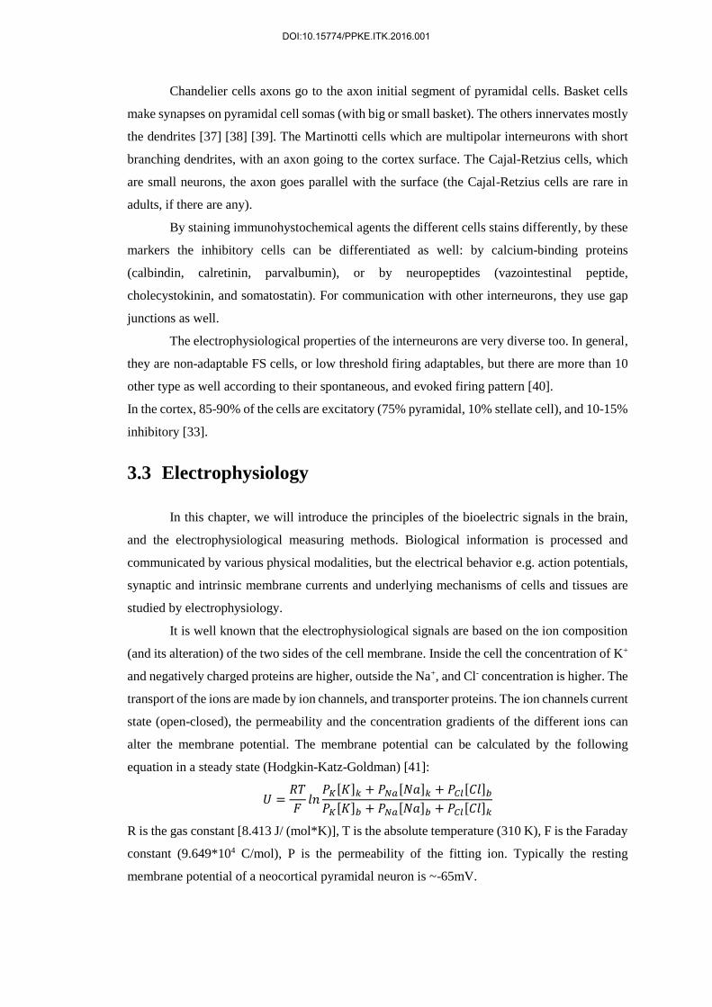
3.3 Electrophysiology
In this chapter, we will introduce the principles of the bioelectric signals in the brain,
and the electrophysiological measuring methods. Biological information is processed and
communicated by various physical modalities, but the electrical behavior e.g. action potentials,
synaptic and intrinsic membrane currents and underlying mechanisms of cells and tissues are
studied by electrophysiology.
It is well known that the electrophysiological signals are based on the ion composition
(and its alteration) of the two sides of the cell membrane. Inside the cell the concentration of K+
and negatively charged proteins are higher, outside the Na+, and Cl- concentration is higher. The
transport of the ions are made by ion channels, and transporter proteins. The ion channels current
state (open-closed), the permeability and the concentration gradients of the different ions can
alter the membrane potential. The membrane potential can be calculated by the following
equation in a steady state (Hodgkin-Katz-Goldman) [41]:
𝑈 =𝑅𝑇
𝐹𝑙𝑛
𝑃𝐾[𝐾]𝑘 + 𝑃𝑁𝑎[𝑁𝑎]𝑘 + 𝑃𝐶𝑙[𝐶𝑙]𝑏
𝑃𝐾[𝐾]𝑏 + 𝑃𝑁𝑎[𝑁𝑎]𝑏 + 𝑃𝐶𝑙[𝐶𝑙]𝑘
R is the gas constant [8.413 J/ (mol*K)], T is the absolute temperature (310 K), F is the Faraday
constant (9.649*104 C/mol), P is the permeability of the fitting ion. Typically the resting
membrane potential of a neocortical pyramidal neuron is ~-65mV.
DOI:10.15774/PPKE.ITK.2016.001

If the membrane potential (by some stimuli or by the effect of some presynaptic cell)
moves in the positive direction, it is called depolarization, if it goes more into the negative than
the process is called hyperpolarization. Chemical or electrical signaling between cells are made
by synapses. If some information (for example action potential) comes from the presynaptic cell
then Ca2+ flows into the presynaptic terminal and neurotransmitters are released to the synaptic
gap, then the neurotransmitters connects to the receptors of the postsynaptic cell. If it is an
excitatory neurotransmitter like glutamate, then the postsynaptic cell depolarizes (Excitatory
postsynaptic potential occurs (EPSP)) by the Na+ inflow, if it is an inhibitory neurotransmitter
like GABA then it hyperpolarizes (Inhibitory postsynaptic potential occurs (IPSP)) by the Cl-
channels opening. If these potentials reach the soma, and the summarized EPSP-s and the IPSP-
s go beyond a threshold depolarization level, then action potential (AP) occurs, and thereafter
the AP is transmitted towards the axon terminal [19] [42] [43] [44]. The axon initial segment is
the place of the AP initiation, because of the vast amount of voltage dependent Na+ channels.
The AP conduction on the axon is described by the Hodgkin Huxley equation:
𝐶𝑚
𝑑𝑉𝑚
𝑑𝑡+ 𝐼𝑖𝑜𝑛 = 𝐼𝑒𝑥𝑡
Cm is the membrane capacitance, Vm is the intracellular potential, t is time, Iion is the current
going through the membrane, Iext is the external current applied. The ion current which goes
through the membrane channels consist of the Na, K, and leaking currents (Figure 2.).
𝐶𝑚
𝑑𝑉𝑚
𝑑𝑡= 𝐼𝑁𝑎 + 𝐼𝐾 + 𝐼𝑙 + 𝐼𝑒𝑥𝑡
The membrane can be modeled by a circuit. In this model there are some conductances which
should be the result of open membrane channels. Many ion channel has gates which can block
the ion flow through. If these gates are open then by the in- or outflow of the corresponding ion
(depending on the concentration gradient) the membrane potential alters closer to the
corresponding ions equilibrium potential.
Figure 2. Left) The different channel conductance changes during the action potential. Right) The axon’s (membrane,
channels) replacement wiring diagram. G = conductance, I = ioncurrent, E = reverse potential, L = leaking [41] [45].
DOI:10.15774/PPKE.ITK.2016.001

3.3.1 Electrical properties of the brain
In this chapter the basic properties of bioelectric field potentials and characteristic brain
rhythms emerging during different vigilance states in humans will be described. The electric
fields generated by neuronal activity can be measured with various electrophysiological
methods. Electroencephalography (EEG), which is one of the most widely used techniques to
record the potential changes in the brain, will be the main method through which the features of
the different neuronal oscillations are demonstrated in this chapter.
Field potentials recorded with the EEG on the scalp represent the summation of the
synchronous synaptic activity of a myriad of cortical neurons which have similar spatial
orientation. On the level of the single neuron, excitatory postsynaptic activity originating on the
dendrites generates an inward current flow into the cell (active sink) at the site of the origin,
with a simultaneous outward current on the soma (passive source), latter acting as a return
current [46]. The electric field generated by this current dipole can be detected with voltage
recording electrodes. In a simplistic view, the field potentials registered with the EEG are the
resultant of thousands of spatially and temporally superponed dipoles. If the activity of the
neurons is temporally synchronized, then the recorded EEG signal contains high amplitude
waves with low frequency (synchronized activity). On the other hand, if the activity is
temporally asynchronous, low amplitude waves with high frequency can be detected
(desynchronized activity). However, the amplitude and frequency content of the EEG signals
depends on various factors: e.g. the age of the patient, the vigilance state, and certain diseases
can alter these properties as well.
Based on the frequency of the recorded bioelectric field potentials, we can characterize
different brain rhythms or oscillations, including the first-discovered and well-known alpha
waves [43].
1. Delta (1-4 Hz): Delta waves belong to the brain rhythms with the lowest frequency.
The activity of neuronal populations is highly synchronized during delta oscillations,
therefore high amplitude waves can be recorded on the entire scalp. This particular
brain rhythm arises in the thalamus and neocortex during the deepest stage (slow-wave
sleep) of the non-rapid eye movement (NREM) sleep in adults. High amount of delta
waves recorded in the awake state usually refers to pathological conditions (e.g. brain
tumor).
2. Theta (4-8 Hz): Theta waves are faster compared to the delta rhythm. In humans theta
rhythm can be recorded with the EEG during stage 2 of the NREM sleep, but it can
occur in meditation in the limbic cortical areas as well [43]. There is another type of
theta rhythm which was observed in the hippocampus of rodents during exploration of
their environment and during rapid eye movement (REM) sleep [46].
DOI:10.15774/PPKE.ITK.2016.001

3. Alpha (8-13 Hz): The alpha waves were discovered by Hans Berger, the inventor of
the EEG. This brain rhythm can be recorded on the occipital sites of the EEG during
periods of eyes closed, but they are present during stage 1 of the NREM sleep as well
[46].
4. Beta (13-30): Desynchronized activity with higher frequency oscillations (>13), such
as beta and gamma rhythms are the characteristic features of the awake state. Beta
waves are present in adults on the frontal and central cortical areas and are associated
with active thinking, attention, focusing and problem solving [46].
5. Gamma (30-80 Hz): The neuronal mechanism underlying the gamma waves is actively
researched, and this phenomenon may have a major role in the conscious perception
(e.g. the binding problem, see ref. [32] [46] [47] [48] [49]. These waves are the
hallmark of the awake and attentive brain, where desynchronized activity with low
amplitude EEG signals can be observed.
3.3.2 Brain electric recording techniques
Measurements of brain electrical properties require a connective medium between the
tissue and the recording device. The connective medium is called electrode, which is formed by
an electron conductor placed in an electrolyte [50] [51]. In order to measure potential
differences, such as brain electric potentials, at least two electrodes are needed. One electrode
is always placed over active tissue. Placement of the other electrode can be also over active
tissue, in which case the recording is bipolar. When the second electrode is placed over a zero
potential area as a reference electrode, the recording is called monopolar. Both arrangements
are widely used in research, so several different properties should be carefully taken into account
when choosing one of them for a specific experiment [50].
Brain electric potentials can be both recorded from outside and from inside the brain
(Figure 3.). EEG recorded from the scalp and ECoG recorded from the brain surface are the two
typical recording techniques for measuring potentials outside the brain. Extracellular and
intracellular recordings are the two main types of electric potential measurements performed in
the brain.
Extracellular recordings are carried out by placement of a recording electrode in the
extracellular medium. Recording electrodes can be metal wires, silicon microprobes with metal
recording contacts or glass micropipettes filled with electrolyte solution and connected to an
Ag/AgCl electrode. Extracellularly, local field potentials (LFP), multiunit (MUA) and single
unit activity (SUA) can be measured. SUA is obtained from MUA recordings when the electrode
arrangement, such as tetrode configuration, allows for sorting of recorded spikes based on their
DOI:10.15774/PPKE.ITK.2016.001

waveform characteristics [50]. MUA and SUA are only recorded from neurons in close
proximity of the electrode, since action potential waveforms quickly vanish in the highly
conductive extracellular medium [50] [52]. Using multichannel extracellular electrodes enables
recording activity from larger brain areas, such as several cortical layers or multiple subcortical
nuclei simultaneously. Extracellular recordings can be performed both in vitro in prepared brain
slices or tissue cultures and in vivo in anesthetized or freely moving subjects. Potentials recorded
extracellularly are in the µV range.
Intracellular recordings are performed inside a single cell. For this purpose glass
micropipettes are always used. The two main types of intracellular recording electrodes are
sharp and patch electrodes. A sharp electrode has a tip less than 1 µm thick which can easily
penetrate the cell membrane [53] [54]. Sharp recordings are mostly performed in brain slices
and carried out in current clamp mode. Current clamp mode means injecting constant current
into the cell and measuring resulting membrane potential changes, which are in the mV range.
Sharp recordings are used to measure whole-cell membrane potential dynamics but are not able
to record single channel potential changes [53] [54]. For this purpose the patch recording
technique is widely used. Patch electrode tips are thicker than the tips of sharp electrodes; their
thickness is about 1 µm [53] [54]. In contrast to the sharp electrode technique, patch electrodes
do not penetrate the cell membrane but form a tight seal on a small patch of the membrane.
There are four different types of patch recording [53] [54]. Firstly, cell attached technique means
that the patch pipette is tightly attached to the intact cell membrane with negative pressure. In
contrast, whole-cell patch recordings are carried out by attaching the recording micropipette to
the cell membrane and then ripping the membrane patch inside the micropipette. The other two
patch clamp techniques, inside-out and outside-out patch clamp, are performed on membrane
patches ripped off the cell. The difference between the two techniques is the side of membrane
facing the electrolyte solution in the micropipette. In patch clamp recordings, both current and
voltage clamp modes are used. Opposed to current clamp mode, in voltage clamp mode the
amount of injected current required for keeping the voltage of the membrane patch at a constant
level is measured [53] [54]. Single cell recordings during hippocampal SPA described in this
dissertation were made using whole-cell patch clamp in current clamp mode.
DOI:10.15774/PPKE.ITK.2016.001

Figure 3. Recording techniques (EEG: Electroencephalogram, AEP: Auditory Evoked Potential, EcoG:
Electrocorticogram, EP: Evoked Potential, FP: Field Potential, RP: Resting Potential, PSP: Post-synaptic Potential
[51]
3.3.3 In vitro and in vivo human brain tissue preparations for electrical recordings
Electrical recordings from human brain tissue can be either made from different tissue
preparations or from the brain of a living patient. In vitro preparations have the advantage of
easy manipulation but provide limited access to mechanisms in an intact brain [53]. The easiest
way to measure electrical potentials from brain cells is the recording from isolated brain cell
cultures. These cultures can be tested in many different conditions by just changing the
ingredients of their bathing solution. Heterologous expression systems are cell cultures that
express a foreign gene coding for example an ion channel [53]. These systems allow for easy
testing of different ion channels in a controlled way. However, in vitro tissue preparations
closest to the intact brain are acute brain slices. These slices are kept in a solution called ACSF,
which closely resembles the cerebrospinal fluid in the brain. Acute brain slices contain small
networks of neurons so that measurements of these slices can reveal valuable information about
functions of neuronal networks in the brain [53]. Results of this dissertation were obtained from
recordings performed on human acute brain slices.
While all of the previously described extracellular and intracellular recording
techniques can easily be used in in vitro tissue preparations, in humans in vivo only extracellular
recordings can be performed. However, in vivo recordings have the great advantage of recording
from a tissue in its whole physiological environment. Human in vivo recordings are always
carried out in subjects undergoing therapeutic brain surgery [55] [56]. Brain surgeries are
DOI:10.15774/PPKE.ITK.2016.001

preceded by simultaneous video and EEG recordings and also by different brain imaging
sessions to localize the area for surgery. During surgery, different recording electrodes are
implanted onto the surface of the brain, such as grid and strip electrodes, or into the brain, such
as the thumbtack electrode [55] [56](Figure 4.). Recordings by such electrodes are of great value
since they are obtained from tissue that is later removed and used in in vitro recordings. This
allows for direct comparison of data recorded from different tissue preparations [55] [56].
Figure 4. Subdurally implantable thumbtack- (A and C), and grid electrode (B) [55].
3.3.4 EEG graphoelements
EEG graphoelements are manifestations of brain electrical activity recorded on the
scalp. These elements are results of both spontaneous and evoked potential changes in the brain.
Graphoelements help in categorizing and describing EEG recordings, thus providing a powerful
tool for both researchers and clinicians. In 1924, Hans Berger described the first EEG
graphoelement as the alpha wave, which is measured from occipital areas during wakefulness
with eyes closed [57] [58]. As such, the definition of alpha wave is as old as the EEG recording
technique itself. Since then, several different EEG graphoelements were introduced.
DOI:10.15774/PPKE.ITK.2016.001

From a healthy brain, various categories of graphoelements can be recorded [48].
Regular rhythmical oscillations are mostly recorded during slow wave sleep and characterized
by low frequency and high amplitude waves. Certain frequency sinusoidal waves are
characterized by a single frequency and appear as a sinusoid-like wave on the recording. One of
the most easily recognized certain frequency sinusoidal wave is the alpha wave. In many cases,
certain frequency waves do not appear and disappear instantly but form waxing and waning
oscillation snippets, called spindles. These spindles can be observed in many cases, such as sleep
spindles during the deepening phase of slow wave sleep or alpha spindles during eyes closing
in wakefulness. The most prevalent EEG graphoelements during a resting wakefulness with
open eyes are the irregular arhythmical EEG waves. These consist of various frequencies and
reflect the ongoing activity of the wake brain. More complicated graphoelements which consist
of many different frequency waves are called complexes. The most well-known complex in the
EEG is the so called K-complex which can be observed during stage-2 non-REM sleep in
humans [48].
In addition to the graphoelements recorded from a healthy brain, several EEG
graphoelements can be indicative of pathological phenomena (Figure 5.). The most prevalent
pathological graphoelements are the sharp waves, spikes, spike and wave complexes, polyspikes
and polyspike and wave complexes [48]. These elements play a key role in diagnosing and
localizing pathologies such as epilepsy.
Figure 5. Examples of the above mentioned EEG graphoelements [48]
DOI:10.15774/PPKE.ITK.2016.001

3.4 Epilepsy
Epilepsy is one of the earliest recognized neurological disorders. The Babylonians wrote
down most of the seizure types, but they thought that it is some kind of evil spirit taking
possession of the body.
The first person to claim that epilepsy is a kind of brain disorder was Robert Bentley
Todd in 1849: he suggested that the seizures were electrical discharges from the brain.
John Hughlings Jackson was the one who had made this approach popular among the public at
large, and with his guidance Victor Horsley was the first who performed craniotomy to cure
epilepsy. This was the beginning of the discipline of epilepsy surgery.
The first antiepileptic medication -phenobarbital- was invented in 1912, and till now
this is the most widely used medicine in the pharmacological treatment of epilepsy [3] [59] [60]
[61] [62].
In 1935 Wilder Penfield and Herbert Jasper were the first who made awake EEG
assisted surgeries in the Montreal Neurological Institute [63]. They stimulated different cortical
areas during the surgeries and mapped the evoked responses (like movement of the mouth etc.),
or what sensations the patients experienced.
Epilepsy is a frequent neurological disorder; approximately 65 million people suffer
from epilepsy worldwide. Epilepsy affects seriously the quality of life of the patients and their
families. Epilepsy is not curable, but large scale of antiepileptic drugs are available, which can
attenuate or stop the seizures. Epilepsy is frequently accompanied by other psychiatric disease
patterns like depression, psychotic symptoms, personality disorder, anxiety, cognitive failures
and a higher suicide rate compared to the non-epileptic population [59] [60] [61] [62]. This can
be explained either by the disease itself or by the side effects of the different antiepileptic drugs.
There is an increased mortality (2-10 times higher) in the case of epileptic patients. However,
this is not the consequence of the brain disorder, but the injuries connected to the seizures [64]
[65].
It is important to be mentioned that an epileptic seizure is not equal to epilepsy. On one
hand, a seizure can be a symptom or a momentary signal. It can be synchronous or excessive,
but it is always the consequence of some pathologic activity of the brain. On the other hand,
epilepsy is a long lasting susceptibility to seizures by the dysfunctional behavior of the brain.
On the neuronal network level epileptic seizures manifest as states of pathological
hyperexcitability and hypersynchronous activity of large populations of neurons with
concomitant synaptic reorganization of the affected brain region [2] [66].
The epileptogenecity of the different brain regions are diverse. Neocortical neuron
populations are especially capable to produce excessive, synchronous firing during
physiological conditions which is indispensable in several processes like formation of memory.
DOI:10.15774/PPKE.ITK.2016.001

Therefore, they have the potential to display extensive hypersynchronous firing under
pathological conditions [67].
According to the most well-known hypothesis, epilepsy is linked to an impaired balance
of excitation and inhibition in the affected brain region [2]. The role of an altered GABA-
mediated inhibition in epileptogenesis and seizure activity has been studied for decades, as well
as the sprouting of certain excitatory pathways [8] [68].
There are a lot of syndromes of epilepsy, and the International League Against Epilepsy
tried to distinguish the different types. There are two main categories: they are either separable
by the seizure focus, or by the etiology of the seizures/epilepsy.
When separating by the focus, there are focal/partial seizures (the epileptogenic zone is
well localized), and there are generalized seizures (both of the hemispheres are involved in the
emergence). Regarding etiology, there are idiopathic, or symptomatic epilepsies. Symptomatic
epilepsies occur along with other disorders of the central nervous system. There is not any other
disease before the occurrence of idiopathic epilepsies; they are the result of some kind of
heritable susceptibility.
The seizures which are connected to a specific brain region are called partial seizures.
Partial seizures which do not involve disturbance of the consciousness are the simplex partial
seizures. Seizures involving the frontal and temporal lobe are the complex seizures (mostly with
loss of consciousness). If the motor cortex of both hemispheres are involved, than grand mal
seizure occurs (generalized tonic-clonic-seizure). When the area of the epileptic seizure is not
definable then it’s a generalized seizure, with symmetrical motoric phenomenons appearing in
the EEG signal, the seizure appears on systems which innervates areas on both hemispheres.
There are some generalized seizures during which no or only a few excitation signs
appear (petit mal). These seizures cut off extensive areas for a few seconds with cortical spike
trains [3] [59] [60] [61] [62]. Focal seizures are most often of temporal lobe origin [3] [69], but
frontal, occipital or parietal lobes are also frequently the focus of the seizures.
Despite the large variety of antiepileptic drugs available, a considerable part of the
patients are resistant to pharmacological treatment. In case of temporal lobe epilepsy, the
percentage of drug resistant patients is extremely high [70] [71].
Nowadays, the surgical treatment emerges in some type of epilepsy. The progression of
the surgeries is due to the advances of imaging techniques. With the ever growing influence and
quality of the imaging techniques, there is a possibility now to gain much more insight into the
localization, extension, and pathological nature of the epileptogenic brain disorder in a variety
of lesion types. The vascular malformations diagnostics have improved greatly in recent years,
and by the characteristic MR signals of an epileptogenic cavernome gives us the opportunity to
surgically treat it. MR is good for recognizing epileptogenic sub-acute, or chronic encephalitis.
DOI:10.15774/PPKE.ITK.2016.001

Some of the newest application of fMRI is that it can localize the motoric and speaking functions
areas, so it gives the opportunity to make a surgery close to these areas.
The development of subdural electrodes allows a much better localization of the seizure
onset zone, where the scalp electrodes are not sufficient enough. Using the two techniques
together the seizure onset zone can be connected to a specific anatomical structure in the brain.
Since the majority of partial epilepsies are of temporal lobe origin, temporal lobectomy is
performed most often, during which the parts of the hippocampus and the temporal lobe are
removed [3] [4] [72] [73]. After the surgery the majority of the patients is seizure free, or has
seizures less frequently.
3.4.1 Tumor based epilepsies
20-45%of tumor patients have some kind of epileptic event. The age, the place of the lesion,
the pathology of the tissue can all influence the seizure’s appearance. The etiology of the tumor
caused epilepsies are quite diverse [3] [59] [60] [61] [62] [74] [75].
- Central nervous systems disorders
o Primary brain tumor: glial, neuronal
o Increase in the number of excitatory neurotransmitters, increase of the pH
o Morphological alterations in the tissue (abnormal neuronal migration), receptor
binding site alteration
o Brain metastasis (lung-, breast melanoma)
- Toxic, metabolic effects
o Medicine side effect
o Liver- or renal failure
o Non-central nervous system infection
o Electrolyte disorder
- Treatment caused seizures
o Chemotherapy
o Other agents
[3] [59] [60] [61] [62] [74] [75] [76].
The tumor caused epilepsy treatment needs to be cautious, because the treatment can
cause seizure as well, or the different agents can effect each other. Yet, antiepileptics are often
used in case of these patients (mostly monotherapy to eliminate interactions). Surgery comes
only after unsuccessful medical treatments. First, the primary brain tumor or metastasis is
removed and after this the epileptogenic zone. Sometimes the chemotherapy and radiotherapy
can offer solution for some of the problems.
DOI:10.15774/PPKE.ITK.2016.001

3.4.2 Cortical dysgenesis
Cortical dysgenesis derives from a disorder in the brain’s development. These
alterations are hard to detect without a high-resolution MRI. 24% of the epilepsies are due to
cortical dysgenesis [77] [78] [79].
The cortical cells are developed from the neuroblasts of the ventricular zone near the
developing brain’s midline. These cells divide, differentiate and migrate continuously before
birth. More than 25% of the primer neurons will die by programmed cell death. Cortical
dysgenesis is a disorder that can occur in the whole pregnancy but mostly between the 7-16th
week. The pathology depends more on the occurring time of the defect, than its cause. MRI can
be used for the detection of the structural difference, but not the etiology of it [77] [79].
A useable classification of the dysgenesis caused epilepsies was based on MRI, which
takes into account the histological properties (many times the cause is a subependymal or
subcortical heterotopy). Misplaced cell groups can be formed by abnormal migrating endpoint,
excessive migrating, or the absence of the programmed cell death before birth.
3.4.3 SPA and interictal activity
For patients with pharmacoresistant focal epilepsy, resective surgery provides a good
treatment alternative. The possibility of examining the removed epileptogenic zone
revolutionized epilepsy research, as it raised the opportunity of measuring the activity of single
neurons in a physiological or quasi-physiological state. In the experiment described in this
manuscript, this is important, because our aim was not just to record and analyze LFP changes
caused by the cells, we also wanted to know the underlying mechanisms involved in SPA
generation. For this purpose, it was examined how all the separate cells are responding to the
activity that is recorded using a laminar microelectrode. It has been shown that interictal spikes
don’t depend on age, gender, pathology, histology, or used antiepileptics [6].
Distinct from these pathologic interictal events, spontaneously occurring synchronous
population activity could be detected in vitro in brain slices obtained from resected human
epileptic neocortices, subiculum, and hippocampus [6] [80] [81] [82] [83].The emergence of
synchronized events in the neocortex is probably based on the complex interactions between
and within the neural network’s inhibitory and excitatory components.
The work of Köhling et al. [6] involved the investigation of human neocortical tissues
resected during epilepsy surgery. They investigated the role of glutamatergic and GABAergic
synaptic transmission, as well as the role of voltage gated calcium channels in the generation of
the spontaneous activity they describe [6]. In their work, the extracellular field potential gradient
DOI:10.15774/PPKE.ITK.2016.001

was measured in the cortical layers II and V. However, Köhling et.al. measured this activity
only in tissue obtained from epilepsy patients. Their characteristics of the spontaneous field
potentials were:
- amplitude 20-323 μV (72+-13 μV)
- duration 20-200ms (151+-18ms)
- repetition rate 4-108 per minute (43+-4 per minute)
- monophasic [6]
While some have argued that SPA is distinct from the pathologic interictal events
occurring in epilepsy patients [84], it is still controversial whether SPA is epileptic, or whether
it can be found in physiological conditions.
Many studies demonstrated the importance of transmembrane calcium currents and the
effects of glutamate, and GABA during spontaneous field potential transients [82] [83] [85]. It
has been shown, that an increase in the Mg2+ concentration reduces the recurrence frequency of
spontaneous population activity, but application of APV does not, which points to a calcium-
antagonistic effect of Mg2+. Calcium currents play a crucial role in the generation of spontaneous
activity, and epileptiform activity induced in experimental models and in seizures in epilepsy
patients [6]. Blocking the non-NMDA type glutamate receptors, the GABAA receptors, or
calcium channels can suppress this type of activity, but blocking the NMDA type or GABAB
receptors does not have any effect on these field potential transients [6] [80] [86] [87].
Our group’s preliminary results [83] [85] [88] indicate that an activity similar to
interictal spikes (as in Köhling et. al. [6]) is detectable in non-epileptic tissue (derived from deep
brain tumor patients, non-epileptic part not infiltrated by the tumor (Figure 6.)).
Using current source density (CSD) analysis the flowing currents between cell
compartments can be estimated. Using CSD analysis, it is possible to evaluate which neuronal
populations generate the changes in the field potential. During spontaneous interictal discharges
in vitro, the current sinks are mostly located in layers II. and III. (positive charges flowing into
the cells) even after Mg2+ withdrawal. However, where spontaneous activity did not occur, the
Mg2+ withdrawal, or Bicuculline (GABAA receptor antagonist) application caused the sinks to
spread over the whole extent of the cortex, especially in layer V. [87].
DOI:10.15774/PPKE.ITK.2016.001

Figure 6. Example recording of SPA from an epileptic (A), and from a tumor (B) patient. The SPA discharges are
marked with an asterisk (*). SPA is mostly generated in the supragranular layers, shows increased cell firing
(indicated by an increase in MUA), and the occurrence of High frequency oscillations during the LFP discharge
[83] [85] [88].
In the present study, we want to further investigate the origins of this SPA.
Measurements performed with an extracellular laminar multielectrode provide the desired
spatial information on how the different cortical layers respond.
However, this approach does not yield extensive information on single cell activity.
Thus, it is difficult to address the question of cellular mechanisms, as it is not feasible to patch
each of the cells to obtain cell specific information. Question addressed in the present study are:
How are the cells involved in the generation of SPA? What proportion of cells is active during
SPA? Which types of cells are active (neurons, interneurons, glial cells) during SPA? When are
they most active (before/during/after the LFP transient)?
Since SPA can occur both in epileptic and in healthy tissues, we decided to investigate
the differences in how the healthy and versus the pathological tissue generate a very similar
activity. To be able to answer the questions stated above, our research group used 2-photon
microscopy. In addition, histological analysis of the tissue is included (cell labeling and staining,
followed by light and electron microscopy and 3D reconstruction) to address the question of
morphological differences between epileptic and non-epileptic tissue.
3.5 Two-photon microscopy
Neuroscientists have become more and more interested in two-photon microscopy over
the last decades because of the numerous advantages. Two-photon microscopy allows high
resolution imaging in living tissue compared to confocal microscopy. Another great advantage
DOI:10.15774/PPKE.ITK.2016.001

of this method is that it makes achievable to image even in great depths (several hundred microns
1 millimeter compared to 30-50 um) by, solving the light scattering problem causing
deterioration of optical signals. The improvements of the fluorescent staining and marking
techniques in the last few years allowed the functional exploration of neuronal activity from
single cell (or subcellular) activity, through cellular networks, to even the measurement of a
cortical column (with 3D scanning technology). In this chapter the principles of two photon
microscopy will be introduced and its application in the field of neuroimaging will also be
discussed.
3.5.1 Theoretical background of two-photon microscopy
In 1931 Maria Göppert-Mayer set the theorem of two-photon absorption [89]. However,
because of technological reasons it was experimentally confirmed in the 1960s. With the
production of ultra-short pulsed lasers, two-photon microscopes became designable [90].
Fluorescence techniques addressed the problems of neurobiology. Currently, two-
photon microscopy became an essential tool for the examination of biological tissues (living or
fixed), because the fluorescent objects can be visualized selectively (even with small
fluorophore concentration) with good signal-to-noise ratio. Light microscopy is able to track
spatially complex dynamics [91] over a great depth, and can resolve single synapses [17] [92].
For light microscopy intact cortical tissue is challenging, but if it is possible neurons should be
studied in their natural domain.
The drawback of conventional fluorescent microscopy is that in thick (over 100 μm)
samples contrast and resolution are degraded because of the scattering of the tissue [93] [94].
By confocal microscopy some of the scattering effects were overcame by the pinhole detectors,
because it rejects fluorescence from off-focus locations [93] [94]. However, scanning a section
excites the whole specimen, and thus could damage it. Furthermore, pinhole rejects the scattered
signal photons emerging from the focus. In great depth confocal microscopy loses untolerably
high amount of signal photons by the scattering [93] [95]. Signal loss can be compensated by
increased fluorescence excitation, but it can lead to photobleaching and phototoxicity.
The theory of two-photon excitation is based on the concept when two low-energy photons hits
a fluorescent molecule causing an electronic transition to a higher-energy state. Each photon
carries the half of the energy necessary to the excitation, and results in the emission of a
fluorescent photon (the photon is at a higher energy than either of the excitatory ones (Figure
DOI:10.15774/PPKE.ITK.2016.001

7.)).
Figure 7. Left: Single and two-photon excitation of a fluorescent molecule (from the excitation it goes up to an excited
energy state, and when it descends back to the original a photon is emitted). Right: the focus of the laser beam by
confocal or two-photon (marked with white arrow) technology [96].
In a focused laser, the intensity is highest in the focus, and degrades quadratically by the
distance. A fluorescent molecules excitation probability has a quadratic dependence on light
intensity, resulting in an excitation exclusively only a small diffraction-limited focal volume
(Figure 7.). If the objective (used to focus the beam) has a high numerical aperture (NA), most
of the fluorescence excitation occurs in a focal volume of ~0.1 mm3 [97]. This way the point
spread function’s axial spread is significantly lower than for single-photon excitation.
Photobleaching and photodamage are greatly reduced to the tissue, and no fluorescence is
emitted from out of focus locations (automatic optical sectioning), so there is no need for out of
focus rejection strategy (like in confocal microscopy). The signal to noise ratio (SNR) is greatly
increased by the collection of scattered photons, in contrast to confocal microscopy where they
are rejected.
The most commonly used fluorophores have excitation spectra in the 400–500 nm
range, whereas the laser used to excite the two-photon fluorescence lies in the ~700–1000 nm
(infrared) range. The living tissue scatters light more in the visible wavelengths than in the
infrared, thus fluorescent objects in living tissue can be examined in greater depth [98] [99].
Nowadays it ranges from hundreds of micrometers to a millimeter.
DOI:10.15774/PPKE.ITK.2016.001

For its above mentioned advantages two-photon microscopy became a unique tool for
imaging samples in depth (especially in vivo), or in local photochemistry. However, on
transparent or very thin slices two-photon microscopy is not as effective as confocal or wide
field fluorescence microscopy since the achievable spatial resolution is reduced by the longer
wavelengths.
Two-photon functional imaging, and calcium imaging have an extensive background
[100], either focusing on its technical aspects [97] [99] [101] [102], on its applications [103]
[104] [105] [106], or on its place in the wider context of the various recent technological
developments, which provide tools for the "reverse engineering" of the brain [107].
3.5.2 Hardware of a two photon microscope
Two-photon microscopy is typically implemented in a simple laser scanning
microscope. The setup consist of the following main elements: laser source, scanner, objective
and detectors (Figure 8.).
.
Figure 8. Schematic drawing showing the optical design and modular nature of the 2P microscope used. Modules: 1
- Dispersion compensation. 2 – Laser beam positioning. 3 – CCD/2P switcher. 4 – CCD camera. 5 – Upper detectors
(PMT – photomultiplier; Dic. - dichroic mirror). 6 – Perfusion chamber and focusing. 7 – Lower detectors. 8 –
Infrared lamp (IR). 9 – Transmission infrared detector (TIR). Dic: Dichroic mirror. Source: [108].
DOI:10.15774/PPKE.ITK.2016.001

The typical differences between two-photon and confocal microscopes are the laser (see
below) and the fluorescence detection path. In two-photon microscopy, all useful fluorescence
photon collected by the objective. The optimal solution is to project the objective back-aperture
onto the photosensitive area of the photodetector [109] [110]. Fluorescence photons can appear
to originate from a large effective field of view because the multiple scattering before exiting
the tissue [108]. In confocal microscopy, the epifluorescent light passes back through the scan
mirrors and through a pinhole before detection [93].
In two-photon microscopy the laser beam is directed into the microscope through an
epifluorescent light path. To focus in the specimen, the excitation light is passed through a
dichroic mirror to the microscope objective. Two-photon induced fluorescence occurs only at
the focal spot. By scanning the fluorescent volume with a galvanometer-driven scanner, the
images can be constructed. The emission signal is collected by the same objective and reflected
to the detector by the dichroic mirror. A barrier filter is also needed to attenuate the scattered
excitation light. To ensure maximal detection efficiency high-sensitivity detectors and
electronics are used.
3.5.3 Laser Sources
Opposed to confocal microscopy (continuous wave emitting lasers) two photon
microscopy requires pulsed lasers (for precise technical details see the corresponding part of the
methods section).
Two-photon excitation efficiency increases with the inverse of the pulse duration, if the
average pulse repetition rate, and power are constant. The most commonly employed lasers for
two photon microscopy are mode-locked Ti:sapphire lasers. Mostly the excitation light is
tunable between 700-1000nm [112].
Pulse rates should balance the onset of saturation and the fluorophore’s excitation
efficiency. This criterion is met by the used laser for most common molecules (e.g. Ti:sapphire
laser), that is fortunate because mode-locked lasers pulse rate is difficult to change. For stable
excitation rate, it is essential that a constant number of pulses arrives on a pixel. More than 100
pulses arrive on a pixel, assuming a pulse rate of 100 MHz and 1 μs dwell time, so the number
of pulses per pixel will be stable at the 1% level [112].
The imaging depth is determined by scattering: with increasing depth, a smaller fraction
of the incident photons are delivered to the focus. Since rays entering the brain at higher angles
have longer paths to reach the focus they are more likely to be scattered, causing a loss of
resolution with imaging depth [105]. SNR is also worsened with depth, because extensive
scattering and absorption occurs on the lower wavelength emitted light.
DOI:10.15774/PPKE.ITK.2016.001

The contrast and the localized excitation worsen with increasing depth. Because of the
out of focus fluorescence generated on the surface, a depth limit is imposed on imaging. In gray
matter it is ~ 1mm [112].
3.5.4 Scanning Methods (x-y plane)
The most widespread solution is the use of mirrors moved by galvanometers.
Galvanometric mirrors allow rapid, arbitrary positioning of the focus in the focal plane (x-y)
[113] [114]. In raster scanning application, galvanometric mirrors, allow zooming, image
rotation and have excellent optical properties. Their major drawback is their relatively slow
speed (>1 ms per line); raster scanning of a typical image requires ~1 s. Speed can only be
increased at the cost of the spatial resolution and the extent of covered area [113] [114]. Since
many neurophysiological processes take place on the millisecond time scale, faster scanning
methods are required.
“Line scan” is an alternative scanning method. The laser does not scan through the
whole 2D plane (like in raster scan), instead the laser scans only the lines which were specified
by the user on the structure of interest. For example multiple dendritic spines can be imaged
within 1 ms [115]. However, it has to be mentioned that the cost of scanning speed is the loss
of spatial information. During the experiment the selection and adjustment of the trajectories
should efficiently be made. This can be problematic, because only one fixed z plane at a time
can be used, while the neural structures do not necessarily fall in the same plane. Another way
to accelerate the scanning is to jump between regions of interests (ROIs) by maximal scanning
speed between ROIs of the mirrors. With this method no time is wasted for the extracellular
structures. Still line scans can be wasteful when the ROIs are sparsely distributed. The scanning
speed depends on the acceleration/deceleration rate of the motors. All ROI based scanning
method, such as line scan, need extensive software support which is not always provided by the
microscope manufacturer.
An attractive alternative is presented by acousto-optic (AO) deflector. AO scanning
technology is another solution for rapidly change beam focusing excluding mechanical
movement, and AO technology is adapted in many two-photon applications [116] [117] [118]
[119] [120] [121] [122] [123]. Acousto-optic deflectors are transparent crystals, in which an
optical grating is created by applying sound waves to them. Periodic change in the refractive
index of the medium is caused by sound-generated pressure fluctuations, and the changes
behaves like optical grating (the light diffracts according to the period-and the wavelength of
the light). The frequency of the sound wave determines the spacing of the grating, and thus the
angle of diffraction of the laser beam. Since the laser beam can be diverted without mechanical
DOI:10.15774/PPKE.ITK.2016.001

movements, speed of the positioning is vastly enhanced compared to the galvanometric
approach. These optical materials are dispersive, leading to temporal and angular dispersion,
which, worsens the excitation efficiency, point spread function and image quality for typical
pulse durations (< 1 ps) [118]. With gratings, prisms and/or additional acousto-optic devices the
compensation can be made [124] [125] [126]. Acousto-optic deflectors have also been proposed
[117] for tackling scanning also along the z axis [102] [121] [122] [123] [127].
3.5.5 Objectives
The objective is a very important part of the microscope, because it generates the laser
focus required for localization of excitation. There is a wide range of available choices from
objectives in the market which are suitable for a two-photon microscope.
Important criterias are:
1. Numerical aperture, has to be large because it determines the resolution and the angle
for fluorescence light collection
2. Magnification, according to the desired field of view;
3. Large working distance (2 mm is ideal), especially in in vivo applications and
simultaneous electrophysiology measurements;
4. Excellent transmission efficiency in visible wavelengths and the near-infrared.
The laser beam should expand to “fill” the microscope objective’s back aperture, this
way it focuses the light into the sample. Since the laser beam has a Gaussian radial intensity
profile, laser beam is said to “overfill” the back aperture of the objective when the latter accepts
the central part of the beam up to the radius at which its intensity decays to 13.7% (1/e2). In this
case, 84% of the beam’s power is transmitted and lateral and axial resolution [97] are nearly as
good as with a uniform excitation beam (92 and 96%, respectively). Using a narrower beam
increases the amount of power transmitted, but reduce the effective numerical aperture (NA)
leading to a loss of resolution [97] [99].
It is important to note that increasing depth leads to reduced effective NA: large angle
photons travel longer distances than those near to the optical axis and often they are scattered or
absorbed. Underfilling the objective might be a good solution, if the observed objects are large
enough and their detectability does not suffer by the reduced accuracy. As the size of the focal
volume grows with decreasing effective NA, the peak intensity decreases, resulting a decrease
in photobleaching and photodamage.
3.5.6 Detectors
DOI:10.15774/PPKE.ITK.2016.001
Two-photon microscopy demands large photosensitive areas (can be millimeters) [111],
so avalanche photodiodes are excluded. Important factors are gain, quantum efficiency, dark
noise, absorption spectra, and absorption angle of the detector. Photomultiplier tubes (PMTs)
are the best for two-photon microscope applications (the different type’s performance
comparison can be found in [97]).
In two-photon microscopy, with spatially ultra-selective excitation, optical partitioning
can be achieved. So all photons should be collected, even the scattered ones, emitted by an
excited fluorescent molecule. The excitation light has larger wavelength than the fluorescence,
so from lower depths the fluorescent photons are greatly scattered thus photons appearing on
the surface are negligible below several hundred μm. Thus, large NA and low magnification
objectives (for example 20x at a NA of 1.0) are the best choice to catch as many photons as
possible. Moreover, large sensitive area photomultipliers (PMTs) are positioned to detect as
much as possible. In our in vitro experiments all excited photons can be useful (and can be
detected) either scattered in the backward (“epi”- collection geometry) or forwards (“trans”-
collection geometry) direction.
3.6 Neuronal calcium signals
Tissues can be considered as semi-infinite by in vivo imaging, therefore forward-
collection blocked, which makes an effective backward-detection really important. “Whole-
area” configuration is used in most cases. It means that to the PMT’s sensitive area the
objective’s back is projected and this way it allows all objective collected photons to end up on
a detector [128].
The role of Ca2+ dynamics in neuronal signaling has ever growing interest by
neuroscientists during the last decade. There are basically two types of Ca2+ imaging techniques.
One there are the classical chemical fluorescent Ca2+ indicators, the other is the genetically
encoded protein-based Ca2+ indicators. In this study (in human) only the Ca2+ indicators can be
used in vitro [11].
Calcium ions generate versatile intracellular signals for many functions in almost every
cell type [129]. Intracellular calcium signals (in the nervous system) regulate processes from the
time scale of neurotransmitter release (microsecond) to gene transcription, (minutes/ hours)
[130]. For the function of these signals the amplitude, the time course, and the local action site
are essential. There are many types of neuronal Ca2+ signaling, sources of Ca2+ influx are Ca2+-
permeable α-amino-3-hydroxy-5-methyl-4-isoxazolepropionic acid (AMPA) and N-methyl-D-
aspartate (NMDA) glutamate- type receptors, voltage-gated Ca2+ channels (VGCC), nicotinic
acetylcholine receptors (nAChR), and transient receptor potential type C (TRPC) channels [11].
DOI:10.15774/PPKE.ITK.2016.001

Ryanodine receptors (RyR) and inositol trisphosphate receptors (IP3R) mediates the Ca2+
release from internal stores is mediated. Metabotropic glutamate receptors (mGluR) can
generate inositol trisphosphate [11]. Sodium-calcium exchanger (NCX), plasma membrane
calcium ATPase (PMCA) and sarco-/endoplasmic reticulum calcium ATPase (SERCA) can
mediate Ca2+ efflux. Mitochondria is important for neuronal Ca2+ homeostasis [11].
In this research understanding the connection between the AP and the Ca2+ is very
important the theoretical background will be shortly summarized in this paragraph. There are
many researches, showing the connection between the AP and the Ca2+ inflow, especially in the
neurotransmitter release to the synaptic cleft, the dendritic Ca2+ transients, backpropagating AP,
etc… [131] [132] [133] [134] [135] [136].
When we speak about the importance of Ca2+ dynamics in the neuronal signals we
should speak about axonal- somatic-, and dendritic Ca2+ response.
Neurotransmitter molecules are stored in vesicles in the presynaptic terminal of neurons.
Ca2+ inflow plays a key role in neurotransmitter release. It is inhibited when Ca2+ channels are
blocked. Upon the arrival of AP to the axon terminal voltage dependent Ca2+ channels open and
Ca2+ flows from the extracellular fluid into the presynaptic neuron’s cytosol via the
concentration gradient this sudden Ca2+ influx is followed by the docking and fusion of
neurotransmitter vesicles to the presynaptic neuron’s cell membrane through SNARE proteins
resulting in a rapid neurotransmitter release. So it can be said that in the axon when there is an
action potential, then an inward Ca2+ concentration change follows it [131].
We need to speak about dendritic Ca2+ transients, because in two-photon microscopy
the cells soma, and the dendritic tree is what can be easily imaged by scanning methods (like it
was described before in the scanning methods). Dendritic Ca2+ transients can be related to
passive somatic depolarization propagation or active AP propagation.
By somatic voltage-clamp, dendritic Ca2+ imaging and somatic excitation experiments, high
Ca2+ transients were detected in both the initial, and the distal dendrites (backpropagating action
potential). But when the Na+-dependent AP propagation was blocked, then the Ca2+ transients
degraded in the initial and vanished in the distal dendrites. So the local Ca2+ entry mediated by
action potentials was reflected in the Ca2+ transients [131] [132] [133] [134] [135].
It was also shown, that the intracellular dendritic Ca2+ rise could influence the synaptic
integration by the downreguation of the NMDA receptor-mediated responses [133].
Based on the literature it can be assumed, that whenever there is a Ca2+ response in the cell: if it
is measured from the soma it probably reports an AP, if it measured in the dendrites then it refers
to a postsynaptic response or an AP [131] [132] [133] [134] [135] [136] (Figure 9.).
DOI:10.15774/PPKE.ITK.2016.001

Figure 9. Representative Ca2+ responses recorded by two-photon Ca2+ imaging. 16 bulk loaded (OGB-1-AM, SR101)
human neocortical neuron/interneuron were selected by line scan for spontaneous recording, the scanning time were
20 s. The vertical axis is time, the horizontal is how the laser go through the placed lines in the line-scan, and the
more red the color is, the higher the Ca2+ fluorescence response. Two of the cells responded in this case (two of the
ROI-s are marked by purple and blue).
3.6.1 Calcium indicators
The first generation of fluorescent calcium indicators consisted of quin-2, fura-2, indo-
1, and fluo-3. Quin-2 can be excited by ultraviolet light (339 nm). Quin-2 was the first of this
group to be used in biological experiments [137] [138]. However, to overcome cellular
autofluorescence, Quin-2 needs to be used at high intracellular concentrations [139]. Fura-2
became very popular among neuroscientists [140], is in many ways superior to quin-2 and Fura-
2 is excited at ~350 - 380 nm and shows significantly larger calcium-dependent fluorescence
than quin-2. Furthermore, fura-2 allows more quantitative calcium measurements [141].
Nowadays Oregon Green BAPTA and fluo-4 dye families [142] are widely used in
neuroscience. They provide a large signal-to-noise ratio and are relatively easy to implement.
The introduction of protein-based genetically encoded calcium indicators (GECIs) was the next
great breakthrough [143].
3.6.2 Calcium Imaging
Two-photon imaging started after the bulk loading, and/or whole cell configuration achieved on
the two-photon laser scanning system. Spatially normalized and projected Ca2+ response can be
calculated by the raw line-scan, F(d,t) using the
DOI:10.15774/PPKE.ITK.2016.001

∆𝐹
𝐹=
(𝐹(𝑑, 𝑡) − 𝐹0(𝑑))
𝐹0(𝑑)
formula where d denote to the distance along the curve and t to time. F0(d) is the average/
background-corrected prestimulus fluorescence as a function of distance along the curve. Ca2+
responses are projected as function of t and d and Kd color coded (yellow to red show increasing
Ca2+ responses, 0–63 % ∆F/F). In the experiments the relative fluorescence value was converted
to Ca2+ concentration [144] [145] [146]
∆[𝐶𝑎2+]
𝐾𝑑=
𝑓𝑚𝑎𝑥
𝑓0(1 −
1
𝑅𝑓)
𝛿𝑓
(𝛿𝑓𝑚𝑎𝑥 − 𝛿𝑓)𝛿𝑓𝑚𝑎𝑥
[147], where ∆[Ca2+] is the change in the intracellular calcium concentration, δƒ denotes ∆F/F,
Rf ( = ƒmax/ ƒ min) is the dynamic range of the dye and KD is the affinity of the indicator.
DOI:10.15774/PPKE.ITK.2016.001

4 Materials and methods
First of the main goals (and Thesis I.: A method has been developed for the two-photon
Ca2+ imaging of human neocortical tissue) of this research was to make the methodology that
permits the deeper understanding of the events behind SPA. This was not an easy task, because
not just the methodology should be made, but organizing the teamwork of the different research
groups, and the surgeons, were a big portion of the work.
First the contact with the surgeons and the ever refreshing information of the surgeries (of the
National Institute of Clinical Neuroscience, Budapest, Hungary) and the patient’s approval of
the research should be organized. Then the preparing the solutions, and the experimental design
should be made for every experiment.
The experimental work with the Two-photon microscope should be learned (from the
research group of the Femtonics Ltd (Budapest Hungary) lab in the Pázmány Péter Catholic
University, Budapest, Hungary), the experimental time of the different groups in the same lab
were organized. The preparation of the two-photon microscope for the experiment were made
(the set-up of the laser, the preparation of the ACSF circulation system (with a double flow
chamber) in the two-photon microscope, the setup of the laminar electrode recording system
(the data transmission system, and the recoding electrode integration to the two-photon
microscope), the preparation of the micropipettes, and the imaging dyes). And all the used
programs should be learned, not just for the experiments, but for the analysis as well.
The preparation of the slices for histological analyses were included, too. The possibility
of the microscope usage for light- transmission electron microscopy, and 3D reconstruction
were organized.
Organizing all these people and all these pre setting should be together to make the
preparation of this methodology possible.
The training of our research group (Hungarian Academy of Sciences, Research Centre
for Natural Sciences, Institute of Cognitive Neuroscience and Psychology, research group of
Comparative Psychophysiology, Budapest Hungary) for the experiment were prepared and
made for the future work as well.
From now on this methodology will be described.
DOI:10.15774/PPKE.ITK.2016.001

4.1 Patients
In the present study – investigating human neocortical tissue slices – we included 3
patients with therapy resistant focal epilepsy (Pts 4, 5, 7, age: 26-52 years) and 3 patients with
brain tumor but without epilepsy (Pts 1, 2, 6, age: 64-78 years). Additionally, there were two
patients who had tumor associated epilepsy (Pt 3, 8, age: 27, 71 years). Tissue samples derived
from the temporal (Pts 2, 4, 5, 6, 7, 8), parietal (Pt 1) and frontal (Pt 3) lobes [148]. The seizure
focus was identified by multimodal studies including video-EEG monitoring, magnetic
resonance imaging. Brain tumor was diagnosed by computed tomography and/or magnetic
resonance imaging. The patients had subcortical tumors. We examined the neocortex above the
tumor, which needed to be removed for surgical technical reasons. Patients underwent their
surgery in the National Institute of Clinical Neuroscience in Budapest, Hungary. All patients
gave written consent approved by the Regional and Institutional Committee of Science and
Research Ethics of Scientific Council of Health (ETT TUKEB 20680-4/2012/EKU) in
accordance with the Declaration of Helsinki.
Neocortical samples were obtained from a total of 45 patients (age range: 19-83 years
mean±SD: 53±17 years; n=9 epileptic patients, age range: 19-53 years, mean±standard
deviation (SD): 32±10 years; n=8 tumor associated epileptic patients, age range: 27-71 years,
mean±standard deviation (SD): 48±18 years; and n=28 tumor patients, age range: 27-83 years,
mean±SD: 60±14 years) [148].
We could not achieve satisfactory recordings in several cases (in n=17 cases the slices
seemed dead, and there was no activity in any type of recording, n=6 the cells looked intact, but
after the bulk loading they seemed dead, in n=6 cases some SUA, or MUA were detectable in
the laminar recordings, but in the two-photon there was not any detectable Ca2+ response from
the cells)(n=2 epileptic, n=6 tumor associated epileptic and n=21 tumor patients), and therefore
these patients were excluded from the data analysis. We always followed our standardized
protocol (see below) but the tissue quality was unacceptable in these latter cases [148].
Criteria for acceptable tissue quality were the following 1) cellular and/or population activity
on the LFPg or LFP recordings, 2) at least 25% of the cells looked healthy on the picture
acquired with the transmission infrared mode of the two-photon microscope [148].
DOI:10.15774/PPKE.ITK.2016.001

4.2 Tissue preparation
Tissue was transported from the operating room to the laboratory in a cold, oxygenated
solution containing (in mM) 248 D-sucrose, 26 NaHCO3, 1 KCl, 1 CaCl2, 10 MgCl2, 10 D-
glucose and 1 phenol red, equilibrated with 5% CO2 in 95% O2. Neocortical slices of 500 µm
thickness were cut with a vibratome (Leica 1000 S). They were transferred into a dual perfusion
chamber mounted (Figure 10.) on a two-photon microscope. In this type of chamber artificial
cerebrospinal fluid (ACSF) is allowed to flow both above and below the slice, resulting in a
better oxygen supply, and in a better maintained network oscillations (Hájos et al., 2009;
Chiovini et al., 2010; Chiovini et al., 2014). Slices were perfused with a warm (36°C) ACSF
containing (in mM) 124 NaCl, 26 NaHCO3, 3.5 KCl, 1 MgCl2, 1 CaCl2, and 10 D-glucose,
equilibrated with 5% CO2 in 95% O2. High flow rate of the bathing medium was used to
maintain the optimal oxygenation level of the tissue [108] [148] [149].
Figure 10. A) The schematic of the double perfusion chamber. B) The bubble catcher’s schematic (it is connected in
the tubes between the oxygenated ACSF bottle, and the double flow chamber). C) The sectional picture of the double
perfusion system, and how it fit in to the two-photon microscope system [105].
Bulk loading of 1.75 mM Oregon Green 488 BAPTA-1 AM (OGB-AM), 350 μM
sulforhodamine 101 (SR-101) and 20% Pluronic F-127 in DMSO was applied on the tissue
(Invitrogen, Carlsbad, CA) to visualize neurons and glial cells, respectively [148] [150] [151]
[152] (Figure 11.).
DOI:10.15774/PPKE.ITK.2016.001

Figure 11. Two-photon image of bulk loaded human tissue. OGB-1-AM (green), SR-101 (red) were applied into the
tissue. The neurons, and interneurons take up the OGB-1-AM, the glial cells (mostly astrocytes) take up the SR-101
(the dead cells are red as well).
4.3 Electrophysiology recordings
4.3.1 Laminar recordings The extracellular local field potential gradient (LFPg) was recorded with a laminar
multielectrode array (24 channels, distance between contacts: 100 or 150 µm, [56] [83] [153]
[154] using a custom made voltage gradient amplifier (pass-band 0.01 Hz to 10 kHz). The 24
channel laminar probe was connected to the data transmission system’s head stage. The
electrode was fixed to the 2-photon microscopes built-in micromanipulator, to maneuver the
probe above the slice (Figure 12.) [148].
Figure 12. An example of how the laminar electrode is placed on a slice (this slice is just for the example from a rat).
A) the objective, B) 24 channel laminar electrode, C) the suction of the ASCF circulation, the double flow chamber
is visible too and on the slice there is a holder grid preventing the slice from floating away, D) the 24 contact of the
DOI:10.15774/PPKE.ITK.2016.001

laminar eletctrode, E) the scematic of the electrode design (http://www.neuronelektrod.hu/elektrod-
tipusok/acelcso-alapu-elektrodok/agyszelet-elektrod-hajlitott.html).
The laminar multielectrode was placed on the surface of the neocortical slice,
perpendicularly to the pial surface. This way, the whole extent of the examined region was
covered by the array, so that extracellular recordings were made from each neocortical layer.
4.3.2 Data transmission systems
In the experiments two types of data transmission systems were used, which both
fulfilled the necessary requirements for the measurements.
In the beginning of the experiments a National Instruments (NI) device based data
transmission system were used. The signals were digitized with a 32 channel, 16-bit resolution
analog-to-digital converter (National Instruments, Austin, TX) at 20 kHz sampling rate (using
home made routines for Lab View7 (National Instruments, Austin, TX))(Figure 13.) [148].
Figure 13. Left NI usb-6353 X series data acquisition device. Right. Gefen USB 400 FO (Gefen, LLC, Chatsworth,
CA).
The data transmission system (Figure 14.) consists of a Head Stage (Gain 10x, Dc, 24
channel + reference), that connects with the Main Amplifier through ribbon cable (Gain 100x,
0.1 Hz-6kHz band, ±3.2 V Lithium battery). After the amplification the next step is the A/D
conversion with the above mentioned NI device (24 channel, 16 bit/ 20-40kHz/ channel), the
A/D converter is supplied by a +14.4 V Lithium battery. The A/D converter sends a +5 V power
supply and the digital signals to the Gefen USB 400 FO Optical USB links sender side (Figure
13.) (USB to optical conversion in the sender side, send the data through fiber optic cables and
its back conversion happened on the receiver side). The optical USB receiver sends the signals
to a computers USB port, and we can save the signals with a home made LabWiev software
(.cnt format) [148].
In the second half of the experiments an Intan RHD2000 Evaluation System (Figure
14.) was used. The Intan RHD2000 Evaluation System has open-source hardware and software
to record biopotential signals from up to 256 low-noise amplifier channels using RHD2000
series of digital electrophysiology chips (for detailed information check
DOI:10.15774/PPKE.ITK.2016.001

http://www.intantech.com/). Here we use only 24 channel because of the electrodes, and the
system fulfills the expectations for this research.
Head Stage Main Amplifier
A/D Converter
USB portOptical USB link reciever
Optical USB link sender
Fiber optic cable
+5V USB Cable
USB
Cable
Ribbon
cable
Ribbon
cable
Figure 14. Left) The sketch of the NI data transmission system. Right) The Intan RHD2000 evaluation system, the
interface cable, and the 2132 amplifier board (Intan Technologies, LLC., Los Angeles, California).
4.4 Micropipettes
4.4.1 Local field potential
Local field potential (LFP) signals were recorded with an additional glass patch clamp
electrode (Sutter Instruments BF120-69-10) filled with ACSF on the sites where the largest SPA
was detected with the laminar multielectrode (Figure 15.) [148].
Figure 15. An example of SPA in a LFP extracellular micropipette recording (bottom zoomed in to a 10s section).
DOI:10.15774/PPKE.ITK.2016.001

4.4.2 Intracellular
Intracellular recording in human tissue is not an easy task. Compared to the yung (some
days old) rodents, which are usually used in in vitro measurement, in human tissue it is much
harder to make a good patch. Beacause the wide wariety of age (26-78 year old), the glial cells
around the neurons which are not visible on the camera pictures, etc…
Intracellular patch-clamp recordings were made with glass electrodes (5-9 MΩ) (Sutter
Instruments BF120-69-10) filled with (in mM) 125 potassium gluconate, 20 KCl, 10 Hepes, 10
Di-Tris-salt phosphocreatine, 0.3 Na-GTP, 4 Mg-ATP, 10 NaCl, 0.008 biocytin, completed with
0.06 Oregon green BAPTA-1 (OGB-1) and 0.1 Alexa594 (Invitrogen, Carlsbad, CA).
Electrophysiological recordings were made using a MultiClamp 700B Amplifier (Axon
Instruments, Foster City, CA), and Digidata 1440 digitizer (Axon Instruments, Foster City, CA).
Data acquisition was performed by using pClamp8 (Axon Instruments, Foster City, CA) and a
custom made program written in MATLAB (The MathWorks, Natick, MA). Cells were held at
-65 mV in current clamp recordings. Ramp test was made of the patched cells (Figure 16.) [148].
The LFP, LFPg and whole cell current clamp recording was simultaneously registered with Ca2+
line scans (Figure 17.) [148].
Figure 16. After a successful patch there were a ramp test to see whether the cell is active, and if so then what type
of cell we have. A,B) two typical pyramidal cells. C) bursting pyramidal cell. D) interneuron.
DOI:10.15774/PPKE.ITK.2016.001

Figure 17. A) Simultaneous recording of LFP, Ca2+ imaging, and IC from the same slice and time. * marks the SPA.
B) The bulk loaded slice and the patched cell.
4.5 Multiphoton imaging
Two-photon imaging was performed using a laser scanning system Femto2D-uncage
(Femtonics, Budapest, Hungary). The imaging laser wavelength was set to 800 nm and to 840
nm in the uncaging experiments (Mai Tai HP Deep See, SpectraPhysics, Santa Clara, CA or
Chameleon, Coherent, Santa Clara, CA). To minimize photodamage, the laser intensity was
always kept at the minimum required to attain sufficient signal-to-noise ratio [145]. We injected
bulk loading of OGB-1-AM (green) and sulforodamine-101 in the tissue, for differentiating the
neuron and interneurons from the glial cells, and in half an hour the cells have taken up the cell
permeable dyes. Free line scans were placed on the somas to check whether the cells have Ca2+
responses, or later following the curvature of long dendritic segments to monitor the
backpropagating action potentials (AP) and uncaging induced Ca2+ signals [127] (Figure 18.).
To measure population activity, neuronal somata were scanned with constant speed using lines
which slightly extended the somata to decrease scanning-induced noise artifacts, while
intermediate sections were jumped over within 100 µs using a spline interpolated path [155]. As
all unnecessary background areas were avoided in this way, the method provided an increased
signal-to-noise ratio and higher measurement speed. The scanning time of about 6 ms allowed
us to analyze the spatiotemporal properties of Ca2+ compartments along long dendritic segments
[148].
For single cell recordings we used additional OGB-1 (green) and Alexa594 (red) in the
patch pipette for several reasons. First, the amount of OGB-1 taken by the cells is usually enough
to mark only the cell body. Therefore, additional OGB-1 was injected through the pipette which
is diffused into the dendrites within a few minutes. This allowed us to perform dendritic Ca2+
recordings. Second, transmission infrared mode of the microscope is usually used to perform
patch clamp recordings in slice preparations, which has its visibility limits around 50 µm from
DOI:10.15774/PPKE.ITK.2016.001

the surface of the slice. The fluorescent mode of the two-photon microscope was used to reach
the bulk loaded cells located in deeper regions of the slice, which does not allow seeing unfilled
electrodes. Alexa594 is used for further technical reasons. The green detector is usually earlier
saturated than the red detector. Our recording system has its own safety switch shutting down
the detector before getting damaged, which results in the loss of the picture in the green channel,
while the red is still available. Furthermore, in some cases, we increased the signal-to-noise ratio
of our recordings by subtracting the red channel signal from the green channel signal. At the
end of each experiment, z-stack images of the area of interest were taken. To monitor recording
conditions and the potential photodamage we repetitively measured Ca2+ responses induced by
a burst of five action potentials (900 pA, 5ms, 50Hz) (Figure 19.) [148].
We used a Femto2D microscope in a combination with Axon amplifiers (see above).
Both the microscope and the electrophysiological equipment were shielded, therefore no electric
artifact was observed during scanning. We used an external shutter to switch on and off the laser
which generated a small artifact at the beginning and at the end of the imaging and lines
scanning. These artifacts could be easily recognized and were removed from the recordings.
Figure 18. A) Maximum intensity z-projection of a neuronal population from a human neocortical slice loaded with
OGB-1-AM Ca2+ indicator and sulforhodamine. White lines and numbers indicate regions of interest selected for
multiple line scanning, and are covering nine neurons. B) Ca2+ response of 9 out of 13 cells recorded along the white
lines, in top panel. C) Spatial average of Ca2+ response in region (=cell) #6. D) Ca2+ response in region #6 as in A,
but recorded with a higher spatial discretization.
DOI:10.15774/PPKE.ITK.2016.001

Figure 19. Ca2+ response in the dendrite for a BP5AP excitation protocol (the picture shows the MES software line-
scan viewer window, the two-photon image and the scanning line in the right top, the Ca2+ response in the left, and
the different ROIs on Ca2+ responses in the right bottom).
DOI:10.15774/PPKE.ITK.2016.001

4.6 Multiphoton glutamate uncaging
In uncaging experiments the bath solution was exchanged to ACSF containing 4-
Methoxy-7-Nitroindolinyl (MNI)-caged L-glutamate trifluoroacetate (MNI-glutamate TFA; 2.5
mM; Femtonics, Budapest, Hungary). Fast photolysis of caged glutamate was performed with
ultrafast, pulsed lasers at 720 nm. Laser intensity was controlled with an electro-optical
modulator in the Femto2D-uncage microscope. A motorized beam stabilization unit provided
100 nm radial and 300 nm axial overlap of the imaging and uncaging point spread functions.
The overlap was monitored by the transmitted infrared detector. Chromatic aberration was
compensated for at the focal plane. Free line scanning of the dendritic segments was interleaved
with uncaging periods when galvano mirrors jumped to the pre-selected locations (within 60-80
µs jump time) and returned back to the scanning trajectory thereafter (Figure 20.). The distance
of uncaging locations from the activated dendritic segments was also monitored by measuring
the fluorescence artifact and keeping it below a given critical value (~2000 analog-to-digital
converter unit at 90% photomultiplier (PMT) saturation) during photostimulations. The
uncaging evoked artifact was relatively small due to the strong infrared filtering before detectors
and we also used the motion artifact correction program module of the Femto2D microscope.
With the use of this program module we were able to correct for the sample drift during
measurements as well as to keep the relative distance between dendrite and uncaging locations
[148]. Therefore the overlap between uncaging locations and the dendritic segment was minimal
which decreased the fluorescent artifact. In the uncaging measurements we used BPAP or
BP5AP protocols:
- At 1000ms the shutter of the uncaging laser opens for 0.1-1ms. The laser goes through
each uncaging point with ~100 Hz, but this is based on ROI-s in the process, because
we need an EPSP excitation not an AP. With an EPSP it is easier to quantify the Ca2+
response, by deleting the uncage excitation points one by one.
- At 2500ms one or five (depends on the protocoll) action potential evoked by somatic
current injection through the micropipette (900 pA, 5ms, 50Hz) (see in Figure 27.).
DOI:10.15774/PPKE.ITK.2016.001

Figure 20. A) Dendritic segment with the uncaging locations (white spots). Blue line indicates the scanning path of
free line scanning. B) Dendritic Ca2+ response, recorded along the blue line in the middle, was normalized to the
background fluorescence level and plotted as a function of dendritic distance and time. Ca2+ response was evoked by
two-photon glutamate uncaging in the white points. Uncaging time is indicated by black arrowhead.
4.7 Data analysis
LFPg data analysis was performed with Neuroscan Edit 4.5 program (Compumedics
Neuroscan, Charlotte, NC), LFP and patch clamp recordings were analyzed using the pClamp
8, or MES (Femtonics) program. Home written programs for Matlab were used to analyze data
of two-photon imaging (MES (Femtonics)). MES program was written to control the two-
photon microscope in the measurement, and has lots of features for that (camera mode two-
photon mode, linescan, z-stack, etc…) [96] [119], but on top of that it has an analysis program
part as well. MES analysis handle well the Ca2+ imaging data and the recordings from the
micropipettes (extracellular, or intracellular) can be imported.
The MES analysis software is very good in event related studies, like uncaging
experiments, or episodic stimulation, they are easy to handle (for example: all of the episodes
can be loaded, and preprocessed at the same time with one command, because one can set the
event time, and on these signals, use a loads of built in filtering, averaging, etc…) [148].
The spontaneous recordings are a little bit harder to handle, because you need to search
your signal for each event to preprocess (like in this research for the average of the signals one
need to search for every event (SPA peak), and set a new window for it (and on all the connected
DOI:10.15774/PPKE.ITK.2016.001

Ca2+ signals or patch) to prepare it for further analysis), but one can easily get the amplitudes,
and the latencies of the events.
The LFP signal was band pass filtered between 1 and 30 Hz to determine the peak of
the SPA. The peak of the SPA was set as time zero for further event related analyses.
During intracellular Ca2+ signal detection, the baseline was set for each cell at 120-80
ms before the peak of each SPA. The Ca2+ transient was considered to be significantly increased
when it passed two times the standard deviation (SD) of the baseline interval. We determined
for each cell, during each SPA event, whether its Ca2+ signal passed this significance level.
So to summarize the steps of the signal preprocessing:
- import the LFP signal from the .abf files to the Ca2+ responses in MES program
- low pass filter the LFP recording (Bessel 30Hz)
- filter the Ca2+ signals (Gaussian filtering)
- SPA peakdetection
- windowing the signals (around the SPA events) and aligning the SPA peaks
- baselining the Ca2+ responses
- gathering the amplitudes, and the latencies of the events
- defining the 2SD threshold
- separating the cells which datakluster (see below in Table 1.) it fits
After these steps the more complex analyses can take place (averaging, comparisons,
statistics, etc…).
4.8 Cell morphology
Biocytin was passively diffused into the cell through the patch pipette, during
intracellular recording. After recording, slices were maintained for at least 30 minutes in the
recording chamber, and then fixed overnight in 4% paraformaldehyde with 15% picric acid in
0.1 M phosphate buffer (PB, pH 7.4) at 4°C. The slices were resectioned (Leica 1200 S) at 60
µm and freeze-thawed above liquid N2 in 0.1 M PB containing 30% sucrose. Endogenous
peroxidase activity was blocked by 1% H2O2 in Tris-buffered saline for 10 min. Cells containing
biocytin were revealed with the ABC reaction (avidin-biotinylated horseradish complex, Vector,
1.5 h, 1:250) using 3,3’-diaminobenzidine-tetrahydrochloride (DAB, Sigma, St. Louis, MO,
0.05 M in Tris-buffer pH 7.6) as the chromogen. Sections were osmicated (20 min, 0.5% OsO4),
dehydrated in ethanol, and mounted in Durcupan (ACM; Fluka, Buchs, Switzerland).
One biocytin-filled neuron was chosen to be digitally reconstructed in three dimensions
using the NeuroLucida system (MicroBrightField Inc. Williston, VT). Shrinkage correction
factor of 1.33 was used in x and y dimensions [156]. Shrinkage in z dimension was measured in
10 randomly chosen points in the section containing the cell body, and was averaged. A
shrinkage correction factor of 1.41 was applied in z dimension [148].
DOI:10.15774/PPKE.ITK.2016.001

4.9 Electron microscopy
After light microscopic examination (and three dimensional reconstruction of the
chosen filled cell) areas of interest were re-embedded and sectioned for electron microscopy
with a Leica ultramicrotome (Leica EM UT7). Ultrathin (~60 nm) serial sections were collected
on Formvar-coated single slot grids, stained with lead citrate, and examined with a Hitachi 7100
(Hitachi, Tokyo, Japan) transmission electron microscope [148].
DOI:10.15774/PPKE.ITK.2016.001

5 Results
In the following the new scientific results will be presented. Let there be a summary of
the theses again, but the results will be shown in an arrangement of the steps of the measurement,
because it is easier to describe the results by that way.
Thesis I: A method has been developed for the two-photon Ca2+ imaging of human
neocortical tissue (see in the Materials and methods section).
Thesis II: The extracellular recording system was successfully combined with the two-
photon microscope system. This way the epileptic and non-epileptic human neocortical neurons
Calcium responses during SPA was compared.
Thesis III: The functional coupling of LFP, Calcium responses and intracellular activity
in human neocortical interneurons and pyramidal cells during SPA will be demonstrated.
Thesis IV: The electrophysiological and imaging measurements were succesfully
combined with anatomical reconstruction of the intracellularly loaded cells, to gain more
information of the morphology of the loaded cells.
Thesis V: The electron microscopic ultrastructure of the filled and reconstructed
pyramidal cell will be described at electron microscopic level.
5.1 Recording the spontaneous network activity by
simultaneous Ca2+ imaging and field-potential
measurements
The local field potential (LFP) was recorded in 69 human neocortical slices from 17
patients (32 slices from 8 tumor patients, 17 slices from 5 epileptic patients, and 20 from 4 tumor
associated epileptic patients). SPA was detected in 15 slices (9 slices from 4 tumor patients, 4
slices from 3 epileptic patients, and 2 slices from 1 tumor associated epileptic patient) by using
the following procedure. The multielectrode was placed on the surface of the slice,
perpendicular to the pial surface, allowing electrophysiological recording from all neocortical
layers. The slices were mapped to localize the areas generating SPA by recording every 300-
400 µm from one end of the slice to the other end (Figure 21.) [148].
DOI:10.15774/PPKE.ITK.2016.001

ch1
8500 ms30 V
Figure 21. Laminar recordings were made in the human neocortex in vitro. The 24 channel multielectrode array was
placed perpendicularly to the pial surface and covered the whole width of the neocortex, permitting recordings from
each layer. LFPg recordings from 8 channels are shown on this figure. Spontaneous population activity (SPA, marked
with black triangles) emerged in layer I-III in the neocortex of Pt 1.
After mapping the neocortical slices with the laminar multielectrode, regions where
SPA could be detected with LFPg recording were chosen for further two-photon Ca2+ imaging
and intracellular patch clamp recordings. Bulk loading was performed on the sites where SPA
had the largest LFPg amplitude, and additional extracellular local field potential (LFP) signals
were recorded with a glass patch pipette filled with ACSF at the site of the bulk loaded cells.
This way we could effectively record the SPA generation associated Ca2+ signals with two-
photon imaging in human neuronal populations (Figure 22.). The multielectrode array measured
neuronal activity in the entire width of the examined neocortical region, near the site of the bulk
loading. We simultaneously recorded the LFP signal of SPAs and the Ca2+ signals of the loaded
neurons in 6 slices. In the remaining 4 slices SPA could not be detected after bulk loading. In
the slices with detectable SPA a frame scan was taken after bulk loading, then cells were selected
for fast measurement (Figure 23.) and were measured using the multiple line scanning method
[127] [148].
DOI:10.15774/PPKE.ITK.2016.001

Figure 22. A) Average of LFP from a whole measurement of a tumor patient. B) The connected cells Ca2+ responses,
from a whole measurement of a tumor patient. There are two of the cells that had good responses in the whole
experiment. The LFP is filtered (Bessel 30 Hz low pass), and the Ca2+ responses are smoothed (Gaussian average).
Dashed orange line is the 2SD. Because of the averaging the second cell seems here not to pass the 2SD limit, but by
the reliably responding cell definition below, the cell was in this group.
C) Average of LFP from a whole measurement of an epileptic patient. D) The connected cells Ca2+ responses, from
a whole measurement of an epileptic patient. There are 3 cells that had good responses in the whole experiment. The
LFP is filtered (Bessel 30 Hz low pass), and the Ca2+ responses are smoothed (Gaussian average). Dashed orange
line is the 2SD. Because of the averaging the third cell seems here not to pass the 2SD limit, but by the reliably
responding cell definition below, the cell was in this group.
Figure 23. Two-photon image of a human neuronal population loaded with OGB-1-AM Ca2+ dye for population
imaging. A pyramidal neuron was patch-clamped (IC, intracellular recording) and filled with OGB-1 (60 µM) and
Alexa594 (100 µM) through the recording pipette. Neuronal somata were scanned with constant speed along the
white lines while intermediate sections were jumped over within 100 µs using a spline interpolated path (multiple
line scanning).
DOI:10.15774/PPKE.ITK.2016.001

The advantage of this method is that it increases the product of measurement speed and
signal collection efficiency [119] resulting in high measurement speed an increased signal-to-
noise ratio (Figure 23, 24). We simultaneously recorded the LFP signal of SPAs and the somatic
Ca2+ signal of 31 neurons in 2 slices from tumor patients, and 55 neurons in 4 slices from
epileptic patients (Table 1).
A relative increase in Ca2+ signal from the baseline larger than 2x standard deviation
(SD) of the baseline was taken as significant Ca2+ response. Baseline was measured from -120
to -80 ms before the peak of every SPA event. The neurons showing at least one significant Ca2+
response were taken as responding cells. Occasionally responding cells showed increased Ca2+
signal during <20% of the SPA events, non-reliably responding cells responded to 20-40% of
the SPA events, whereas reliably responding cells showed Ca2+ responses to >40% of the SPA
events. With this method, we identified 21 silent cell (68%), 4 occasionally (13%), 1 non-
reliably (3%) and 5 reliably responding (16%) cells in the tumor tissue. The distribution of the
responding cells was considerably different in epileptic tissue: we found 19 silent cells (35%),
20 occasionally (36%), 11 non-reliably (20%) and 5 reliably (9%) responding cells (Table 1)
[148].
DOI:10.15774/PPKE.ITK.2016.001

Table 1.
Examination of cellular activity during SPA with two-photon Ca2+ imaging in epileptic and non-epileptic tissue.
Patient/Slice Number
of SPAs
Number
of
recorded
cells
Number
of silent
cells
(0%)
Number of
occasionally
responding
cells (<20%)
Number of
non-
reliably
responding
cells (20-
40%)
Number of
reliably
responding
cells
(≥40%)
Pt 1 (tumor)
slice 1
8 13 7 2 1 3
Pt 2 (tumor)
slice 1
15 18 14 2 0 2
Pt 4 (epileptic)
slice 2
79 26 4 16 3 3
Pt 4 (epileptic)
slice 3
13 15 5 3 6 1
Pt 5 (epileptic)
slice 1
15 4 2 0 2 0
Pt 5 (epileptic)
slice 2
12 10 8 1 0 1
Pt 8 (t
associated e)
slice 4
7 23 11 6 3 3
Tumor 31 21 (68%) 4 (13%) 1 (3%) 5 (16%)
Epileptic 55 19 (35%) 20 (36%) 11 (20%) 5 (9%)
T associated E 23 11 (48%) 6 (26%) 3 (13%) 3 (13%)
DOI:10.15774/PPKE.ITK.2016.001

5.2 Intracellular recordings
Based on the Ca2+ responses of the cells within region of interest, we chose non-reliably
or reliably responding neurons for further intracellular recording. Whole cell (n=7 neurons) or
loose patch clamp (n=2 neurons) recordings were made to reveal the electrophysiological
activity of the given cell. In these cases LFP, intracellular recordings and Ca2+ signals of the
patched and the neighboring cells were simultaneously detected (Figure 21, 23, 24). Based on
the morphology revealed by the fluorescent dyes, electrophysiological recording was made from
3 pyramidal cells and 6 interneurons.
We examined the somatic and dendritic Ca2+ responses of both interneurons (n=4,
Figure 25.) and pyramidal cells (n=3, Figure 26.), together with their somatic
electrophysiological activity [148].
Figure 24. Left) Simultaneous LFP, Ca2+ signal (Ca2+) and loose patch clamp recording during three successive
spontaneous SPA events (black triangles). Ca2+ transients show the responses of eight neurons from the eighteen
recorded shown Figure 23. Different colors of the Ca2+ signals represent different cells. Note that three cells were
responding to SPAs, but the other cells did not show increased Ca2+ levels. The intracellularly recorded cell (IC),
shown in Figure 23 was burst firing during SPA, which is also reflected in a simultaneous increase in the intracellular
Ca2+ level (green line). Note the trial-to-trial variability in relative Ca2+ responses between neurons.
Right) LFP signal of a SPA event (black triangle) on an enlarged view with the corresponding Ca2+ responses
recorded from the neuronal population shown in Figure 23. and the simultaneously recorded loose-patch signal. Note
the large Ca2+ signal during the somatic AP burst associated to the SPA event (green line in the middle).
As it has been described in animal tissue [157] [158] positive correlation between the
number of somatic action potentials and the amplitude of the dendritic Ca2+ signal was observed.
Briefly, bursts of action potentials generated in pyramidal cells (n=2 cells) and multiple action
potentials detected in interneurons (n=2 cells) resulted in larger dendritic Ca2+ increase than
single action potentials. A detailed future study is needed to exactly correlate somatic
electrophysiological recording with the somatic and dendritic Ca2+ signal of both human
pyramidal cells and interneurons (Figure 25, 26).
DOI:10.15774/PPKE.ITK.2016.001

Figure 25. A) Maximum intensity z-projection image of a human aspiny neocortical interneuron with a dendritic
segment selected for free line scanning (white dashed line). Only the red PMT channel data are shown. B) Ca2+
response measured along the white dashed line plotted as a function of distance along the dendrite and time.
Responses were spatially normalized to the background fluorescence level.
C) Spatial integral of the dendritic Ca2+ response shown B. D) Simultaneously recorded somatic membrane potential.
Dashed gray lines mark the initiation and termination of short temporal intervals with high AP number and dotted
lines mark single APs. Note the synchronous increase in average dendritic Ca2+ response during the multiple APs.
Figure 26. A) Maximum intensity z-projection image of a human neocortical pyramidal cell, red channel data. B)
Enlarged view of the dendritic segment shown in the white box in Left. Blue line indicates free line scan. C) Dendritic
Ca2+ responses (cyan line) averaged along the blue line in B. Simultaneously recorded somatic membrane potential
responses. Enlarged view of AP bursts (blue). The amplitude of the Ca2+ signal shows correspondence to the number
of APs recorded in the cell body. Note, that the rising phase of the pyramidal cell dendritic Ca2+ signal is steeper than
that of the interneuron shown on Figure 25.
Measurement of input-output functions of cortical pyramidal cells and interneurons is
important to understand dendritic integration and neuronal computation [115] [127] [159] [160]
[161]. As human neurons have more complex dendritic branching compared to animals, (see the
dendritic length of our reconstructed pyramidal cell, [162], we expect a more complex human
dendritic arithmetic. Two-photon uncaging is widely used to investigate neuronal input-output
functions and postsynaptic signal integration. As in animal models [127] [163], we could use
DOI:10.15774/PPKE.ITK.2016.001

spatially and temporally clustered input pattern to activate short dendritic segments via
glutamate uncaging and measured the postsynaptic Ca2+ response using free line scanning and
somatic whole cell recording (Figure 27) [148].
Figure 27. A Maximum intensity z-projection image of a human neocortical interneuron. Only the red channel data
are shown. B) Dendritic segment with the uncaging locations (white spots). Blue line indicates the scanning path of
free line scanning. C) Dendritic Ca2+ response, recorded along the blue line in the middle, was normalized to the
background fluorescence level and plotted as a function of dendritic distance and time. Ca2+ response was evoked by
two-photon glutamate uncaging in the white points. Uncaging time is indicated by black arrowhead.
D) Spatial average of five Ca2+ responses detected in the dendrite shown in C. E) Simultaneously recorded somatic
membrane potential. Note that both the uncaging evoked EPSP (black arrowhead) and the somatic current injection
induced AP was associated with an increase in dendritic Ca2+ level.
5.3 Anatomy
Intracellularly recorded cells were filled with biocytin (n=6) and were processed for
anatomy. The successfully filled neurons showed the morphology of either pyramidal cells
(n=2) or interneurons (n=2). The pyramidal cells displayed a long and thick apical dendrite and
numerous thin basal dendrites (Figure 28), the interneurons appeared as small multipolar cells
with shorter smooth dendrites (Figure 30). The whole dendritic and axonal arbor of one well
filled neocortical layer III. pyramidal cell was chosen to be reconstructed in three dimensions
(from Pt 7, Figure 29.). Out of the 4 filled cells, this was the only neuron having an apparently
complete (and well filled) dendritic arbor, as well as filled axons. Two other cells were not
completely filled, i.e. they possessed pale dendritic segments and had no filled axons. The cell
body of one neuron was close to the surface of the slice (within 50 µm) and part of its dendritic
tree was cut during slice preparation.
DOI:10.15774/PPKE.ITK.2016.001

The apical dendrite of the reconstructed cell was 4310 µm long, the sum of the length
of its basal dendrites was 13478 µm and the length of all the axonal segments was 3875 µm
long. It far exceeds the dendritic length of pyramidal cells in monkey temporal cortex, even
though they were labelled in vivo [148] [164]. Pyramidal cells of the rodent neocortex also
possess considerably shorter dendritic lengths (see www.neuromorpho.org, [165] [166] [167]).
Figure 28. A) Maximum intensity z-projection of a population of human neurons loaded with OGB-1-AM dye. The
neuron corresponding to region #6 (also shown in Figure 18) was whole-cell recorded and loaded through the
recording pipette with the green Ca2+ dye OGB-1, the red Alexa594 and biocytin.
B) Light micrograph of the cell #6 shown left, processed for anatomy. The axon initial segment is marked with arrow.
50 µm
Figure 29. The dendritic (blue) and axonal (pink) arbor of the pyramidal cell #6 in Figure 28 was reconstructed in
three dimensions.
DOI:10.15774/PPKE.ITK.2016.001

Figure 30. Light microscopy image of an interneuron shown in Figure 25. The cell was filled with biocytin and was
processed for anatomy following the two-photon experiment.
5.4 Electron microscopy
We examined the filled and reconstructed pyramidal cell at electron microscopic level.
Large vacuoles were found in the cell body and the dendrites of the cell (Figure 31), while
outside these areas mitochondria and other organelles such as endoplasmic reticulum seemed to
be intact. We found numerous axon terminals forming either asymmetrical (presumably
excitatory) or symmetrical (presumably inhibitory) synapses on the dendrites of the filled cell.
We could not find synapses innervating the cell body of this pyramidal cell, but we observed
several symmetrical synapses terminating on its axon initial segment. The axon terminals of the
filled cell formed asymmetrical synapses with non-stained dendrites and spines [148].
DOI:10.15774/PPKE.ITK.2016.001

Figure 31. The electron microscopic investigation of the same cell (Figure 18, 28 #6) showed large empty spaces
(vacuoles) in the cell body. The neighboring neuron (identified on Figure 18 as #9) is a healthy pyramidal cell without
large somatic vacuoles.
We hypothesized that the presence of vacuoles is the result of our methodological
procedure. First, applying OGB-AM and SR-101 for bulk loading may change the structure of
the neurons. Second, the long time (several hours) spent in the recording chamber might also
affect the survival of the cells. And third, patch clamp procedure (mechanical damage caused
by the pipette, as well as the intracellular use of a high concentration of the fluorophores
Alexa594 and OGB-AM) might also trigger changes in cellular ultrastructure. To test these
hypotheses we made further electron microscopic examinations. First, we examined 62 non-
filled cells (45 neurons and 17 glial cells) in the vicinity of the biocytin-filled cell. Based on the
low magnification frame scan taken during the two-photon experiment, these cells were located
within the region of bulk loading. We could not see large vacuoles in any of the bulk loaded
cells. Next, we checked 61 cells (43 neurons and 18 glial cells) in the same slice, in a region
where bulk loading was not performed. Both blocks were re-embedded from neocortical layer
III. of Pt. 7, with a distance of ~5mm between them. None of the non-loaded cells displayed
similar vacuoles in their somata. We made further experiments to test the hypothesis that several
DOI:10.15774/PPKE.ITK.2016.001

hours of in vitro conditions might induce the formation of somatic vacuoles. We therefore re-
embedded one block from Pt. 4, from a slice which spent 6 hours in the recording chamber and
an other block from the same tissue sample (from the same part of the gyrus) which was fixed
immediately after the cutting procedure. We examined 35 neurons and 22 glial cells from the
recorded tissue slice and 43 neurons and 25 glial cells from the immediately fixed tissue sample.
Large vacuoles were not observed in these cells [148].
DOI:10.15774/PPKE.ITK.2016.001

6 Conclusion
Two-photon Ca2+ imaging is widely used to reveal sub- and suprathreshold neuronal
activity in rodent neocortical and hippocampal slice preparations [11]. Somatic, dendritic and
axonal Ca2+ signals were also correlated with somatic electrophysiological and local field
potential recordings in these animal models [108] [168], but nothing is known about the
intracellular Ca2+ signaling of single human neurons and neuronal populations. The aim of the
present technical report is to demonstrate that these fundamental measurements can be achieved
in human neurons following similar methodological procedures to those used in animals.
Furthermore, we wished to show that combining different electrophysiological and optical
methods in human neocortical slice preparations can give valuable information about cellular
and network properties of cortical synchronization processes.
Recording in human brain tissue is very valuable in order to gain information about
characteristics of human neurons and relate it to animal models. The present study is the first to
show that Ca2+ dynamics of human neurons is comparable to those found in animals. We
demonstrate that the use of appropriate methodological procedures provide high quality data
about the somatic and dendritic Ca2+ signals of individual neurons and populations of human
neocortical cells. During our experiments we noticed the high variability of tissue quality, even
though we followed our standardized protocol. Several reasons might account for this
phenomenon, usually not reported in studies using animals. The age of the patients varied from
young adults to elderly (19 to 83 years), while research groups working on animal models
usually use young animals of the same age group. Furthermore, we cannot exclude the
possibility that differences in the pathology and in surgery conditions of our patients might also
account for the considerable variance of tissue quality. We concluded that valuable
electrophysiological, two-photon Ca2+ imaging and anatomical results could be obtained if the
tissue quality was acceptable. Here we have adopted and used an improved version of dual
perfusion chamber [108] [149], which provided excellent tissue oxygenation to maintain
network activity and allowed simultaneous imaging and two-photon uncaging experiments
during population activity. The high signal-to-noise ratio obtained in our measurements has not
only been enhanced by the high numerical aperture of the water-immersion objectives but also
by the use of our multiple line scanning method.
The techniques used in our study are complementary in several ways: two-photon Ca2+
imaging records the activity of large populations of neighboring neurons although at low
temporal scale, whereas multiple channel electrophysiology records the activity of a few cells
distributed along the entire width of the cortex and at high temporal scale. This allows us to
examine larger and more complex neuronal populations than any of the mentioned technique
alone. In summary, one of the main advantages of our combined method is that it allows
DOI:10.15774/PPKE.ITK.2016.001

simultaneous optical and electrophysiological examination of human neurons and neuronal
assemblies with high spatial and temporal resolution. Subsequent anatomy is a useful tool to
reveal differences in the fine structure of the human cortex related to the pathology of the patient,
or to the capability of SPA generation. Anatomical examination of intracellularly filled human
neurons could reveal possible differences between cells participating vs. not participating in the
generation of SPA, between cells located in regions where SPA is present vs. regions outside of
SPA, as well as between cells derived from epileptic vs. tumor patients.
Our study reported a technical difficulty associated to Ca2+ imaging of living cells.
Although our intracellularly labeled cells looked healthy under light microscope, we observed
large autophagic vacuoles in the somatodendritic compartment of the examined neuron at
electron microscopic level (see also supplementary material of [169]). Our electron microscopic
studies suggest that this phenomenon is attributed to photodamage. Oxygen radicals generated
during illumination and photobleaching of intracellular fluorophores [170] [171] induce
ultrastructural changes in the cell, such as inactivation of proteins [170] [172] [173] and
formation of autophagic vacuoles [174]. This phenomenon is exploited in a developing powerful
technique called Chromophore-Assisted Laser Inactivation (CALI), used as a potent cell biology
technique and as a therapeutic tool in cancer research (for review see [173]). At the same time,
Ca2+ imaging caused photodamage has never been directly addressed in neuronal tissue. We
tried to minimize photodamage by using line scans and by keeping laser intensity at the
minimum required to attain sufficient signal-to-noise ratio. We could not see changes in the
physiology of the neurons during recordings, neither signs of cell degeneration at the light
microscope, but photodamage became evident when examined with electron microscopy.
We performed simultaneous correlated somatic whole-cell, local field potential and
intracellular Ca2+ measurements during conditions when the network of human neurons showed
synchronous discharges. Electrophysiological recordings of synchronous population events in
the human neocortex were already performed in vitro, describing the responses of single neurons
[6]. Our multimodal approach allows us to record the simultaneous activity of large neuronal
populations together with the intracellular response of selected single neurons. In addition, Ca2+
imaging of neuronal populations revealed the relatively high percentage of silent cells (35% of
the cells in epileptic and 67% in tumor tissue) which were unnoticeable in electrophysiological
recordings. We demonstrated that higher proportions of neurons participate in the generation of
SPA in slices from epileptic (65% of the cells) than from tumor (32% of the cells) patients
(Table 1). The ratio of cells responding to >20% of the SPA events is also higher in epileptic
tissue (29% vs. 19% in epileptic vs. tumor tissue), even if the proportion of reliably responding
cells was lower in epileptic tissue. This suggests that in the human epileptic neocortex more
neurons are contributing to network synchrony, although with a lower precision. This network
phenomenon is similar to the cellular properties observed in epileptic rats [175], where an
DOI:10.15774/PPKE.ITK.2016.001

enhanced synaptic activity and a lower spike-timing reliability have been shown to induce
synchronies related to epilepsy (fast ripples).
The epilepsies are a serious health problem affecting large percentage of human
populations during their lifetime. Our multi-modal and multi-scale approach could help to
clarify the abnormalities in cellular and network properties that underlie this pathology,
providing both a better understanding of the disease and, eventually, contributing to better
therapeutic approaches to the treatment of neocortical epilepsies. Future therapeutic strategies
that consider data from human neural tissue will better facilitate the development of new, more
efficient drugs or other treatments that prevent epileptic seizures and/or alleviate epilepsy
caused damage. The detailed analysis of human epileptic tissue is required to promote
pharmaceutical research, but also crucial for the development of new, more realistic animal
models. Animal models are necessary to better understand the mechanisms, causes and
consequences of epilepsy. However, results derived from animal models must be compared and
contrasted with human data if they are to provide valuable information about human disease.
DOI:10.15774/PPKE.ITK.2016.001

7 Future plans
We want to move forward with these experiments and have more informations of the
mechanisms behind SPA. For this we need more successful simultaneous LFP, Ca2+ imaging,
and intracellular recordings.
There are two ways we are developing the laminar electrode: - polyimid based, and silicon
based. We want to tets and use these newer versions of the electrode in our future experiments.
For the exact and precise statistics we need more simultaneous recordings (and enough
events) from each patient groups (we think that at least 5-5 patient from each group will be
enough but 10-10 would be much more precise), avoiding the misinterpretation of the statistical
results. We want to make correlation from the Ca2+ signals and the SPA events between the
different patiet groups, coherence, and causality investigation, and some statistical tests.
We want to further investigate the above mentioned vacuoles detected with Transmission
Electron Microscopy, and how they occurred. We will study the different parts of our
experimental methodology which is the responsible for it. We will investigate lasser effect on
simple tissue, on bulk loaded tissue, and on filled cells, on different laser energy levels, and on
different laser exposure times. With this investigation we want to know if there is a limit of the
laser energy and exposure time for our research.
We want to have more informations of the morphologyical differences of the cells of
different patients groups (if there are any) so we need more filled cells to reconstruct,
andinvestigated under Transmission Electron Micrscope.
For more informations from the human neurons behaviour, we want to investigate the
dendritic integration of the spontaneous, and induced (uncaging, or electrical stimulated)
signals.
DOI:10.15774/PPKE.ITK.2016.001

8 Acknowledgements
I would like to acknowledge here the people who made this work possible: first of all
my supervisor István Ulbert for all the guidance, instructions to learn the different
electrophysiological measurements and imaging methods and the possibilities he gave me
during this years.
I would like to thank the Group of Comparative Psychophysiology in the Institute of
Cognitive Neuroscience and Psychology, Research Centre for Natural Sciences, Hungarian
Academy of Sciences, (for the advices, and answers during this research, and this thesis would
be much more disorganized without their constructive criticism), especially Lucia Wittner for
introducing me to the field, and the principles of this study, and Kinga Tóth for training me the
slice preparation techniques, and histology.
I would like to thank Balázs Rózsa and the researchers (of company Femtonics, and
Institute of Cognitive Neuroscience and Psychology, Research Centre for Natural Sciences,
Hungarian Academy of Sciences, Budapest, Hungary) Attila Kaszás, Dénes Pálfi, and Balázs
Chiovini, who taught me the use of the Two-photon microscope, and Calcium imaging
techniques.
I would like to acknowledge the surgeons (of National Institute of Clinical
Neuroscience, Budapest, Hungary) especially Loránt Erőss, and Attila Bagó for the surgeries to
this research.
Last, but not least, I would like to thank my family for all the support they gave me in
these years.
Supported by:
Hungarian-French grant TÉT_10-1-2011-0389, GOP-1.1.1-08/1-2008-0085, Swiss-Hungarian
grant SH/7/2/8, KTIA (KMR_12-1-2012-0214), Hungarian grant OTKA PD91151, Bolyai
Research Fellowship (to L.W.), KTIA_NAP_13 and TÁMOP 4.2.1.B11/2/KMR2011-0002.
DOI:10.15774/PPKE.ITK.2016.001

9 References
[1] J. Corsellis and B. S. Meldrum, The pathopysyology of epilepsy, London: Edward Arnold, 1976.
[2] R. S. Fisher, W. v. E. Boas, W. Blume, C. Elger, P. Genton, P. Lee and J. J. Engel, "Epileptic seizures and epilepsy: definitions proposed by the International League Against Epilepsy (ILAE) and the International Bureau for Epilepsy (IBE)," Epilepsia, vol. 46, pp. 470-472, 2005.
[3] M. J. Morell, "Epilepsia," Orvostudomány, vol. 8(1), pp. 1-18, 1997.
[4] D. D. Spencer and S. S. Spencer, "Surgery for epilepsy.," Neurol Clin, pp. 313-30, 1985.
[5] P. A. Schwartzkroin, "Cellular electrophysiology of human epilepsy.," Epilepsy Res, pp. 185-92, 1994.
[6] R. Kohling, A. Lucke, H. Straub, E. J. Speckmann, I. Tuxhorn, P. Wolf, H. Pannek and F. Oppel, "Spontaneous sharp waves in human neocortical slices excised from epileptic patients.," Brain, pp. 121 ( Pt 6):1073-1087., 1998.
[7] D. D. Spencer and S. S. Spencer, "Hippocampal resections and the use of human tissue in defining temporal lobe epilepsy syndromes.," Hippocampus, pp. 243-9, 1994.
[8] Z. Maglóczky and T. F. Freund, "Impaired and repaired inhibitory circuits in the epileptic human hippocampus.," Trends Neurosci., pp. 334-40, 2005.
[9] P. A. Schwartzkroin and W. D. Knowles, "Intracellular study of human epileptic cortex: in vitro maintenance of epileptiform activity?," Science, pp. 223:709-712, 1984.
[10] A. K. Roopun, J. D. Simonotto, M. L. Pierce, A. Jenkins, C. Nicholson, I. S. Schofield, R. G. Whittaker, M. Kaiser, M. A. Whittington, R. D. Traub and M. O. Cunningham, "A nonsynaptic mechanism underlying interictal discharges in human epileptic neocortex.," Proc Natl Acad Sci U S A, vol. 107, pp. 338-343, 2010.
[11] C. Grienberger and A. Konnerth, "Imaging calcium in neurons.," Neuron, vol. 73, pp. 862-885, 2012.
[12] G. S. Belinsky, M. T. Rich, C. L. Sirois, S. M. Short, E. Pedrosa, H. M. Lachman and S. D. Antic, "Patch-clamp recordings and calcium imaging followed by single-cell PCR reveal the developmental profile of 13 genes in iPSC-derived human neurons.," Stem Cell Res, vol. 12, pp. 101-118, 2014.
[13] W. Boesmans, M. A. Martens, N. Weltens, M. M. Hao, J. Tack, C. Cirillo and P. V. Berghe, "Imaging neuron-glia interactions in the enteric nervous system.," Front Cell Neurosci, vol. 7, p. 183, 2013.
[14] M. Navarrete, G. Perea, L. Maglio, J. Pastor, R. G. de Sola and A. Araque, "Astrocyte calcium signal and gliotransmission in human brain tissue.," Cereb Cortex, pp. 23:1240-1246., 2013.
[15] C. Economo and G. N. Koskinas, Die Cytoarchitektonik der Hirnrinde des erwachsenen Menschen., Berlin: J. Springer, 1925.
[16] C. Economo and L. C. Triarhou, Cellular structure of the human cerebral cortex., Basel: Karger Publishers, 2009.
[17] R. Y. Cajal, Histology of the Nervous System, two volumes [1909-1911], Oxford University Press, 1995.
[18] G. J. Tortora and B. Derrickson, Principles Of Anatomy And Physiology, 13th ed, Hoboken: John Wiley & Sons, 2012.
DOI:10.15774/PPKE.ITK.2016.001

[19] J. Szentágothai and M. Réthelyi, Funkcionális anatómia, Budapest: Medicina Könyvkiadó, 2002.
[20] K. Brodman, Vergleichende Lokalisationslehre der Groshirnrinde, Leipzig: Verlag von Johann Ambrosius Barth, 1909.
[21] J. Talairach and P. Tournoux, Co-planar Stereotaxic Atlas of the Human Brain, New York: Thieme Medical, 1988.
[22] L. Triarhou, "A proposed number system for the 107 cortical areas of Economo and Koskinas, and Brodmann area correlations," Stereotact Funct Neurosurg, vol. 85, pp. 204-215, 2007.
[23] P. Roland and K. Zilles, "Structural divisions and functional fields in the human cerebral cortex," Brain Research Reviews, vol. 26, pp. 87-105, 1998.
[24] J. Szabadics, C. Varga, G. Molnar, S. Olah, P. Barzo and G. Tamas, "Excitatory effect of GABAergic axo-axonic cells in cortical microcircuits.," Science, vol. 311, pp. 233-235, 2006.
[25] B. W. Connors, M. J. Gutnick and D. A. Prince, "Electrophysiological properties of neocortical neurons in vitro.," J Neurophysiol, vol. 48, pp. 1302-1320, 1982.
[26] B. W. Connors and M. J. Gutnick, "Intrinsic firing patterns of diverse neocortical neurons.," Trends Neurosci, vol. 13, pp. 99-104, 1990.
[27] A. Nunez, F. Amzica and M. Steriade, "Electrophysiology of cat association cortical cells in vivo: intrinsic properties and synaptic responses.," J Neurophysiol, vol. 70, pp. 418-430, 1993.
[28] M. Steriade, I. Timofeev and F. Grenier, "Natural waking and sleep states: A view from inside neocortical neurons.," J Neurophysiol, vol. 85, pp. 1969-1985, 2001.
[29] M. Steriade and I. Timofeev, "Neuronal plasticity in thalamocortical networks during sleep and waking oscillations.," Neuron, vol. 37, pp. 563-576, 2003.
[30] Y. Chagnac-Amitai, H. J. Luhmann and D. A. Prince, "Burst generating and regular spiking layer 5 pyramidal neurons of rat neocortex have different morphological features.," J Comp Neurol, vol. 296, pp. 598-613, 1990.
[31] Y. Nishimura, H. S. K. Kitagawa, M. Asahi, K. Itoh, K. Yoshioka, T. T. T. Asahara and T. Yamamoto, "The burst firing in the layer III and V pyramidal neurons of the cat sensorimotor cortex in vitro.," Brain Res, vol. 727, pp. 212-216, 1996.
[32] M. Steriade and R. W. McCarley, Brainstem control of wakefulness and sleep., New York: Plenum Press, 2005.
[33] M. Abeles, Corticonics : neural circuits of the cerebral cortex., Cambridge: Cambridge University Press, 1991.
[34] E. M. Kasper, A. U. Larkman, J. Lubke and C. Blakemore, "Pyramidal neurons in layer 5 of the rat visual cortex. I. Correlation among cell morphology, intrinsic electrophysiological properties, and axon targets.," J Comp Neurol, vol. 339, pp. 459-474, 1994.
[35] J. Lubke, V. Egger, B. Sakmann and D. Feldmeyer, "Columnar organization of dendrites and axons of single and synaptically coupled excitatory spiny neurons in layer 4 of the rat barrel cortex.," J Neurosci, vol. 20, pp. 5300-5311, 2000.
[36] C. D. Gilbert and T. N. Wiesel, "Morphology and intracortical projections of functionally characterised neurones in the cat visual cortex.," Nature, vol. 280, pp. 120-125, 1979.
[37] H. Markram, M. Toledo-Rodriguez, Y. Wang, A. Gupta, G. Silberberg and C. Wu, "Interneurons of the neocortical inhibitory system.," Nat Rev Neurosci, vol. 5, pp. 793-807, 2004.
DOI:10.15774/PPKE.ITK.2016.001

[38] P. Somogyi, G. Tamas, R. Lujan and E. H. Buhl, "Salient features of synaptic organisation in the cerebral cortex.," Brain Res Brain Res Rev, vol. 26, pp. 113-135, 1998.
[39] A. Peters and E. G. Jones, Cerebral cortex, New York: Plenum Press, 1984.
[40] I. N. G. Petilla, G. A. Ascoli, L. Alonso-Nanclares, S. A. Anderson, G. Barrionuevo, R. Benavides-Piccione, A. Burkhalter, G. Buzsaki, B. Cauli, J. Defelipe, A. Fairen, D. Feldmeyer, G. Fishell, Y. Fregnac, T. F. Freund, D. Gardner, E. P. Gardner and J. Goldberg, "Petilla terminology: nomenclature of features of GABAergic interneurons of the cerebral cortex.," Nat Rev Neurosci, vol. 9, pp. 557-568, 2008.
[41] M. Nelson and J. Rinzel, The Hodgkin-Huxley Model, New York: Springer, 1990.
[42] E. Lábos, "Az Elektrofiziológia Fejlődésének Állomásai," Fiz. Szle, vol. 6, p. 195, 1996.
[43] J. A. Kiernan and M. L. Barr, Barr’s the Human Nervous System: An Anatomical Viewpoint 9th ed, Lippincott Williams & Wilkins, 2009.
[44] L. Sherwood, Human Physiology: From Cells to Systems, Cengage Learning, 2010.
[45] F. Bezanilla, "Review: The action potential: From voltage-gated conductances to molecular structures," Biol. Res., vol. 39, p. 425–435, 2006.
[46] S. Sanei and J. A. Chambers, EEG Signal Processing, John Wiley & Sons, 2007.
[47] J. Kropotov, Quantitative EEG, Event-Related Potentials and Neurotherapy., London: Academic Press, 2008.
[48] E. Niedermeyer and F. H. L. da Silva, Electroencephalography: Basic Principles, Clinical Applications, and Related Fields 5th edition, Philadelphia: Lippincott Williams & Wilkins, 2005.
[49] S. Sanei, Adaptive Processing of Brain Signals, Chichester: John Wiley & Sons, 2013.
[50] F. Bretschneider and J. R. de Weille, Introduction to Electrophysiological Methods and Instrumentation., Oxford: Elsevier Ltd, 2006.
[51] R. Cooper, J. W. Osselton and J. C. Shaw, EEG technology, Butterworth-Heinemann, 1974.
[52] C. Gold, D. A. Henze, C. Koch and G. Buzsáki, "On the Origin of the Extracellular Action Potential Waveform: A Modeling Study," Journal of Neurophysiology, vol. 95, no. 5, pp. 3113-3128, 2006.
[53] M. Carter and J. C. Shieh, Guide to Research Techniques in Neuroscience, Oxford: Elsevier Inc, 2010.
[54] S. Veitinger, The Patch-Clamp Technique: An Introduction, Science Lab, 2011.
[55] R. Csercsa, D. B. D. Fabo, L. Wittner, L. Eross, L. Entz, A. Solyom, G. Rasonyi, A. Szucs, A. Kelemen, R. Jakus, V. Juhos, L. Grand, A. Magony, P. Halasz, T. F. Freund, Z. Magloczky, S. S. Cash, L. Papp, G. Karmos and E. Halgren, "Laminar analysis of slow wave activity in humans.," Brain, vol. 133, pp. 2814-29, 2010.
[56] D. Fabó, Z. Maglóczky, L. Wittner, A. Pék, L. Erőss, S. Czirják, J. Vajda, A. Sólyom, G. Rásonyi, A. Szűcs, A. Kelemen, V. Juhos, L. Grand, B. Dombovári, P. Halász, T. F. Freund, E. Halgren, G. Karmos and I. Ulbert, "Properties of in vivo interictal spike generation in the human subiculum.," Brain, vol. 131, pp. 485-499, 2008.
[57] P. Gloor, "Hans Berger on Electroencephalography," in American Journal of EEG Technology, 1969, pp. 1-8.
[58] H. Berger, "Über das Elektrenkephalogramm des Menschen.," Arch. Psychiat. Nervenkr., vol. 87, p. 527–570, 1929.
[59] P. Halász and A. Fogarasi, Epilepszia esetkönyv: Sikerek, kudarcok, tanulságok, Budapest: GARBO Kiadó, 2010.
DOI:10.15774/PPKE.ITK.2016.001

[60] P. Halász, Epilepszia: ablak az agyra, Budapest: GARBO KIADÓ, 2007.
[61] P. Halász, Epilepszia, Budapest: Medicina könyvkiadó ZRT., 2008.
[62] P. Halász and P. Rajna, A felnőttkori epilepszia EEG atlasza - The EEG of adulthood epilepsy, Innomark, 1990.
[63] W. Penfield and H. Jasper, Epilepsy and the functional anatomy of the human brain, Oxford, England: Little, Brown & Co, 1954.
[64] A. T. B. David, J. Thurman, E. Beghi, C. E. Begley, W. A. H. Jeffrey, R. Buchhalter, D. Ding, D. C. Hesdorffer, K. L. L. Kazis, R. Kobau, B. Kroner, D. Logroscino, M. T. Medina, C. R. Newton, A. S. A. Pascal, P. M. Preux, J. W. Sander, T. T. W. Theodore and S. Weibe, "Standards for epidemiologic studies and surveillance of epilepsy," Epilepsia, 2011.
[65] P. Shackleton and R. G. J. Westendorp, "Mortality in patients with epilepsy: 40 years of follow up in a Dutch cohort study," J. Neurol. Neurosurgery, Psychiatry, vol. 66, p. 636–640, 1999.
[66] R. C. Green, "Neuropathology and behavior in epilepsy.," in Epilepsy and Behavior, Wiley-Liss, Inc, 1991, pp. 345-359.
[67] L. M. de la Prida and A. J. Trevelyan, "Cellular mechanisms of high frequency oscillations in epilepsy: on the diverse sources of pathological activities.," Epilepsy Res, vol. 97, pp. 308-17, 2011.
[68] Z. Maglóczky, "Sprouting in human temporal lobe epilepsy: excitatory pathways and axons of interneurons.," Epilepsy Res, vol. 89, pp. 52-59, 2010.
[69] J. H. Margerison and J. A. Corsellis, "Epilepsy and the temporal lobes. A clinical, electroencephalographic and neuropathological study of the brain in epilepsy, with particular reference to the temporal lobes.," Brain, vol. 89, pp. 499-530, 1966.
[70] H. J. Meencke and G. Veith, "Hippocampal sclerosis in epilepsy," in H. Luders (Ed.), Epilepsy Surgery, New York, Raven Press, 1991, pp. 705-715.
[71] L. A. Miller, D. G. Munoz and M. Finmore, "Hippocampal sclerosis and human memory," Arch Neurol, pp. 391-394, 1993.
[72] N. Nakasato, M. F. Lévesque and T. L. Babb, "Seizure outcome following standard temporal lobectomy: correlation with hippocampal neuron loss and extrahippocampal pathology.," J Neurosurg, pp. 194-200, 1992.
[73] T. H. Swanson, "The pathophysiology of human mesial temporal lobe epilepsy," J Clin Neurophysiol., pp. 2-22, 1995.
[74] E. K. Avila, “Tumor Associated Epilepsy,” in Clinical Management and Evolving Novel Therapeutic Strategies for Patients with Brain Tumors, T. Lichtor, Ed. InTech, 2013.
[75] K. F. Rajneesh and D. K. Binder, "Tumor-associated epilepsy," Neurosurg. Focus, vol. 27, p. E4, 2009.
[76] J. Grewal, H. K. Grewal and A. D. Forman, "Seizures and epilepsy in cancer: etiologies, evaluation, and management," Curr. Oncol. Rep., vol. 10, pp. 63-71, 2008.
[77] A. A. Raymond, M. Cook, D. R. Fish and S. D. Shorvon, "Cortical dysgenesis in adults with epilepsy," in Magnetic Resonance Scanning and Epilepsy, vol. 264, Plenum Press, 1994, pp. 89-94.
[78] J. Janszky, "Az epilepszia diagnózisa," Ideggyogy. Sz.,, vol. 57, p. 157–164, 2004.
[79] S. Shorvon, "I of Cortical Dysgenesis," Epilepsia, vol. 38, p. Suppl. 13–18, 1997.
DOI:10.15774/PPKE.ITK.2016.001

[80] I. Cohen, V. Navarro, S. Clemenceau, M. Baulac and R. Miles, "On the origin of interictal activity in human temporal lobe epilepsy in vitro.," Science, vol. 298, pp. 1418-1421, 2002.
[81] C. Wozny, A. Knopp, T. N. Lehmann, U. Heinemann and J. Behr, "The subiculum: a potential site of ictogenesis in human temporal lobe epilepsy.," Epilepsia, vol. 46, pp. 17-21, 2005.
[82] G. Huberfeld, L. Wittner, S. Clemenceau, M. Baulac, K. Kaila, R. Miles and C. Rivera, "Perturbed chloride homeostasis and GABAergic signaling in human temporal lobe epilepsy.," J Neurosci, vol. 27, p. 9866 –9873, 2007.
[83] L. Wittner, G. Huberfeld, S. Clémenceau, L. Eross, E. Dezamis, L. Entz, I. Ulbert, M. Baulac, T. F. Freund, Z. Maglóczky and R. Miles, "The epileptic human hippocampal cornu ammonis 2 region generates spontaneous interictal-like activity in vitro.," Brain, vol. 132, p. 3032–3046, 2009.
[84] M. R. Karlócai, Z. Kohus, S. Káli, I. Ulbert, G. Szabo, Z. Máté, T. F. Freund and G. A. I, "Physiological sharp wave-ripples and interictal events in vitro: what’s the difference?," Brain, vol. 137, pp. 463-85, 2014.
[85] L. Wittner, Z. Jakab, P. Vaci and I. Ulbert, "Patterns of sharp-wave ripple complexes in the rat hippocampus in vitro," J. Neurosci.
[86] R. Köhling, J. M. Höhling, H. Straub, D. Kuhlmann, U. Kuhnt, I. Tuxhorn, A. Ebner, P. Wolf, H. W. Pannek, A. Gorji and E. J. Speckmann, "Optical monitoring of neuronal activity during spontaneous sharp waves in chronically epileptic human neocortical tissue.," J. Neurophysiol, vol. 84, pp. 2161-2165, 2000.
[87] R. Köhling, M. Qü, K. Zilles and E. J. Speckmann, "Current source density profiles associated with sharp waves in human epileptic neocortical tissue," Neuroscience, vol. 94, p. 1039–1050, 1999.
[88] K. T. Hofer, Á. Kandrács, I. Ulbert, I. Pál, C. Szabó, L. Héja and L. Wittner, "The hippocampal CA3 region can generate two distinct types of sharp wave-ripple complexes, in vitro.," Hippocampus, vol. 25, p. 169–186, 2015.
[89] M. Göppert-Mayer, "Über Elementarakte mit zwei Quantensprüngen.," Annalen der, vol. 401, pp. 273-294, 1931.
[90] W. Denk, J. H. Strickler and W. W. Webb, "Two-photon laser scanning fluorescence microscopy," Science, vol. 248 , pp. 73-76 , 1990.
[91] A. Grinvald and R. Hildesheim, "Vsdi: a New Era in Functional Imaging of Cortical Dynamics," Nature Reviews Neuroscience, vol. 5, pp. 874-885, 2004.
[92] W. Müller and J. A. CONNOR, "Dendritic spines as individual neuronal compartments for synaptic Ca2+ responses," Nature, vol. 354, pp. 73 - 76 , 1991.
[93] J. A. Conchello and J. W. Lichtman, "Optical sectioning microscopy," Nature Methods, vol. 2, pp. 920-931, 2005.
[94] W. Denk and K. Svoboda, "Photon upmanship: why multiphoton imaging is more than a gimmick," Neuron, vol. 18, p. 351–357, 1997.
[95] V. E. Centonze and J. G. White, "Multiphoton excitation provides optical sections from deeper within scattering specimens than confocal imaging.," Biophys J, vol. 75, p. 2015–2024, 1998.
[96] B. Rózsa, Hippokampális interneuronok dendritikus Ca2+ szignalizációjának mérése 2-foton pásztázó mikroszkóp technológiával (Doktori értekezés (Ph.D.)), 2006.
[97] W. R. Zipfel, R. M. Williams and W. W. Webb, "Nonlinear magic: multiphoton microscopy in the biosciences," Nat Biotechnol, vol. 21, pp. 1369-1377, 2003.
DOI:10.15774/PPKE.ITK.2016.001

[98] B. A. Flusberg, E. D. Cocker, W. Piyawattanametha, J. C. Jung, E. L. Cheung and M. J. Schnitzer, "Fiber-optic fluorescence imaging.," Nat Methods, vol. 2, pp. 941-950, 2005.
[99] F. Helmchen and W. Denk, "Deep tissue two-photon microscopy," Nat Methods, vol. 2, pp. 932-940, 2005.
[100] R. D. Frostig, In Vivo Optical imaging of brain function, Boca Raton: FL: CRC Press, 2009.
[101] H. J. Koester, D. Baur, R. Uhl and S. W. Hell, "Ca2+ fluorescence imaging with pico- and femtosecond two-photon excitation: signal and photodamage," Biophys J, vol. 77, p. 2226–2236, 1999.
[102] W. Göbel and F. Helmchen, "In vivo calcium imaging of neural network function," Physiology, vol. 22, pp. 358-365 , 2007.
[103] M. Hubener and T. Bonhoeffer, "Visual cortex: two-photon excitement," Curr. Biol., vol. 15, p. R205–R208, 2005.
[104] O. Garaschuk, R. I. Milos, C. Grienberger, N. Marandi, H. Adelsberger and A. Konnerth, "Optical monitoring of brain function in vivo: from neurons to networks," Pflugers Arch, vol. 453, pp. 385-396, 2006.
[105] K. Svoboda and R. Yasuda, "Principles of two-photon excitation microscopy and its applications to neuroscience.," Neuron, vol. 50, pp. 823-839, 2006.
[106] J. N. Kerr and W. Denk, "Imaging in vivo: watching the brain in action.," Nat. Rev. Neurosci., vol. 9, pp. 195-205, 2008.
[107] D. H. O'Connor, D. Huber and K. Svoboda, "Reverse engineering the mouse brain," Nature, vol. 461, pp. 923-929, 2009.
[108] B. Chiovini, G. F. Turi, G. Katona, A. Kaszás, F. Erdélyi, G. Szabó, H. Monyer, A. Csákányi, E. S. Vizi and B. Rózsa, "Enhanced dendritic action potential backpropagation in parvalbumin-positive basket cells during sharp wave activity.," Neurochem Res, vol. 35, pp. 2086-2095, 2010.
[109] W. Denk, J. R. Holt, G. M. Shepherd and D. P. Corey, "Calcium imaging of single stereocilia in hair cells: Localization of transduction channels at both ends of tip links," Neuron, vol. 15, p. 1311–1321, 1995.
[110] Z. F. Mainen, R. Malinow and K. Svoboda, "Synaptic calcium transients in single spines indicate that NMDA receptors are not saturated," Nature, vol. 399, pp. 151-155 , 1999.
[111] M. Oheim, E. Beaurepaire, E. Chaigneau, J. Mertz and S. Charpak, "Two-photon microscopy in brain tissue: parameters influencing the imaging depth," J Neuroscience Methods, vol. 111, p. 29–37, 2001.
[112] P. Theer, M. T. Hasan and W. Denk, "Two-photon imaging to a depth of 1000 micron in living brains by use of a Ti:Al2O3 regenerative amplifier," Opt Lett, vol. 28, p. 1022–1024, 2003.
[113] G. Y. Fan, H. Fujisaki, A. Miyawaki, R. K. Tsay, R. Y. Tsien and M. H. Ellisman, "Video-rate scanning two-photon excitation fluorescence microscopy and ratio imaging with cameleons," Biophys J, vol. 76, pp. 2412-2420, 1999.
[114] Q. T. Nguyen, N. Callamaras, C. Hsieh and I. Parker, "Construction of a two-photon microscope for video-rate Ca(2+) imaging.," Cell Calcium, vol. 30, pp. 383-393, 2001.
[115] A. Losonczy and J. C. Magee, "Integrative properties of radial oblique dendrites in hippocampal CA1 pyramidal neurons," Neuron, vol. 50, pp. 291-307, 2007.
[116] A. Kaplan, N. Friedman and N. Davidson, "Acousto-optic lens with very fast focus scanning," Opt Lett, vol. 26, pp. 1078-1080, 2001.
DOI:10.15774/PPKE.ITK.2016.001

[117] G. D. Reddy and P. Saggau, "Fast three-dimensional laser scanning scheme using acousto-optic deflectors," J Biomed Opt, vol. 10(6), p. 064038, 2005.
[118] V. Iyer, T. M. Hoogland and P. Saggau, "Fast functional imaging of single neurons using random-access multiphoton (RAMP) microscopy," J Neurophysiol, vol. 95, pp. 535-545 , 2006.
[119] B. Rózsa, G. Katona, E. S. Vizi, Z. Varallyay, A. Saghy, L. Valenta, P. Maak, J. Fekete, A. Banyasz and R. Szipocs, "Random access three-dimensional two-photon microscopy.," Appl Opt, vol. 46, pp. 1860-1865, 2007.
[120] D. Vucinic and T. J. Sejnowski, "A compact multiphoton 3D imaging system for recording fast neuronal activity," PLoS One, vol. 2(8), pp. e699. 1-12, 2007.
[121] G. Duemani Reddy, K. Kelleher, R. Fink and P. Saggau, "Three-dimensional random access multiphoton microscopy for functional imaging of neuronal activity," Nat Neurosci, vol. 11, pp. 713-720, 2008.
[122] B. F. Grewe, F. F. Voigt, M. van 't Hoff and F. Helmchen, "Fast two-layer two-photon imaging of neuronal cell populations using an electrically tunable lens.," Biomed Opt Express, vol. 2, pp. 2035-2046, 2011.
[123] P. A. Kirkby, K. M. S. Nadella and R. A. Silver, "A compact Acousto-Optic Lens for 2D and 3D femtosecond based 2-photon microscopy," Opt Express, vol. 18, pp. 13720-13744, 2010.
[124] B. K. Ngoi, K. Venkatakrishnan, L. E. Lim and B. Tan, "Angular dispersion compensation for acousto-optic devices used for ultrashort-pulsed laser micromachining.," Opt Express, vol. 9, pp. 200-206, 2001.
[125] J. D. Lechleiter, D. T. Lin and I. Sieneart, "Multi-photon laser scanning microscopy using an acoustic optical deflector.," Biophys J, vol. 83, p. 2292–2299, 2002.
[126] V. Iyer, B. E. Losavio and P. Saggau, "Compensation of spatial and temporal dispersion for acousto-optic multiphoton laser-scanning microscopy.," J Biomed Opt, vol. 8(3), pp. 460-471 , 2003.
[127] G. Katona, A. Kaszas, G. F. Turi, N. Hajos, G. Tamas, E. S. Vizi and B. Rozsa, "Roller Coaster Scanning reveals spontaneous triggering of dendritic spikes in CA1 interneurons," Proc Natl Acad Sci, vol. 108, p. 2148–2153, 2011.
[128] C. A. Combs, A. Smirnov, D. Chess, D. B. McGavern, J. L. Schroeder, J. Riley, S. S. Kang, M. Lugar-Hammer, A. Gandjbakhche, J. R. Knutson and R. S. Balaban, "Optimizing multiphoton fluorescence microscopy light collection from living tissue by noncontact total emission detection (epiTED).," J Microsc, vol. 241, pp. 153-161, 2011.
[129] M. J. Berridge, P. Lipp and M. D. Bootman, "The versatility and universality of calcium signalling.," Nat Rev Mol Cell Biol, vol. 1, pp. 11-21, 2000.
[130] M. J. Berridge, M. D. Bootman and H. L. Roderick, "Calcium signalling: Dynamics, homeostasis and remodelling.," Nat Rev Mol Cell Biol, vol. 4, pp. 517-529 , 2003.
[131] G. Stuart, N. Spruston, B. Sakmann and M. Häusser, "Action potential initiation and backpropagation in neurons of the mammalian CNS," Cell Press Trends in Neuroscience, vol. 20, p. 125–131, 1997.
[132] B. R. Christie, J. C. Magee and D. Johnston, "The role of dendritic action potentials and Ca2+ influx in the induction of homosynaptic long-term depression in hippocampal CA1 pyramidal neurons," Learning and Memory, vol. 3, pp. 160-169 , 1996.
[133] J. C. Magee and D. Johnston, "A synaptically controlled, associative signal for Hebbian plasticity in hippocampal neurons," Science, vol. 275, pp. 209-213 , 1997.
DOI:10.15774/PPKE.ITK.2016.001

[134] D. B. Jaffe, D. Johnston, N. Lasser-Ross, J. E. Lisman, H. Miyakawa and W. N. Ross, "The spread of Na+ spikes determines the pattern of dendritic Ca2+ entry into hippocampal neurons," Nature, vol. 357, pp. 244 - 246, 1992.
[135] H. Markram, P. J. Helm and S. B, "Dendritic calcium transients evoked by single back-propagating action potentials in rat neocortical pyramidal neurons," Journal of Physiology, vol. 485, pp. 1-20, 1995.
[136] F. Helmchen, K. Imoto and B. Sakmann, "Ca2+ buffering and action potential-evoked Ca2+ signaling in dendrites of pyramidal neurons," Cell Press Biophyical journal, vol. 70(2), p. 1069–1081., 1996.
[137] T. Pozzan, P. Arslan, R. Y. Tsien and T. J. Rink, "Anti-immunoglobulin, cytoplasmic free calcium, and capping in B lymphocytes," Cell Biol, vol. 94, pp. 335-340 , 1982.
[138] R. Y. Tsien, T. Pozzan and T. J. Rink, "Calcium homeostasis in intact lymphocytes: cytoplasmic free Ca2+ monitored with a new, intracellularly trapped fluorescent indicator," Cell biol, vol. 94, pp. 325-334 , 1982.
[139] R. Y. Tsien and T. Pozzan, "Measurement of cytosolic free Ca2+ with quin-2 practical aspects," Methods Enzymol., vol. 172, p. 230–261, 1989.
[140] G. Grynkiewitz, M. Poenie and R. Y. Tsien, "A new generation of Ca2+ indicators with greatly improved fluorescence properties," J. Biol. Chem., vol. 260, pp. 3440-3450., 1992.
[141] E. Neher, "The use of fura-2 for estimating Ca2+ buffers and Ca2+ fluxes," Neuropharmacology, vol. 34, p. 1423–1442, 1995.
[142] R. M. Paredes, J. C. Etzler, L. T. Watts, W. Zheng and J. D. Lechleiter, "Chemical calcium indicators.," Methods, vol. 46, p. 143–151, 2008.
[143] A. Miyawaki, J. Llopis, R. Heim, J. M. McCaffery, J. A. Adams, M. Ikura and R. Y. Tsien, "Fluorescent indicators for Ca2+ based on green fluorescent proteins and calmodulin," Nature, vol. 388, pp. 882-887 , 1997.
[144] G. Tamas, A. Lorincz, A. Simon and J. Szabadics, "Identified sources and targets of slow inhibition in the neocortex," Science, vol. 299 , pp. 1902-1905, 2003.
[145] B. Rozsa, T. Zelles, S. E. Vizi and B. Lendvai, "Distance-dependent scaling of calcium transients evoked by backpropagating spikes and synaptic activity in dendrites of hippocampal interneurons," J Neurosci, vol. 24, p. 661– 670, 2004.
[146] B. Rozsa, G. Katona, A. Kaszas, R. Szipocs and S. E. Vizi, "Dendritic nicotinic receptors modulate backpropagating action potentials and long-term plasticity of interneurons.," Eur J Neurosci, vol. 27, p. 364–377, 2008.
[147] M. Maravall, Z. F. Mainen, B. L. Sabatini and K. Svoboda, "Estimating intracellular calcium concentrations and buffering without wavelength ratioing.," Biophys. J., vol. 78, p. 2655–2667, 2000.
[148] B. P. Kerekes, K. Tóth, A. Kaszás, B. s Chiovini, Z. Szadai, G. Szalay, D. Pálfi, A. Bagó, K. Spitzer, B. Rózsa, I. Ulbert and L. Wittner, "Combined two-photon imaging, electrophysiological, and anatomical investigation of the human neocortex in vitro.," Neurophotonics, vol. 1, pp. 011013-1-10, 2014.
[149] N. Hájos, T. J. Ellender, R. Zemankovics, E. O. Mann, R. Exley, S. J. Cragg, T. F. Freund and O. Paulsen, "Maintaining network activity in submerged hippocampal slices: importance of oxygen supply," Eur J Neurosci, vol. 29, p. 319–327, 2009.
[150] A. Nimmerjahn, F. Kirchhoff, J. N. Kerr and F. Helmchen, "Sulforhodamine 101 as a specific marker of astroglia in the neocortex in vivo.," Nat Methods, vol. 1, pp. 31 - 37, 2004.
[151] J. N. Kerr, D. Greenberg and F. Helmchen, "Imaging input and output of neocortical networks in vivo," Proc Natl Acad Sci, vol. 102, p. 14063–14068, 2005.
DOI:10.15774/PPKE.ITK.2016.001

[152] G. Katona, G. Szalay, P. Maák, A. Kaszás, M. Veress, D. Hillier, B. Chiovini, E. S. Vizi, B. Roska and B. Rózsa, "Fast two-photon in vivo imaging with three-dimensional random-access scanning in large tissue volumes," Nat Methods, vol. 9, p. 201–208, 2012.
[153] I. Ulbert, E. Halgren, G. Heit and G. Karmos, "Multiple microelectrode-recording system for human intracortical applications.," J Neurosci Methods, vol. 106, pp. 69-79, 2001.
[154] I. Ulbert, Z. Maglóczky, L. Erőss, S. Czirják, J. Vajda, L. Bognár, S. Tóth, Z. Szabó, P. Halász, D. Fabó, E. Halgren, T. F. Freund and G. Karmos, "In vivo laminar electrophysiology co-registered with histology in the hippocampus of patients with temporal lobe epilepsy," Exp Neurol, vol. 187, p. 310–318, 2004.
[155] A. Lőrincz, B. Rózsa, G. Katona, E. S. Vizi and G. Tamás, "Differential distribution of NCX1 contributes to spine-dendrite compartmentalization in CA1 pyramidal cells.," Proc Natl Acad Sci, vol. 104, p. 1033–1038, 2007.
[156] L. Wittner, D. A. Henze, L. Záborszky and G. Buzsáki, "Three-dimensional reconstruction of the axon arbor of a CA3 pyramidal cell recorded and filled in vivo.," Brain Struct Funct, vol. 212, pp. 75-83, 2007.
[157] N. L. Rochefort, O. Garaschuk, R. I. Milos, M. Narushima, N. Marandi, B. Pichler, Y. Kovalchuk and A. Konnerth, "Sparsification of neuronal activity in the visual cortex at eye-opening.," Proc Natl Acad Sci, vol. 106, p. 15049–15054, 2009.
[158] H. Lutcke, M. Murayama, T. Hahn, D. J. Margolis, S. Astori, S. M. Z. A. Borgloh, W. Gobel, Y. Yang, W. Tang, S. Kugler, R. Sprengel, T. Nagai, A. Miyawaki, M. E. Larkum, F. Helmchen and M. T. Hasan, "Optical recording of neuronal activity with a genetically-encoded calcium indicator in anesthetized and freely moving mice.," Front Neural Circuits, vol. 4, pp. 1-12, 2010.
[159] M. E. Larkum, T. Nevian, M. Sandler, A. Polsky and J. Schiller, "Synaptic integration in tuft dendrites of layer 5 pyramidal neurons: a new unifying principle," Science, vol. 325, pp. 756-760 , 2009.
[160] T. Abrahamsson, L. Cathala, K. Matsui, R. Shigemoto and D. A. Digregorio, "Thin dendrites of cerebellar interneurons confer sublinear synaptic integration and a gradient of short-term plasticity.," Neuron, vol. 73, p. 1159–1172, 2012.
[161] K. Vervaeke, A. Lőrincz, Z. Nusser and R. A. Silver, "Gap junctions compensate for sublinear dendritic integration in an inhibitory network.," Science, vol. 335, pp. 1624-1628 , 2012.
[162] G. N. Elston, R. Benavides-Piccione, A. Elston, B. Zietsch, J. Defelipe, P. Manger, V. Casagrande and J. H. Kaas, "Specializations of the granular prefrontal cortex of primates: implications for cognitive processing," Anat Rec A Discov Mol Cell Evol Biol, 2006.
[163] B. Chiovini, G. F. Turi, G. Katona, A. Kaszás, D. Pálfi, P. Maák, G. Szalay, M. F. Szabó, G. Szabó, Z. Szadai, S. Káli and B. Rózsa, "Dendritic Spikes Induce Ripples in Parvalbumin Interneurons during Hippocampal Sharp Waves," Neuron, vol. 82, p. 908–924, 2014.
[164] H. Duan, S. L. Wearne, J. H. Morrison and P. R. Hof, "Quantitative analysis of the dendritic morphology of corticocortical projection neurons in the macaque monkey association cortex," Neuroscience, vol. 114, p. 349–359, 2002.
[165] T. P. Wong, G. Marchese, M. A. Casu, A. Ribeiro-da-Silva, A. C. Cuello and Y. De Koninck, "Loss of presynaptic and postsynaptic structures is accompanied by compensatory increase in action potential-dependent synaptic input to layer V neocortical pyramidal neurons in aged rats.," J Neurosci, vol. 20(22), pp. 8596-8606;, 2000.
DOI:10.15774/PPKE.ITK.2016.001

[166] M. Marx and D. Feldmeyer, "Morphology and physiology of excitatory neurons in layer 6b of the somatosensory rat barrel cortex," Cereb Cortex, vol. 23 (12), pp. 2803-2817, 2013.
[167] K. I. van Aerde and D. Feldmeyer, "Morphological and Physiological Characterization of Pyramidal Neuron Subtypes in Rat Medial Prefrontal Cortex," Cereb Cortex, vol. 25, pp. 788-805, 2013.
[168] H. J. Koester and B. Sakmann, "Calcium dynamics associated with action potentials in single nerve terminals of pyramidal cells in layer 2/3 of the young rat neocortex," J Physiol, vol. 529, p. 625—646, 2000.
[169] N. Holderith, A. Lőrincz, K. G. B. Rózsa, A. Kulik, M. Watanabe and Z. Nusser, "Release probability of hippocampal glutamatergic terminals scales with the size of the active zone.," Nat Neurosci, vol. 15, p. 988–997, 2012.
[170] D. G. Jay, "Selective destruction of protein function by chromophore-assisted laser inactivation," Proc Natl Acad Sci, vol. 85, p. 5454–5458, 1988.
[171] M. Grabenbauer, W. J. Geerts, J. Fernadez-Rodriguez, A. Hoenger, A. J. Koster and T. Nilsson, "Correlative microscopy and electron tomography of GFP through photooxidation.," Nat Methods, vol. 2, pp. 857 - 862, 2005.
[172] F. S. Wang and D. G. Jay, "Chromophore-assisted laser inactivation (CALI): probing protein function in situ with a high degree of spatial and temporal resolution.," Trends Cell Biol, vol. 6, p. 442–445, 1996.
[173] K. Jacobson, Z. Rajfur, E. Vitriol and K. Hahn, "Chromophore-assisted laser inactivation in cell biology," Trends Cell Biol, vol. 18, p. 443–450, 2008.
[174] J. J. J. Reiners, P. Agostinis, K. Berg, N. L. Oleinick and D. Kessel, "Assessing autophagy in the context of photodynamic therapy.," Autophagy, vol. 6, pp. 7-18, 2010.
[175] G. Foffani, Y. G. Uzcategui, B. Gal and L. M. de la Prida, "Reduced spike-timing reliability correlates with the emergence of fast ripples in the rat epileptic hippocampus," Neuron, vol. 55, p. 930–941, 2007.
DOI:10.15774/PPKE.ITK.2016.001

10 Publications
10.1 Papers Combined two-photon imaging, electrophysiological, and anatomical investigation of the
human neocortex in vitro.
Bálint Péter Kerekes, Kinga Tóth, Attila Kaszás, Balázs Chiovini, Zoltán Szadai, Gergely
Szalay, Dénes Pálfi, Attila Bagó, Klaudia Spitzer, Balázs Rózsa, István Ulbert, Lucia Wittner
Neurophotonics 1:(1) pp. 111. (2014)
In vivo validation of the electronic depth control probes
Balázs Dombovári, Richárd Fiáth, Bálint Péter Kerekes, Emília Tóth, Lúcia Wittner,
Domonkos Horváth, Karsten Seidl, Stanislav Herwik, Tom Torfs, Oliver Paul, Patrick Ruther,
Herc Neves and István Ulbert*
Biomed Tech, 2013, DOI 10.1515/bmt-2012-0102
A novel multisite silicon probe for high quality laminar neural recordings
Grand L, Pongrácz A, Vázsonyi E, Márton G, Gubán D, Fiáth R, Kerekes B P, Karmos G,
Ulbert I, Battistig G
Sensors and Actuators A: Physical 166:(1) pp. 1421. (2011)
Torfs T, Aarts A A A, Erismis M A, Aslam J, Yazicioglu R F, Seidl K, Herwik S, Ulbert I,
Dombovari B, Fiáth R, Kerekes B P, Puers R, Paul O, Ruther P, Van Hoof C, Neves H P
Twodimensional multichannel neural probes with electronic depth control
IEEE Transactions on Biomedical Circuits and Systems 5:(5) pp. 403412. (2011)
10.2 Posters Multimodal analysis of the human cortical synchronous population activity in vitro
Kerekes B P, Kaszás A, Tóth K, Chiovini B, Szalay G, Pálfi D, Spitzer K, Ulbert I, Lucia W,
Rózsa B
Magyar Idegtudományi Társaság XIV. Konferenciája, 2013. Jan. 17-19., Budapest,
Magyarország
Analysis of the human cortical spontaneous synchronous population activity in vitro based on
multimodal experiments
Bálint Péter Kerekes, Kinga Tóth, Attila Kaszás, Balázs Chiovini, Gergely Szalay, Zoltán
Szadai, Dénes Pálfi, Klaudia Spitzer, Balázs Rózsa, István Ulbert, Lucia Wittner
DOI:10.15774/PPKE.ITK.2016.001

8th FENS Forum of European Neuroscience, 2012. Jul. 14-18., Barcelona, Spanyolország
Spontán populációs aktivitás vizsgálata kombinált két foton és elektrofiziológiai módszerekkel
Bálint Péter Kerekes, Kinga Tóth, Attila Kaszás, Balázs Chiovini, Gergely Szalay, Zoltán
Szadai, Dénes Pálfi, Attila Bagó, Balázs Rózsa, István Ulbert, Lucia Wittner
A Magyar Idegsebészeti Társaság 2014. évi Nemzeti Kongresszusa, 2014.nov.20-22 Budapest
Magyarország
Simultaneous Electrophysiology and Ca-imaging of human cortical population activity in vitro
Bálint Péter Kerekes, Kinga Tóth, Attila Kaszás, Balázs Chiovini, Gergely Szalay, Zoltán
Szadai, Dénes Pálfi, Klaudia Spitzer, Balázs Rózsa, István Ulbert, Lucia Wittner
Neuroscience 2013, Society for Neuroscience, 43nd Annual Meeting, 2013. Nov. 5-15., San
Diego, USA
Torfs T, Aarts A, Erismis M A, Aslam J, Yazicioglu R F, Puers R, Van Hoof C, Neves H, Ulbert
I, Dombovari B, Fiath R, Kerekes B P, Seidl K, Herwik S, Ruther P
Two-dimensional multichannel neural probes with electronic depth control: 2010 IEEE
Biomedical Circuits and Systems Conference, BioCAS 2010
10.3 Presentations Simultaneous Electrophysiology and Ca-imaging of human cortical synchronous population
activity in vitro
Kerekes BP, Kaszás A, Tóth K, Chiovini B, Szalay G, Pálfi D, Spitzer K, Ulbert I, Lucia W,
Rózsa B
Neuronus 2014 IBRO & IRUN Neuroscience Forum, 2014.ápr.25-28 Krakkow, Lengyelország
A method to analyze the human cortical spontaneous synchronous population activity in vitro
Kerekes BP, Kaszás A, Tóth K, Chiovini B, Szalay G, Pálfi D, Spitzer K, Ulbert I, Lucia W,
Rózsa B
Kálmán Erika Doktori Konferencia 2014, 2014. december 10-12. Budapest Magyarország
Else
Multimodal analysis of the human cortical spontaneous synchronous population activity in vitro
Bálint Péter Kerekes
Pázmány Péter Catholic University PhD Proceedings 8: pp. 133136. (2013)
DOI:10.15774/PPKE.ITK.2016.001

Towards combining cortical electrophysiology, fMR measurements and 2photon microscopy
Bálint Péter Kerekes
Pázmány Péter Catholic University PhD Proceedings pp. 8588. (2012)
Towards combining cortical electrophysiology and fMR measurements
Bálint Péter Kerekes
Pázmány Péter Catholic University PhD Proceedings pp. 912. (2011)
DOI:10.15774/PPKE.ITK.2016.001