(Colorimetric) ab155898 Activity Assay Kit 6 ...
28
Copyright © 2017 Abcam. All rights reserved Version 6 Last updated 19 December 2018 ab155898 6-Phosphofructokinase Activity Assay Kit (Colorimetric) For the rapid, sensitive and accurate measurement of 6- phosphofructokinase (PFK) activity in various samples. View kit datasheet: www.abcam.com/ab155898 (use www.abcam.cn/ab155898 for China, or www.abcam.co.jp/ab155898 for Japan) This product is for research use only and is not intended for diagnostic use.
Transcript of (Colorimetric) ab155898 Activity Assay Kit 6 ...

Copyright © 2017 Abcam. All rights reserved
Version 6 Last updated 19 December 2018
ab1558986-Phosphofructokinase Activity Assay Kit (Colorimetric)
For the rapid, sensitive and accurate measurement of 6-phosphofructokinase (PFK) activity in various samples.
View kit datasheet: www.abcam.com/ab155898(use www.abcam.cn/ab155898 for China, or www.abcam.co.jp/ab155898 for Japan)
This product is for research use only and is not intended for diagnostic use.

Copyright © 2017 Abcam. All rights reserved
Table of Contents
1. Overview 1
2. Protocol Summary 2
3. Precautions 3
4. Storage and Stability 3
5. Limitations 4
6. Materials Supplied 4
7. Materials Required, Not Supplied 5
8. Technical Hints 6
9. Reagent Preparation 7
10. Standard Preparation 9
11. Sample Preparation 10
12. Assay Procedure 12
13. Calculations 14
14. Typical Data 16
15. Quick Assay Procedure 18
16. Troubleshooting 19
17. Interferences 21
18. Notes 22

ab155898 PFK Activity Assay Kit (Colorimetric) 1
1. Overview
6-Phosphofructokinase Activity Assay Kit (Colorimetric) (ab155898) provides a simple and sensitive method to measure total 6-phosphofructokinase (PFK) activity in cell and tissue extracts. The assay is based in the conversion by PFK of fructose-6-phosphate to fructose-diphosphate, in presence of ATP. In the presence of a specific substrate and enzyme mix, The ADP produced from the reaction is converted to AMP and NADH, which in turn converts the colorless probe to a colored product with strong absorbance at OD 450 nm. The color intensity is proportional to the PFK activity present in the sample.The assay can detect phosphofructokinase activity from as low as 1 mU PFK in solution.
PFK1Fructose-6-phosphate F-1,6-dP + ADP
PFK2 F-2,6-dP + ADP
Enzyme mix probeADP + Substrate AMP + NAPDH Color
(OD 450 nm)
Phosphofructokinase (PFK, EC 2.7.1.11) is a key glycolytic enzyme and plays a major regulatory role during glycolysis. This enzyme is present in bacteria, plants and animals. There are 2 types of phosphofructokinases: PFK1 and PFK2. In the presence of ATP, PFK1 catalyze the conversion of fructose-6-phosphate to fructose-1,6-diphosphate, whereas PFK2 catalyzes the conversion to fructose-2,6-diphosphate. PFK1 has three major isoforms in mammals: PFK1-M (muscle), PFK1-L (liver) and PFK1-P (platelet). In humans, PFK deficiency causes glycogen storage disease, also called Tarui’s disease, which is characterized by exercise-induced muscle weakness and cramps. On the other hand, increased PFK activity contributes to cancer cell proliferation and tumorigenicity. Early detection of abnormal phosphofructokinase activity is crucial for diagnosis, prediction and therapeutic strategy.

ab155898 PFK Activity Assay Kit (Colorimetric) 2
2. Protocol Summary
Standard curve preparation
Sample preparation
Add reaction mix
Measure absorbance (OD450 nm) for 20-60 minutes at 37°Cin kinetic mode*
*For kinetic mode detection, incubation time given in this summary is for guidance only

ab155898 PFK Activity Assay Kit (Colorimetric) 3
3. Precautions
Please read these instructions carefully prior to beginning the assay.
All kit components have been formulated and quality control tested to function successfully as a kit.
We understand that, occasionally, experimental protocols might need to be modified to meet unique experimental circumstances. However, we cannot guarantee the performance of the product outside the conditions detailed in this protocol booklet.
Reagents should be treated as possible mutagens and should be handled with care and disposed of properly. Please review the Safety Datasheet (SDS) provided with the product for information on the specific components.
Observe good laboratory practices. Gloves, lab coat, and protective eyewear should always be worn. Never pipet by mouth. Do not eat, drink or smoke in the laboratory areas.
All biological materials should be treated as potentially hazardous and handled as such. They should be disposed of in accordance with established safety procedures.
4. Storage and Stability
Store kit at -20°C in the dark immediately upon receipt. Kit has a storage time of 1 year from receipt, providing components have not been reconstituted.Refer to list of materials supplied for storage conditions of individual components. Observe the storage conditions for individual prepared components in the Materials Supplied section.Aliquot components in working volumes before storing at the recommended temperature.
Note: Reconstituted components are stable for 2 months.

ab155898 PFK Activity Assay Kit (Colorimetric) 4
5. Limitations
Assay kit intended for research use only. Not for use in diagnostic procedures.
Do not mix or substitute reagents or materials from other kit lots or vendors. Kits are QC tested as a set of components and performance cannot be guaranteed if utilized separately or substituted.
6. Materials Supplied
Item Quantity
Storage temperature
(before prep)
Storage temperatur
e (after prep)
Assay Buffer 27 mL -20°C -20°C
PFK Substrate (lyophilized) 1 vial -20°C -20°C
ATP (lyophilized) 1 vial -20°C -20°C
PFK Enzyme Mix (lyophilized) 1 vial -20°C -20°C
PFK Developer (lyophilized) 1 vial -20°C -20°C
NADH Standard (lyophilized) 1 vial -20°C -20°C
Positive Control (lyophilized) 1 vial -20°C -20°C

ab155898 PFK Activity Assay Kit (Colorimetric) 5
7. Materials Required, Not Supplied
These materials are not included in the kit, but will be required to successfully perform this assay: Microplate reader capable of measuring absorbance at
OD 450 nm Double distilled water (ddH2O) PBS Pipettes and pipette tips, including multi-channel pipette Assorted glassware for the preparation of reagents and buffer
solutions Tubes for the preparation of reagents and buffer solutions 96-well plate with clear flat bottom Dounce homogenizer (if using tissue) (Optional) BCA protein assay kit (reducing agent compatible):
we recommend using BCA protein assay kit reducing agent compatible (microplate) (ab207003)
(Optional) 10 kD Spin Column (ab93349) – to filter sample lysates if background noise is very high

ab155898 PFK Activity Assay Kit (Colorimetric) 6
8. Technical Hints
This kit is sold based on number of tests. A “test” simply refers to a single assay well. The number of wells that contain sample, control or standard will vary by product. Review the protocol completely to confirm this kit meets your requirements. Please contact our Technical Support staff with any questions.
Selected components in this kit are supplied in surplus amount to account for additional dilutions, evaporation, or instrumentation settings where higher volumes are required. They should be disposed of in accordance with established safety procedures.
Avoid foaming or bubbles when mixing or reconstituting components.
Avoid cross contamination of samples or reagents by changing tips between sample, standard and reagent additions.
Ensure plates are properly sealed or covered during incubation steps.
Ensure all reagents and solutions are at the appropriate temperature before starting the assay.
Samples generating values that are greater than the most concentrated standard should be further diluted in the appropriate sample dilution buffer.
Make sure all necessary equipment is switched on and set at the appropriate temperature.

ab155898 PFK Activity Assay Kit (Colorimetric) 7
9. Reagent Preparation
Briefly centrifuge small vials at low speed prior to opening.
9.1 PFK Assay Buffer (50 mL):Ready to use as supplied. Equilibrate to room temperature before use. Store at -20°C.
9.2 PFK Substrate (lyophilized, 1 vial):Reconstitute the PFK substrate in 220 µL Assay Buffer. Aliquot so that you have enough to perform the desired number of assays. Store at -20°C. Avoid freeze/thaw cycles. Once reconstituted, use within two months. Keep on ice while in use.
9.3 ATP (lyophilized, 1 vial):Reconstitute ATP in 220 µL ddH2O. Aliquot so that you have enough to perform the desired number of assays. Store at -20°C. Avoid freeze/thaw cycles. Once reconstituted, use within two months. Keep on ice while in use.
9.4 PFK Enzyme Mix (lyophilized, 1 vial):Reconstitute PFK enzyme mix in 220 µL Assay Buffer. Aliquot so that you have enough to perform the desired number of assays. Store at -20°C. Avoid freeze/thaw cycles. Once reconstituted, use within two months. Keep on ice while in use.
9.5 PFK Developer (lyophilized, 1 vial):Reconstitute PFK developer in 220 µL ddH2O. Aliquot so that you have enough to perform the desired number of assays. Store at -20°C. Avoid freeze/thaw cycles. Once reconstituted, use within two months. Keep on ice while in use.
9.6 NADPH Standard (lyophilized, 1 vial):Reconstitute NADPH Standard in 40 μL Assay Buffer to generate 10 mM NADPH Standard solution. Aliquot standard so that you have enough volume to perform the desired number of assays. Store at –20°C. Use within 2 months. Keep on ice while in use.

ab155898 PFK Activity Assay Kit (Colorimetric) 8
9.7 Positive control (lyophilized, 1 vial):Reconstitute positive control in 100 µL Assay Buffer. Aliquot so that you have enough to perform the desired number of assays. Store at -20°C. Avoid freeze/thaw cycles. Once reconstituted, use within two months. Keep on ice while in use.

ab155898 PFK Activity Assay Kit (Colorimetric) 9
10.Standard Preparation
Always prepare a fresh set of standards for every use. Discard working standard dilutions after use as they do not store
well.
10.1 Prepare a 1 mM NADPH working standard solution (1:10 dilution) by adding 10 μL 10 mM NADPH Standard to 90 μL Assay Buffer.
10.2 Using 0.2 mM NADPH working standard, prepare standard curve dilution as described in the table in a microplate or microcentrifuge tubes:
Standard#
NADPH 1 mM
Standard (µL)
Assay Buffer (µL)
Final volume
standard in well (µL)
End amount
NADPH in well
(nmol/well)
1 0 150 50 0
2 6 144 50 2
3 12 138 50 4
4 18 132 50 6
5 24 126 50 8
6 30 120 50 10
Each dilution has enough amount of standard to set up duplicate readings (2 x 50 µL).

ab155898 PFK Activity Assay Kit (Colorimetric) 10
11.Sample Preparation
General sample information: We recommend performing several dilutions of your sample to
ensure the readings are within the standard value range. We recommend that you use fresh samples. If you cannot
perform the assay at the same time, we suggest that you snap freeze your samples in liquid nitrogen upon extraction and store them immediately at -80°C. When you are ready to test your samples, thaw them on ice and proceed with the Sample Preparation step. Be aware however that this might affect the stability of your samples and the readings can be lower than expected.
11.1 Cell lysates:11.1.1 Harvest the amount of cells necessary for each assay (initial
recommendation: 2 x 10⁶ cells). 11.1.2 Wash cells with cold PBS.11.1.3 Resuspend cells in 100 µL ice cold Assay buffer (containing
protease inhibitors).11.1.4 Homogenize cells quickly by pipetting up and down a few
times.11.1.5 Keep on ice for 10 minutes.11.1.6 Centrifuge sample for 5 minutes at 4°C at 12,000 x g using a
cold microcentrifuge to remove any insoluble material.11.1.7 Collect supernatant and transfer to a new tube.11.1.8 Keep on ice.11.1.9 Optional: measure protein amount in the sample. Initial
recommendation for reaction: 100 µg protein/well.
11.2 Tissue lysates:11.2.1 Harvest the amount of tissue necessary for each assay (initial
recommendation ~20 mg).11.2.2 Wash tissue with cold PBS.11.2.3 Homogenize tissue in 100 μL of ice cold Assay Buffer
(containing protease inhibitors) with a Dounce homogenizer sitting on ice, with 10 – 15 passes.
11.2.4 Centrifuge sample for 5 minutes at 4°C at 12,000 x g using a cold microcentrifuge to remove any insoluble material.
11.2.5 Collect supernatant and transfer to a new tube.

ab155898 PFK Activity Assay Kit (Colorimetric) 11
11.2.6 Keep on ice.11.2.7 Optional: measure protein amount in the sample. Initial
recommendation for reaction: 100 µg protein/well.
Note: if sample background control wells show very high signal, you might need to remove interfering molecules with a 10 kD Spin Column (ab93349). Prewet the spin column with ddH2O and spin down ddH2O for
2 minutes at 10,000 x g at 4°C in a cold microcentrifuge. Remove ddH2O from upper and bottom reservoirs.
Add 100 µL sample lysate and spin down for 10 minutes at 10,000 x g at 4°C. Discard filtrate and collect upper fraction.
Note: We suggest using different volumes of sample to ensure readings are within the standard curve range.

ab155898 PFK Activity Assay Kit (Colorimetric) 12
12.Assay Procedure
Equilibrate all materials and prepared reagents to room temperature prior to use.
We recommend that you assay all standards, controls and samples in duplicate.
Prepare all reagents, working standards, and samples as directed in the previous sections.
A positive control is provided to ensure the assay is working correctly. It shouldn’t be used as standard or to extrapolate enzyme activity from the sample.
If sample background control wells show high signal, we recommend performing an additional filtration step with our 10 kD Spin Column (ab93349) to remove interfering molecules (See Section 11 for more details).
Note: Small molecules such as glucose, ADP or NADPH present in cell or tissue extracts can generate background in this assay. We recommend that you set up Sample Background Controls to control for background noise.
12.1 Reaction wells set up: Standard wells = 50 µL standard dilutions. Sample wells = 1-50 µL samples (adjust volume to 50 µL/well with
Assay Buffer). Note: we recommend testing 100 µg protein per well. Sample Background Control wells = 1-50 µL samples (adjust
volume to 50 µL/well with Assay Buffer). Note: we recommend testing 100 µg protein per well. Positive control wells = 10-20 µL PFK positive control (adjust
volume to 50 µL/well with Assay Buffer).

ab155898 PFK Activity Assay Kit (Colorimetric) 13
12.2 PFK Reaction mix:12.2.1 Prepare 50 µL of PFK Reaction Mix and Background Mix for
each reaction. Prepare a master mix to ensure consistency.
Component Reaction Mix (µL)
Background Reaction Mix (µL)
Assay Buffer 42 44
PFK Enzyme Mix 2 2
PFK Developer 2 2
ATP 2 2
PFK Substrate 2 0
12.2.2 Add 50 µL of Reaction Mix into each standard, positive control and sample wells.
12.2.3 Add 50 µL of Background Reaction Mix into the background control sample wells.
12.2.4 Mix thoroughly.12.3 Measurement:12.3.1 Measure output at OD 450 nm on a microplate reader in
kinetic mode for at least 20-60 minutes at 37°C protected from light.
Note: Incubation time depends on the PFK activity in the samples. We recommend measuring OD in a kinetic mode, and choosing two time points (T1 and T2) to calculate the PFK activity of the samples. For standard curve, do not subtract A2 from A1. Standard curve can also be read in end point mode (i.e. at the end of incubation time).

ab155898 PFK Activity Assay Kit (Colorimetric) 14
13.Calculations
Samples producing signals greater than that of the highest standard should be further diluted in appropriate buffer and reanalyzed, then multiply the concentration found by the appropriate dilution factor.
Use only the linear rate for calculation.
13.1 Standard curve calculation:13.1.1 Subtract the mean absorbance value of the blank
(Standard #1) from all standard and sample readings. This is the corrected absorbance.
13.1.2 Average the duplicate reading for each standard.13.1.3 Plot standard curve readings and draw the line of the best fit
to construct the standard curve (most plate reader software or Excel can do this step). Calculate the trend line equation based on your standard curve data (use the equation that provides the most accurate fit).
13.2 Measurement of PFK activity in the sample:13.2.1 For all reaction wells (including background control samples),
choose two time points (T1 and T2) in the linear phase of the reaction progress curves and obtain the corresponding OD values at those points (OD1 and OD2)
13.2.2 Calculate ΔOD for sample as follows:
ΔOD450nm = A2 – A1
13.2.3 Determine the background corrected change in fluorescence intensity for each well of sample by subtracting the ΔOD value of the background control (BC).
13.2.4 PFK activity (nmol/min/mL or mU/mL) in the test samples is calculated as:

ab155898 PFK Activity Assay Kit (Colorimetric) 15
𝑃𝐹𝐾 𝐴𝑐𝑡𝑖𝑣𝑖𝑡𝑦 = ( 𝐵∆𝑇𝑥 𝑉) ∗ 𝐷
Where:B = amount of NADPH in sample well calculated from standard curve (nmol).ΔT = linear phase reaction time T2 – T1 (minutes).V = original sample volume added into the reaction well (mL).D = sample dilution factor.
PFK activity can also be expressed as mU/µg of total protein in the sample.
Unit definition:1 Unit PFK activity = amount of PFK that will generate 1.0 µmol of NADPH per minute at pH 7.4 at 37°C.

ab155898 PFK Activity Assay Kit (Colorimetric) 16
14.Typical Data
Typical standard curve – data provided for demonstration purposes only. A new standard curve must be generated for each assay performed.
Figure 1. Typical NADH standard calibration curve.

ab155898 PFK Activity Assay Kit (Colorimetric) 17
Figure 2. Kinetic curves showing PFK activity detection in HeLa cell lysate (green), bovine liver lysate (red) and positive control included in the kit (orange). Blank (Standard #1) is shown in blue.
ab155898 PFK Activity Assay Kit (Colorimetric) 18
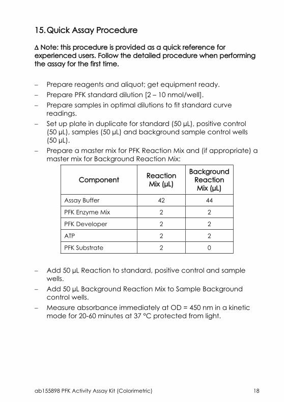
15.Quick Assay Procedure
Note: this procedure is provided as a quick reference for experienced users. Follow the detailed procedure when performing the assay for the first time.
Prepare reagents and aliquot; get equipment ready. Prepare PFK standard dilution [2 – 10 nmol/well]. Prepare samples in optimal dilutions to fit standard curve
readings. Set up plate in duplicate for standard (50 µL), positive control
(50 µL), samples (50 µL) and background sample control wells (50 µL).
Prepare a master mix for PFK Reaction Mix and (if appropriate) a master mix for Background Reaction Mix:
Component Reaction Mix (µL)
Background Reaction Mix (µL)
Assay Buffer 42 44
PFK Enzyme Mix 2 2
PFK Developer 2 2
ATP 2 2
PFK Substrate 2 0
Add 50 µL Reaction to standard, positive control and sample wells.
Add 50 µL Background Reaction Mix to Sample Background control wells.
Measure absorbance immediately at OD = 450 nm in a kinetic mode for 20-60 minutes at 37 °C protected from light.

ab155898 PFK Activity Assay Kit (Colorimetric) 19
16.Troubleshooting
Problem Reason Solution
Use of ice-cold buffer
Buffers must be at assay temperature
Plate read at incorrect
wavelength
Check the wavelength and filter settings of instrument
Assay not working
Use of a different microplate
Colorimetric: clear platesFluorometric: black wells/clear
bottom platesLuminometric: white wells/clear
bottom platesCells/tissue samples not homogenized
completely
Use Dounce homogenizer, increase number of strokes
Samples used after multiple free/ thaw
cycles
Aliquot and freeze samples if needed to use multiple times
Use of old or inappropriately stored samples
Use fresh samples or store at - 80°C (after snap freeze in liquid nitrogen)
till use
Sample with erratic readings
Presence of interfering
substance in the sample
Check protocol for interfering substances; deproteinize samples
Improperly thawed components
Thaw all components completely and mix gently before use
Allowing reagents to sit for extended
times on ice
Always thaw and prepare fresh reaction mix before use
Lower/higher readings in
samples and standards Incorrect
incubation times or temperatures
Verify correct incubation times and temperatures in protocol

ab155898 PFK Activity Assay Kit (Colorimetric) 20
Problem Reason Solution
Pipetting errors in standard or reaction mix
Avoid pipetting small volumes (< 5 µL) and prepare a master mix
whenever possibleAir bubbles formed
in wellPipette gently against the wall of
the tubes
Standard readings do not follow a linear
pattern Standard stock is at incorrect
concentration
Always refer to dilutions described in the protocol
Measured at incorrect
wavelengthCheck equipment and filter setting
Samples contain interfering substances
Troubleshoot if it interferes with the kitUnanticipated
resultsSample readings
above/ below the linear range
Concentrate/ Dilute sample so it is within the linear range

ab155898 PFK Activity Assay Kit (Colorimetric) 21
17. Interferences
These chemical or biological materials will cause interferences in this assay causing compromised results or complete failure: Small molecules such as glucose, NADH or ADP present in the
sample: we recommend setting up a background control well to correct for noise interference.
RIPA buffer: SDS may interfere with the enzymatic reaction.

ab155898 PFK Activity Assay Kit (Colorimetric) 22
18.Notes

ab155898 PFK Activity Assay Kit (Colorimetric) 23

ab155898 PFK Activity Assay Kit (Colorimetric) 24

ab155898 PFK Activity Assay Kit (Colorimetric) 25

Copyright © 2017 Abcam. All rights reserved
Technical Support
Copyright © 2017 Abcam, All Rights Reserved. The Abcam logo is a registered trademark. All information / detail is correct at time of going to print.
[email protected] | [email protected] | [email protected] | [email protected] | 91-114-65-60
[email protected] Deutsch: 043-501-64-24 | Français: 061-500-05-30UK, EU and [email protected] | +44(0)1223-696000
[email protected] | 877-749-8807US and Latin [email protected] | 888-772-2226
Asia Pacific [email protected] | (852) [email protected] | +86-21-5110-5938 | [email protected] | +81-(0)3-6231-0940Singapore [email protected] | 800 188-5244
[email protected] | +61-(0)3-8652-1450New Zealand [email protected] | +64-(0)9-909-7829