Color stability, water sorption and cytotoxicity of ... · 278 Color stability, water sorption and...
10
278 Color stability, water sorption and cytotoxicity of thermoplastic acrylic resin for non metal clasp denture Dae-Eun Jang 1 , Ji-Young Lee 1 , Hyun-Seon Jang 2 , Jang-Jae Lee 3 , Mee-Kyoung Son 1 * 1 Department of Prosthodontics, School of Dentistry, Chosun University, Gwangju, Republic of Korea 2 Department of Oral Pathology, School of Dentistry, Chosun University, Gwangju, Republic of Korea 3 Department of Computer Science and Statistics, College of Natural Sciences, Chosun University, Gwangju, Republic of Korea PURPOSE. The aim of this study was to compare the color stability, water sorption and cytotoxicity of thermoplastic acrylic resin for the non-metal clasp dentures to those of thermoplastic polyamide and conventional heat-polymerized denture base resins. MATERIALS AND METHODS. Three types of denture base resin, which are conventional heat-polymerized acrylic resin (Paladent 20), thermoplastic polyamide resin (Bio Tone), thermoplastic acrylic resin (Acrytone) were used as materials for this study. One hundred five specimens were fabricated. For the color stability test, specimens were immersed in the coffee and green tee for 1 and 8 weeks. Color change was measured by spectrometer. Water sorption was tested after 1 and 8 weeks immersion in the water. For the test of cytotoxicity, cell viability assay was measured and cell attachment was analyzed by FE-SEM. RESULTS. All types of denture base resin showed color changes after 1 and 8 weeks immersion. However, there was no significant difference between denture base resins. All specimens showed significant color changes in the coffee than green tee. In water sorption test, thermoplastic acrylic resin showed lower values than conventional heat-polymerized acrylic resin and thermoplastic polyamide resin. Three types of denture base showed low cytotoxicity in cell viability assay. Thermoplastic acrylic resin showed the similar cell attachment but more stable attachment than conventional heat-polymerized acrylic resin. CONCLUSION. Thermoplastic acrylic resin for the non-metal clasp denture showed acceptable color stability, water sorption and cytotoxicity. To verify the long stability in the mouth, additional in vitro studies are needed. [J Adv Prosthodont 2015;7:278-87] KEY WORDS: Denture base; Color stability; Water sorption; Cytotoxicity http://dx.doi.org/10.4047/jap.2015.7.4.278 http://jap.or.kr J Adv Prosthodont 2015;7:278-87 INTRODUCTION As patient expectations about cosmetic dentistry have been increasing, dissatisfaction among patients with removable partial dentures is also rising because of the unpleasant appearance of metal frameworks, especially with regard to the exposure of a metal clasp. In response to this trend, the non-metal clasp denture was introduced as an alternative in which the clasp is fabricated with a thermoplastic polymer instead of the conventional heat-polymerized polymethyl- methacrylate (PMMA) resin. Non-metal clasp dentures are made of elastic denture base resins instead of conventional metals, and they have the following advantages: enhanced esthetic appeal, no met- al-induced allergic reactions, no need for additional prosth- odontic treatment or tooth reduction, and reduced chair time. 1 Because of such esthetic and clinical benefits, they are preferred for the cases of anterior partial denture, growing patients, temporary dentures after implant surgery, Corresponding author: Mee-Kyoung Son Department of Prosthodontics, School of Dentistry, Chosun University, 303, Pilmun-daero, Dong-gu, Gwangju 61452, Republic of Korea Tel. 82 62 220 3825: e-mail, [email protected] Received November 14, 2014 / Last Revision February 23, 2015 / Accepted March 20, 2015 © 2015 The Korean Academy of Prosthodontics This is an Open Access article distributed under the terms of the Creative Commons Attribution Non-Commercial License (http://creativecommons. org/licenses/by-nc/3.0) which permits unrestricted non-commercial use, distribution, and reproduction in any medium, provided the original work is properly cited. pISSN 2005-7806, eISSN 2005-7814 This study was supported by research funds from Chosun University Dental Hospital 2014.
Transcript of Color stability, water sorption and cytotoxicity of ... · 278 Color stability, water sorption and...
278
Color stability, water sorption and cytotoxicity of thermoplastic acrylic resin for non metal clasp denture
Dae-Eun Jang1, Ji-Young Lee1, Hyun-Seon Jang2, Jang-Jae Lee3, Mee-Kyoung Son1* 1Department of Prosthodontics, School of Dentistry, Chosun University, Gwangju, Republic of Korea2Department of Oral Pathology, School of Dentistry, Chosun University, Gwangju, Republic of Korea3Department of Computer Science and Statistics, College of Natural Sciences, Chosun University, Gwangju, Republic of Korea
PURPOSE. The aim of this study was to compare the color stability, water sorption and cytotoxicity of thermoplastic acrylic resin for the non-metal clasp dentures to those of thermoplastic polyamide and conventional heat-polymerized denture base resins. MATERIALS AND METHODS. Three types of denture base resin, which are conventional heat-polymerized acrylic resin (Paladent 20), thermoplastic polyamide resin (Bio Tone), thermoplastic acrylic resin (Acrytone) were used as materials for this study. One hundred five specimens were fabricated. For the color stability test, specimens were immersed in the coffee and green tee for 1 and 8 weeks. Color change was measured by spectrometer. Water sorption was tested after 1 and 8 weeks immersion in the water. For the test of cytotoxicity, cell viability assay was measured and cell attachment was analyzed by FE-SEM. RESULTS. All types of denture base resin showed color changes after 1 and 8 weeks immersion. However, there was no significant difference between denture base resins. All specimens showed significant color changes in the coffee than green tee. In water sorption test, thermoplastic acrylic resin showed lower values than conventional heat-polymerized acrylic resin and thermoplastic polyamide resin. Three types of denture base showed low cytotoxicity in cell viability assay. Thermoplastic acrylic resin showed the similar cell attachment but more stable attachment than conventional heat-polymerized acrylic resin. CONCLUSION. Thermoplastic acrylic resin for the non-metal clasp denture showed acceptable color stability, water sorption and cytotoxicity. To verify the long stability in the mouth, additional in vitro studies are needed. [ J Adv Prosthodont 2015;7:278-87]
KEY WORDS: Denture base; Color stability; Water sorption; Cytotoxicity
http://dx.doi.org/10.4047/jap.2015.7.4.278http://jap.or.kr J Adv Prosthodont 2015;7:278-87
INTRODUCTION
As patient expectations about cosmetic dentistry have been
increasing, dissatisfaction among patients with removable partial dentures is also rising because of the unpleasant appearance of metal frameworks, especially with regard to the exposure of a metal clasp. In response to this trend, the non-metal clasp denture was introduced as an alternative in which the clasp is fabricated with a thermoplastic polymer instead of the conventional heat-polymerized polymethyl-methacrylate (PMMA) resin.
Non-metal clasp dentures are made of elastic denture base resins instead of conventional metals, and they have the following advantages: enhanced esthetic appeal, no met-al-induced allergic reactions, no need for additional prosth-odontic treatment or tooth reduction, and reduced chair time.1 Because of such esthetic and clinical benefits, they are preferred for the cases of anterior partial denture, growing patients, temporary dentures after implant surgery,
Corresponding author: Mee-Kyoung SonDepartment of Prosthodontics, School of Dentistry, Chosun University, 303, Pilmun-daero, Dong-gu, Gwangju 61452, Republic of KoreaTel. 82 62 220 3825: e-mail, [email protected] November 14, 2014 / Last Revision February 23, 2015 / Accepted March 20, 2015
© 2015 The Korean Academy of ProsthodonticsThis is an Open Access article distributed under the terms of the Creative Commons Attribution Non-Commercial License (http://creativecommons.org/licenses/by-nc/3.0) which permits unrestricted non-commercial use, distribution, and reproduction in any medium, provided the original work is properly cited.
pISSN 2005-7806, eISSN 2005-7814
This study was supported by research funds from Chosun University Dental Hospital 2014.
The Journal of Advanced Prosthodontics 279
and elderly or handicapped patients who are vulnerable to the complex procedure of conventional treatments.
Polyamide resins, polycarbonate resins, and polyethylene terephthalates have been introduced as representative ther-moplastic denture base resin materials for the fabrication of non-metal clasp dentures.2 Of these, the polyamide res-in, commonly referred to as nylon polymer, was introduced in the 1950s as a denture base material.3-5
Polyamide resin is produced from the polymerization of diamine and dibasic acid; while PMMA is amorphous, poly-amides are crystalline polymers with properties suitable for high-quality elastic denture materials, such as low solubility, high thermal resistance and flexibility, high strength, and superior moldability.5-7 Despite these advantages, unlike common PMMA acrylic resins, polyamide resins have diffi-culties in reline and repair when relining becomes necessary because of resorption of the alveolar bone under the den-ture base.
To overcome this drawback, the thermoplastic PMMA resin, which is produced by using an injection molding meth-od, has recently been introduced for the fabrication of non-metal clasp dentures. Acrytone, its key component, contains PMMA components that are found in auto- and heat-polymerized relining resins; this allows it to be applied for relining, thereby overcoming the drawback of existing elas-tic dentures made of polyamide resins.8,9
The required properties for good denture base materials include esthetic appeal, color stability, dimensional stability, low water sorption, ease of fabrication, easy to repair, and biocompatibility.8-10 Of these, color stability has a long-term influence on the esthetics of denture base resins. Denture base resins prone to pigmentation and discoloration are often a cause of patient dissatisfactions and denture refab-rication.11 Therefore, in order to ensure satisfactory long-term use of dentures, it is crucial to use denture base mate-rials that can maintain color stability against external pig-mentation factors in various oral environments.
Water sorption of a denture base resin can trigger dis-coloration and halitosis as well as dimensional instability, leading to internal stress and ultimately to cracks or failure of the denture.12,13 In other words, a high water sorption rate tends to affect the material properties and consequent-
ly reduce the service life of a denture within the oral cavity; therefore, it is crucial to use materials with minimum possi-ble water sorption rates.14
A denture base comes into direct contact with the oral mucosa, therefore, the use of biocompatible materials is essential for avoiding hypersensitivity reactions or the release of toxic substances. A serious problem associated with conventional acrylic resins was allergic reactions trig-gered by residual monomers after polymerization.15 To address this problem, materials and polymerization meth-ods that reduce or eliminate monomers have been intro-duced, and the biocompatibilities of denture base materials is considered a very important quality.
These fundamental requirements for denture base mate-rials also apply to the resins used for non-metal clasp den-ture fabrication to ensure the long-term success of the den-tal prosthesis and the oral health of the patient. This led to early research into the properties of the denture base mate-rials for non-metal clasp dentures and their long-term sta-bility in the oral cavity environment. However, the related studies still have very limited scope, and only a few studies have explored color stability, water sorption rate, and cyto-toxicity in various oral cavity environments. Although the properties of these materials are currently unclarified, they are being used in clinical settings based merely on the pref-erence or experience of the clinician.
Therefore, this study aimed to assess the feasibility of the clinical use of denture base resins for non-metal clasp dentures by comparing the color stability, water sorption rate, and cytotoxicity of the currently used thermoplastic acrylic resins with those of thermoplastic polyamide and conventional heat-polymerized acrylic resins.
MATERIALS AND METHODS
In this study, three types of denture base resins are used: 1) Paladent 20, a PMMA-based conventional heat-polymerized acrylic resin; 2) Bio Tone, a thermoplastic polyamide resin; and 3) Acrytone, a PMMA-based thermoplastic acrylic res-in. The compositions of these materials are provided in Table 1.
Table 1. Materials and components used in the study
Brand names Main components
ManufacturerPowder Liquid
Paladent 20 PMMA MMA Heraeus Kulzer, Hanau, Germany
Bio Tone Polyamide - High–Dental-Japan, Osaka, Japan
Acrytone PMMA MMA High–Dental-Japan, Osaka, Japan
Color stability, water sorption and cytotoxicity of thermoplastic acrylic resin for non metal clasp denture

280
Specimens fabrication: A total of one hundred five test specimens (35 specimens for each type of denture base res-ins) in a disc form with 20-mm diameter and 1-mm thick-ness were prepared according to the polymerization meth-ods specified by manufacturers (Table 2). The fabricated specimens were polished according to the methods recom-mended for each denture base material type.
Color stability test: Twenty pairs of specimens for each type of denture base resins (total 60 specimens) were used for the color stability test. Specimens of each type were assigned serial numbers of 1 through 20, and the number was written on the side of the specimen that would not be used for the color stability test. These specimens were sub-divided into 2 groups (n=10) and were immersed in two different beverages; coffee and green tea.
The coffee solution was prepared by filtering 250 mL of boiled distilled water mixed with 15 g of coffee powder (Maxim original, Dongsuh, Seoul, Korea) 10 minutes of stirring, and the green tea solution was prepared by steep-ing 5 green tea bags (Dongsuh, Korea) 10 minutes in 250 mL of boiled distilled water.
Specimens of each type of resin were immersed in 50 ml of each beverage in 200 mL glass beakers, while taking great care to avoid contact between the specimens within the beaker. The beakers with the immersed specimens were stored in a dark room at room temperature for 8 weeks.
The specimen colors were measured with a spectral color-imeter (SpectroShadeTM Micro, MHT, Niederhasli, Switzerland). The color assessment steps were as follows: 1) baseline col-or measurement and recording prior to immersion; 2) inter-im color measurement and recording after 1 week of immersion, whereupon the specimens were removed, rinsed with distilled water, and dried with tissue paper, and 3) final color measurement and recording after 8 weeks, performed as described for 2). Each specimen was measured three times at randomly selected spots and the mean value was recorded to reduce potential error due to measurement at various areas. The differences between the baseline and post-immersion colors were measured according to the CIE
Lab system. These differences are expressed as CIE color difference (ΔE) values.Themethod for calculating thesevalues is described in the following section.16
ΔΕ*=[(ΔL*)2+(Δa*)2+(Δb*)2]1/2
L*:indicateWhite-Black,ΔL=Lt - L0a*:indicateRedness-Greenness,Δa=at - a0b*:indicateYellowness-Blueness,Δb=bt - b0(Lt, at, bt : values at the time of measurement after immersion / L0, a0, b0 : values before immersion)
The resulting color stability data were analyzed by using repeated measures ANOVA to validate the main effects and interaction among materials, solutions, and time depen-dence. The material-solution interaction effects on the results of 1-week and 8-week immersions were assessed by using two-way ANOVA. The material-dependent differenc-es in color stability after 1-week and 8-week immersions were analyzed by using the Student’s t-test. The solution-dependent differences in color stability after 1-week and 8-week immersions were analyzed by using one-way ANOVA, followed by the Newman-Keull’s multiple com-parison test.
Water sorption test: Ten specimens for each type of den-ture base resins (total 30 specimens) were used for the water sorption test. Prior to the test, the diameter and thickness of each specimen were re-measured by using an electronic dig-ital caliper to calculate the precise volume (V). The weight of each specimen was measured with an electronic digital balance after storing the specimens in a drying chamber for 24 hours. The weight was recorded when the digital reading remained at a constant value (W0). After measuring the baseline volumes and weights, we stored the specimens in a constant-temperature water tank (37ºC) filled with distilled water.
The weights were measured after 1 week and 8 weeks of immersion, whereupon 5 specimens for each type were removed from the tank, dried with tissue paper, and left to stand on the electronic digital balance for 30 seconds before
Table 2. Molding and curing methods of denture base resins by manufacturers
Denture base resins Molding method Curing method
Paladent 20 Compression moldingHeat processed at 80°C/15 min,Boiling water/20 min
Bio Tone Injection molding300°C meltingInjection at 0.7 MPaCooling under pressure
Acrytone Injection molding
260°C melting/25 minInjection at 0.7 MPaCooling under pressure(30 min in air, 30 min in water)
J Adv Prosthodont 2015;7:278-87

The Journal of Advanced Prosthodontics 281
reading the indicated value (W1). We performed a second measurement after the specimens had been stored in a drier for 24 hours after the first measurement was taken; the val-ue was read when the balance remained at a constant value for 30 sec (W2). The water sorption rate was defined as the mass by which each specimen increased per unit volume after immersion,17 calculated as described in the following paragraph.18
WaterSorption:(ΔWsorp) = (W2 - W1) / V (µg/mm3)
The data on the time-dependent water sorption rates measured after 1 week and 8 weeks of immersion were ana-lyzed by using repeated measures ANOVA to test the main effects and interaction of the material and time. The mate-rial-dependent differences in water sorption after the same immersion time (1 week and 8 weeks) were analyzed by using one-way ANOVA, followed by Tukey’s multiple com-parison test, and the time-dependent differences in the water sorption rate for each material were analyzed with the paired t-test.
Cytotoxicity test: Cell viability assay and cell attachment analysis were performed to verify the cytotoxicity of the denture base resins by using fifteen specimens (5 specimens for each). We pretreated the specimens by cleaning them an ultrasonic cleaner filled with pure acetone solution for 10 min, followed by washing with alcohol and rinsing with dis-tilled water. Each sample was then separately wrapped and sterilized in a high-pressure high-temperature sterilizer in preparation for cell culture experiments.
Cell isolation and culture: Human gingival fibroblasts (hGF) were isolated from patient tissue that had been resected for a second-stage implant procedure, and cultured in Dulbecco’s Modified Eagle’s Medium (DMEM) contain-ing 10% fetal bovine serum (FBS) and 1× antibiotic/anti-mycotic. The cells were cultured in a cell culture incubator (37ºC, 95% humidity) containing 5% CO2. The media was renewed every 2 days. The 5th passage human gingival fibro-blasts were used.
Preparations for cell viability test: The cells were seeded in a 12-well plate at a density of 1.8 × 10⁴ cells/well and were cultured for 24 hours in an incubator. The specimens were put into the wells (1 specimen/well) and were cultured for 1 day and 6 days. The cells were cultured in DMEM
containing 10% FBS and 1× antibiotic/antimycotic. The EZ-Cytox Enhanced Cell Viability Assay Kit (Daeillab ser-vice co., Seoul, South Korea) was used to measure the effects of the test specimens on cell viability of the gingival fibroblasts. The test was performed in compliance with the guidelines provided by the manufacturer. After removing the specimens applied to the cells, 10 μL of EZ-Cytoxreagent were added toeach well and cultured the cells for 4 h. Subsequently, the optical density (OD) at a wavelength of 450 nm was measured by using a microplate reader (Bio-Tek Instrument, Winooski, VT, USA).
Preparations for cell attachment analysis: In order to determine the time-dependence of the ability of the cells to attach to the surface of each specimen, the specimens were placed in a 12-well plate and the cultured cells were seeded in each well (6 × 10⁴ cells/well) that contained a specimen. The samples were incubated for 1, 6, and 10 days in DMEM containing 10% FBS and 1× antibiotic/antimycot-ic. At each measured point, the cells that had been incubat-ed on each specimen were fixed with 2.5 % glutaraldehyde, and the extent of adhesion was observed with a FE-SEM (HITACHI S-4800, Tokyo, Japan) at ×1,000 magnification on 1, 6, and 10 days after incubation.
RESULTS
Resultsof colorstability:ThemeanΔEvalues,whichindi-cate the color differences exhibited by the three types of denture base resin specimens when immersed in coffee and green tea after 1 week (T0T1) and 8 weeks (T0T2), are shown in Table 3.
As a results of the repeated ANOVA, there was no sig-nificant interaction effect among materials, solutions, and time dependence (P>.05). Under two-way ANOVA analysis, the material-solution interaction effect on the different times also showed no significant difference (P>.05). Therefore, material-dependent and solution-dependent differences after 1 and 8-week immersions were analyzed respectively by using Student T-test and one-way ANOVA.
Statistical analysis revealed no material-dependent color difference among the denture base materials after 1 week of immersion (ΔΕT0T1); however, a significantly greater color difference (ΔΕ)was observed for Paladent 20 andAcrytone in coffee to green tea (P<.001, P=.004 for each),
Table 3. Color changes after 1 and 8 weeks immersion in coffee and tea (ΔE Mean ± SD)
Denture base resins1 week 8 week
Coffee Tea P value Coffee Tea P value
Paladent 20 5.648 ± 0.291 1.636 ± 0.547 <.001 5.624 ± 2.211 2.885 ± 0.958 .022
Bio Tone 5.274 ± 3.235 2.058 ± 0.869 .103 8.578 ± 1.765 3.196 ± 0.877 .002
Acrytone 4.072 ± 1.013 1.912 ± 0.990 .004 8.724 ± 2.979 3.567 ± 1.243 .003
P value 0.305 0.658 0.295 0.883
Color stability, water sorption and cytotoxicity of thermoplastic acrylic resin for non metal clasp denture
282
whereas no beverage-dependent difference was exhibited by Bio Tone (P=.103).
In general, greater color differences were exhibited after 8 weeks of immersion. While no significant material-depen-dentcolordifferences(ΔΕT0T2) were observed (P>.05), all three types of denture base resin showed greater color dif-ferences in coffee than in green tea (P=.003, P=.002, P=.022 for each).
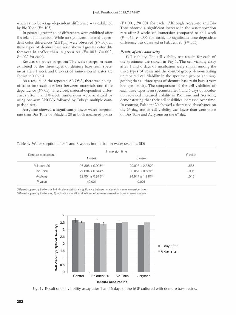
Results of water sorption: The water sorption rates exhibited by the three types of denture base resin speci-mens after 1 week and 8 weeks of immersion in water are shown in Table 4.
As a results of the repeated ANOVA, there was no sig-nificant interaction effect between materials and time dependence (P>.05). Therefore, material-dependent differ-ences after 1 and 8-week immersions were analyzed by using one-way ANOVA followed by Tukey’s multiple com-parison test,.
Acrytone showed a significantly lower water sorption rate than Bio Tone or Paladent 20 at both measured points
(P<.001, P=.001 for each). Although Acrytone and Bio Tone showed a significant increase in the water sorption rate after 8 weeks of immersion compared to at 1 week (P=.045, P=.006 for each), no significant time-dependent difference was observed in Paladent 20 (P=.563).
Results of cell cytotoxicityCell viability: The cell viability test results for each of
the specimens are shown in Fig. 1. The cell viability assay after 1 and 6 days of incubation were similar among the three types of resin and the control group, demonstrating unimpaired cell viability in the specimen groups and sug-gesting that all three types of denture base resin have a very low cytotoxicity. The comparison of the cell viabilities of each three types resin specimen after 1 and 6 days of incuba-tion revealed increased viability in Bio Tone and Acrytone, demonstrating that their cell viabilities increased over time. In contrast, Paladent 20 showed a decreased absorbance on the 6th day, and its cell viability was lower than were those of Bio Tone and Acrytone on the 6th day.
Table 4. Water sorption after 1 and 8 weeks immersion in water (Mean ± SD)
Denture base resinsImmersion time
P value1 week 8 week
Paladent 20 28.306 ± 0.923aA 29.025 ± 2.530aA .563
Bio Tone 27.694 ± 0.644aA 30.057 ± 0.539aB .006
Acytone 22.904 ± 0.875bA 24.917 ± 1.210bB .045
P value <0.001 0.001
Different superscript letters (a, b) indicate a statistical significance between materials in same immersion time.Different superscript letters (A, B) indicate a statistical significance between immersion times in same material.
Fig. 1. Result of cell viability assay after 1 and 6 days of the hGF cultured with denture base resins.
J Adv Prosthodont 2015;7:278-87

The Journal of Advanced Prosthodontics 283
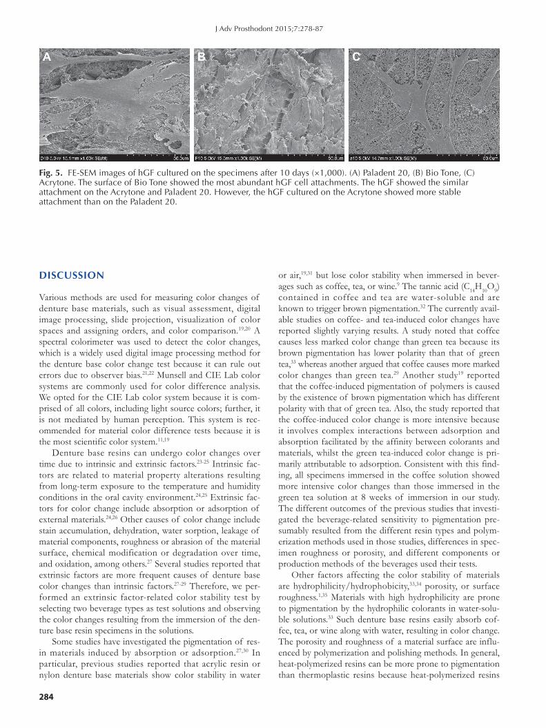
Cell attachments: In SEM observation, Bio Tone showed the smoothest pre-test surface (Fig. 2) and the most efficient cell attachment on 1 day of incubation, while Acrytone and Paladent 20 showed moderate and poor cell attachment, respectively (Fig. 3), and the richest hGF cell attachment on
6 days of incubation (Fig. 4). On 10 days incubation, Bio Tone still showed the most abundant cell attachment, whereas Acrytone and Paladent 20 showed the similar degrees of cell attachment, with Acrytone showing a more stable pattern than Paladent 20 (Fig. 5).
Fig. 2. FE-SEM images of the specimen’s surface before cell attachments (×1,000). (A) Paladent 20, (B) Bio Tone, (C) Acrytone. The specimen of Bio Tone showed the smoothest surface. The specimen of Paladent 20 showed rough surface patterns and small pores.
Fig. 3. FE-SEM images of hGF cultured on the specimens after 1 day (×1,000). (A) Paladent 20, (B) Bio Tone, (C) Acrytone. The surface of Bio Tone showed most abundant hGF cell attachments. The surface of Acrytone showed moderate cell attachment appearance, and Paladent 20 showed a relatively weak cell attachments.
Fig. 4. FE-SEM images of hGF cultured on the specimens after 6 days (×1,000). (A) Paladent 20, (B) Bio Tone, (C) Acrytone. The surface of Bio Tone showed the most abundant hGF cell attachments. The hGF showed a multi-layer morphology on the Bio Tone surface.
Color stability, water sorption and cytotoxicity of thermoplastic acrylic resin for non metal clasp denture
284
DISCUSSION
Various methods are used for measuring color changes of denture base materials, such as visual assessment, digital image processing, slide projection, visualization of color spaces and assigning orders, and color comparison.19,20 A spectral colorimeter was used to detect the color changes, which is a widely used digital image processing method for the denture base color change test because it can rule out errors due to observer bias.21,22 Munsell and CIE Lab color systems are commonly used for color difference analysis. We opted for the CIE Lab color system because it is com-prised of all colors, including light source colors; further, it is not mediated by human perception. This system is rec-ommended for material color difference tests because it is the most scientific color system.11,19
Denture base resins can undergo color changes over time due to intrinsic and extrinsic factors.23-25 Intrinsic fac-tors are related to material property alterations resulting from long-term exposure to the temperature and humidity conditions in the oral cavity environment.24,25 Extrinsic fac-tors for color change include absorption or adsorption of external materials.24,26 Other causes of color change include stain accumulation, dehydration, water sorption, leakage of material components, roughness or abrasion of the material surface, chemical modification or degradation over time, and oxidation, among others.27 Several studies reported that extrinsic factors are more frequent causes of denture base color changes than intrinsic factors.27-29 Therefore, we per-formed an extrinsic factor-related color stability test by selecting two beverage types as test solutions and observing the color changes resulting from the immersion of the den-ture base resin specimens in the solutions.
Some studies have investigated the pigmentation of res-in materials induced by absorption or adsorption.27,30 In particular, previous studies reported that acrylic resin or nylon denture base materials show color stability in water
or air,19,31 but lose color stability when immersed in bever-ages such as coffee, tea, or wine.9 The tannic acid (C14H10O9) contained in coffee and tea are water-soluble and are known to trigger brown pigmentation.32 The currently avail-able studies on coffee- and tea-induced color changes have reported slightly varying results. A study noted that coffee causes less marked color change than green tea because its brown pigmentation has lower polarity than that of green tea,33 whereas another argued that coffee causes more marked color changes than green tea.29 Another study19 reported that the coffee-induced pigmentation of polymers is caused by the existence of brown pigmentation which has different polarity with that of green tea. Also, the study reported that the coffee-induced color change is more intensive because it involves complex interactions between adsorption and absorption facilitated by the affinity between colorants and materials, whilst the green tea-induced color change is pri-marily attributable to adsorption. Consistent with this find-ing, all specimens immersed in the coffee solution showed more intensive color changes than those immersed in the green tea solution at 8 weeks of immersion in our study. The different outcomes of the previous studies that investi-gated the beverage-related sensitivity to pigmentation pre-sumably resulted from the different resin types and polym-erization methods used in those studies, differences in spec-imen roughness or porosity, and different components or production methods of the beverages used their tests.
Other factors affecting the color stability of materials are hydrophilicity/hydrophobicity,33,34 porosity, or surface roughness.1,35 Materials with high hydrophilicity are prone to pigmentation by the hydrophilic colorants in water-solu-ble solutions.33 Such denture base resins easily absorb cof-fee, tea, or wine along with water, resulting in color change. The porosity and roughness of a material surface are influ-enced by polymerization and polishing methods. In general, heat-polymerized resins can be more prone to pigmentation than thermoplastic resins because heat-polymerized resins
Fig. 5. FE-SEM images of hGF cultured on the specimens after 10 days (×1,000). (A) Paladent 20, (B) Bio Tone, (C) Acrytone. The surface of Bio Tone showed the most abundant hGF cell attachments. The hGF showed the similar attachment on the Acrytone and Paladent 20. However, the hGF cultured on the Acrytone showed more stable attachment than on the Paladent 20.
J Adv Prosthodont 2015;7:278-87
The Journal of Advanced Prosthodontics 285
have a higher porosity. According to previous studies on the surface roughness of resins, denture base resins of the PMMA acrylic group have a significantly smoother surface than resins of other groups35; further, if polished by using the same method, the polyamide resin shows a more inten-sive pigmentation than the PMMA acrylic resin because of the greater surface roughness of the polyamide resin. However, no such differences in surface roughness were observed in our study because each type of denture base res-in specimens was polished with their own polishing burs according to manufacturer’s recommendation, and some polyamide specimens even showed smoother surfaces. These complex issues presumably contributed to the observed lack of significant differences in color changes among the three types of denture base resin immersed in the same beverage solution. These results are consistent with those of previous studies that compared the color stability between heat-polymerized denture base resins and nylon denture base resins.19
The water sorption rate of materials affects their color stability as well as their physical properties. Acrylic resins absorb water for a prolonged length of time because of the polar property of the resin molecules, whereby the extent of water sorption is determined in proportion to the resin components with high polarity, which form hydrogen bonds with water molecules.36 The absorbed water infiltrates into the resin polymers and triggers reversible or irreversible bond breakages between weak molecular interchain bond, thereby causing deterioration of the mechanical properties of the materials, such as hardness, flexural strength, and fatigue limit, as well as dimensional stability.34,37-40 Therefore, the International Organization for Standardization (ISO 1567:1999)41 specifies the maximum water sorption ability of heat-polymerizedresinmaterialsas32μg/mm3. Most of the previous studies on the water sorption abilities of acryl-ic resins have demonstrated a low water sorption rate of 10-25 μg/mm.3,16,37,38,42 All denture base resins used in our study also showed water sorption rates ranging between 17 and25μg/mm3, thereby meeting the pertinent ISO standards.
The earlier polyamide resins had limited properties regarding deformation, water sorption, surface roughness, and polishing.4,5 Polyamide-group denture base resins are subjected to water sorption between molecular chains due to the hydrophilicity of the many amide bonds that form the main chains of the resins, resulting in high water sorp-tion rates. To overcome this drawback, recently available polyamide-type denture base materials were developed with the aim of reducing water absorption by controlling the amide-group concentration.1,43 As a result, recent studies using polyamide resins with lowered water sorption rates reported that no differences in mean water sorption rate were observed between heat-polymerized resins and nylon resins.44 Similarly, Bio Tone, of the polyamide group, was resistant to hydrogen bonding, thereby demonstrating water sorption rates similar to those of common heat-polymer-ized acrylic resins in our study. Further, the presence of residual monomers is reported to affect water sorption and
expansion.45 In this regard, Paladent 20 is likely to have more residual monomers than Acrytone because the polymerization method for Paladent 20 employs compres-sion whereas that for Acrytone employs pressure injection. This presumably explains why Acrytone showed a lower water sorption rate than Paladent 20 in our study.
Even polymerized denture base resins can release resid-ual monomers or toxic materials such as formaldehyde, methacrylic acid, and benzoic acid, which can cause irrita-tion, inflammation, or allergic reactions in the oral tissues.13 The amount of such residual monomers are known to be influenced by the type, curing method, and thickness of denture base resins; therefore, the tissue irritation induced by the denture base resin can be reduced by addressing these influential factors.46-48 The majority of studies on the cytotoxicity of the resins currently used as denture base materials reported that these resins are not toxic49 and do not suppress cell growth.50 The three types of denture base resin materials tested in our study revealed that they have a negligible influence on cell viability and adhesion; hence, these materials are not believed to be cytotoxic.
This in-vitro study involved a limited analysis of color stability, water sorption and cytotoxic properties for the denture base materials used. For clinical application, further investigations regarding other mechanical and biological properties studies and warranting the long-term effect in vivo were still required.
CONCLUSION
The conclusion of this study indicates that thermoplastic acrylic resins used as materials for non-metal clasp dentures are applicable in the oral cavity environment because their color stability, water sorption rate, and cytotoxicity are simi-lar to those of the thermoplastic polyamide and conven-tional heat-polymerized acrylic resins. However, to verify the long stability and elastic properties of thermoplastic acrylic resins in the mouth, additional in vitro studies are still needed.
ORCID
Dae-Eun Jang http://orcid.org/0000-0002-0493-5052Ji-Young Lee http://orcid.org/0000-0003-4615-4657Hyun-Seon Jang http://orcid.org/0000-0003-1231-3816Jang-Jae Lee http://orcid.org/0000-0001-8787-9940Mee-Kyoung Son http://orcid.org/0000-0001-9225-1744
REFERENCES
1. Takabayashi Y. Characteristics of denture thermoplastic res-ins for non-metal clasp dentures. Dent Mater J 2010;29:353-61.
2. Truong VT, Thomasz FG. Comparison of denture acrylic resins cured by boiling water and microwave energy. Aust Dent J 1988;33:201-4.
3. Yunus N, Rashid AA, Azmi LL, Abu-Hassan MI. Some flex-
Color stability, water sorption and cytotoxicity of thermoplastic acrylic resin for non metal clasp denture
286
ural properties of a nylon denture base polymer. J Oral Rehabil 2005;32:65-71.
4. Matthews E., Smith DC. Nylon as a denture base material. Br Dent J 1955;98:231-7.
5. Watt DM. Clinical assessment of nylon as a partial denture base material. Br Dent J 1955;98:238-44.
6. Katsumata Y, Hojo S, Hamano N, Watanabe T, Yamaguchi H, Okada S, Teranaka T, Ino S. Bonding strength of autopo-lymerizing resin to nylon denture base polymer. Dent Mater J 2009;28:409-18.
7. Soygun K, Bolayir G, Boztug A. Mechanical and thermal properties of polyamide versus reinforced PMMA denture base materials. J Adv Prosthodont 2013;5:153-60.
8. Kim JH, Choe HC, Son MK. Evaluation of adhesion of re-line resins to the thermoplastic denture base resin for non-metal clasp denture. Dent Mater J 2014;33:32-8.
9. Cil ingir A, Bilhan H, Geckil i O, Sulun T, Bozdag E, Sunbuloglu E. In vitro comparison of two different materials for the repair of urethan dimethacrylate denture bases. J Adv Prosthodont 2013;5:396-401.
10. Nam KY. Characterization and bacterial anti-adherent effect on modified PMMA denture acrylic resin containing plati-num nanoparticles. J Adv Prosthodont 2014;6:207-14.
11. Hersek N, Canay S, Uzun G, Yildiz F. Color stability of den-ture base acrylic resins in three food colorants. J Prosthet Dent 1999;81:375-9.
12. Hiromori K, Fujii K, Inoue K. Viscoelastic properties of denture base resins obtained by underwater test. J Oral Rehabil 2000;27:522-31.
13. Wong DM, Cheng LY, Chow TW, Clark RK. Effect of pro-cessing method on the dimensional accuracy and water sorp-tion of acrylic resin dentures. J Prosthet Dent 1999;81:300-4.
14. Cucci AL, Vergani CE, Giampaolo ET, Afonso MC. Water sorption, solubility, and bond strength of two autopolymeriz-ing acrylic resins and one heat-polymerizing acrylic resin. J Prosthet Dent 1998;80:434-8.
15. Lefebvre CA, Knoernschild KL, Schuster GS. Cytotoxicity of eluates from light-polymerized denture base resins. J Prosthet Dent 1994;72:644-50.
16. Hunter RS, Harold RW. The measurement of appearance. 2nd ed. New York; John Wiley; 1987.
17. Miettinen VM, Vallittu PK, Docent DT. Water sorption and solubility of glass fiber-reinforced denture polymethyl meth-acrylate resin. J Prosthet Dent 1997;77:531-4.
18. Tuna SH, Keyf F, Gumus HO, Uzun C. The evaluation of water sorption/solubility on various acrylic resins. Eur J Dent 2008;2:191-7.
19. Sepúlveda-Navarro WF, Arana-Correa BE, Borges CP, Jorge JH, Urban VM, Campanha NH. Color stability of resins and nylon as denture base material in beverages. J Prosthodont 2011;20:632-8.
20. Paul S, Peter A, Pietrobon N, Hämmerle CH. Visual and spectrophotometric shade analysis of human teeth. J Dent Res 2002;81:578-82.
21. Bagheri R, Burrow MF, Tyas M. Influence of food-simulating solutions and surface finish on susceptibility to staining of aesthetic restorative materials. J Dent 2005;33:389-98.
22. Hu X, Johnston WM, Seghi RR. Measuring the color of max-illofacial prosthetic material. J Dent Res 2010;89:1522-7.
23. Villalta P, Lu H, Okte Z, Garcia-Godoy F, Powers JM. Effects of staining and bleaching on color change of dental composite resins. J Prosthet Dent 2006;95:137-42.
24. Goiato MC, Santos DM, Haddad MF, Pesqueira AA. Effect of accelerated aging on the microhardness and color stability of flexible resins for dentures. Braz Oral Res 2010;24:114-9.
25. Wilson NH, Burke FJ, Mjör IA. Reasons for placement and replacement of restorations of direct restorative materials by a selected group of practitioners in the United Kingdom. Quintessence Int 1997;28:245-8.
26. Abu-Bakr N, Han L, Okamoto A, Iwaku M. Color stability of compomer after immersion in various media. J Esthet Dent 2000;12:258-63.
27. Asmussen E. Factors affecting the color stability of restor-ative resins. Acta Odontol Scand 1983;41:11-8.
28. Polyzois GL, Yannikakis SA, Zissis AJ. Color stability of visi-ble light-cured, hard direct denture reliners: an in vitro inves-tigation. Int J Prosthodont 1999;12:140-6.
29. Buyukyilmaz S, Ruyter IE. Color stability of denture base polymers. Int J Prosthodont 1994;7:372-82.
30. Chan KC, Fuller JL, Hormati AA. The ability of foods to stain two composite resins. J Prosthet Dent 1980;43:542-5.
31. Lai YL, Lui HF, Lee SY. In vitro color stability, stain resis-tance, and water sorption of four removable gingival flange materials. J Prosthet Dent 2003;90:293-300.
32. Guler AU, Yilmaz F, Kulunk T, Guler E, Kurt S. Effects of different drinks on stainability of resin composite provisional restorative materials. J Prosthet Dent 2005;94:118-24.
33. Um CM, Ruyter IE. Staining of resin-based veneering mate-rials with coffee and tea. Quintessence Int. 1991;22:377-86.
34. Ferracane JL. Hygroscopic and hydrolytic effects in dental polymer networks. Dent Mater 2006;22:211-22.
35. de Freitas Fernandes FS, Pereira-Cenci T, da Silva WJ, Filho AP, Straioto FG, Del Bel Cury AA. Efficacy of denture cleansers on Candida spp. biofilm formed on polyamide and polymethyl methacrylate resins. J Prosthet Dent 2011;105:51-8.
36. Malacarne J, Carvalho RM, de Goes MF, Svizero N, Pashley DH, Tay FR, Yiu CK, Carrilho MR. Water sorption/solubili-ty of dental adhesive resins. Dent Mater 2006;22:973-80.
37. Barsby MJ. A denture base resin with low water absorption. J Dent 1992;20:240-4.
38. Ristic B, Carr L. Water sorption by denture acrylic resin and consequent changes in vertical dimension. J Prosthet Dent 1987;58:689-93.
39. Patel MP, Braden M. Heterocyclic methacrylates for clinical applications. III. Water absorption characteristics. Biomaterials 1991;12:653-7.
40. Woelfel JB, Paffenbarger GC, Sweeney WT. Some physical properties of organic denture base materials. J Am Dent Assoc 1963;67:499-504.
41. ISO 1567:1999. Dentistry - Denture base polymers. 42. Arima T, Murata H, Hamada T. The effects of cross-linking
agents on the water sorption and solubility characteristics of denture base resin. J Oral Rehabil 1996;23:476-80.
J Adv Prosthodont 2015;7:278-87

The Journal of Advanced Prosthodontics 287
43. Hargreaves AS. Nylon as a denture-base material. Dent Pract Dent Rec 1971;22:122-8.
44. Kurtulmus H, Kumbuloglu O, Aktas RT, Kurtulmus A, Boyacioglu H, Oral O, User A. Effects of saliva and nasal se-cretion on some physical properties of four different resin materials. Med Oral Patol Oral Cir Bucal 2010;15:e969-75.
45. Dixon DL, Breeding LC, Ekstrand KG. Linear dimensional variability of three denture base resins after processing and in water storage. J Prosthet Dent 1992;68:196-200.
46. Lefebvre CA, Schuster GS, Marr JC, Knoernschild KL. The effect of pH on the cytotoxicity of eluates from denture base resins. Int J Prosthodont 1995;8:122-8.
47. Koda T, Tsuchiya H, Yamauchi M, Ohtani S, Takagi N, Kawano J. Leachability of denture-base acrylic resins in arti-ficial saliva. Dent Mater 1990;6:13-6.
48. McCabe JF, Basker RM. Tissue sensitivity to acrylic resin. A method of measuring the residual monomer content and its clinical application. Br Dent J 1976;140:347-50.
49. Vallittu PK, Ekstrand K. In vitro cytotoxicity of fibre-poly-methyl methacrylate composite used in dentures. J Oral Rehabil 1999;26:666-71.
50. Hensten-Pettersen A, Wictorin L. The cytotoxic effect of denture base polymers. Acta Odontol Scand 1981;39:101-6.
Color stability, water sorption and cytotoxicity of thermoplastic acrylic resin for non metal clasp denture