Color Doppler ultrasonography targeted reconstruction ... · 2 flaps partial necrosis that needed...
10
REVIEW Color Doppler ultrasonography targeted reconstruction using pedicled perforator flaps—a systematic review and meta-analysis Rami Mossad Ibrahim 1,2 & Gudjon Leifur Gunnarsson 3 & Javed Akram 1 & Jens Ahm Sørensen 1 & Jørn Bo Thomsen 1,4 Received: 2 May 2018 /Accepted: 11 June 2018 /Published online: 29 June 2018 # The Author(s) 2018 Abstract Background Flaps are increasingly popularized in reconstructive surgery and there is need to test and increase their reliability. Color Doppler ultrasound has been stated to be valuable in flap planning. The aim of this study was to conduct a systematic review and meta-analysis of the literature of Color Doppler ultrasound targeted pedicled perforator flaps and provide information on outcomes and complication rates. Method A systematic review and meta-analysis were conducted for articles published until April 2017 in PubMed and Embase. We aimed to include randomized clinical trials, meta-analysis, prospective studies, case-control studies, and cohort studies written in English. We included studies where CDU was used to identify the perforator(s) prior to surgery. We evaluated the quality of the included studies using checklists recommended by the Cochrane group. Results From the initial 219 studies, only 12 studies using Color Doppler targeted pedicled perforator flaps in 252 cases met the inclusion and exclusion criteria. Eleven of these were case series and one a prospective study. The incidence of major compli- cations was 8% (21/252) and minor complications was 14%, comprising of mostly necrosis 8% (24/252) and venous congestion 8% (21/252). Conclusions The reconstructive success rate following pedicled perforator flap reconstruction targeted by CDU appears to be high and the procedure provides a wide scope of applications and margin of safety. It is evident that the risk of venous congestion is 11 times greater in the lower extremities than the truncus, a finding that needs further attention in future studies. Level of Evidence: Level IV, risk/prognostic study Keywords CDU . Perforator . Reconstruction . Pedicled . Flaps Introduction Knowledge about perforator anatomy has led to an increased use of pedicled perforator flaps for reconstruction throughout the body [1]. Pedicled perforator flaps allow the surgeon to relocate local tissue and facilitate a simple reorganization, which enables an optimal cosmetic and functional reconstruc- tive outcome. They provide a fast and simple, single-stage so- lution and offer an alternative to microsurgery or skin graft [2]. Handheld Doppler and color Doppler ultrasonography (CDU) have been shown to be useful to identify perforators and aid in the planning of flap reconstructions [2]. CDU provides additional visual information about avail- able soft tissue, vessel flow patterns, vessel course through the soft tissue as well as perforator size and location. However, the use of CDU is not widely reported for use in the planning of pedicled perforator flaps reconstruction [3]. The aim of this systematic review was to evaluate the existing literature regarding color Doppler ultrasonography * Rami Mossad Ibrahim Gudjon Leifur Gunnarsson [email protected] Javed Akram [email protected] Jens Ahm Sørensen [email protected] Jørn Bo Thomsen [email protected] 1 Department of Plastic Surgery, Odense University Hospital, Odense, Denmark 2 Plastic Surgery Department, Herlev Hospital, Copenhagen, Denmark 3 Department of Plastic Surgery, Telemark Hospital, Skien, Norway 4 Department of Plastic Surgery, Lillebaelt Hospital, Vejle and Odense University Hospital, Odense, Denmark European Journal of Plastic Surgery (2018) 41:495–504 https://doi.org/10.1007/s00238-018-1435-y
Transcript of Color Doppler ultrasonography targeted reconstruction ... · 2 flaps partial necrosis that needed...
REVIEW
Color Doppler ultrasonography targeted reconstruction using pedicledperforator flaps—a systematic review and meta-analysis
Rami Mossad Ibrahim1,2& Gudjon Leifur Gunnarsson3
& Javed Akram1& Jens Ahm Sørensen1
& Jørn Bo Thomsen1,4
Received: 2 May 2018 /Accepted: 11 June 2018 /Published online: 29 June 2018# The Author(s) 2018
AbstractBackground Flaps are increasingly popularized in reconstructive surgery and there is need to test and increase theirreliability. Color Doppler ultrasound has been stated to be valuable in flap planning. The aim of this study was toconduct a systematic review and meta-analysis of the literature of Color Doppler ultrasound targeted pedicled perforatorflaps and provide information on outcomes and complication rates.Method A systematic review and meta-analysis were conducted for articles published until April 2017 in PubMed and Embase.We aimed to include randomized clinical trials, meta-analysis, prospective studies, case-control studies, and cohort studieswritten in English. We included studies where CDU was used to identify the perforator(s) prior to surgery. We evaluated thequality of the included studies using checklists recommended by the Cochrane group.Results From the initial 219 studies, only 12 studies using Color Doppler targeted pedicled perforator flaps in 252 cases met theinclusion and exclusion criteria. Eleven of these were case series and one a prospective study. The incidence of major compli-cations was 8% (21/252) and minor complications was 14%, comprising of mostly necrosis 8% (24/252) and venous congestion8% (21/252).Conclusions The reconstructive success rate following pedicled perforator flap reconstruction targeted by CDU appears to behigh and the procedure provides a wide scope of applications and margin of safety. It is evident that the risk of venous congestionis 11 times greater in the lower extremities than the truncus, a finding that needs further attention in future studies.Level of Evidence: Level IV, risk/prognostic study
Keywords CDU . Perforator . Reconstruction . Pedicled . Flaps
Introduction
Knowledge about perforator anatomy has led to an increaseduse of pedicled perforator flaps for reconstruction throughoutthe body [1]. Pedicled perforator flaps allow the surgeon torelocate local tissue and facilitate a simple reorganization,which enables an optimal cosmetic and functional reconstruc-tive outcome. They provide a fast and simple, single-stage so-lution and offer an alternative to microsurgery or skin graft [2].
Handheld Doppler and color Doppler ultrasonography(CDU) have been shown to be useful to identify perforatorsand aid in the planning of flap reconstructions [2].
CDU provides additional visual information about avail-able soft tissue, vessel flow patterns, vessel course throughthe soft tissue as well as perforator size and location.However, the use of CDU is not widely reported for use inthe planning of pedicled perforator flaps reconstruction [3].
The aim of this systematic review was to evaluate theexisting literature regarding color Doppler ultrasonography
* Rami Mossad Ibrahim
Gudjon Leifur [email protected]
Javed [email protected]
Jens Ahm Sø[email protected]
Jørn Bo [email protected]
1 Department of Plastic Surgery, Odense University Hospital,Odense, Denmark
2 Plastic Surgery Department, Herlev Hospital, Copenhagen, Denmark3 Department of Plastic Surgery, Telemark Hospital, Skien, Norway4 Department of Plastic Surgery, Lillebaelt Hospital, Vejle and Odense
University Hospital, Odense, Denmark
European Journal of Plastic Surgery (2018) 41:495–504https://doi.org/10.1007/s00238-018-1435-y
used to identify perforators for pedicled perforator flap recon-struction and evaluate the associated risk of major and minorcomplications.
Methods
This systematic review was conducted according to the rec-ommendations outlined in the Cochrane Handbook for re-views [4] and the PRISMA statement (Preferred ReportingItems for Systematic Reviews and Meta-Analyses) [5].
Literature search
We performed a literature review regarding the use of CDU inthe preoperative assessment and planning of pedicled perfora-tor flap reconstruction in April 2017 in the PubMed andEMBASE databases, using the search string:
((CDU OR color doppler ultrasound OR color doppler ul-trasonography OR ultrasound)) AND (perforator flap) AND(pedicled)
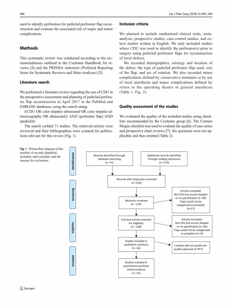
The search yielded 71 studies. The retrieved articles werereviewed and their bibliographies were scanned for publica-tions relevant for this review (Fig. 1).
Inclusion criteria
We planned to include randomized clinical trials, meta-analysis, prospective studies, case-control studies, and co-hort studies written in English. We only included studieswhere CDU was used to identify the perforator(s) prior tosurgery using pedicled perforator flaps for reconstructionof local defects.
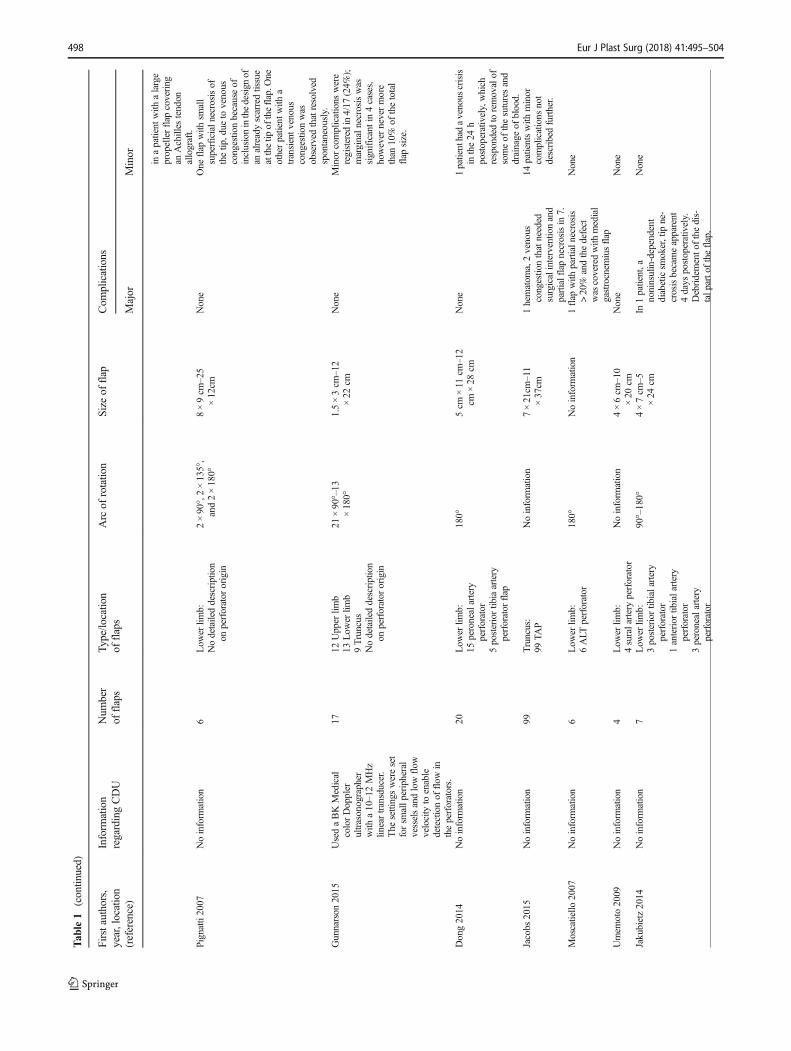
We recorded demographics, etiology and location ofthe defect, the type of pedicled perforator flap used, sizeof the flap, and arc of rotation. We also recorded minorcomplications defined by conservative treatment or by useof local anesthesia and major complications defined byreturn to the operating theater in general anesthesia(Table 1, Fig. 2).
Quality assessment of the studies
We evaluated the quality of the included studies using check-lists recommended by the Cochrane group [6]. The CarmenMogas checklist was used to evaluate the quality of case seriesand prospective chart reviews [7]. Six questions were not ap-plicable and thus omitted (Table 2).
Fig. 1 Prisma flow diagram of thenumber of records identified,included, and excluded, and thereasons for exclusions
496 Eur J Plast Surg (2018) 41:495–504
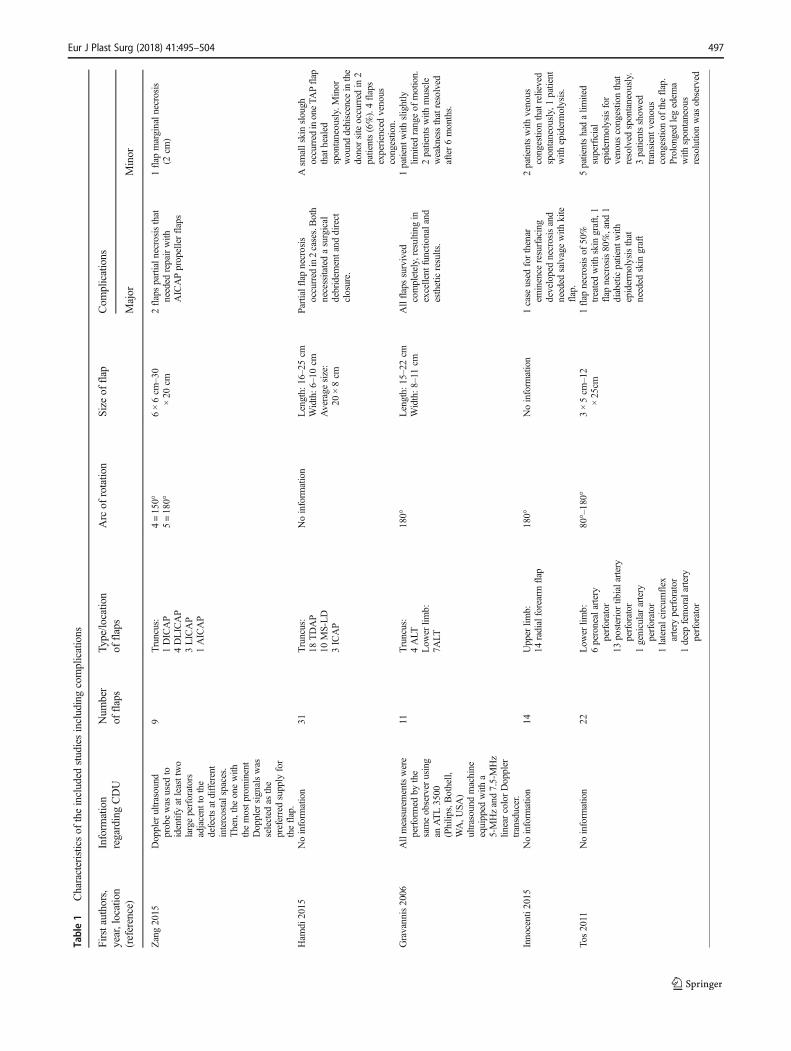
Table1
Characteristicsof
theincluded
studiesincludingcomplications
Firstauthors,
year,location
(reference)
Inform
ation
regardingCDU
Num
ber
offlaps
Type/lo
catio
nof
flaps
Arc
ofrotatio
nSizeof
flap
Com
plications
Major
Minor
Zang2015
Doppler
ultrasound
probewas
used
toidentifyatleasttwo
largeperforators
adjacent
tothe
defectsatdifferent
intercostalspaces.
Then,theonewith
themostp
rominent
Doppler
signalswas
selected
asthe
preferredsupply
for
theflap.
9Truncus:
1DICAP
4DLICAP
3LICAP
1AICAP
4=150°
5=180°
6×6cm
–30
×20
cm2flapspartialn
ecrosisthat
needed
repairwith
AICAPpropellerflaps
1flap
marginaln
ecrosis
(2cm
)
Ham
di2015
Noinform
ation
31Truncus:
18TDAP
10MS-LD
3ICAP
Noinform
ation
Length:
16–25cm
Width:6
–10cm
Average
size:
20×8cm
Partialflapnecrosis
occurred
in2cases.Both
necessitatedasurgical
debridem
entand
direct
closure.
Asm
allskinslough
occurred
inoneTA
Pflap
thathealed
spontaneously.Minor
wound
dehiscence
inthe
donorsiteoccurred
in2
patients(6%).4flaps
experiencedvenous
congestio
n.Gravannis2006
Allmeasurementswere
performed
bythe
sameobserver
using
anATL3500
(Philip
s,Bothell,
WA,U
SA)
ultrasound
machine
equipped
with
a5-MHzand7.5-MHz
linearcolorDoppler
transducer.
11Truncus:
4ALT
Low
erlim
b:7A
LT
180°
Length:
15–22cm
Width:8
–11cm
Allflapssurvived
completely,resulting
inexcellent
functio
naland
estheticresults.
1patient
with
slightly
limitedrangeof
motion.
2patientswith
muscle
weaknessthatresolved
after6months.
Innocenti2
015
Noinform
ation
14Upper
limb:
14radialforearm
flap
180°
Noinform
ation
1case
used
forthenar
eminence
resurfacing
developednecrosisand
needed
salvagewith
kite
flap.
2patientswith
venous
congestio
nthatrelieved
spontaneously,1patient
with
epidermolysis.
Tos2011
Noinform
ation
22Low
erlim
b:6peronealartery
perforator
13posteriortib
ialartery
perforator
1genicularartery
perforator
1lateralcircumflex
artery
perforator
1deep
femoralartery
perforator
80°–180°
3×5cm
–12
×25cm
1flap
necrosisof
50%
treatedwith
skin
graft,1
flap
necrosis80%,and
1diabeticpatient
with
epidermolysisthat
needed
skin
graft
5patientshadalim
ited
superficial
epidermolysisfor
venous
congestionthat
resolved
spontaneously.
3patientsshow
edtransientv
enous
congestionof
theflap.
Prolongedlegedem
awith
spontaneous
resolutio
nwas
observed
Eur J Plast Surg (2018) 41:495–504 497
Tab
le1
(contin
ued)
Firstauthors,
year,location
(reference)
Inform
ation
regardingCDU
Num
ber
offlaps
Type/lo
catio
nof
flaps
Arc
ofrotatio
nSizeof
flap
Com
plications
Major
Minor
inapatient
with
alarge
propellerflap
covering
anAchilles
tendon
allograft.
Pignatti2007
Noinform
ation
6Low
erlim
b:Nodetaileddescription
onperforator
origin
2×90°,2×135°,
and2×180°
8×9cm
–25
×12cm
None
One
flap
with
small
superficialn
ecrosisof
thetip
,due
tovenous
congestio
nbecauseof
inclusioninthedesign
ofan
alreadyscarredtissue
atthetip
oftheflap.O
neotherpatient
with
atransientv
enous
congestio
nwas
observed
thatresolved
spontaneously.
Gunnarson
2015
UsedaBKMedical
colorDoppler
ultrasonographer
with
a10–12MHz
lineartransducer.
The
settingswereset
forsm
allp
eripheral
vesselsandlowflow
velocity
toenable
detectionof
flow
intheperforators.
1712
Upper
limb
13Low
erlim
b9Truncus
Nodetaileddescription
onperforator
origin
21×90°–13
×180°
1.5×3cm
–12
×22
cmNone
Minor
complications
were
registered
in4/17
(24%
);marginaln
ecrosiswas
significantin4cases,
however
nevermore
than
10%
ofthetotal
flap
size.
Dong2014
Noinform
ation
20Low
erlim
b:15
peronealartery
perforator
5posteriortib
iaartery
perforator
flap
180°
5cm
×11
cm–12
cm×28
cmNone
1patienthadavenous
crisis
inthe24
hpostoperatively,which
respondedto
removalof
someof
thesuturesand
drainage
ofblood.
Jacobs
2015
Noinform
ation
99Truncus:
99TA
PNoinform
ation
7×21cm
–11
×37cm
1hematom
a,2venous
congestio
nthatneeded
surgicalinterventio
nand
partialflapnecrosisin7.
14patientswith
minor
complications
not
describedfurther.
Moscatiello
2007
Noinform
ation
6Low
erlim
b:6ALT
perforator
180°
Noinform
ation
1flap
with
partialn
ecrosis
>20%
andthedefect
was
coveredwith
medial
gastrocnem
iusflap
None
Umem
oto2009
Noinform
ation
4Low
erlim
b:4suralarteryperforator
Noinform
ation
4×6cm
–10
×20
cmNone
None
Jakubietz2014
Noinform
ation
7Low
erlim
b:3posteriortib
ialartery
perforator
1anterior
tibialartery
perforator
3peronealartery
perforator
90°–180°
4×7cm
–5×24
cmIn
1patient,a
noninsulin-dependent
diabeticsm
oker,tip
ne-
crosisbecameapparent
4days
postoperatively.
Debridemento
fthedis-
talp
arto
ftheflap,
None
498 Eur J Plast Surg (2018) 41:495–504
Statistical analysis
We conducted a meta-analysis for outcomes of complications;any necrosis, venous congestion, and flap loss. We calculatedproportions with a 95% confidence interval (CI) based on arandom-effects model due to the heterogeneous nature of thestudies [8]. The heterogeneity was investigated using chi-squared and the I2 statistics. All statistical analyses were con-ducted using Stata/IC 14.0 (StataCorp LP) and supervised bya statistician at Odense University Hospital.
Results
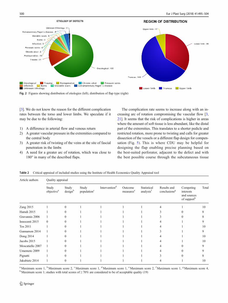
We evaluated 71 studies from the research databases and 170by assessing the reference lists (Fig. 1). We included 12 stud-ies, 11 case series/retrospective chart reviews, and one pro-spective study. The studies described 252 CDU targeted ped-icled perforator flaps used for reconstruction in 246 patients;72 male, 153 female, and 21 gender not described [3, 9–19].The mean age was 53 (36–79) years. The defects needingreconstruction were located in the upper limb in 17/252 cases(7%), lower limb 86/252 (34%), and trunk 149/252 (59%)(Fig. 2). The reconstructive goal was achieved in 247/252(98%) cases. The size of the flaps used for reconstructionwas reported in 240/252 (95%) cases and varied from 4.5 to600 cm2. In the upper limb, the size of the flaps varied be-tween 4,5 and 136 cm2, 40 and 600 cm2 in the torso, and 15and 400 cm2 in the lower limb. The main indication for recon-struction was an oncological defect 155/252 (61%), post-traumatic 31/252 (12%), and other surgery 15/252 (6%)(Fig. 2).
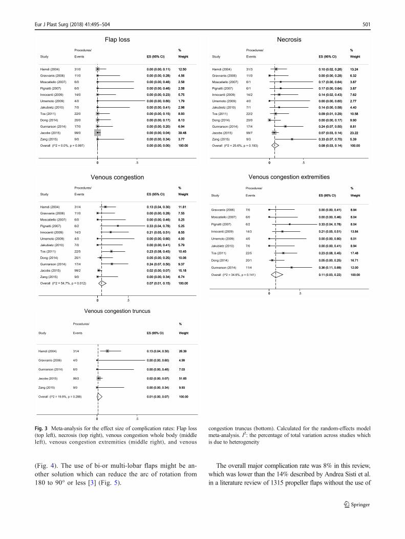
Surgical revision in general anesthesia was needed due tomajor complications in 21/252 (8%) cases. The re-operationswere performed due to necrosis 16/252, venous congestion 2/252, hematoma 1/252, and epidermolysis 2/252 (Table 2).There were 36 cases of minor complications (14%) (Table2). The most frequent was venous congestion 19/252 followedby tip necrosis 13/252, wound dehiscence 2/252, and otherreasons 2/252. The meta-analysis yielded summarized com-plication rates of 0% flap loss, 8% any necrosis, 7% venouscongestion throughout the whole body, 11% venous conges-tion in the extremities, and 1% venous congestion in truncus(Fig. 3).
Discussion
Venous congestion was the most common complication in thisseries, which coincides with previous reports using pedicledperforator flaps for reconstruction [3, 20]. The risk of venouscongestion was significantly higher in the lower extremities(11%) than in the torso (1%), as described previously (Fig. 3)T
able1
(contin
ued)
Firstauthors,
year,location
(reference)
Inform
ation
regardingCDU
Num
ber
offlaps
Type/lo
catio
nof
flaps
Arc
ofrotatio
nSizeof
flap
Com
plications
Major
Minor
negativ
epressure
therapy,andskin
graft.
In1patient
with
periph-
eralvascular
diseasede-
velopedsuperficial
epidermolysisin
both
tipsof
flap,w
hich
also
required
skin
graftin
g10
days
afterthefirst
surgery.
DICAPdorsal
intercostalartery
perforator,DLICAPdorsolateral
intercostalartery
perforator,LICAPlateralintercostalartery
perforator,AICAPanterior
intercostalartery
perforator,TD
AP/TAP
thoracodorsalarteryperforator,M
S-LD
musclesparring
latissimus
dorsi,ICAPintercostalarteryperforator,A
LTanterolateralthigh
Eur J Plast Surg (2018) 41:495–504 499
[3]. We do not know the reason for the different complicationrates between the torso and lower limbs. We speculate if itmay be due to the following:
1) A difference in arterial flow and venous return2) A greater vascular pressure in the extremities compared to
the central body3) A greater risk of twisting of the veins at the site of fascial
penetration in the limbs4) A need for a greater arc of rotation, which was close to
180° in many of the described flaps.
The complication rate seems to increase along with an in-creasing arc of rotation compromising the vascular flow [3,21]. It seems that the risk of complications is higher in areaswhere the amount of soft tissue is less abundant, like the distalpart of the extremities. This translates to a shorter pedicle andrestricted rotation, more prone to twisting and calls for greaterdissection of the vessels or a different flap design for compen-sation (Fig. 5). This is where CDU may be helpful fordesigning the flap enabling precise planning based onthe best-suited perforator, adjacent to the defect and withthe best possible course through the subcutaneous tissue
Table 2 Critical appraisal of included studies using the Institute of Health Economics Quality Appraisal tool
Article authors Quality appraisal
Studyobjectivea
Studydesignb
Studypopulationc
Interventiond Outcomemeasurese
Statisticalanalysisf
Results andconclusionsg
Competinginterestsand sourcesof supporth
Total
Zang 2015 1 0 1 1 1 1 4 1 10
Hamdi 2015 1 0 1 1 1 1 3 0 8
Gravannis 2006 1 0 1 1 1 1 3 0 8
Innocenti 2015 0 0 1 1 1 1 4 1 9
Tos 2011 1 0 1 1 1 1 4 1 10
Gunnarson 2014 1 0 1 1 1 1 3 1 9
Dong 2014 1 0 1 1 1 1 4 1 10
Jacobs 2015 1 0 1 1 1 1 4 1 10
Moscatiello 2007 1 0 1 1 1 1 4 0 9
Umemoto 2009 1 0 1 1 1 1 4 0 9
Pignatti 1 0 1 1 1 1 3 0 8
Jakubietz 2014 1 0 1 1 1 1 4 1 10
aMaximum score 1, bMaximum score 2, cMaximum score 1, dMaximum score 1, eMaximum score 2, fMaximum score 1, gMaximum score 4,hMaximum score 1; studies with total scores of ≥ 70% are considered to be of acceptable quality (19)
Fig. 2 Figures showing distribution of etiologies (left), distribution of flap type (right)
500 Eur J Plast Surg (2018) 41:495–504
(Fig. 4). The use of bi-or multi-lobar flaps might be an-other solution which can reduce the arc of rotation from180 to 90° or less [3] (Fig. 5).
The overall major complication rate was 8% in this review,which was lower than the 14% described by Andrea Sisti et al.in a literature review of 1315 propeller flaps without the use of
Overall (I^2 = 25.6%, p = 0.193)
Jakubietz (2010)
Tos (2011)
Innocenti (2009)
Moscatiello (2007)
Zang (2015)
Umemoto (2009)
Study
Dong (2014)
Pignatti (2007)
Gravvanis (2006)
Gunnarson (2014)
Hamdi (2004)
Jacobs (2015)
7/1
22/2
14/2
6/1
9/3
4/0
Events
20/0
6/1
11/0
17/4
Procedures/
31/3
99/7
0.08 (0.03, 0.14)
0.14 (0.00, 0.58)
0.09 (0.01, 0.29)
0.14 (0.02, 0.43)
0.17 (0.00, 0.64)
0.33 (0.07, 0.70)
0.00 (0.00, 0.60)
ES (95% CI)
0.00 (0.00, 0.17)
0.17 (0.00, 0.64)
0.00 (0.00, 0.28)
0.24 (0.07, 0.50)
0.10 (0.02, 0.26)
0.07 (0.03, 0.14)
100.00
4.40
10.58
7.62
3.87
5.39
2.77
Weight
9.90
3.87
6.32
8.81
%
13.24
23.22
00 .5
Necrosis
Overall (I^2 = 0.0%, p = 0.997)
Hamdi (2004)
Jakubietz (2010)
Jacobs (2015)
Umemoto (2009)
Pignatti (2007)
Innocenti (2009)
Moscatiello (2007)
Gravvanis (2006)
Dong (2014)
Study
Zang (2015)
Tos (2011)
Gunnarson (2014)
31/0
7/0
99/0
4/0
6/0
14/0
6/0
11/0
20/0
Procedures/
Events
9/0
22/0
17/0
0.00 (0.00, 0.00)
0.00 (0.00, 0.11)
0.00 (0.00, 0.41)
0.00 (0.00, 0.04)
0.00 (0.00, 0.60)
0.00 (0.00, 0.46)
0.00 (0.00, 0.23)
0.00 (0.00, 0.46)
0.00 (0.00, 0.28)
0.00 (0.00, 0.17)
ES (95% CI)
0.00 (0.00, 0.34)
0.00 (0.00, 0.15)
0.00 (0.00, 0.20)
100.00
12.50
2.98
39.48
1.79
2.58
5.75
2.58
4.56
8.13
%
Weight
3.77
8.93
6.94
00 .5
Flap loss
Overall (I^2 = 54.7%, p = 0.012)
Jacobs (2015)
Moscatiello (2007)
Gunnarson (2014)
Dong (2014)
Tos (2011)
Pignatti (2007)
Jakubietz (2010)
Umemoto (2009)
Zang (2015)
Study
Hamdi (2004)
Gravvanis (2006)
Innocenti (2009)
99/2
6/0
17/4
20/1
22/5
6/2
7/0
4/0
9/0
Events
31/4
11/0
14/3
Procedures/
0.07 (0.01, 0.15)
0.02 (0.00, 0.07)
0.00 (0.00, 0.46)
0.24 (0.07, 0.50)
0.05 (0.00, 0.25)
0.23 (0.08, 0.45)
0.33 (0.04, 0.78)
0.00 (0.00, 0.41)
0.00 (0.00, 0.60)
0.00 (0.00, 0.34)
ES (95% CI)
0.13 (0.04, 0.30)
0.00 (0.00, 0.28)
0.21 (0.05, 0.51)
100.00
15.18
5.25
9.37
10.06
10.45
5.25
5.79
4.00
6.74
Weight
11.81
7.55
8.55
%
00 .5
Venous congestion
Overall (I^2 = 34.6%, p = 0.141)
Dong (2014)
Pignatti (2007)
Gunnarson (2014)
Innocenti (2009)
Tos (2011)
Jakubietz (2010)
Gravvanis (2006)
Moscatiello (2007)
Study
Umemoto (2009)
20/1
6/2
11/4
14/3
22/5
7/0
7/0
6/0
Events
Procedures/
4/0
0.11 (0.03, 0.22)
0.05 (0.00, 0.25)
0.33 (0.04, 0.78)
0.36 (0.11, 0.69)
0.21 (0.05, 0.51)
0.23 (0.08, 0.45)
0.00 (0.00, 0.41)
0.00 (0.00, 0.41)
0.00 (0.00, 0.46)
ES (95% CI)
0.00 (0.00, 0.60)
100.00
16.71
8.04
12.00
13.84
17.48
8.94
8.94
8.04
Weight
%
6.01
0.11 (0.03, 0.22)
0.05 (0.00, 0.25)
0.33 (0.04, 0.78)
0.36 (0.11, 0.69)
0.21 (0.05, 0.51)
0.23 (0.08, 0.45)
0.00 (0.00, 0.41)
0.00 (0.00, 0.41)
0.00 (0.00, 0.46)
ES (95% CI)
0.00 (0.00, 0.60)
100.00
16.71
8.04
12.00
13.84
17.48
8.94
8.94
8.04
Weight
%
6.01
00 .5
Venous congestion extremities
Overall (I^2 = 19.9%, p = 0.288)
Study
Hamdi (2004)
Jacobs (2015)
Gunnarson (2014)
Zang (2015)
Gravvanis (2006)
Events
31/4
99/2
6/0
9/0
4/0
Procedures/
0.01 (0.00, 0.07)
ES (95% CI)
0.13 (0.04, 0.30)
0.02 (0.00, 0.07)
0.00 (0.00, 0.46)
0.00 (0.00, 0.34)
0.00 (0.00, 0.60)
100.00
Weight
26.39
51.65
7.03
9.93
4.99
%
0.01 (0.00, 0.07)
ES (95% CI)
0.13 (0.04, 0.30)
0.02 (0.00, 0.07)
0.00 (0.00, 0.46)
0.00 (0.00, 0.34)
0.00 (0.00, 0.60)
100.00
Weight
26.39
51.65
7.03
9.93
4.99
%
00 .5
Venous congestion truncus
Fig. 3 Meta-analysis for the effect size of complication rates: Flap loss(top left), necrosis (top right), venous congestion whole body (middleleft), venous congestion extremities (middle right), and venous
congestion truncus (bottom). Calculated for the random-effects modelmeta-analysis. I2: the percentage of total variation across studies whichis due to heterogeneity
Eur J Plast Surg (2018) 41:495–504 501
CDU [20]. However, we cannot use these results to concludethat the use of CDU is associated with an overall lower compli-cation rate although it may show a trend.
Interestingly, most of the included studies were small studiesincluding 20 patients or less. Thus, the complication rates in thisreview, major 8% and minor 14%, have to be considered in thecontext of a learning curve setup. Better results should be expect-ed once the learning curve is surpassed [11]. The summed majorcomplication rate of the five smallest studies in this review was16% compared to 8%overall, which is in accordancewith Jiga etal. and Panse et al., who found that the overall outcome can beexpected to improve while the complication rate decrease overtime [22, 23].
The use of CDU for detection of perforators is observ-er dependent, which can be exemplified by two studiesusing CDU for detection of perforators for the harvest ofthe radial forearm flap. CDU was found to be extremelyuseful for detecting perforators for the radial forearm flapin one of these studies, yet the other study describeddifficulties using CDU to identify the perforators, be-cause the signal from the radial artery shielded visuali-zation of the perforators [18, 24]. It is therefore impor-tant to facilitate the correct use of CDU, which enablesthe surgeon to plan and design the pedicled perforatorflap for reconstruction using the best available tissue ad-jacent to the defect, allowing for the least possible arc of
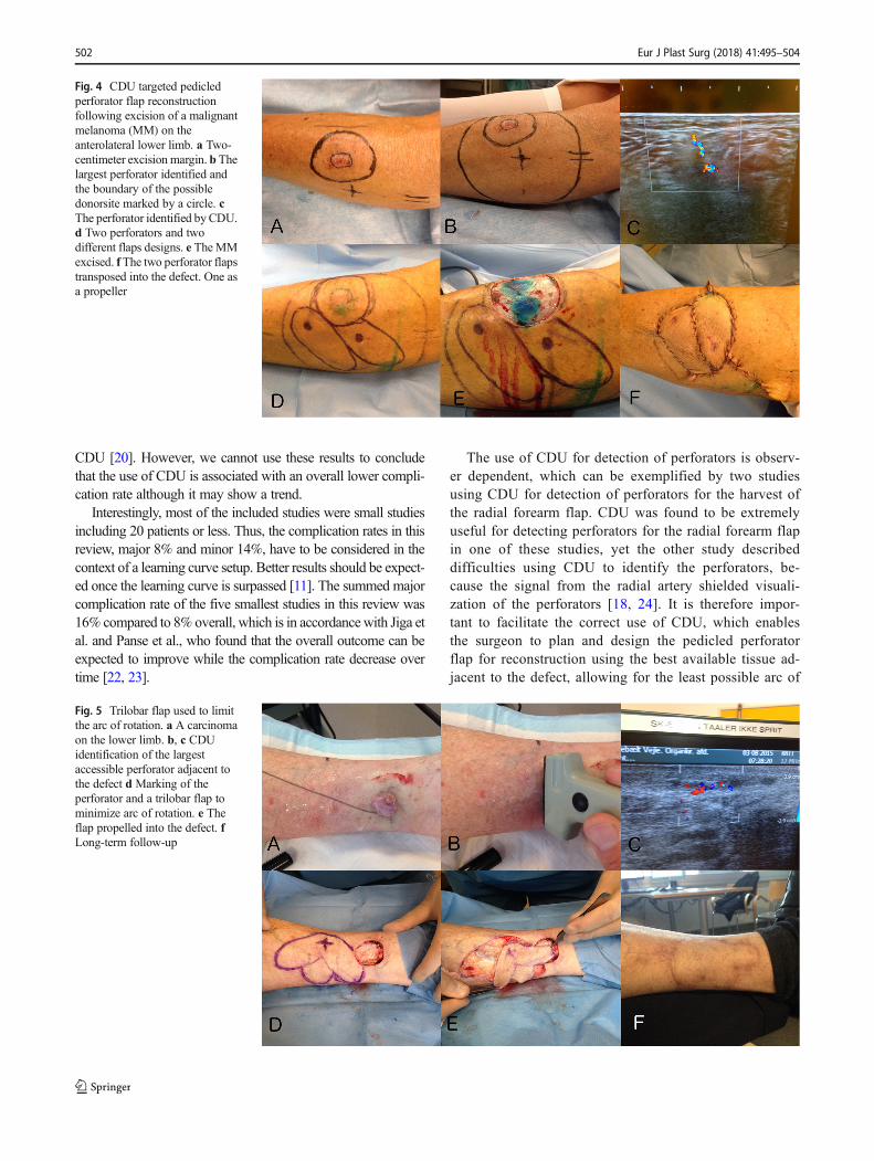
Fig. 5 Trilobar flap used to limitthe arc of rotation. a A carcinomaon the lower limb. b, c CDUidentification of the largestaccessible perforator adjacent tothe defect d Marking of theperforator and a trilobar flap tominimize arc of rotation. e Theflap propelled into the defect. fLong-term follow-up
Fig. 4 CDU targeted pedicledperforator flap reconstructionfollowing excision of a malignantmelanoma (MM) on theanterolateral lower limb. a Two-centimeter excision margin. b Thelargest perforator identified andthe boundary of the possibledonorsite marked by a circle. cThe perforator identified byCDU.d Two perforators and twodifferent flaps designs. e TheMMexcised. fThe two perforator flapstransposed into the defect. One asa propeller
502 Eur J Plast Surg (2018) 41:495–504
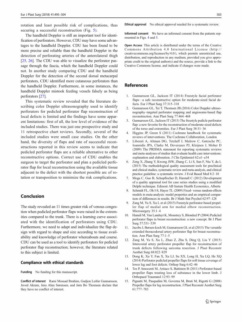
rotation and least possible risk of complications, thussecuring a successful reconstruction (Fig. 5).
The handheld Doppler is still an important tool for identi-fication of perforators. However, CDUmay have some advan-tages to the handheld Doppler. CDU has been found to bemore precise and reliable than the handheld Doppler in thedetection of perforating arteries of the anterolateral thigh[25, 26]. The CDU was able to visualize the perforator pas-sage through the fascia, which the handheld Doppler couldnot. In another study comparing CDU and the handheldDoppler for the detection of the second dorsal metacarpalperforators, CDU identified more cutaneous perforators thanthe handheld Doppler. Furthermore, in some instances, thehandheld Doppler mistook feeding vessels falsely as beingperforators [27].
This systematic review revealed that the literature de-scribing color Doppler ultrasonography used to identifyperforators for pedicled perforator flap reconstruction oflocal defects is limited and the findings have some appar-ent limitations: first of all, the low level of evidence of theincluded studies. There was just one prospective study and11 retrospective chart reviews. Secondly, several of theincluded studies were small case studies. On the otherhand, the diversity of flaps and rate of successful recon-structions reported in this review seems to indicate thatpedicled perforator flaps are a reliable alternative to otherreconstructive options. Correct use of CDU enables thesurgeon to target the perforator and plan a pedicled perfo-rator flap for local reconstruction using the available tissueadjacent to the defect with the shortest possible arc of ro-tation or transposition to minimize the risk complications.
Conclusion
The study revealed an 11 times greater risk of venous conges-tion when pedicled perforator flaps were raised in the extrem-ities compared to the trunk. There is a learning curve associ-ated with the identification of perforators using CDU.Furthermore, we need to adapt and individualize the flap de-sign with regard to shape and size according to tissue avail-ability and knowledge of perforator whereabouts and course.CDU can be used as a tool to identify perforators for pedicledperforator flap reconstruction; however, the literature relatedto this subject is limited.
Compliance with ethical standards
Funding No funding for this manuscript.
Conflict of interest Rami Mossad Ibrahim, Gudjon Leifur Gunnarsson,Javed Akram, Jens Ahm Sørensen, and Jørn Bo Thomsen declare thatthey have no conflict of interest.
Ethical approval No ethical approval needed for a systematic review.
Informed consent We have an informed consent from the patients rep-resented in Figs. 4 and 5.
Open Access This article is distributed under the terms of the CreativeCommons At t r ibut ion 4 .0 In te rna t ional License (h t tp : / /creativecommons.org/licenses/by/4.0/), which permits unrestricted use,distribution, and reproduction in any medium, provided you give appro-priate credit to the original author(s) and the source, provide a link to theCreative Commons license, and indicate if changes were made.
References
1. Gunnarsson GL, Jackson IT (2014) Freestyle facial perforatorflaps—a safe reconstructive option for moderate-sized facial de-fects. Eur J Plast Surg 37:315–318
2. Gunnarsson GL, Tei T, Thomsen JB (2016) Color Doppler ultraso-nography–targeted perforator mapping and angiosome-based flapreconstruction. Ann Plast Surg 77:464–468
3. Gunnarsson GL, Jackson IT (2015) The freestyle pedicle perforatorflap: a new favorite for the reconstruction of moderate-sized defectsof the torso and extremities. Eur J Plast Surg 38:31–36
4. Higgins JP, Green S (2011) Cochrane handbook for systematicreviews of interventions. The Cochrane Collaboration, London
5. Liberati A, Altman DG, Tetzlaff J, Mulrow C, Gøtzsche PC,Ioannidis JPA, Clarke M, Devereaux PJ, Kleijnen J, Moher D(2009) The PRISMA statement for reporting systematic reviewsand meta-analyses of studies that evaluate health care interventions:explanation and elaboration. J Clin Epidemiol 62:e1–e34
6. Zeng X, Zhang Y, Kwong JSW, Zhang C, Li S, Sun F, Niu Y, du L(2015) The methodological quality assessment tools for preclinicaland clinical studies, systematic review andmeta-analysis, and clinicalpractice guideline: a systematic review. J Evid Based Med 8:2–10
7. Moga C, Guo B, Schopflocher D, Harstall C (2012) Developmentof a quality appraisal tool for case series studies using a modifiedDelphi technique. Edmont AB Instute Health Economics, Alberta
8. Schmidt FL, Oh I-S, Hayes TL (2009) Fixed- versus random-effectsmodels in meta-analysis: model properties and an empirical compar-ison of differences in results. Br J Math Stat Psychol 62:97–128
9. Zang M, Yu S, Xu L et al (2015) Freestyle perforator-based propel-ler flap of medial arm for medial elbow reconstruction.Microsurgery 35:1–4
10. HamdiM, Van Landuyt K,Monstrey S, Blondeel P (2004) Pedicledperforator flaps in breast reconstruction: a new concept. Br J PlastSurg 57:531–539
11. Jacobs J, Børsen-kochM, GunnarssonGL et al (2015) The versatileextended thoracodorsal artery perforator flap for breast reconstruc-tion. Ann Plast Surg 77:1–5
12. Zang M, Yu S, Xu L, Zhao Z, Zhu S, Ding Q, Liu Y (2015)Intercostal artery perforator propeller flap for reconstruction oftrunk defects following sarcoma resection. J Plast ReconstrAesthet Surg 68:822–829
13. Dong K, Xu Y, Fan X, Xu LJ, Su XX, Long H, Xu LQ, He XQ(2014) Perforator pedicled propeller flaps for soft tissue coverage oflower leg and foot defects. Orthop Surg 6:42–46
14. Tos P, Innocenti M, Artiaco S, Battiston B (2011) Perforator-basedpropeller flaps treating loss of substance in the lower limb. JOrthopaed Traumatol 12:93–99
15. Pignatti M, Pasqualini M, Governa M, Bruti M, Rigotti G (2008)Propeller flaps for leg reconstruction. J Plast Reconstr Aesthet Surg61:777–783
Eur J Plast Surg (2018) 41:495–504 503
16. Jakubietz R, Meffert RH, Jakubietz MG (2010) Reconstruction ofsoft tissue defects of the achilles tendonwith rotation flaps, pedicledpropeller flaps and free perforator flaps. Microsurgery 1:1–6
17. Moscatiello F, Carrera A, Moscatiello F et al (2007) TheBpropeller^ distal anteromedial thigh perforator flap. Anatomicstudy and clinical applications. J Plast Reconstr Aesthet Surg 60:1323–1330
18. Innocenti M, Baldrighi C, Delcroix L, Adani R (2009) Local per-forator flaps in soft tissue reconstruction of the upper limb.Handchir Mikrochir Plast Chir 41:315–321
19. Umemoto Y, Adachi Y, Ebisawa K (2005) The sural artery perfo-rator flap for coverage of defects of the knee and tibia. Scand J PlastReconstr Surg Hand Surg 39:209–213
20. Sisti A, D’Aniello C, Fortezza L, Tassinari J, Cuomo R et al (2016)Propeller flaps: a literature review. In Vivo (Brooklyn) 30:351–373
21. Paik J, Pyon J-K (2016) Risk factor analysis of freestyle propellerflaps. J Reconstr Microsurg 33:026–031
22. Jiga LP, Barac S, Taranu G, Blidisel A, Dornean V, Nistor A,Stoichitoiu T, Geishauser M, Ionac M (2010) The versatility ofpropeller flaps for lower limb reconstruction in patients with
peripheral arterial obstructive disease: initial experience. AnnPlast Surg 64:193–197
23. Panse N, Sahasrabudhe P (2014) Free style perforator based pro-peller flaps: simple solutions for upper extremity reconstruction!Indian J Plast Surg 47:77–84
24. Matei I, Georgescu A, Chiroiu B et al (2008) Harvesting of forearmperforator flaps based on intraoperative vascular exploration: clini-cal experiences and literature review. Microsurgery 28:321–330
25. Lethaus B, Loberg C, Kloss-Brandstätter A, Bartella AK, Steiner T,Modabber A, Hölzle F, Teichmann J (2017) Color duplex ultraso-nography versus handheld Doppler to plan anterior lateral thighflaps. Microsurgery 37:388–393
26. Cheng H-T, Lin F-Y, Chang SC-N (2013) Diagnostic efficacy ofcolor Doppler ultrasonography in preoperative assessment of an-terolateral thigh flap cutaneous perforators: an evidence-based re-view. Plast Reconstr Surg 131:471e–473e
27. Nanno M, Kodera N, Tomori Y, Hagiwara Y, Takai S (2017) ColorDoppler ultrasound assessment for identifying perforator arteries ofthe second dorsal metacarpal flap. J Orthop Surg 25:230949901668474
504 Eur J Plast Surg (2018) 41:495–504