RESEARCH ARTICLE Color vision and learning in the monarch ...
Upload
saleh-al-naimiCategory
view
74download
2
The International Journal of Periodontics & Restorative Dentistry

Every opaque object receives light,the three primary light colors insome ratio. Some objects reflect allof the light they receive, and othersabsorb it almost totally.1 Most“opaque” objects absorb partiallyand reflect the rest. The dominantwavelength/s reflected back to theeye is the perceived color of the ob-ject. Natural teeth have many opti-cal characteristics that increase thecomplexity of what we see. Under-standing these characteristics willimprove our ability to describe them.
Albert Munsell described coloras a three-dimensional phenome-non. He described the three dimen-sions as hue, value (brightness), andchroma (saturation).
Hue
Hue is the quality that distinguishesone family of colors from another. Itis specified as the dominant range ofwavelengths in the visible spectrumthat yields the perceived color, eventhough the exact wavelength of theperceived color may not be present.2
Hue is a physiologic and psychologic
Shade Matching in RestorativeDentistry: The Science and Strategies
James Fondriest, DDS*
Closely matching natural teeth with an artificial restoration can be one of the mostchallenging procedures in restorative dentistry. Natural teeth vary greatly in colorand shape. They reveal ample information about patients’ background and person-ality. Dentistry provides the opportunity to restore unique patient characteristics orreplace them with alternatives. Whether one tooth or many is restored, the abilityto assess and properly communicate information to the laboratory can be greatlyimproved by learning the language of color and light characteristics. It is only possi-ble to duplicate in ceramic what has been distinguished, understood, and commu-nicated in the shade-matching process of the natural dentition. This article will givethe reader a better understanding of what happens when incident light hits the sur-face of a tooth and give strategies for best assessing and communicating this to thedental laboratory. (Int J Periodontics Restorative Dent 2003;23:xxx–xxx.)
*Visiting Faculty, LD Pankey Institute, Key Biscayne, Florida.
*Reprint requests: Dr James Fondriest, 560 Oakwood Avenue, Suite 200,Lake Forest, Illinois 60045. e-mail: [email protected]
3
Volume 23, Number 5, 2003

interpretation of a sum of wave-lengths. In dental terms, hue is rep-resented by A, B, C, or D on the com-monly used Vita Classic shade guide.
Value
Value, or brightness, is the amount oflight returned from an object.Munsell described value as a white-to-black gray scale. Bright objectshave lower amounts of gray, andlow-value objects have largeramounts of gray and will appeardarker. The brightness of a crown isusually increased in two ways: byusing lighter porcelain (loweringchroma), or by increasing the reflec-tivity of the surface. Lowering valuemeans diminished light returns fromthe object illuminated; more light isbeing absorbed, scattered else-where, or transmitted through.
Chroma
Chroma is the saturation, intensity, orstrength of the hue. Envision placingred food dye into a glass of water.Each time more of the same colordye is added, the intensity increases,but it is the same red color (hue). Asmore dye is added, the mixture alsoappears darker, so the increase inchroma has a corresponding changein value. As chroma is increased, thevalue is decreased; chroma andvalue are inversely related. Highernumbers on the Vita Classic shadeguide represent increased chroma.
Other optical properties
Translucency
In dental ceramics, we try to imitatethe appearance of the tooth as asum of all its visual dimensions.Human teeth are characterized byvarying degrees of translucency,which can be defined as the gradientbetween transparent and opaque.Generally, increasing the translu-cency of a crown lowers its valuebecause less light returns to the eye.With increased translucency, light isable to pass the surface and is scat-tered within the body of porcelain.The translucency of enamel varieswith the angle of incidence, surfaceluster, wavelength, and level of de-hydration. With a translucent enamellayer, the ceramist achieves colordepth and the illusion of a vital nat-ural tooth.
Fluorescence
Ultraviolet (UV) light can have a dra-matic effect on the level of vitalityexhibited by restorations. With thecharacteristic of fluorescence, theylook brighter and more alive.Fluorescence is the absorption oflight by a material and the sponta-neous emission of light in a longerwavelength.3 In a natural tooth, itprimarily occurs in the dentinbecause of the higher amount oforganic material present.1,4–6
Ambient near-UV light is absorbedand fluoresced back as light primar-ily in the blue end of the spectrum,but it will occur at all wavelengths.
The more the dentin fluoresces, thelower the chroma.1,7 Fluorescentpowders are added to crowns toincrease the quantity of lightreturned back to the viewer, blockout discolorations, and decreasechroma.7 This is especially benefi-cial in high-value shades, as it canraise value without negatively affect-ing translucency when placed withinthe dentin porcelain layers.
Opalescence
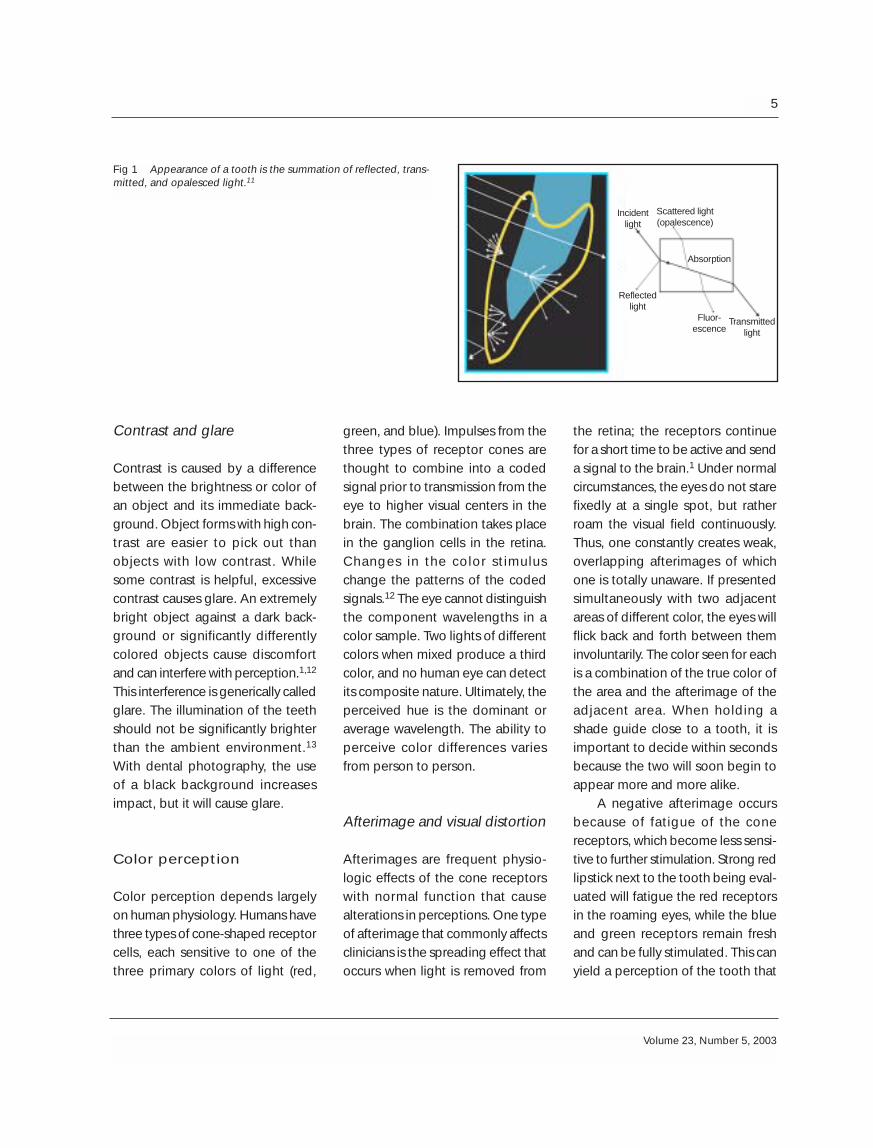
Opalescence is the phenomenon inwhich a material appears to be onecolor when light is reflected from itand another color when light is trans-mitted through it.8 A natural opal isan aqueous disilicate that breakstransilluminated light down into itscomponent spectrum by refraction.Opals act like prisms and refract(bend) different wavelengths to vary-ing degrees. The shorter wave-lengths bend more and require ahigher critical angle to escape anoptically dense material than thereds and yellows. The hydroxyap-atite (HA) crystals of enamel also actas prisms. Wavelengths of light havedifferent degrees of translucencythrough teeth and dental materials.When illuminated, opals and enamelwill transilluminate the reds and scat-ter the blues within its body; thus,enamel appears bluish even thoughit is colorless.1,4,9 The opalescenteffects of enamel brighten the toothand give it optical depth and vital-ity10 (Fig 1).
4
The International Journal of Periodontics & Restorative Dentistry
Contrast and glare
Contrast is caused by a differencebetween the brightness or color ofan object and its immediate back-ground. Object forms with high con-trast are easier to pick out thanobjects with low contrast. Whilesome contrast is helpful, excessivecontrast causes glare. An extremelybright object against a dark back-ground or significantly differentlycolored objects cause discomfortand can interfere with perception.1,12
This interference is generically calledglare. The illumination of the teethshould not be significantly brighterthan the ambient environment.13
With dental photography, the useof a black background increasesimpact, but it will cause glare.
Color perception
Color perception depends largelyon human physiology. Humans havethree types of cone-shaped receptorcells, each sensitive to one of thethree primary colors of light (red,
the retina; the receptors continuefor a short time to be active and senda signal to the brain.1 Under normalcircumstances, the eyes do not starefixedly at a single spot, but ratherroam the visual field continuously.Thus, one constantly creates weak,overlapping afterimages of whichone is totally unaware. If presentedsimultaneously with two adjacentareas of different color, the eyes willflick back and forth between theminvoluntarily. The color seen for eachis a combination of the true color ofthe area and the afterimage of theadjacent area. When holding ashade guide close to a tooth, it isimportant to decide within secondsbecause the two will soon begin toappear more and more alike.
A negative afterimage occursbecause of fatigue of the conereceptors, which become less sensi-tive to further stimulation. Strong redlipstick next to the tooth being eval-uated will fatigue the red receptorsin the roaming eyes, while the blueand green receptors remain freshand can be fully stimulated. This canyield a perception of the tooth that
green, and blue). Impulses from thethree types of receptor cones arethought to combine into a codedsignal prior to transmission from theeye to higher visual centers in thebrain. The combination takes placein the ganglion cells in the retina.Changes in the color stimuluschange the patterns of the codedsignals.12 The eye cannot distinguishthe component wavelengths in acolor sample. Two lights of differentcolors when mixed produce a thirdcolor, and no human eye can detectits composite nature. Ultimately, theperceived hue is the dominant oraverage wavelength. The ability toperceive color differences variesfrom person to person.
Afterimage and visual distortion
Afterimages are frequent physio-logic effects of the cone receptorswith normal function that causealterations in perceptions. One typeof afterimage that commonly affectsclinicians is the spreading effect thatoccurs when light is removed from
5
Volume 23, Number 5, 2003
Fig 1 Appearance of a tooth is the summation of reflected, trans-mitted, and opalesced light.11
Scattered light (opalescence)
Incidentlight
Reflectedlight
Fluor-escence
Absorption
Transmittedlight

is too blue-green. The afterimagewill always be the complementarycolor of what you are observing.Give the eyes a break with neutralgray backgrounds. Kulzer’s smallintraoral gray shields screen back-ground color glare; 18% reflectivegray cards are the photographicindustry standard achromatic back-ground.14 Blue backgrounds are notappropriate because they causeafterimages and will bias perceptionto its complementary color, orange.Some advocate use of a blue back-ground15–18 to make the eyes moresensitive to yellow-orange, but thisselectively fatigues one type of coneand does not make the others anymore sensitive. An 18% reflectivegray card is an excellent backgroundfor photographic evaluation of hueand chroma.19 It has no comple-mentary colors (thus, no afterimagesare produced), and it is brightenough to limit the contrast ofbrightness between the teeth andbackground.
Proper environment forcolor rendering
The ability to perform shade selec-tion depends on how well the eyesperceive the details of teeth. Factorsdetermining the visibility of thesedetails include ambient light quality,luminance (light quantity), size, con-trast, and glare. Establishing theproper environment for evaluationrequires an understanding of these“seeability” factors. Dental-unitlights are commonly used for colorrendering. Most are incandescent
and emit light high in the red-yellowspectrum and low at the blue end.Therefore, illuminating opaque sam-ples of red, yellow, and blue underan incandescent light source showsthat red and yellow are quite strong,or highly saturated, while blue isweaker and more difficult to see.
The translucency of enamel is afunction of wavelength. The longerthe wavelength, the higher thetranslucency. Therefore, enamel ismore translucent in light rich in yel-low and red (eg, incandescent light)and will show more dentin, makingthe tooth appear redder, with ahigher chroma and lower value thanit actually has.20 Under an ordinarycool white fluorescent source, whichis high in the green-yellow spectrumwith some strong but narrow blue-spectrum spikes, the reds and violetsare less apparent. Some fluorescentbulbs have full color content andrender color more accurately. Theambient light quality of the opera-tory must be maintained with artifi-cial lighting (natural light conditionsvary); it is commonly measured bythe color temperature and ColorRendering Index (CRI). When black iron is heated gradually,it will begin to glow, first with a redhue, then yellow, white, and blue.
Color temperature is the meanwavelength of the ambient light. Theideal color temperature for color ren-dering is 5,500 K. Light at this tem-perature can be described as havinga medium-temperature feel and isconsidered “white” light.
Not all wavelengths need berepresented to produce white light;it can be simply produced by mixing
the three primary light colors.Ambient light is a varying assemblyof many different wavelengths.Artificial lighting can approach whitelight (5,500 K), but the full spectrumof wavelengths is not necessarilypresent. The reflected colors (wave-lengths) of a tooth cannot be seen ifthose wavelengths are not present inthe ambient light spectrum.2,16 Ifambient conditions have only a smallrange of the spectrum of light wave-lengths, all that is reflected back arethe wavelengths present. If red lightis not fairly represented in the spec-trum, the reds in the object to bematched will not be visible.
CRI is the measure of the com-pleteness of the light spectrum. Ameasure of 100 indicates that theentire visible and near-UV light spec-trum is present. Although the close-to-visible UV spectrum cannot beseen, it is commonly absorbed andfluoresced out at wavelengths in thevisible spectrum. The average incan-descent dental-unit lamp has a CRIof 75 and averages 3,800 K.12,18,21 ACRI greater than 93 is preferred.Ideally, both the clinician and labo-ratory technician should have bal-anced full-spectrum lighting condi-tions.
Metamerism
Metamerism occurs when restora-tions match in one light but displaya different color in other light condi-tions.14 One object may have theability to reflect more red thananother. However, if there is no redrange in the light source, they will
6
The International Journal of Periodontics & Restorative Dentistry

appear the same; when viewedunder a light source containing red,they will appear different. The colorseen depends on the nature of thelight source illuminating the object.The color of an opaque object is thesum of the wavelengths that reflectoff it. Light spectrum reflectancegraphs measure the percentage ofreflectance of all the near-UV andvisible-light spectrum off of a mate-rial. Porcelain might reflect light offits surface exactly as enamel in onepart of the spectrum, but under dif-ferent illumination the two objectsthat previously looked identicalmight look different. The closer thecurves of the two materials to bematched, the more successful thecolor match will be.22 Use of opaquesurface stains to correct mismatcheswill increase metamerism. Whenreconstructing a tooth with dentalporcelain, mimicking the layers ofthe tooth employing materials withthe same optical properties (spectralreflectance curves) will minimizemetamerism.
Light intensity
The intensity of the light conditionsis also important. If the amount oflight (measured in foot-candles orlumens per foot2) is too small, finedetails are missed and the eye hasdifficulty perceiving hue. Usually, theceiling lighting in the dental opera-tory is not intense enough to seeeverything. With teeth that havesubtle color variations, proper inten-sity is needed. Too great an intensityand glare decrease the accuracy of
Reflections of light
It is important to realize that hue andchroma are fifth or sixth in impor-tance on the list of things to matchwhen constructing a prostheticreplacement. One must be fairlyclose to someone to detect subtledifferences in hue; yet shape, sur-face morphology, value, and translu-cency disparities can be seen from 4or 5 feet or more. Violating confor-mity of the unique characteristics ofthe natural dentition will causeunwanted prominence of therestoration.16,26–28 These character-istics determine how light isreflected, transmitted, or scattered,affecting its hue, chroma, value, andtranslucency.16,29,30 The appearanceof teeth is mostly determined byhow light interacts with the curvedand varied surfaces. Attractiveprosthodontic replacement startswith a consistent silhouette andshape of the buccal surface becausethese factors determine how themajority of light will be reflected.
An observer only sees an objectwhen light comes from that object.Surfaces perpendicular to the viewersend the most light back. The reflec-tive surfaces of a tooth will not returnas much light to the eyes if they arenot perpendicular to them. Becausea viewer mainly sees the surfaces ofa tooth perpendicular to him or her,the perceived width and length canbe manipulated by bending or flat-tening surfaces. The practitioner canmake a tooth look narrower orshorter by decreasing the width orlength of the direct buccal reflectivesurface.31
color rendering. Dental-unit lightsshould not be used for color ren-dering; they are too bright and causeglare. Glare fatigues the eyes; ren-dering shades immediately afterusing a dental-unit light is also con-traindicated.
The ideal luminosity for dentalshade matching is 75 to 250 foot-candles.13,15,18,23–25 To have 175foot-candle intensity at the level ofthe dental chair, ten to twelve 4-footbulbs would be needed in a 10 ft �10 ft room with 8-foot ceilings.1,13,24
The diffusion panels covering fluo-rescent bulbs are also importantbecause they screen out wave-lengths. As they age, the panelschange what wavelengths theyabsorb. The best diffusers, prefer-ably the egg-crate type, do not filterout any wavelengths of the spec-trum. Using 10 to 12 color-correctedbulbs on the ceiling will yield morelight in the operatory than would beconsidered comfortable. Portablehigh-quality light units, such as theVident light, are ideal. Shade match-ing with photography lessens, butdoes not obviate, the need for spe-cial lighting. The proper shade tabsstill need to be selected.
7
Volume 23, Number 5, 2003

Reflection from a smooth, mir-ror-like surface results in a clear, well-defined image. Such a “specularreflection” returns a high percent-age of direct, nondiffused light, andif strongly illuminated, will bebrighter and stand out. Smoothingthe texture of the buccal surface willmake teeth appear lighter andbrighter and is therefore a primarydeterminant of value. The morereflective the surface, the morewavelengths return to the eyes; theadditive combination of more wave-lengths yields whiter light (huechange). Brighter objects appearcloser to the viewer, so a restorationthat is too light appears to “jump outat you.” Lowering the value makesobjects appear further away. Rough-ening texturally the specular high-lights of a too-bright crown will makeit blend better. Most teeth have ir-
regular surfaces with convexities andconcavities. The convexities tend towear and become smooth, withspecular reflective characteristics.The visual impact of a tooth comesfrom these specular highlights thatgive the tooth its visual shape.Concavities tend to be unpolishedand collect light by reflecting inward,diffusing the light and returning lessto the eyes32 (Figs 2 and 3).
Surface texture
After shape, surface morphology isthe next most important parameterof a good match. Surface morphol-ogy affects how light interplays withthe tooth surface, and thus the qual-ity and quantity of light that returnsto your eyes. A roughened surfacetexture will not yield as well-defined
an image and will scatter the light;the individual wavelengths bend dif-ferently, yielding a substantially dif-ferent spectrum.33 Texture can bebroken down into subgroups: verti-cal, horizontal, and malformations(Aiba N, personal communication,June 2001). Vertical surface texturesare primarily composed of theheights of contour of the develop-mental lobes and marginal ridges(Fig 4). The specular highlightsreflecting off these heights tend toform the visual outline of the tooth.
Perichymata, the fine, trans-verse, wavelike grooves believed tobe external manifestations of thestriae of retzius,6,34 are horizontaltextures. The striae, or lines, of retz-ius are the result of the layering man-ner in which the deposition ofenamel takes place (Fig 5). Perichy-mata can be abraded with age, often
8
The International Journal of Periodontics & Restorative Dentistry
Fig 4 (left) Heavy vertical surface textureshighlighted by specular reflections off theheights of contour.
Fig 5 (right) Heavy horizontal surface tex-tures sit on top of the vertical textures andthus follow in and out of the concavities.
Fig 2 (left) Double reflection and absorp-tion of light in the fissures and concavitiescause diminution of light coming out ofthese areas.
Fig 3 (right) Light is reflected more inbulging and curved areas, which are gen-erally more worn and polished.

resulting in horizontal grooves sep-arated by distances much greaterthan the original perichymata tra-versing the tooth. These groovescan be convexities and/or concavi-ties, and they stretch in a flat to Ushape (bottom of U toward gingiva)across the buccal surfaces of themaxillary incisors. These horizontalundulations get flatter and closertogether going gingivally (Aiba N,personal communication, June2001); they never cross, and they gocircumferentially. There tends to bemore stippling of these textures gin-givally. The concentrations of de-chromatized white enamel so oftenfound in younger, more superficiallayers of enamel are often associ-ated with horizontal textures. Hori-zontal textures are formed on top ofvertical textures; the horizontal pat-terns follow into the concavitiesformed by the vertical textures, butthe vertical are not affected by thehorizontal. When texturizing restora-tions, carve the vertical textures firstand then overlay the horizontal ones.
Malformations are the third tex-tural group and include stippling,cracks, chips, and other surface aber-rations. Surface texture can be gen-eralized as being heavy, medium, orlight. At eruption, teeth have theirroughest surface texture. With age,these surface features graduallywear. As the wear process contin-ues, all signs of the perichymata areusually lost, and even the definitionof the developmental lobes is oblit-erated.
Because of the impact they have onthe optical properties of the tooth,the prudent practitioner will notethese properties in the lab prescrip-tion.
Tooth color characteristics
In a newly erupted tooth, the super-ficial layers of enamel are the mostopaque. These layers frequentlyappear as though they have a whitefrost. This enamel may have a higherorganic component (Eubank J, per-sonal communication, 2001; andBoyde36), is less mineralized, and hasmore empty space between theenamel crystals, all causing in-creased opacity.9,36 It has a very lowluster caused by the pronounced rodendings from enamel deposition. Asthese top layers wear off, the under-lying enamel is less opaque. Thechroma of a tooth, which primarilycomes from dentin, will be lower ina young tooth because of the mask-ing effect of the enamel. The naturalthickness of enamel is greatest atthe incisal and lowest at the cervicalaspect; therefore, chroma is greatestat the cervical and decreases towardthe incisal aspect.37 As the enamelgets thinner with age, the dentinbecomes more obvious and thetooth becomes less monochromatic.Young enamel is also more perme-able and will dehydrate quickly. Thedeeper layers of enamel have fewerair spaces and are more highly min-eralized. This deeper enamel is moretranslucent.35
When light enters a tooth, it mayreflect off many surfaces within the
Luster
There is an order of magnitude ormore size difference between lusterand texture. Reducing the surfaceluster of a piece of clear windowglass by wet sanding or etching willproduce a frosty white look. As lighthits the surface of the etched glass,it scatters irregularly, causing anincrease in opacity. The light is notcarried off and away from the sur-face, but rather reflected. As theglass becomes less translucent, thevalue goes up. The net effect is thatmore light returns to the viewer asthe luster goes down. A decrease inluster creates a similar change inenamel opacity as dehydration andbleaching. It is important to notethat surface texture, not luster, deter-mines specular reflection. Youngteeth tend to have a much lower lus-ter but still have flat areas that allowfor specular reflections. Polishing therough glaze off a porcelain restora-tion is a subtle way to lower value bymaking the porcelain clearer andmore translucent.35 Super-polishedflat surfaces can appear brightbecause reflection, but they alsohave more translucency because thelight is not scattered at the surface.When a surface defect is polisheddown approaching the wavelengthof light, it disappears.
A surface can have differentcombinations of texture and luster. Aheavy surface texture will produce alower value by redirecting reflectionsaway from the viewer or with doubleinward reflections, and a high surfaceluster also makes a tooth or crowndarker and more translucent.
9
Volume 23, Number 5, 2003

tooth before it exits, substantiallychanging its character. The morescattering in the enamel, the higherthe value.5,9
The hues of natural teeth tend tobe in the yellow to yellow-orangerange. If one were to place the rain-bow on a line, the A shade is moretoward the red end of the yellowspectrum, and the B shade is moreto the green end of the yellow spec-trum. Most teeth are closer to A onthe Vita Classic shade guide, butthere is a much wider spectrum ofnatural hues and values than mostshade guides provide.15,38–43 Dif-ferent teeth in the arch can belong todifferent hue families.44 The caninesare the most red, then the centrals,and then the lateral incisors.45 Thecervical hues are always more redthan the middle or incisal thirds ofthe anterior teeth.44 Older teeth aremore red44 because of the loss ofenamel thickness and opacity.
Value is mainly determined byqualities of the enamel layer in theform of reflectivity and opacity. Asthe superficial layers of the enamelsurface are worn, the translucencygoes up and the dentin becomesmore visible and dentinal chromabegins to influence value more. Toraise the value in a restoration thatneeds to be highly translucent(translucency normally drops value),the brightness needs to be built intothe dentin instead of the enamel.Value is typically lowest at the cervi-cal, then at the incisal, and highest inthe middle third of the tooth.37 Valueincreases going medially from themaxillary canines to the centralincisors.44,45
Translucency is greatest in lateralincisors; therefore, opalescence (pri-marily in translucent enamel) is mostevident in them. The mammelonsand interproximal contact areas usu-ally show the most blue opalescencebecause there is no opaque dentinbehind them to reflect back the redand yellow wavelengths. Caninesshow very little translucency. Re-member that the maxillary caninesare often one to two full chromasteps higher than the maxillary in-cisors and will sometimes give a bet-ter clue to the average hue family.The hue and chroma of natural teethare not constant. If a laboratory usesthe same porcelain for all of theteeth in an arch, the mouth will lookflat.35 A natural three dimensionalitycan be developed with chroma gra-dients getting darker from the cen-tral incisors to the posterior.46
Bleached teeth
Bleaching teeth will cause a changein hue, chroma, value, and translu-cency. Bleaching causes dehydra-tion and the brightening or removalof pigmented organic material frombetween the HA crystals, which sig-nificantly changes how light interactswith the enamel and, with prolongedbleaching, the dentin. Commonclear glass is relatively transparent.When crushed into smaller pieces,the glass that remains becomesopaque. If water is added to the pileof broken glass, it becomes moretranslucent again. Dehydrationincreases the opacity of the enamel.Light no longer can go from HA crys-
tal to crystal. Less translucencycauses more reflection, so the toothis brighter.5 The hue changes be-cause of a change in the reflectancespectrum of the enamel.47 Recentlybleached teeth are not color stable;shade matching should be delayedfor at least 1 month. The rebound ofbleaching is mostly due to the rehy-dration of enamel. Bleached teethdehydrate much faster than otherteeth, so shade rendering should becompleted prior to any treatment.
Guidelines for matching
Clinicians can create circumstancesto allow for better viewing of theteeth. If the quality of a match isjudged by shape, surface morphol-ogy, value, translucency, hue, andchroma, finding superior ways toassess these is imperative. The chro-matic portion of value and the hueand chroma require the freshest eyesand should be evaluated first.Remember that value or brightnesscomes from two sources, the chromaof the tooth and the surface reflec-tivity. The chromatic portion is eval-uated with a value guide in subduedlight conditions.18,29 The rods in theeyes are sensitive to lightness/dark-ness, or the value scale, even withsmall amounts of light. The conesonly become activated with higherlight levels. When the cones arefunctioning, the hue and chromaseen will confuse value discrimina-tion. Once the chromatic portion ofvalue is measured, raise the light lev-els to normal light conditions. Thislight level is still too low for hue
10
The International Journal of Periodontics & Restorative Dentistry

determination but is perfect for fur-ther value evaluations. Now viewteeth with the lips relaxed (indirectlight) and reflected (with direct lightat 90 degrees). If there is a greatvalue drop when shadows are caston teeth by the upper lip, or with apolarized light filter, the predomi-nant brightness source is superficial,caused by high surface reflectivity.After value is determined, hue andchroma are selected.
Environmental conditions arecritical to the proper selection of hueand chroma. Create a neutral-col-ored environment. Extraorally, brightclothing and the color of the walls inthe operatory and lab can alter colorperception. Peri- and intraorally, lip-stick and the red oral tissue back-ground fatigue the cones, yieldingcomplementary afterimages. Thebest extra- and intraoral back-grounds for hue and chroma selec-tion are neutral gray.48,49 Neutral grayhas no complementary color and isrestful to the cones. This is even morecritical with aged teeth that have aglossy surface that reflects the shadeof any color placed in close proxim-ity.13,21,34,49 Use a gray bib or towelto cover the patient’s clothes,50 andremove, retract, or cover any lipstick.
Provide the ideal lighting con-ditions while hue rendering. No mat-ter what technique is used, withouta light source that approaches 5,500K and has a CRI of 93 and the properluminosity, a superior match is diffi-cult for the clinician and lab. Viewingteeth under diffuse illumination willminimize the distortion of thereflected light. Reflection from thespecular surfaces of a tooth reveals
natural teeth exhibit increased red-ness and lower translucency at thecervical aspect,21,37 and this must benoted in the prescription. Because ofeye fatigue, first impressions are thebest. To prevent hue accommoda-tion, do not stare at the teeth formore than a few seconds.18 Mosthumans have eye dominance, andone eye will preferentially perceiveshade.46 In addition, difficulties canarise where the tooth being exam-ined differs considerably in size fromthe specimen on the shade guide. Avariation in color perception canoccur, with the larger area appearingbrighter and more vivid.53
Different light wavelengthsreflect off a rough surface in differentways. Shades should be evaluatedby looking at the tooth at differentangles, or vectoring.14,26,46 It is wiseto hold the shade guide on bothsides of the tooth at different vectors(Aiba N, personal communication,June 2001). Because of the curvedtranslucent surfaces found on teeth,the anisotrophic properties ofenamel, and the complex layering ofthe tooth structure, vectoring willallow the operator to identify col-orations within the layers of the toothand better visualize the translucentareas. Sometimes, the value of thegingival and incisal thirds of a toothis seen as lower than it actually isbecause of the natural curvature ofthe tooth.49 These are all limitationsof the new mechanical shade-takingdevices that register reflected lightpredominantly from surfaces per-pendicular to them.
Translucency is best evaluatedwith a black background. The best
more of the color of the illuminatinglight than the color of the tooth.20
Consider using a portable Videntlight with a rheostat that can controlthe light intensity and give a diffuseillumination.
Miller40 has suggested arrang-ing the Vita Classic shade guide byhue with the A and B hues at oppo-site ends and C and D in the mid-dle.39 C and D have hues in betweenA and B40 on the linear rainbow(chroma and value are manipulatedto yield different looks). Whenchoosing the hue family, use the A-4 and B-4 or A-2 and B-2 tabs, whichfacilitates the process of eliminationby using tabs with the greatest huespreads.18 The chroma is very low forshades A1 and B1; it can be difficultto distinguish the proper hue familyusing these tabs. Compare the high-est chroma tab in each hue familywith the maxillary canines. If in doubtas to the hue family, choose A (MillerL, personal communication, Feb2001; and Smith and Wilson51). Mostnatural teeth have more red than theB family. Perhaps as much as 80% ofnatural teeth are a closer match tothe A hue family.52
Hold the shade tab incisal edgeto the incisal edges of the teeth. Thiseffectively isolates the shade tabsfrom the teeth so they do not reflectonto each other (Aiba N, personalcommunication, June 2001; andRay21), reducing afterimages. Whenchoosing the hue with a shade tab,look to the midbuccal aspect of thetooth. Differences between theshade tabs and the natural color ofthe teeth increase near the root.Compared to the Vita shade guide,
11
Volume 23, Number 5, 2003

way to evaluate translucent areas isto look for the opalescent blues. Thetranslucent enamel transmits redsand yellows and holds in blues. Ablack background prevents the redsfrom the back of the mouth fromremixing with the blues. When draw-ing proximal translucence, ask thepatient to turn from right to left,which allows a better analysis.
Shade map in a 3-D drawing allthat is seen. Use multiple views (eg,90-degree straight buccal, straightincisal/occlusal). Break the labial faceof the crown into zones. Describethe surface texture as vertical, hori-zontal, or with malformations, andwhether it is heavy or light. The pre-operative models will help duplicatethese contours, although the lusterand texture can be better docu-mented photographically. Be spe-cific when describing the reflectancepattern and heights of contour onthe prescription form. These surfacefeatures determine the characterand amount of light reflected34 andthe amount of light that enters thetooth. The surface morphology of acrown should be designed to simu-late the contralateral tooth.
All shade guide selection shouldbe done before turning on the den-tal-unit light. This light is too brightand causes eye fatigue.46 Anotherreason to do shade selection beforetreatment is dehydration.
Photos for shade rendering
Many methods have been describedto facilitate the transfer of shadeinformation to the lab. It is difficult to
accurately describe a complex, mul-tilayered, multitextured, 3-D colorscheme of varying opacities with a 2-D shade guide system.40 In addition,9.3% of male dentists have a colorvision defect, and most of them donot receive help with matching fromsomeone trained in color science.54
The best way to communicateto a laboratory is with color-accurate35-mm slides. Use a color-correctedprofessional-quality film (eg, KodakEPN-100, E100-S, or EPP; EastmanKodak), and use a good photo labfor developing. An accurate clinicalphotograph can document numer-ous details that would be missed bythe eyes. Use a color-corrected flash.
The teeth should be dry whenevaluating value, translucency, andsurface geography to allow unre-stricted observation of surface.Surface geography and value shotsshould be taken at 90 degrees fromthe surface. The teeth can be wettedfor hue and chroma evaluation tolimit the influence of surface mor-phology (Fig 6).
Arrange the matching tab in themiddle and use the tabs one chromastop higher on the right and onestop lower on the left. Note thatshade guides are being used not tomeasure sum total color, but ratherthe color of each layer of the tooth.Use as many tabs as there are colorsin the tooth. If more than one huefamily in a tooth/arch is visible, photoall the tabs that seem to match.Suggest ratios to the lab in the pre-scription. Vector shots. Try to keepthe tabs at the same distance as theteeth from the camera; if broughtcloser, they will appear brighter.
The reflections produced at 90degrees reduce the ability to colorrender the tooth and/or shade tab(Fig 7). The cardboard PenslerShields should be kept 5 to 15 mmfrom the teeth (Fig 8).
It is easier to identify the translu-cent areas of a tooth with a blackbackground behind the incisors (Fig9). This will stop any light reflectedfrom inside the mouth from reenter-ing the enamel (thus adding itselfback in), which would lessen thevisual impact of the bluing in translu-cent areas.55 Bracket the camera Fstops. Closing down the lens in-creases contrast and allows bettervisualization of the internal struc-tures. Lower light helps identify thecoloration within the different layersof the tooth and better viewing oftranslucent areas.56 A black back-ground is not useful in hue andchroma selection, as it increasesglare.12 Because of the confusinginfluence of hue and chroma in theshade tabs, value can be more eas-ily evaluated in low light or withblack-and-white film.55
Remember to take incisal orocclusal shots. The older the patient,the higher the chroma of these areas.Also take photos with shoulder andincisal porcelain tabs. Although themedia image of teeth has a limitedchroma gradient going gingivally, ifone wants the single central to dis-appear, this information is needed. Ifan all-ceramic restoration is to beused, photograph the preparedteeth. Keep the prepared teeth wetfor these pictures.
An extension tube allows formore magnification of the charac-
12
The International Journal of Periodontics & Restorative Dentistry

terizations. Take photos at 1:1 scale.The technician can then use calipersto measure exactly where to placecharacterizations. Always documentthe shade tabs visible in the slide byincluding the tab number in the pho-tograph or writing it down on theslide border. If the crown does notmatch, rephoto with the mismatchedcrown in the mouth.
Conclusion
Knowing how light interplays withthe surface and internal layers ofteeth is beneficial in the creation ofan artificial replacement of toothstructure. Faithfully matching theoptical properties of each layerincreases the likelihood of a goodmatch and decreases the problem ofmetamerism. What the practitionersees is very difficult to communicateto the laboratory technician withoutthe proper language. Both clinicianand technician need to understandthe nomenclature of visual effects.Clinicians should create the optimalenvironmental circumstances in theshade-matching process so that allthat there is to see can be seen with-out distortion. When matchingteeth, the shape, surface geogra-phy, value, translucency, chroma,and hue are all important character-istics. Intraoral photography is thebest device to communicate thesefactors to the laboratory.
13
Volume 23, Number 5, 2003
Fig 6 For chroma and hue, take shots at60 to 70 degrees from surface with incisaledge of tooth down and away from cam-era; keep tabs at uniform distance fromtooth surface; vector shots; use 18% reflec-tive gray background; include occlusal andincisal shots; and bracket F stops.
Fig 7 Shade guides perpendicular to theflash have reflections rendering them use-less for hue and chroma evaluation. Onenever knows what reflections will appear ina slide until it is developed, so take pic-tures at many angles.
Fig 8 Gray background is too far behindthe teeth; it appears to be black, whichincreases contrast and glare. Gingivalnecks are also partially cropped, not allow-ing total view of the teeth.
Fig 9 For translucency, use a black back-ground; underexpose shots; take shotsfrom 60 degrees to surface (from above);and vector shots.
References
1. Overheim D. Light and Color. New York:John Wiley, 1982.
2. Rossing TD, Chiaverina CJ. Light Science:Physics and the Visual Arts. New York:Springer, 1999.
3. McLaren E. Luminescent veneers. JEsthet Dent 1997;9:3–12.
4. Winter R. Visualizing the natural denti-tion. J Esthet Dent 1993;5:103–117.
5. Cornell D, Winter R. Manipulating lightwith the refractive index of an all-ceram-ic material. Pract Periodontics AesthetDent 1999;11:913–917.
6. Orban BJ. Oral Histology and Embryol-ogy, ed 6. St Louis: Mosby, 1976.
7. McLaren E. The 3D-master shade-match-ing system and the skeleton buildup tech-nique: Science meets art and intuition.Quintessence Dent Technol 1999;22:55–68.
8. Sundar V, Amber PL. Opals in nature. JDent Technol 1999;16:15–17.
9. ten Bosch JJ, Coops JC. Tooth color andreflectance as related to light scatteringand enamel hardness. J Dent Res 1995;74:374–380.
10. Garber DA, Adar P, Goldstein RE, SalamaH. The quest for the all-ceramic restora-tion. Quintessence Dent Technol 2000;23:27–36.
11. Meyenberg K. Dental esthetics: A Euro-pean perspective. J Esthet Dent 1994;6:274–281.
12. Rainwater C. Light and Color. Racine, WI:Golden Press, 1971:100–118.
13. Preston JD, Ward LC, Bobrick M. Lightand lighting in the dental office. DentClin North Am 1978;22:431–451.
14. Pensler AV. Shade selection: Problemsand solutions. Compend Contin EducDent 1998;19:387–396.
15. Sproull R. Color matching in dentistry.Part I. J Prosthet Dent 1973;29:416–424.
16. Glick K. Color and shade selection incosmetic dentistry: Part III. Establishingthe proper environment and technique. JAm Acad Cosmet Dent 1994;10:14–20.
17. Mathews TG. A method for shade selec-tion. Quintessence Dent Technol 1980;11:101–105.
18. Miller LL. Esthetic dentistry developmentprogram. J Esthet Dent 1994;6:47–60.
19. Pensler AV. Photography in the dentalpractice. Quintessence Dent Technol1983;14:855–858.
20.O’Brien W. Double layer effect and otheroptical phenomena related to esthetics.Dent Clin North Am 1985;29:667–673.
21. Ray NJ. Some aspects of colour andcolour matching in dentistry. J Irish DentAssoc 1994;40:16–19.
22. Sproull R. Color matching in dentistry.Part III. Color control. J Prosthet Dent1974;31:146–154.
23. Preston J, Bergen S. Color Science andDental Art. St Louis: Mosby, 1980:31–45.
24. Wozniak WT, Moser JB. How to improveshade matching in the dental operatory.Council on Dental Materials, Instruments,and Equipment. J Am Dent Assoc 1981;102:209–210.
25. Barna GJ, Taylor JW, King GF, Pelleu GBJr. The influence of selected light inten-sities on color perception within the colorrange of natural teeth. J Prosthet Dent1981;46:450–453.
26. McLaren EA. Provisionalization and the 3-D communication of shape and shade.Contemp Esthet Restorative Pract 2000;5:48–60.
27. Saleski C. Color, light, and shade match-ing. J Prosthet Dent 1972;27:263–268.
28. Ubassy G. Shape and Color. Chicago:Quintessence, 1993.
29. Glick KL. Color management of cosmet-ic restorations. Curr Opin Cosmet Dent1995:36–40.
30. Williamson RT, Breeding LC. Make lustertabs for use in matching texture of porce-lain surfaces. J Prosthet Dent 1993;69:536–537.
31. Rufenacht C. Fundamentals of Esthetics.Chicago: Quintessence, 1992.
32. Ubassy G. Shape and Color: The Key toSuccessful Ceramic Restorations.Chicago: Quintessence, 1993:197–204.
14
The International Journal of Periodontics & Restorative Dentistry
33. Obregon A, Goodkind RJ, SchwabacherWB. Effects of opaque and porcelain sur-face texture on the color of ceramomet-al restorations. J Prosthet Dent 1981;46:330–340.
34. Ancowitz S, Torres T, Rostami H. Texturingand polishing: The final attempt at valuecontrol. Dent Clin North Am 1998;42:607–613.
35. Geller W. Polishing porcelain makes acrown smoother, more translucent, andimproves the color. Quintessence DentTechnol 1983;7:384–387.
36. Boyde A. Microstructure of enamel. CibaFound Symp 1997;205:19–31.
37. Hasegawa A, Ikeda I, Kawaguchi S. Colorand translucency of in vivo natural centralincisors. J Prosthet Dent 2000;83:418–423.
38. Freedman G. Color communication. JCan Dent Assoc 1994;60:695–699.
39. Miller LL. Shade matching. J Esthet Dent1993;5:143–153.
40. Miller LL. Shade selection. J Esthet Dent1994;6:47–60.
41. Sproull RC. Color matching in dentistry.Part II: Practical applications of the orga-nization of color. J Prosthet Dent 1973;29:556–566.
42. Preston J. Current status of shade selec-tion and color matching. Quintessence Int1985;16:47–58.
43. Schwabacher WB, Goodkind RJ, Lua MJ.Interdependence of the hue, value, andchroma in the middle site of anteriorhuman teeth. J Prosthodont 1994;3:188–192.
44. Goodkind RJ, Schwabacher WB. Use of afiber-optic colorimeter for in vivo colormeasurements of 2830 anterior teeth. JProsthet Dent 1987;58:535–542.
45. Zhao Y, Zhu J. In vivo color measurementof 410 maxillary anterior teeth. Chinese JDent Res 1998;1(3):49–51.
46. McCullock AJ, McCullock RM. Communi-cating shades: A clinical and technicalperspective. Dent Update 1999;26:247–252.
47. Russell MD, Gulfraz M, Moss BW. In vivomeasurement of colour changes in naturalteeth. J Oral Rehabil 2000;27:786–792.
48. Lemire P, Burk B. Color in dentistry.Hartford, CT: Ney, 1975.
49. Jun S. Communication is vital to producenatural looking metal ceramic crowns. JDent Technol 1997;14(8):15–20.
50. Jun SK. Shade matching and communi-cation in conjunction with segmentalporcelain build-up. Pract PeriodonticsAesthet Dent 1999;11:457–464.
51. Smith PW, Wilson N. Shade selection forsingle-unit anterior metal crowns: A 5-year retrospective study of 2,500 cases.Int J Prosthodont 1998;11:302–306.
52. Touati B, Miara P, Nathanson D. EstheticDentistry and Ceramic Restorations.London: Martin Dunitz, 1993.
53. Schärer P, Rinn LA, Kopp FR. EstheticGuidelines for Restorative Dentistry.Chicago: Quintessence, 1982.
54. Wasson W, Schuman N. Color vision anddentistry. Quintessence Int 1992;23:349–353.
55. Hall NR. Tooth contour selection: Theapplication of colour science to dentalcolour matching. Aust Prosthodont J1991;5:41–46.
56. Polaroid Corporation communiqué:Matching tooth color, subsurface charac-teristics using the macro 5 SLR camera.Dent Prod Rep 2000;2:46–47.
15
Volume 23, Number 5, 2003