Metal Stabilization of Collagen and de Novo Designed Mimetic ...

lable at ScienceDirect
Biomaterials 35 (2014) 9236e9245
Contents lists avai
Biomaterials
journal homepage: www.elsevier .com/locate/biomateria ls
Collagen mimetic peptide engineered M13 bacteriophage for collagentargeting and imaging in cancer
Hyo-Eon Jin a, b, Rebecca Farr a, b, Seung-Wuk Lee a, b, *
a Department of Bioengineering, University of California, Berkeley, CA 94720, USAb Physical Bioscience Division, Lawrence Berkeley National Laboratory, Berkeley, CA 94720, USA
a r t i c l e i n f o
Article history:Received 1 June 2014Accepted 23 July 2014Available online 10 August 2014
Keywords:CollagenBacteriophageCancerTargetingImaging
* Corresponding author. Department of BioengineeBerkeley, CA 94720, USA. Tel.: þ1 510 486 4628; fax:
E-mail address: [email protected] (S.-W. Lee).
http://dx.doi.org/10.1016/j.biomaterials.2014.07.0440142-9612/Published by Elsevier Ltd.
a b s t r a c t
Collagens are over-expressed in various human cancers and subsequently degraded and denatured byproteolytic enzymes, thus making them a target for diagnostics and therapeutics. Genetically engineeredbacteriophage (phage) is a promising candidate for the development of imaging or therapeutic materialsfor cancer collagen targeting due to its promising structural features. We genetically engineered M13phages with two functional peptides, collagen mimetic peptide and streptavidin binding peptide, ontheir minor and major coat proteins, respectively. The resulting engineered phage functions as a ther-apeutic or imaging material to target degraded and denatured collagens in cancerous tissues. Wedemonstrated that the engineered phages are able to target and label abnormal collagens expressed onA549 human lung adenocarcinoma cells after the conjugation with streptavidin-linked fluorescentagents. Our engineered collagen binding phage could be a useful platform for abnormal collagen imagingand drug delivery in various collagen-related diseases.
Published by Elsevier Ltd.
1. Introduction
Collagen is one of the most important extracellular matrix(ECM) proteins that plays a critical role in various biological func-tions including cell attachment, differentiation, and migration, aswell as maintenance of tissue mechanical integrity [1,2]. Abnormalcollagen remodeling occurs in various diseases such as cancer, tis-sue fibrosis, Marfan syndrome, etc. [3e5]. Recently, abnormalcollagen remodeling has attracted a lot of attention in cancer di-agnostics and therapeutics because an increase in abnormalcollagen remodeling is correlated with the long-term survival rateof human cancer patients [5e8]. Moreover, many types of collagenare over-expressed in different cancers: collagen type I in breastcancer and medulloblastoma [7e9], collagen type I and III inovarian cancer [10], collagen type IV in pancreatic cancer [11], andcollagen type IV and VII in colorectal cancer [12]. Therefore,collagen is emerging as a biomarker for cancer diagnosis and as apredictor of cancer prognosis [11].
Collagen is composed of three polypeptide chains, containingthe repeating amino acid motif (Gly-X-Y), where X and Y can be any
ring, University of California,þ1 510 486 6488.
amino acid, to form a triple-helical structure [13]. A single collagenfiber is 300 nm in length and 1.5 nm in width. They form collagenfibrils that exhibit a characteristic repeating banding patternwith aperiodicity of 64e67 nm, depending on the tissue [13,14]. Inter-stitial collagens are resistant to most proteolytic enzymes, butvertebrate collagenases (i.e., matrix metalloproteinases, MMPs)cleave them at a single site, approximately three-quarters of thelength from the N-terminus of the triple helix [15e17]. Theexpression and activity of MMPs are increased in almost every typeof human cancer, and this correlates with advanced tumor stage,increased invasion and metastasis, and shortened survival rates[16e18]. The over-expression of collagen paired with the over-expression of MMPs during cancer progression causes abnormalcollagen remodeling to occur. Therefore, collagen in cancers hashighly degraded, branched, and denatured structures, which differfrom normal collagen structures [19]. These abnormal disruptedcollagen structures can be unique targets for cancer imaging ortherapeutics [5,20].
Genetic engineering of bacteriophages (phages) provides un-precedented opportunities for building nanomaterials forbiomedical applications, including drug/gene delivery and tissueengineering [21e29]. Specifically, M13 phage has a filamentousshape (880 nm in length and 6.6 nm in diameter for wild type),composed of single stranded DNA that is encapsulated by 2700copies of themajor coat protein pVIII and capped on both ends with
H.-E. Jin et al. / Biomaterials 35 (2014) 9236e9245 9237
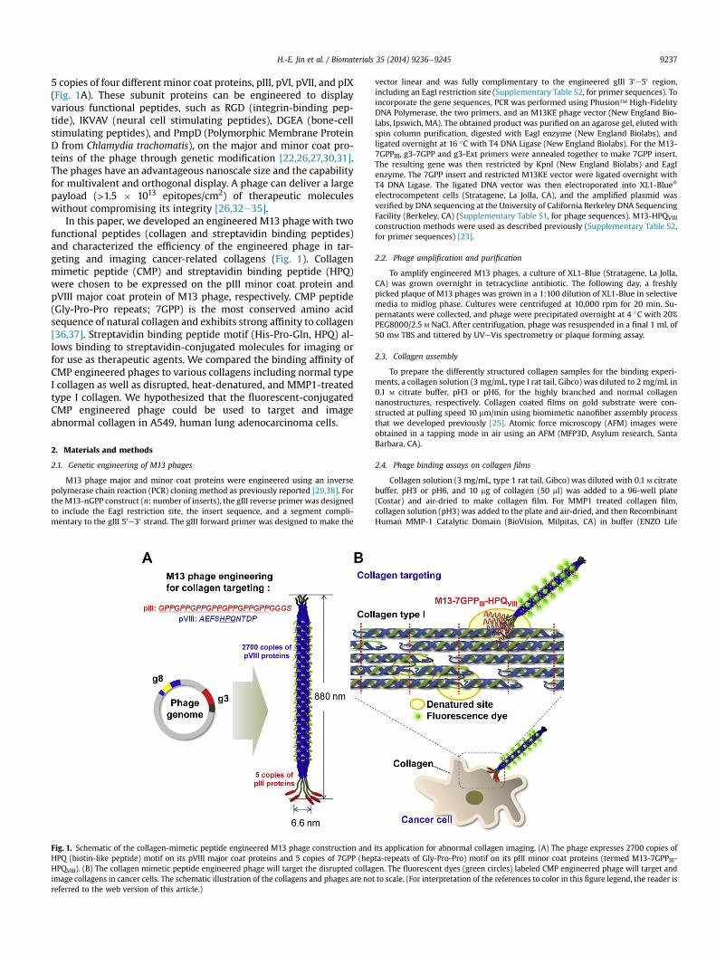
5 copies of four different minor coat proteins, pIII, pVI, pVII, and pIX(Fig. 1A). These subunit proteins can be engineered to displayvarious functional peptides, such as RGD (integrin-binding pep-tide), IKVAV (neural cell stimulating peptides), DGEA (bone-cellstimulating peptides), and PmpD (Polymorphic Membrane ProteinD from Chlamydia trachomatis), on the major and minor coat pro-teins of the phage through genetic modification [22,26,27,30,31].The phages have an advantageous nanoscale size and the capabilityfor multivalent and orthogonal display. A phage can deliver a largepayload (>1.5 � 1013 epitopes/cm2) of therapeutic moleculeswithout compromising its integrity [26,32e35].
In this paper, we developed an engineered M13 phage with twofunctional peptides (collagen and streptavidin binding peptides)and characterized the efficiency of the engineered phage in tar-geting and imaging cancer-related collagens (Fig. 1). Collagenmimetic peptide (CMP) and streptavidin binding peptide (HPQ)were chosen to be expressed on the pIII minor coat protein andpVIII major coat protein of M13 phage, respectively. CMP peptide(Gly-Pro-Pro repeats; 7GPP) is the most conserved amino acidsequence of natural collagen and exhibits strong affinity to collagen[36,37]. Streptavidin binding peptide motif (His-Pro-Gln, HPQ) al-lows binding to streptavidin-conjugated molecules for imaging orfor use as therapeutic agents. We compared the binding affinity ofCMP engineered phages to various collagens including normal typeI collagen as well as disrupted, heat-denatured, and MMP1-treatedtype I collagen. We hypothesized that the fluorescent-conjugatedCMP engineered phage could be used to target and imageabnormal collagen in A549, human lung adenocarcinoma cells.
2. Materials and methods
2.1. Genetic engineering of M13 phages
M13 phage major and minor coat proteins were engineered using an inversepolymerase chain reaction (PCR) cloning method as previously reported [29,38]. FortheM13-nGPP construct (n: number of inserts), the gIII reverse primer was designedto include the EagI restriction site, the insert sequence, and a segment compli-mentary to the gIII 50e30 strand. The gIII forward primer was designed to make the
Fig. 1. Schematic of the collagen-mimetic peptide engineered M13 phage construction andHPQ (biotin-like peptide) motif on its pVIII major coat proteins and 5 copies of 7GPP (hepHPQVIII). (B) The collagen mimetic peptide engineered phage will target the disrupted collagimage collagens in cancer cells. The schematic illustration of the collagens and phages are notreferred to the web version of this article.)
vector linear and was fully complimentary to the engineered gIII 30e50 region,including an EagI restriction site (Supplementary Table S2, for primer sequences). Toincorporate the gene sequences, PCR was performed using Phusion™ High-FidelityDNA Polymerase, the two primers, and an M13KE phage vector (New England Bio-labs, Ipswich, MA). The obtained product was purified on an agarose gel, eluted withspin column purification, digested with EagI enzyme (New England Biolabs), andligated overnight at 16 �C with T4 DNA Ligase (New England Biolabs). For the M13-7GPPIII, g3-7GPP and g3-Ext primers were annealed together to make 7GPP insert.The resulting gene was then restricted by KpnI (New England Biolabs) and EagIenzyme. The 7GPP insert and restricted M13KE vector were ligated overnight withT4 DNA Ligase. The ligated DNA vector was then electroporated into XL1-Blue®
electrocompetent cells (Stratagene, La Jolla, CA), and the amplified plasmid wasverified by DNA sequencing at the University of California Berkeley DNA SequencingFacility (Berkeley, CA) (Supplementary Table S1, for phage sequences). M13-HPQVIII
construction methods were used as described previously (Supplementary Table S2,for primer sequences) [23].
2.2. Phage amplification and purification
To amplify engineered M13 phages, a culture of XL1-Blue (Stratagene, La Jolla,CA) was grown overnight in tetracycline antibiotic. The following day, a freshlypicked plaque of M13 phages was grown in a 1:100 dilution of XL1-Blue in selectivemedia to midlog phase. Cultures were centrifuged at 10,000 rpm for 20 min. Su-pernatants were collected, and phage were precipitated overnight at 4 �C with 20%PEG8000/2.5 M NaCl. After centrifugation, phage was resuspended in a final 1 mL of50 mM TBS and tittered by UVeVis spectrometry or plaque forming assay.
2.3. Collagen assembly
To prepare the differently structured collagen samples for the binding experi-ments, a collagen solution (3 mg/mL, type I rat tail, Gibco) was diluted to 2 mg/mL in0.1 M citrate buffer, pH3 or pH6, for the highly branched and normal collagennanostructures, respectively. Collagen coated films on gold substrate were con-structed at pulling speed 10 mm/min using biomimetic nanofiber assembly processthat we developed previously [25]. Atomic force microscopy (AFM) images wereobtained in a tapping mode in air using an AFM (MFP3D, Asylum research, SantaBarbara, CA).
2.4. Phage binding assays on collagen films
Collagen solution (3 mg/mL, type 1 rat tail, Gibco) was diluted with 0.1 M citratebuffer, pH3 or pH6, and 10 mg of collagen (50 ml) was added to a 96-well plate(Costar) and air-dried to make collagen film. For MMP1 treated collagen film,collagen solution (pH3) was added to the plate and air-dried, and then RecombinantHuman MMP-1 Catalytic Domain (BioVision, Milpitas, CA) in buffer (ENZO Life
its application for abnormal collagen imaging. (A) The phage expresses 2700 copies ofta-repeats of Gly-Pro-Pro) motif on its pIII minor coat proteins (termed M13-7GPPIII-en. The fluorescent dyes (green circles) labeled CMP engineered phage will target andto scale. (For interpretation of the references to color in this figure legend, the reader is
H.-E. Jin et al. / Biomaterials 35 (2014) 9236e92459238
Sciences, Farmingdale, NY) was treated on collagen films in the plate for 2 days atroom temperature. To make gelatin films, the same collagen solutionwas denaturedat 70 �C for 15min, and 10 mg of collagen (50 ml) was added to a 96-well plate and air-dried. The 96-well plates were blocked with 1% BSA in PBS for 1 h and washed threetimes with PBS. Phages (1.0 � 1010 phages/mL) were incubated in collagen film for1 h at 37 �C and washed three times with PBS. We measured binding phages byELISA assay as previously described with modifications [23]. 200 ml of diluted HRP-conjugated anti-M13 monoclonal antibody (GE Healthcare, Piscataway, NJ) (1:5000dilution in blocking buffer) was treated to each well and incubated at room tem-perature for 1 h with agitation. Plates were washed with PBST five times, thenincubated with 3,30 ,5,50-Tetramethylbenzidine (TMB) liquid substrate (SigmaAldrich, St. Louis, MO) at room temperature for 10e30 min and treated with thesame amount of stop reagent (Sigma Aldrich, St. Louis, MO). The absorbance wasmeasured using a microplate reader (Safire, Tecan Group Ltd., M€annedorf,Switzerland) set at 450 nm.
2.5. Phage binding assays on various collagen types
We performed binding assay for various collagen types. Rat collagen type I(Gibco, Island, NY), human collagen type I (Sigma Aldrich, St. Louis, MO), bovinecollagen type II (Sigma Aldrich, St. Louis, MO), human collagen type III (SigmaAldrich, St. Louis, MO), human collagen type IV (Sigma Aldrich, St. Louis, MO), andhuman collagen type V (Sigma Aldrich, St. Louis, MO) were diluted with 100 mM
citrate buffer pH3, and 10 mg of collagen (50 ml) was added to a 96-well plate (Costar)and air-dried. Phages (1.0 � 1010 phages/ml) were incubated in collagens for 1 h at37 �C. ELISA method was same as described above in 2.4. Phage binding assays oncollagen films.
2.6. A549 human lung cancer cell culture
A549, human lung adenocarcinoma cells, were cultured in T-75 flask (Nunc,Rochester, MN) in DMEM containing 10% FBS, 2 mM L-glutamine, 100 U/ml penicillinand 100 mg/ml streptomycin at 37 �C in a humidified 5% CO2 atmosphere, and thegrowth media was changed every 2 days. 5 � 103 cells were seeded in a 96 wellplates for ELISA study, and 1 � 104 cells were seeded in a Lab-Tek®II CC2 ChamberSlide™ (Thermo-Fisher Scientific, Rochester, NY) for collagen imaging. For collageninduction, the cells were incubated in the presence of 5 ng/ml of TGF-b1 and 5 mg/mlof L-ascorbic acid for 72 h [39].
2.7. RT-PCR to measure collagen expression
Total RNA was isolated using Trizol (Invitrogen, Carlsbad, CA) and RT-PCR wasperformed using the SuperScript One-Step RT-PCR kit (Invitrogen). The targettranscript was reverse transcribed at 50 �C for 30 min. For type I collagen, the PCRproduct was amplified using 30 cycles (initial denaturation at 94 �C/2 min followedby PCR amplification, 94 �C/30 s, 58 �C/30 s, and 68 �C/1 min) [39]. PCR productswere visualized on a GelDoc system (Bio-Rad, Hercules, CA) and band density wasanalyzed using NIH ImageJ. The following primers were used for PCR: humancollagen type I forward (50-ACGTCCTGGTGAAGTTGGTC-30), human collagen type Ireverse (50-ACCAGGGAAGCCTCTCTCTC-30), human GAPDH forward (50-GGGCTGCTTTTAACTCTGGT-30), and human GAPDH reverse (50-TGGCAGGTTTTTC-TAGACGG-30).
2.8. Gelatin zymography for matrix metalloproteinases (MMPs) expression inA549 cells
We measured the expression level of MMPs by TGF-b1 and L-ascorbic acidtreatment in the cell culture media using gelatin-based zymography. Cells(1.5 � 104 cells/well) were seeded in a 48-well plate and stimulated with 5 ng/ml ofTGF-b1 and 5 mg/ml of L-ascorbic acid for 72 h, and media were collected. Aftermeasuring the protein concentration by BCA assay, equal amounts (50 mg) of sam-ples were mixed with an equal volume of 2� non-reducing sample buffer (Bio-Rad,Hercules, CA). The samples were applied to a 10% Ready Gel® Zymogram Gel (Bio-Rad, Hercules, CA). After electrophoresis, SDS was removed from the gel by washing3 times for 10 min in 2.5% Triton X-100 solution. Then the gels were incubatedovernight with gentle shaking at 37 �C in buffer [50 mM TriseHCl (pH 7.6), 10 mM
CaCl2, 50 mM NaCl, 0.05% Brij35], and then the gel was stained with 0.25% Coomassieblue R250 in 40% methanol and 10% acetic acid for 2 h at room temperature andsubsequently destained with a 40% methanol, 10% acetic acid solution until thebands became clear.
2.9. Conjugation of streptavidin-Alexa Fluor®488 on the phage for imaging
M13-HPQVIII and M13-7GPPIII-HPQVIII phages (1014 phages each) were mixedwith 100 mL of 2 mg/mL Streptavidin-Alexa Fluor®488 and incubated for 1 h at roomtemperature under gentle mixing. The phages were PEG-precipitated twice andresuspended in 400 mL of PBS. After conjugation, the amount of Streptavidin-AlexaFluor®488 per phage was measured by UVeVis at 269 nm and 495 nm.Streptavidin-Alexa Fluor®488 conjugated phages were used for fluorescence mi-croscopy and fluorescence-activated cell sorting (FACS) analysis.
2.10. Fluorescence microscopy for collagen imaging in A549
Chamber slides were blocked with 1% BSA in the cell culturemedia for 1 h beforephage treatment, and then 1.0� 1010 phages/mL were incubated with cells for 1 h at37 �C. Alexa Fluor® 488 conjugated M13-7GPPIII-HPQVIII phages and Alexa Fluor®
488 conjugated M13-HPQVIII (control) were treated onto the TGF-b1-treated andnon-treated A549 cells for collagen imaging. Cells werewashed three timeswith PBSand fixed in 4% formaldehyde solution for 15 min. Cells were made permeable byincubation of 0.1% Triton X-100 for 3 min and washed with PBS. Actin filaments andnuclei were stained with 100 nM Rhodamine Phalloidin (Cytoskeleton, Inc., Denver,CO) and 300 nM DAPI (Molecular Probes, Eugene, OR), respectively, as counter-staining for all samples. The fluorescence images were collected using an IX71Fluorescence Microscope (Olympus, Tokyo, Japan) and were analyzed using NIHImageJ software.
2.11. FACS analysis
A549 cells (2 � 105 cells) were seeded in a petri dish (35 � 15 mm) and stim-ulated with 5 ng/mL of TGF-b1 and 5 mg/mL of L-ascorbic acid for 72 h. For controlanalysis, A549 cells were seeded in a petri dish (35 � 15 mm) without stimulation.1.0 � 1010 phage particles, Alexa Fluor® 488 conjugated M13-7GPPIII-HPQVIII phagesand Alexa Fluor® 488 conjugated M13-HPQVIII (control), were added to A549 cells(5 � 105 cells) in 1 mL of PBS, and the mixture was incubated for 2 h at 37 �C. Afterincubation, cells were washed three times with PBS. Phage binding was detected byFACScalibur (Becton Dickinson, San Jose, CA). For each sample, 10,000 cells wereanalyzed with FACScalibur using FlowJo software (Treestar, Inc., San Carlos, CA).
2.12. Statistical analysis
All results from in vitro experiments were expressed as mean ± SD of threeindependent experiments. When it was necessary to compare the means betweentreatments, an unpaired Student's t-test was applied to analyze the data. Compar-ison among multiple groups was performed using one-way ANOVA with the Tukeytest. p < 0.05 was considered to be the minimal level of significance.
3. Results
3.1. Construction of the desired CMP engineered M13 phage
We constructed a phage to express both CMP and streptavidinbinding peptide using recombinant DNA techniques. For collagen-targeting purpose, the (GPP)n peptide was engineered on pIII ofM13-Wild (M13-2GPPIII , M13-4GPPIII, M13-6GPPIII, and M13-7GPPIII) (Supplementary Table S1). The phages were engineeredwith the streptavidin binding peptide on pVIII (M13-HPQVIII). Thesestreptavidin binding peptides allow for the conjugation of func-tional motifs such as therapeutic or imaging agents throughstreptavidin conjugation [40,41]. We confirmed the DNA sequencesand locations for both HPQ and (GPP)n peptides (SupplementaryFig. S1). The resulting phage (M13-7GPPIII-HPQVIII) displayed ahigh density of HPQ peptides (2700 copies/phage) on the majorcoat proteins and five copies of the nGPP peptides on the pIII minorcoat proteins (Fig. 1). We used engineered phages with only HPQ onpVIII (M13-HPQVIII) or M13-Wild as controls.
3.2. Binding characterization of the CMP engineered phage oncollagens
In order to measure the binding affinity of the CMP engineeredphage (M13-nGPPIII), we prepared differently structured collagenthin films and characterized the CMP engineered phage binding onthem. Type I collagen self-assembly in vitro is an entropy drivenprocess via loss of surface bound solvent molecules [42]. Collagenfibrils self-assemble into different structures depending on pH andelectrolyte concentrations in the buffered solutions [43,44]. Weprepared two different collagen morphology samples shown inFig. 2A and B. Collagen thin film samples prepared in pH3.0 buff-ered condition exhibited highly disrupted fibrillar structures(Fig. 2A). The collagen films prepared pH6.0 buffered conditionexhibit normal collagen banded structures with the characteristic67 nm collagen band structures (Fig. 2B). Therefore, we used thesetwo films for the collagen model targets in the binding assays.

Fig. 2. Characterization of the CMP engineered phage on collagen films. AFM images of collagen thin films prepared at (A) 0.1 M citrate buffer (pH 3) and (B) 0.1 M citrate buffer (pH6). Collagen bundles are highly disrupted structures at 0.1 M citrate buffer (pH 3) with highly branched structures (A). Normal collagen structure at 0.1 M citrate buffer (pH 6) withcharacteristic collagen bands exhibiting ~67 nm bands (B). (C) CMP engineered phage, M13-6GPPIII and 7GPPIII, bind on disrupted collagen at a significantly higher levels than onnormal collagen (*, p < 0.01). Data represent the mean ± SD of three independent experiments.
H.-E. Jin et al. / Biomaterials 35 (2014) 9236e9245 9239
In order to measure the binding affinity of the CMP engineeredphage, we investigated the CMP engineered phage binding ondisrupted collagen and normal collagen using ELISA assay. Briefly,1.0 � 1010 phages/mL of each phage was incubated on thosecollagen films for 1 h at 37 �C, and the bound phages weremeasured using HRP-conjugated anti-M13 monoclonal antibody.As the CMP length on the phages was increased, the binding ofphages on disrupted collagen was increased. No increase wasobserved for normal collagen (Fig. 2C). M13-6GPPIII and M13-7GPPIII showed significantly higher binding on disrupted collagenthan on normal collagen (p < 0.01) (Fig. 2C).
We investigated the engineered phage binding capability ondisrupted collagen using ELISA assay in the concentration range of2.5 � 106e1.0 � 1013 phages/mL. Based on the ELISA assays, weevaluated the binding affinity of phages by calculating the phageconcentration at which the binding is 50% of maximum binding(Kd: the equilibrium dissociation constant) with the Hill equation,as seen in Fig. 3. The mean Kd value was 1.5 � 1010 phages/mL forM13-7GPPIII and 2.4 � 1011 phages/mL for M13-Wild, suggestingthe affinity of M13-7GPPIII was about 16 times higher than the af-finity of M13-Wild phage on disrupted collagen. As GPP repeats onthe minor coat protein (n ¼ 2, 4, 6, and 7) were increased, the
binding affinity to the disrupted collagen was also enhanced. Themean dissociation constants for the CMP engineered phage (M13-nGPPIII) for the disrupted collagen were 3.9 � 1011, 5.8 � 1010,2.9 � 1010, and 1.5 � 1010 phages/mL for n ¼ 2, 4, 6, and 7,respectively (Fig. 3). There was no significant difference betweenM13-Wild and M13-2GPPIII (Kd, 2.4 � 1011 ± 2.5 � 1010 phages/mLfor M13 wild and 3.9 � 1011 ± 2.0 � 1011 phages/mL for M13-2GPPIII). CMP phage binding to BSA was negligible (Fig. 3). BecausetheM13-7GPPIII exhibited the highest binding affinity for the targetcollagen, we used the M13-7GPPIII for the further characterization.
3.3. Evaluation of the CMP-phage binding on denatured collagens
We characterized collagen binding of M13-7GPPIII phages onvarious collagen structures including normal and denatured colla-gens (disrupted, heat-denatured, andMMP1-treated collagens). Weprepared each collagen films using drop-cast films and character-ized their binding with the M13-7GPPIII by ELISA assay andcompared it to those of M13-Wild (as control). M13-7GPPIII phagewas bound2.1-, 5.6-, 9.4-, and 8.0- timesmore onnormal, disrupted,heat-denatured, and MMP1-treated collagens, respectively, thanM13-Wild phage (Fig. 4). M13-7GPPIII phage can be bound more to

Fig. 3. Binding characterization of the CMP engineered phages. Binding activity of theCMP engineered phages to disrupted rat tail collagen type I or BSA was detected usingHRP-conjugated anti-M13 phage antibody. Solid lines represent fits to the Hill equa-tion. Error bars represent the mean ± SD of three independent experiments.
H.-E. Jin et al. / Biomaterials 35 (2014) 9236e92459240
disrupted collagen than normal collagen and can be bound themostto heat-denatured collagen (unstructured collagen, p < 0.001) andcollagen denatured byMMP1 enzyme treatment (p < 0.001) (Fig. 4).
3.4. Evaluation of the CMP engineered phage binding on differentcollagen types
To further investigate binding characteristics of M13-7GPPIIIphage, we also characterized their binding affinity to various types
Fig. 4. The CMP phage, M13-7GPPIII, binding on rat collagen type I with different de-natured methods. M13-7GPPIII bound significantly more to unstructured collagens(disrupted, MMP1-digested, and heat-denatured) than to normal collagen (*, p < 0.001).
of collagens. We treated M13-7GPPIII and M13-Wild (i.e., control)phages on rat type I, human type I, bovine type II, and human typeIIIeV, and quantitatively measure the bound phage amount usingELISA assay. Because the GPP repeats can be associated to dena-tured parts of collagen fibrils, we tested our engineered phagebinding on fibrillar collagens (collagen type I, II, III, and V), non-fibrillar collagen (collagen type IV) and laminin (ECM protein)[45]. The M13-7GPPIII phage exhibited binding affinity to variouscollagen types (Fig. 5). The M13-7GPPIII showed the best binding oncollagen type I and II. The M13-7GPPIII phage was bound to fibrillarcollagen types, 4.4-, 5.6-, 4.8- times more than M13-Wild phage onrat collagen type I (p < 0.01), human type I (p < 0.05), and bovinetype II (p < 0.05), respectively. M13-7GPPIII phage was bound toother fibrillar collagen types 2.5- and 2.8-times more than M13-Wild phage on human collagen type III (p < 0.05) and V(p < 0.05), respectively. M13-7GPPIII phage was bound to humancollagen type 4, non-fibrillar collagen, 1.9 times more than M13-Wild phage (p < 0.05) (Fig. 5). However, M13-7GPPIII phage wasrarely bound to laminin, an extracellular matrix (ECM) protein,indicating that M13-7GPPIII phage could bind to collagensselectively.
3.5. The CMP engineered phage binding on induced abnormalcollagen in A549 cells
We investigated the targeting capability of the M13-7GPPIIIphage using cancer cell lines with controlled collagen expressionin vitro. We used A549 cells, human lung cancer cells, to controltype I collagen expression by TGF-b1. We then performed bindingassays in a TGF-b1 dependent manner [39]. In order to induce thecollagen expression, the cells were treated with 5 ng/mL of TGF-b1and 5 mg/mL L-ascorbic acid for 72 h, and the effects on collagenexpression were assessed by RT-PCR. The expression of collagentype I was significantly (p < 0.05) stimulated as detected by RT-PCR(Fig. 6A) and increased 1.7 times by TGF-b1 as compared to theexpression in control cells (Fig. 6B). MMPs expression was exam-ined using gelatin zymography to measure whether the collagencondition of A549 cells had similar characteristics to highly dena-tured forms for cancer cell migration in response to TGF-b1 treat-ment [39]. Gelatin zymography was performed using the cell
Fig. 5. Binding characteristics of the CMP phage (M13-7GPPIII) on various collagentypes and laminin. M13-7GPPIII was able to bind various collagen types effectively.Various collagen types (rat collagen type I, human type I, bovine type II, human type III,IV, and V (10 mg/well)) were coated on a 96-well plate. Phage binding was measured byELISA assay. Phage binding is expressed as fold-changes compared to the control. Eacherror bar represents the mean ± SD of three independent experiments. *p < 0.05,**p < 0.01.

H.-E. Jin et al. / Biomaterials 35 (2014) 9236e9245 9241
culture media that were harvested after 72 h of TGF-b1 treatment(5 ng/mL). The samples were applied to a 10% polyacrylamide gelcontaining gelatin without reduction, and proteolytic activity wasdemonstrated by digestion of the gelatin and clearing of the gel.The A549 cells secreted gelatinases with molecular weightsconsistent to an identity of MMP-2 and MMP-9 (Fig. 6C). MMP-2was the major gelatinase expressed, and the TGF-b1 treatmentup-regulated MMP-2 expression in the cell culture media. BasalMMP-9 expression was low, and its expression was not changed byTGF-b1 treatment (Fig. 6C).
TGF-b1 was used to induce abnormal collagen in A549 cells, andphage binding (1.0� 1010 phages/mL) was measured by ELISA assay(Fig. 6D). M13-7GPPIII phage was bound to collagen 1.5 times morethanM13-Wild phage on the surface of A549 cells without collageninduction by TGF-b1. In cancerous, collagen over-expressedA549 cells, M13-7GPPIII phage bound up to 3.7 times more thanthe control phage, while the collagen expression was increased 1.7times (Fig. 6B and D). This enhanced binding of 7GPPIII phagesuggested that the M13-7GPPIII phage could bind more effectivelyto abnormal collagens than to normal collagen (Fig. 6D).
3.6. Fluorescence imaging and FACS analysis for abnormal collagenby the CMP engineered phage
After confirmation of the effective binding of the M13-7GPPIII onthe A549 cells in a TGF-b1 dependent manner, we imaged theA549 cells using the phage engineered with both CMP and strep-tavidin binding HPQ peptides. Streptavidin-Alexa Fluor®488 wasbound to the HPQ peptides of M13-7GPPIII-HPQVIII by the simplemixing of streptavidin-Alexa Fluor®488 (5 mol dye/mole) and HPQ-displayed phages solutions. Loading amounts of streptavidin-Alexa
Fig. 6. Binding characteristics of the CMP phage (M13-7GPPIII) on collagen expressing A5presence of 5 mg/ml L-ascorbic acid for 72 h (A) mRNA expression of collagen type I was dechanges in expression level are expressed as fold-changes compared to the control. Each bsurement of MMP-2 and MMP-9 activities in cell culture media after 72 h of TGF-b1 treatmeMMP-9 activity was unchanged. (D) M13-7GPPIII phage was bound more to TGF-b1-inducedthree independent experiments. **p < 0.001.
Fluor®488 per phage were quantified by the absorbance of thephageeAlexa Fluor®488 conjugate at 269 and 495 nm. Each phagecarried an average of 275 Alexa Fluor®488 molecules. Because fivedyes are conjugated with a streptavidin, the UVeVis result indicatethat ~2% of the HPQ peptides on the pVIII major coat protein werefunctionalized with streptavidin-Alexa Fluor®488 among the 2700copies of the HPQ peptide on major coat protein.
To evaluate the engineered phage as an imaging agent, wetreated Alexa Fluor®488-tagged M13-7GPPIII-HPQVIII phage orAlexa Fluor®488-tagged M13-HPQVIII (control) on A549 cells withor without collagen induction. The same amount of each fluores-cent phage (1.0� 1010 phages/mL) was treated in the three differentwells of A549 cells for 1 h at 37 �C. Representative images areshown in Fig. 7. Control phages (M13-HPQVIII) without collagenbinding motif (7GPP) were not significantly bound to collagens onthe surface of A549 cells both with and without TGF-b1 treatment(Image A3 and C3 in Fig. 7). We observed that the A549 cellswithout TGF-b1 treatment incubated with Alexa Fluor®488-taggedM13-7GPPIII-HPQVIII phage still exhibited phage binding becauseA549 cells without TGF-b1 treatment also expressed some colla-gens (Figs. 6D and Fig. 7-B3). However, the Alexa Fluor®488-taggedM13-7GPPIII-HPQVIII provided the best images of the abnormalcollagens on TGF-b1 treated A549 cells; it was consistent with cellshapes as compared to the actin image (Fig. 7-D3).
Using Alexa Fluor®488-tagged M13-HPQVIII phage (control) andM13-7GPPIII-HPQVIII phage, abnormal collagen induced A549 cellswere detected by FACS. Alexa Fluor®488-tagged M13-HPQVIII phageor M13-7GPPIII-HPQVIII phage were incubated in control A549 cellswithout collagen induction. The fluorescence histogram of M13-7GPPIII-HPQVIII phage incubated A549 cells was slightly increasedwhile the histogram of M13-HPQVIII phage incubated A549 cells
49 cells, induced by TGF-b1. A549 cells were incubated with 5 ng/mL TGF-b1 in thetected by RT-PCR. (B) Densitometric analysis of Fig. 6A was performed by ImageJ. Thear represents the mean ± SD of three independent experiments. *p < 0.05. (C) Mea-nt by enzyme zymography. MMP-2 activity was significantly increased by TGF-b1, andcollagen in A549 cells than in control cells. Each error bar represents the mean ± SD of

Fig. 7. The fluorescent labeled engineered phage (M13 -7GPPIII-HPQVIII) can image collagen in human lung cancer cells, A549. Abnormal collagen in A549 cells were induced bytreatment with 5 ng/mL TGF-b1 in the presence of 5 mg/mL L-ascorbic acid for 72 h (C and D). The control group received no treatment (A and B). Cells were incubated with eitherAlexa Fluor®488 conjugated M13-HPQVIII (as a control) (A and C) or Alexa Fluor®488 conjugated M13-7GPPIII-HPQVIII phage (B and D). (1) Blue: DAPI, nucleus; (2) Red: actin fil-aments; (3) Green: phages-Alexa Fluor®488; (4) composite images of 1, 2, and 3. Scale bar ¼ 100 mm. (For interpretation of the references to color in this figure legend, the reader isreferred to the web version of this article.)
H.-E. Jin et al. / Biomaterials 35 (2014) 9236e92459242
was similar to that of A549 cells without phage incubation in FACShistogram (Fig. 8A). Mean fluorescence intensity (MFI) values incontrol A549 cells without TGF-b1 treatment were 18.1 ± 3.6,12.8 ± 4.0, and 25.1 ± 4.5 for control A549 only, M13-HPQVIII, andM13-7GPPIII-HPQVIII, respectively (Fig. 8A). Percentages ofabnormal collagen positive cells were 1.4 ± 0.4% for M13-HPQVIII
Fig. 8. FACS histogram of induced abnormal collagen detection by Alexa Fluor®488-tagged MGreen: A549 cells without phage, Blue: Alexa Fluor®488 conjugated M13-HPQVIII (control), Rof the references to color in this figure legend, the reader is referred to the web version of
and 2.1± 0.8% forM13-7GPPIII-HPQVIII compared to the control cells(Fig. 8A). Alexa Fluor®488-tagged M13-HPQVIII phage or M13-7GPPIII-HPQVIII phage was incubated in abnormal collagen inducedA549 cells by TGF-b1 treatment. Only the fluorescence histogram ofM13-7GPPIII-HPQVIII phage incubated A549 cells was increased andshifted to right-side in FACS histogram (Fig. 8B). MFI values in
13-7GPPIII-HPQVIII in A549 cells without treatment (A) and with TGF-b1 treatment (B).ed: Alexa Fluor®488-tagged M13-7GPPIII-HPQVIII phage incubation. (For interpretationthis article.)

H.-E. Jin et al. / Biomaterials 35 (2014) 9236e9245 9243
collagen induced A549 cells by TGF-b1 treatment were 19.8 ± 4.1,26.7 ± 5.4, and 121 ± 22.4 for collagen induced A549 cells only,M13-HPQVIII, and M13-7GPPIII-HPQVIII, respectively (Fig. 8B). Per-centages of abnormal collagen positive cells were 4.8 ± 1.2% forM13-HPQVIII and 60.5 ± 8.8% for M13-7GPPIII-HPQVIII compared tothe control cells (Fig. 8B). There was no significant difference be-tween M13-HPQVIII phage incubated A549 cells and A549 cells only(Fig. 8B).
4. Discussion
M13 phage possesses multiple advantageous structural featuresfor biomedical materials. The phage is nonpathogenic to humancells and easily removed from the body with little side-effect[46e48]. Large quantities of identical phage coat proteins can beeasily produced by amplification in bacterial host cells and self-assembled to form a nanofibrous shape. In addition, its minor(pIII, pVI, pVIII and pIX) and major (pVIII) coat proteins can bemodified to display functional peptide motives by genetic orchemical modification [32e35]. In this paper, we developedgenetically engineered M13 phage (M13-7GPPIII-HPQVIII) that couldbe used as an imaging agent to target collagen in cancer cells. Weshowed that by expressing collagen mimetic peptide on the minorcoat protein (M13-7GPPIII) collagen binding of M13-7GPPIII wasdramatically enhanced compared with that of M13-Wild phage(Figs. 2C and 3). Moreover, the addition of streptavidin bindingpeptide (HPQ) on pVIII did not interrupt the function of 7GPP andprovided 2700 copies of motives to bind a streptavidin-conjugatedimaging agent. We were able to successfully image and detect thecancer collagen using M13-7GPPIII-HPQVIII (Figs. 7 and 8). In thefuture, we can use our engineered M13 phage as a drug deliveryvehicle through conjugation with therapeutic drugs.
The hierarchical organization of collagen molecules providessuperior mechanical properties to connective tissues (e.g., liga-ments, tendons, etc.) [49], forms extracellular matrices (e.g., carti-lage, cornea, etc.) [50], and is important for several biologicalfunctions such as tissue-structuring, cell attachment, tissue repair,and control of tissue-related diseases [51,52]. Collagen mimeticpeptides (CMPs) are typically less than 30 amino acids and containthe repeat tripeptide sequences Gly-Pro-Pro and/or Gly-Hyp-Pro.The CMP is known to bind or hybridize with denatured collagenby associating with disentangled parts of the collagen molecules[3,36,37]. Therefore, we hypothesized that CMPs on the M13-7GPPIII phage would bind more to highly denatured collagenstructures present in diseases like cancers than to normal collagens(Fig. 1).
We constructed two different structures of collagen fibrils toreflect normal collagen and disrupted collagen by controlling thepH condition of the buffer solutions used to characterize the CMPengineered phages (Fig. 2A, B) [25,43,53e55]. M13-7GPPIII phageexhibited higher binding on the disrupted collagen film prepared inpH3 than on the normal collagen film in pH6 (p < 0.01) (Fig. 2C). Itexhibited a 16 times increased binding affinity on disruptedcollagen compared to M13-Wild phage (Fig. 3). This indicates thatCMP motives displayed on the M13 phage can be successfullyfunctionalized and can bind to disrupted collagen more than tonormal collagen. Furthermore, in order to emulate more diseaseconditions with disrupted collagen molecular structure, heat-denatured (gelatin) and MMP digested collagen were added tonormal and disrupted rat collagen type I. Heat-denatured collagenis characterized by randomly denatured collagen molecules and anunstructured form through the structural degradation of collagen[56]. MMP can change themolecular structure of collagen in humandisease by cleaving all three polypeptide chains of the 300 nmtriple-helical collagen monomer at a specific recognition site
~225 nm from the N-terminus of collagen molecules. The resultingcollagen molecules cleaved into two collagen fragments havemelting temperatures around 34 �C and can be further denaturedand structurally changed at body temperature [53,57,58]. There-fore, we evaluated M13-7GPPIII phage binding ability to normal anddisrupted forms of rat collagen type I, as well as collagen denaturedby heat or byMMP enzyme digestion. We observed that the level ofM13-7GPPIII binding was as low as the level of M13-Wild bindingon normal collagen and that M13-7GPPIII exhibited approximately5.6e9.4- times higher levels of binding after 37 �C incubation ondisrupted, MMP-1 treated, and heat-denatured (gelatin) collagenscompared to M13-Wild (Fig. 4). This binding result suggested thatM13-7GPPIII phage preferred to bind to denatured collagens than tonormal collagen andmight be used for collagen targeting in diseaseconditions.
We investigated the collagen binding affinity of CMP displayedphages (M13-2GPPIII, 4GPPIII, 6GPPIII, 7GPPIII phages) on disruptedcollagen. The phages which displayed more GPP repeats showed anincreased collagen binding affinity, and the engineered phagesneeded more than 4GPP repeats on their minor coat in order tobind significantly more to disrupted collagen than to normalcollagen (Figs. 2C and 3). We compared the dissociation constants(Kd) of the CMP phages with the Hill equation. As the GPP repeatnumber increased, the binding affinity (Kd) of M13-nGPP phagewasenhanced (Fig. 3). The M13-7GPPIII phage has the best binding af-finity (16 times stronger than M13-Wild) on disrupted collagenthan the other CMP phages (Fig. 4). In order to obtain an evenhigher binding affinity for collagen, we tried to construct longerCMP-displayedM13 phages, such as 8GPP or 10GPP. However, 7GPPwas the longest CMP-displayed M13 phage we could construct sofar. We assume that gene deletion might be occurring while thephage is replicated in Escherichia coli cells because the repeatedGly-Pro-Pro amino acid sequences are made of a high number ofguanine (G) and cytosine (C) repeats in their respective codons (i.e.,GGN-CCN-CCN for GPP amino acids). Highly repeated guanine (G)and cytosine (C) sequences might make hairpin structures whichcan be easily deleted during DNA duplication in E. coli cells un-dergoing phage cloning [59]. For example, when we were con-structing M13-7GPPIII phages, shorter GPP phages were alsoconstructed because of gene deletion (data not shown). Although7GPP is the longest CMP that could be displayed on the minor coatof M13 phage, the Yu group has reported that the binding affinity ofCMP-7 and CMP-10 are similar (1.7 � 10�8
M for CMP-7 and1.4 � 10�8 M for CMP-10, respectively) for disrupted collagen film[60]. Therefore, we expect that the binding affinity of the M13phage with CMP-7 and CMP-10 might be similar [60]. The Meijergroup showed that dendrimers functionalized with collagenbinding peptides in a pentavalent manner could bind 100 timesstronger than that of a monomeric dendrimer [61]. M13 phage alsodisplays five copies of CMP on their minor coat protein. Therefore,we expect that M13-7GPPIII phage could multivalently bind tocollagen and exhibit enhanced binding affinity like pentamericdendrimer.
To conjugate imaging agents on the engineered phage, strep-tavidin binding peptide (HPQ) was displayed on the pVIII majorcoat protein of M13-7GPPIII phage. The HPQ-peptide, which spe-cifically binds to streptavidin, was previously identified usingphage display [40,41,62]. We engineered the phage to display alinear peptide, FSHPQNT (Kd ¼ 125 mM for streptavidin) [40,41]. Itdisplayed 2700 copies of HPQ peptide, densely and uniformly, on itsmajor coat (HPQVIII). We prepared Alexa Flour®488 tagged phagesafter simple mixing of M13-7GPPIII-HPQVIII phage and streptavidin-Alexa Flour®488. We could conjugate ~2% of the HPQ peptides onthe CMP phagewith streptavidin-Alexa Flour®488 for imaging. Thissimple imaging probe modification is an advantageous way to

H.-E. Jin et al. / Biomaterials 35 (2014) 9236e92459244
prepare various imaging agents and could be extended to thera-peutic molecule conjugation.
Human lung cancer cells, A549 cells, were treated with TGF-b1and L-ascorbic acid to induce collagen expression [39]. Not onlycollagen type I was increased in cells, but matrixmetalloproteinase-2 (MMP-2) was also increased in the cell culturemedia, suggesting that collagens on the surface of A549 cells werehighly digested and denatured by MMP-2 after the 72-hr TGF-b1treatment (Fig. 6AeC) [17,39]. M13-7GPPIII-HPQVIII phage attachedto A549 cells with denatured collagens 3.7 times more than toA549 cells without treatment (Fig. 6D). This result indicates thatM13-7GPPIII-HPQVIII phage could attach more to cancer cells andtissues with collagen remodeling than to normal cells. AlexaFlour®488 tagged M13-7GPPIII-HPQVIII phages were successfullybound to abnormal collagen in A549 cells (Fig. 7-D3). Fluorescencesignals of phages were similar to those of actin filaments becausecollagen covered the entire surface of the A549 cells (Fig. 7-B2 andB3, D2 and D3). This suggests that our M13-7GPPIII-HPQVIII phagescould be useful for cancer collagen imaging. Moreover, we detectedthe abnormal collagen in A549 cells using Alexa Flour®488 taggedM13-7GPPIII-HPQVIII phages by FACS (Fig. 8). When AlexaFluor®488-tagged engineered phages were incubated in abnormalcollagen induced A549 cells by TGF-b1 treatment, the fluorescencehistogram of M13-7GPPIII-HPQVIII phage incubated A549 cells wasincreased and shifted to right-side in FACS histogram (Fig. 8B).There was no significant difference between M13-HPQVIII phageincubated A549 cells and A549 cells only (Fig. 8A and B). Therefore,the M13-7GPPIII-HPQVIII phage could selectively distinguish theabnormal collagen over-expressed cancer cells through fluorescentlabeling. In previous research for molecular imaging of cancer usingengineered phages, several peptides were successfully used toimage cancer such as avb3-integrin-binding RGD containing pep-tides [21], VHSPNKK peptide for VCAM-1 (vascular cell adhesionmolecule 1) expressing tumor endothelial cells [63], SPPTGINpeptide for SPARC up-regulated in invasive cancer [64]. In addition,our 7GPP peptide-displayed phage for abnormal collagen might bea potential biomaterial for targeting and for use as imaging agentsin various cancers.
5. Conclusions
In summary, we developed collagen-mimetic peptide (CMP)engineered phage for collagen targeting and imaging in cancers.We genetically engineered M13 phage to stably express both theCMP motif (Gly-Pro-Pro)7 on the minor coat proteins and thestreptavidin-binding peptide (HPQ) on themajor coat proteins. Thisconstruct, M13-7GPPIII-HPQVIII, was able to selectively bind severaltypes of collagen using five copies of the CMP motif. The collagenbinding efficiency of M13-7GPPIII-HPQVIII phage on disruptedcollagen was improved by 16 times compared to that of the M13-Wild phage. Moreover, M13-7GPPIII-HPQVIII phage can bind moreto TGF-b1-induced abnormal collagen in A549 cells than in controlcells. HPQ-peptide on themajor coat protein of M13-7GPPIII-HPQVIIIcan be easily attached to fluorescent imaging agents throughstreptavidin conjugation. We believe that our CMP engineeredphage could be used as a collagen targeting agent to detect andimage the numerous pathological conditions related to abnormalcollagen remodeling in the future.
Acknowledgments
H.E.J. was supported by Basic Science Research Program throughthe National Research Foundation of Korea (NRF) funded by theMinistry of Education, Science and Technology (NRF-2011-357-E00083).
Appendix A. Supplementary data
Supplementary data related to this article can be found online athttp://dx.doi.org/10.1016/j.biomaterials.2014.07.044.
References
[1] Even-Ram S, Yamada KM. Cell migration in 3D matrix. Curr Opin Cell Biol2005;17:524e32.
[2] Gelse K, Poschl E, Aigner T. Collagens e structure, function, and biosynthesis.Adv Drug Deliv Rev 2003;55:1531e46.
[3] Li Y, Foss CA, Summerfield DD, Doyle JJ, Torok CM, Dietz HC, et al. Targetingcollagen strands by photo-triggered triple-helix hybridization. Proc Natl AcadSci U S A 2012;109:14767e72.
[4] Won S, Davies-Venn C, Liu S, Bluemke DA. Noninvasive imaging of myocardialextracellular matrix for assessment of fibrosis. Curr Opin Cardiol 2013;28:282e9.
[5] Provenzano PP, Eliceiri KW, Campbell JM, Inman DR, White JG, Keely PJ.Collagen reorganization at the tumor-stromal interface facilitates local inva-sion. BMC Med 2006;4:38.
[6] Conklin MW, Eickhoff JC, Riching KM, Pehlke CA, Eliceiri KW, Provenzano PP,et al. Aligned collagen is a prognostic signature for survival in human breastcarcinoma. Am J Pathol 2011;178:1221e32.
[7] Saftlas AF, Hoover RN, Brinton LA, Szklo M, Olson DR, Salane M, et al.Mammographic densities and risk of breast cancer. Cancer 1991;67:2833e8.
[8] Saftlas A, Hoover R, Brinton L, Szklo M, Wolfe J. Mammographic densities asindicators of breast-cancer risk. Am J Epidemiol 1988;128:914.
[9] Liang Y, Diehn M, Bollen AW, Israel MA, Gupta N. Type I collagen is overex-pressed in medulloblastoma as a component of tumor microenvironment.J Neurooncol 2008;86:133e41.
[10] Santala M, Simojoki M, Risteli J, Risteli L, Kauppila A. Type I and type IIIcollagen metabolites as predictors of clinical outcome in epithelial ovariancancer. Clin Cancer Res 1999;5:4091e6.
[11] Ohlund D, Lundin C, Ardnor B, Oman M, Naredi P, Sund M. Type IV collagen isa tumour stroma-derived biomarker for pancreas cancer. Br J Cancer2009;101:91e7.
[12] Skovbjerg H, Anthonsen D, Lothe IMB, Tveit KM, Kure EH, Vogel LK. CollagenmRNA levels changes during colorectal cancer carcinogenesis. BMC Cancer2009;9.
[13] Ramshaw JAM, Shah NK, Brodsky B. Gly-X-Y tripeptide frequencies incollagen: a context for host-guest triple-helical peptides. J Struct Biol1998;122:86e91.
[14] Brodsky B, Persikov AV. Molecular structure of the collagen triple helix. AdvProtein Chem 2005;70:301e39.
[15] Nagase H, Visse R. In: Parks WC, Mecham RP, editors. Triple helicase activityand the structural basis of collagenolysis. Extracellular matrix degradation,biology of extracellular matrix. Heidelberg: Springer; 2011. p. 95e122.
[16] Brinckerhoff CE, Matrisian LM. Timeline e matrix metalloproteinases: a tail ofa frog that became a prince. Nat Rev Mol Cell Biol 2002;3:207e14.
[17] Page-McCaw A, Ewald AJ, Werb Z. Matrix metalloproteinases and the regu-lation of tissue remodelling. Nat Rev Mol Cell Biol 2007;8:221e33.
[18] Egeblad M, Werb Z. New functions for the matrix metalloproteinases in cancerprogression. Nat Rev Cancer 2002;2:161e74.
[19] Brinckerhoff CE, Rutter JL, Benbow U. Interstitial collagenases as markers oftumor progression. Clin Cancer Res 2000;6:4823e30.
[20] Provenzano PP, Inman DR, Eliceiri KW, Knittel JG, Yan L, Rueden CT, et al.Collagen density promotes mammary tumor initiation and progression. BMCMed 2008;6:11.
[21] Hajitou A, Trepel M, Lilley CE, Soghomonyan S, Alauddin MM, Marini FC, et al.A hybrid vector for ligand-directed tumor targeting and molecular imaging.Cell 2006;125:385e98.
[22] Bhattarai SR, Yoo SY, Lee SW, Dean D. Engineered phage-based therapeuticmaterials inhibit Chlamydia trachomatis intracellular infection. Biomaterials2012;33:5166e74.
[23] Yoo SY, Merzlyak A, Lee SW. Facile growth factor immobilization platformbased on engineered phage matrices. Soft Matter 2011;7:1660e6.
[24] Farr R, Choi DS, Lee SW. Phage-based nanomaterials for biomedical applica-tions. Acta Biomater 2013;10:1741e50.
[25] Chung WJ, Oh JW, Kwak K, Lee BY, Meyer J, Wang E, et al. Biomimetic self-templating supramolecular structures. Nature 2011;478:364e8.
[26] Merzlyak A, Indrakanti S, Lee SW. Genetically engineered nanofiber-like vi-ruses for tissue regenerating materials. Nano Lett 2009;9:846e52.
[27] Choi DS, Jin HE, Yoo SY, Lee SW. Cyclic RGD peptide incorporation on phagemajor coat proteins for improved internalization by HeLa cells. BioconjugChem 2014;25:216e23.
[28] Smith GP, Petrenko VA. Phage display. Chem Rev 1997;97:391e410.[29] Merzlyak A, Lee SW. Engineering phage materials with desired peptide
display: rational design sustained through natural selection. Bioconjug Chem2009;20:2300e10.
[30] Yoo SY, Kobayashi M, Lee PP, Lee SW. Early osteogenic differentiation ofmouse preosteoblasts induced by collagen-derived DGEA-peptide on nano-fibrous phage tissue matrices. Biomacromolecules 2011;12:987e96.

H.-E. Jin et al. / Biomaterials 35 (2014) 9236e9245 9245
[31] Ghosh D, Lee Y, Thomas S, Kohli AG, Yun DS, Belcher AM, et al. M13-templatedmagnetic nanoparticles for targeted in vivo imaging of prostate cancer. NatNanotechnol 2012;7:677e82.
[32] Yoo SY, Chung WJ, Kim TH, Le M, Lee SW. Facile patterning of geneticallyengineered M13 bacteriophage for directional growth of human fibroblastcells. Soft Matter 2011;7:363e8.
[33] Ghosh D, Kohli AG, Moser F, Endy D, Belcher AM. Refactored M13 bacterio-phage as a platform for tumor cell imaging and drug delivery. ACS Synth Biol2012;1:576e82.
[34] Frenkel D, Solomon B. Filamentous phage as vector-mediated antibody de-livery to the brain. Proc Natl Acad Sci U S A 2002;99:5675e9.
[35] Yoo SY, Oh JW, Lee SW. Phage-chips for novel optically readable tissue en-gineering assays. Langmuir 2012;28:2166e72.
[36] Wang AY, Mo X, Chen CS, Yu SM. Facile modification of collagen directed bycollagen mimetic peptides. J Am Chem Soc 2005;127:4130e1.
[37] Mo X, An YJ, Yun CS, Yu SM. Nanoparticle-assisted visualization of bindinginteractions between collagen mimetic peptide and collagen fibers. AngewChem Int Ed 2006;45:2267e70.
[38] Qi D, Scholthof KB. A one-step PCR-based method for rapid and efficient site-directed fragment deletion, insertion, and substitution mutagenesis. J VirolMethods 2008;149:85e90.
[39] Kasai H, Allen JT, Mason RM, Kamimura T, Zhang Z. TGF-beta 1 induces humanalveolar epithelial to mesenchymal cell transition (EMT). Respir Res 2005;6:56.
[40] Devlin JJ, Panganiban LC, Devlin PE. Random peptide libraries: a source ofspecific protein binding molecules. Science 1990;249:404e6.
[41] Weber PC, Pantoliano MW, Thompson LD. Crystal-structure and ligand-binding studies of a screened peptide complexed with streptavidin.Biochemistry-Us 1992;31:9350e4.
[42] Kadler KE, Hojima Y, Prockop DJ. Assembly of collagen fibrils de novo bycleavage of the type I pC-collagen with procollagen C-proteinase. Assay ofcritical concentration demonstrates that collagen self-assembly is a classicalexample of an entropy-driven process. J Biol Chem 1987;262:15696e701.
[43] Jiang FZ, Horber H, Howard J, Muller DJ. Assembly of collagen into micro-ribbons: effects of pH and electrolytes. J Struct Biol 2004;148:268e78.
[44] Suzuki Y, Someki I, Adachi E, Irie S, Hattori S. Interaction of collagen moleculesfrom the aspect of fibril formation: acid-soluble, alkali-treated, and MMP1-digested fragments of type I collagen. J Biochem 1999;126:54e67.
[45] Bornstein P, Sage H. Structurally distinct collagen types. Annu Rev Biochem1980;49:957e1003.
[46] Projan S. Phage-inspired antibiotics? Nat Biotechnol 2004;22:167e8.[47] Merril CR, Biswas B, Carlton R, Jensen NC, Creed GJ, Zullo S, et al. Long-
circulating bacteriophage as antibacterial agents. Proc Natl Acad Sci U S A1996;93:3188e92.
[48] Zou J, Dickerson MT, Owen NK, Landon LA, Deutscher SL. Biodistribution offilamentous phage peptide libraries in mice. Mol Biol Rep 2004;31:121e9.
[49] Gautieri A, Vesentini S, Redaelli A, Buehler MJ. Hierarchical structure andnanomechanics of collagen microfibrils from the atomistic scale up. Nano Lett2011;11:757e66.
[50] Holmes DF, Gilpin CJ, Baldock C, Ziese U, Koster AJ, Kadler KE. Corneal collagenfibril structure in three dimensions: structural insights into fibril assembly,mechanical properties, and tissue organization. Proc Natl Acad Sci U S A2001;98:7307e12.
[51] Myllyharju J, Kivirikko KI. Collagens and collagen-related diseases. Ann Med2001;33:7e21.
[52] Grinnell F. Fibroblast biology in three-dimensional collagen matrices. TrendsCell Biol 2003;13:264e9.
[53] Orgel JPRO, Irving TC, Miller A, Wess TJ. Microfibrillar structure of type Icollagen in situ. Proc Natl Acad Sci U S A 2006;103:9001e5.
[54] Loo RW, Goh MC. Potassium ion mediated collagen microfibril assembly onmica. Langmuir 2008;24:13276e8.
[55] Fang M, Goldstein EL, Matich EK, Orr BG, Holl MMB. Type I collagen self-assembly: the roles of substrate and concentration. Langmuir 2013;29:2330e8.
[56] Kozlov PV, Burdygina GI. The structure and properties of solid gelatin and theprinciples of their modification. Polymer 1983;24:651e66.
[57] Sarkar SK, Marmer B, Goldberg G, Neuman KC. Single-molecule tracking ofcollagenase on native type I collagen fibrils reveals degradation mechanism.Curr Biol 2012;22:1047e56.
[58] Fligiel SE, Varani J, Datta SC, Kang S, Fisher GJ, Voorhees JJ. Collagen degra-dation in aged/photodamaged skin in vivo and after exposure to matrixmetalloproteinase-1 in vitro. J Invest Dermatol 2003;120:842e8.
[59] Singh VK, Govindarajan R, Naik S, Kumar A. The effect of hairpin structure onPCR amplification efficiency. Mol Biol Today 2000;1:67e9.
[60] Wang AY, Foss CA, Leong S, Mo X, Pomper MG, Yu SM. Spatio-temporalmodification of collagen scaffolds mediated by triple helical propensity. Bio-macromolecules 2008;9:1755e63.
[61] Helms BA, Reulen SWA, Nijhuis S, de Graaf-Heuvelmans PTHM, Merkx M,Meijer EW. High-affinity peptide-based collagen targeting using syntheticphage mimics: from phage display to dendrimer display. J Am Chem Soc2009;131:11683e5.
[62] Katz BA, Cass RT. In crystals of complexes of streptavidin with peptide ligandscontaining the HPQ sequence the pK(a) of the peptide histidine is less than3.0. J Biol Chem 1997;272:13220e8.
[63] Kelly KA, Clemons PA, Yu AM, Weissleder R. High-throughput identification ofphage-derived imaging agents. Mol Imaging 2006;5:24e30.
[64] Kelly KA, Waterman P, Weissleder R. In vivo imaging of molecularly targetedphage. Neoplasia 2006;8:1011e8.