Coexistent herpe simples x encephalitis and malignant ... · Coexistent herpe simples x...
9
Coexistent herpes simplex encephalitis and malignant astrocytoma A clinicopathologic study of three cases Sanford P. Benjamin, M.D.* Lawrence J. McCormack, M.D. M. Eyad Chatty, M.D.f Department of Pathology Donald F. Dohn, M.D. Department of Neurological Surgery Of the multitude of oncogenic viruses known to man, the herpesvirus is most commonly asso- ciated with malignancy. Herpesvirus Type II has been implicated as an oncogenic agent in car- cinoma of the cervix. 1 -® Epstein-Barr virus (EBV), a herpes-like virus, has been found with striking frequency in isolates of Burkitt's lym- phoma, Hodgkin's lymphoma, and nasopharyn- geal carcinoma. 7-18 The purpose of this paper is to discuss the previously unreported entity of coexistent herpesvirus infection of the brain and malignant astrocytoma (glioblastoma multiforme) in three patients. Case reports Case 1. A 57-year-old man was confused and lethar- gic for 4 months before he was admitted to the Cleveland Clinic Hospital for the first time. He had diarrhea which did not respond to tetracycline or penicillin therapy, and a temperature of 100 to 101 F. He was hospitalized 4 weeks in another hospital for treatment of viral pneumonia. After discharge the patient's wife noted that he continued to be confused, repeated questions, and had lapses of memory, especially of recent events. * Fellow, Department of Pathology. f Present address: Sabky Park, Damascus, Syria. 135
Transcript of Coexistent herpe simples x encephalitis and malignant ... · Coexistent herpe simples x...

Coexistent herpes simplex encephalitis and malignant astrocytoma
A clinicopathologic study of three cases
Sanford P. Benjamin, M.D.* Lawrence J . McCormack, M.D.
M. Eyad Chatty, M.D.f
Department of Pathology
Donald F. Dohn, M.D.
Department of Neurological Surgery
Of the multitude of oncogenic viruses known to man, the herpesvirus is most commonly asso-ciated with malignancy. Herpesvirus Type II has been implicated as an oncogenic agent in car-cinoma of the cervix.1-® Epstein-Barr virus (EBV), a herpes-like virus, has been found with striking frequency in isolates of Burkitt's lym-phoma, Hodgkin's lymphoma, and nasopharyn-geal carcinoma.7-18 The purpose of this paper is to discuss the previously unreported entity of coexistent herpesvirus infection of the brain and malignant astrocytoma (glioblastoma multiforme) in three patients.
Case reports
Case 1. A 57-year-old m a n was confused and lethar-gic for 4 months before he was admitted to the Cleveland Clinic Hospital for the first time. H e had diarrhea which did not respond to tetracycline or penicillin therapy, and a temperature of 100 to 101 F. H e was hospitalized 4 weeks in another hospital for treatment of viral pneumonia . After discharge the patient's wife noted that he continued to be confused, repeated questions, and had lapses of memory, especially of recent events.
* Fellow, Department of Pathology. f Present address: Sabky Park, Damascus, Syria.
135
136 Cleveland Clinic Quarterly Vol. 39, No. 3
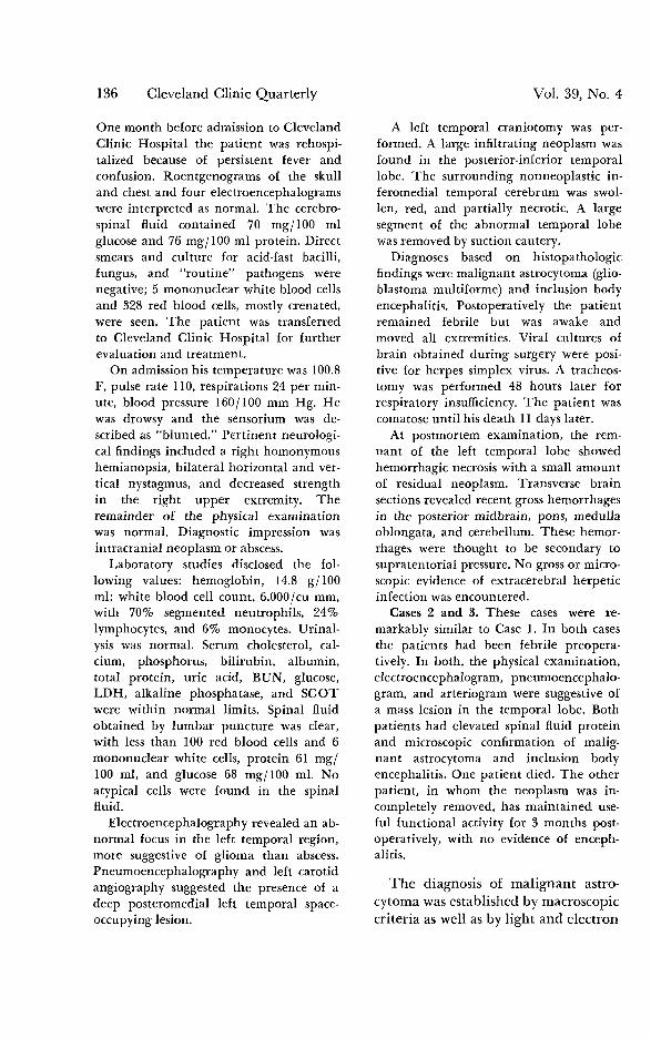
One month before admission to Cleveland Clinic Hospital the patient was rehospi-talized because of persistent fever and confusion. Roentgenograms of the skull and chest and four electroencephalograms were interpreted as normal. The cerebro-spinal fluid contained 70 mg/100 ml glucose and 76 mg/100 ml protein. Direct smears and culture for acid-fast bacilli, fungus, and "routine" pathogens were negative; 5 mononuclear white blood cells and 328 red blood cells, mostly crenated, were seen. The patient was transferred to Cleveland Clinic Hospital for further evaluation and treatment.
On admission his temperature was 100.8 F, pulse rate 110, respirations 24 per min-ute, blood pressure 160/100 mm Hg. He was drowsy and the sensorium was de-scribed as "blunted." Pertinent neurologi-cal findings included a right homonymous hemianopsia, bilateral horizontal and ver-tical nystagmus, and decreased strength in the right upper extremity. The remainder of the physical examination was normal. Diagnostic impression was intracranial neoplasm or abscess.
Laboratory studies disclosed the fol-lowing values: hemoglobin, 14.8 g/100 ml; white blood cell count, 6,000/cu mm, with 70% segmented neutrophils, 24% lymphocytes, and 6% monocytes. Urinal-ysis was normal. Serum cholesterol, cal-cium, phosphorus, bilirubin, albumin, total protein, uric acid, BUN, glucose, LDH, alkaline phosphatase, and SGOT were within normal limits. Spinal fluid obtained by lumbar puncture was clear, with less than 100 red blood cells and 6 mononuclear white cells, protein 61 mg/ 100 ml, and glucose 68 mg/100 ml. No atypical cells were found in the spinaf fluid.
Electroencephalography revealed an ab-normal focus in the left temporal region, more suggestive of glioma than abscess. Pneumoencephalography and left carotid angiography suggested the presence of a deep posteromedial left temporal space-occupying lesion.
A left temporal craniotomy was per-formed. A large infiltrating neoplasm was found in the posterior-inferior temporal lobe. The surrounding nonneoplastic in-feromedial temporal cerebrum was swol-len, red, and partially necrotic. A large segment of the abnormal temporal lobe was removed by suction cautery.
Diagnoses based on histopathologic findings were malignant astrocytoma (glio-blastoma multiforme) and inclusion body encephalitis. Postoperatively the patient remained febrile but was awake and moved all extremities. Viral cultures of brain obtained during surgery were posi-tive for herpes simplex virus. A tracheos-tomy was performed 48 hours later for respiratory insufficiency. The patient was comatose until his death 11 days later.
At postmortem examination, the rem-nant of the left temporal lobe showed hemorrhagic necrosis with a small amount of residual neoplasm. Transverse brain sections revealed recent gross hemorrhages in the posterior midbrain, pons, medulla oblongata, and cerebellum. These hemor-rhages were thought to be secondary to supratentorial pressure. No gross or micro-scopic evidence of extracerebral herpetic infection was encountered.
Cases 2 and 3. These cases were re-markably similar to Case 1. In both cases the patients had been febrile preopera-tively. In both, the physical examination, electroencephalogram, pneumoencephalo-gram, and arteriogram were suggestive of a mass lesion in the temporal lobe. Both patients had elevated spinal fluid protein and microscopic confirmation of malig-nant astrocytoma and inclusion body encephalitis. One patient died. The other patient, in whom the neoplasm was in-completely removed, has maintained use-ful functional activity for 3 months post-operatively, with no evidence of enceph-alitis.
The diagnosis of malignant astro-cytoma was established by macroscopic criteria as well as by light and electron

Winter 1972 Encephalitis and malignant astrocytoma 137
microscopy. Grossly, all three neo-plasms were soft, friable, and grey with areas of necrosis and hemorrhage. Light microscopy demonstrated pseu-dopalisading of tumor cells in the areas of necrosis, astrocytic cellular pleomorphism with giant cell forms, numerous mitotic figures, and prolifer-ation of small blood vessels with capil-lary endothelial proliferation (Fig. 1).
T h e diagnosis of herpes simplex encephalitis was established by: (1) characteristic light microscopic changes (three cases); (2) identification of classic herpesvirus particles by elec-tron microscopy (two cases); and (3) isolation of herpesvirus by tissue cul-ture techniques (two cases).
Light microscopy. As noted during craniotomy or at postmortem examina-tion, in all three cases the encephalitic
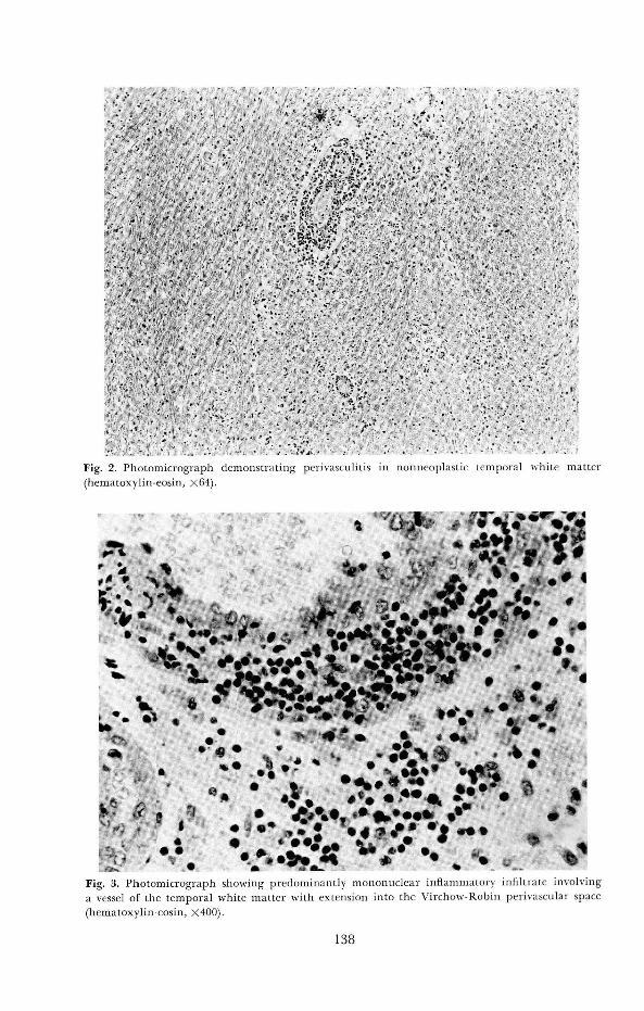
temporal cerebrum was grossly swol-len, partially necrotic and hemor-rhagic, with broadening and flatten-ing of the cerebral gyri. On microscopic examination of Zenker's-fixed, hematoxylin-eosin stained tis-sue, these areas of acute hemorrhagic necrosis demonstrated infiltration of the overlying meninges by lympho-cytes and occasional macrophages. T h e blood vessels of both the cortex and white matter were uniformly sur-rounded by dense aggregates of lym-phocytes, macrophages, rare polymor-phonuclear leukocytes, and plasma cells extending into the perivascular Virchow-Robin space (Figs. 2 and 3). Petechial hemorrhages, and foci of reactive astrocytic and oligodendro-glial gliosis were also noted.
T h e single most important finding
fi' Cr } . ' fe '-J&Ê&iL5-, - t i , - . . V . ... ' . V 1 ' -
•ïii«i. v&WSip.<• - *z • • ••• • ••• . /".'A' •
f .-¿A '' rJ < •
Fig. 1. Phc
ri -v >
ograph of mali: glioblastoma multiforme th promi pseudopalisading about an area of necrosis (hematoxylin-eosin, X64).
• v
Fig. 2. Photomicrograph demonstrating perivasculitis in nonneoplastic temporal white matter (hematoxylin-eosin, X64).
V* % %* t c>t? 1 . V ^ %* . jm -~.m , i f 4 ? r
V• V ^ ^
» m
• • t V* ^ * • a w . •
Fig. 3. Photomicrograph showing predominantly mononuclear inflammatory infiltrate involving a vessel of the temporal white matter with extension into the Virchow-Robin perivascular space (hematoxylin-eosin, X400).
138
Winter 1972 Encephalitis and malignant astrocytoma 139
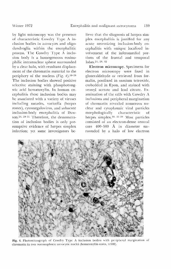
by light microscopy was the presence of characteristic Cowdry Type A in-clusion bodies in astrocytes and oligo-dendroglia within the encephalitic process. T h e Cowdry Type A inclu-sion body is a homogeneous eosino-philic intranuclear sphere surrounded by a clear halo, with resultant displace-ment of the chromatin material to the periphery of the nucleus (Fig. 4).19-28
T h e inclusion bodies showed positive selective staining with phosphotung-stic acid hematoxylin. In human en-cephalitis these inclusion bodies may be associated with a variety of viruses including measles, varicella (herpes zoster), cytomegalovirus, and subacute inclusion-body encephalitis of Daw-son.24' 29~31 Therefore, the demonstra-tion of inclusion bodies is only pre-sumptive evidence of herpes simplex infection; yet some investigators be-
lieve that the diagnosis of herpes sim-plex encephalitis is justified for any acute necrotizing inclusion-body en-cephalitis with unique localized in-volvement of the inferomedial por-tions of the frontal and temporal lobes.24. 28. 32
Electron microscopy. Specimens for electron microscopy were fixed in gluteraldehyde or retrieved from for-malin, postfixed in osmium tetroxide, embedded in Epon, and stained with uranyf acetate and lead citrate. Ex-amination of the cells with Cowdry A inclusions and peripheral margination of chromatin revealed numerous nu-clear and cytoplasmic viral particles morphologically characteristic of herpes simplex.3 1 '3 3"3 8 Most particles consisted of an electron-dense central core 400-500 A in diameter sur-rounded by a halo of low electron
Fig. 4. Photomicrograph of Cowdry Type A inclusion bodies with peripheral margination of chromatin in two nonneoplastic astrocytic nuclei (hematoxylin-eosin, X640).

140 Cleveland Clinic Quarterly Vol. 39, No. 3
Fig. 5. Electron micrograph of intranuclear herpesvirus particles with central core and surround-ing capsid envelopes (X29.800).
density and encapsulated by a thin outer hexagonal membrane known as the capsid (Fig. 5). A few capsids were devoid of their central core. The herpesvirus particles were identified in two cases, primarily in nonneoplastic glial cells, although in each case in-tensive search revealed a few malig-nant astrocytes with intranuclear and intracytoplasmic herpes simplex viral particles.
Viral studies. Viral isolations by tissue culture technique were positive for herpes simplex virus in two cases; attempts at isolating herpes simplex from brain specimens in Case 3 were unrewarding, despite the presence of Cowdry A inclusion bodies and elec-tron microscopic demonstration of viral particles, ft has been suggested that inability to isolate the herpesvirus,
or any other virus, in similar cases of acute necrotizing inclusion body en-cephalitis is an unfortunately common occurrence.2 9 , 3 2 '3 8 Some authors relate this to clironicity of the disease, noting that herpesvirus previously isolated in a brain can no longer be isolated after a certain interval, even though the acute encephalitic process is still clini-cally and histologically active.29
A fourth patient who had a brief history of febrile illness with rapidly progressive neurological deficit was seen on the neurological service. Re-sults of physical examination, electro-encephalography, and arteriography suggested a deep lesion of the right temporal lobe, inferomedial aspect. Af-ter craniotomy, we were able to confirm the diagnosis of malignant astrocytoma and inclusion-body encephalitis in the
Winter 1972 Encephalitis and malignant astrocytoma 141
adjacent temporal cerebrum. Tissue culture techniques and electron micros-copy failed to reveal the presence of virus, thus preventing an objective conclusion as to the exact nature of the inclusion-body encephalitis.
Discussion
T h e large number of animal can-cers with a documented viral etiology suggests that some human cancers may be virus-induced or virus related. Herpesvirus is an especially well-docu-mented agent in neoplastic transfor-mation in lower animals, including Marek's lymphoma of chickens,39 lym-phoma of rabbits and monkeys,40 leu-kemia of guinea pigs,41' 42 and Lucke renal adenocarcinoma of frogs. T h e search for human cancer virus was further stimulated when Burkitt de-scribed a malignant lymphoma of Afri-can children.43 T h e Burkitt lymphoma, as initially described, occurred only in certain geographic areas of Africa, with a distribution overlapping that of certain insect vectors. Burkitt sug-gested that the lymphoma was induced by an infectious agent transmitted by an insect vector. Since Burkitt's initial report, numerous investigators have demonstrated that tissue cultures of Burkitt lymphoma cells contain char-acteristic DNA herpes-like virus.7-17
The fact that latency in the central nervous system is a well recognized in vivo biologic characteristic of herpes simplex virus may bear a relationship to brain neoplasia. Herpes simplex virus differs from other infectious agents in its capacity to remain latent in the central nervous system of man and to be subsequently reactivated.16' 4 4 - 4 6 Latent or so-called "dormant forms" of herpes simplex virus appear to occur within brain cells that are not
actively growing and dividing.45-47
Thus an equilibrium exists between host brain cells and virus. T h e mech-anism for latency is not known, yet herpesvirus latency appears to occur in cells not actively growing. This is an appealing premise on which to base the hypothesis that some un-identified factor related to mitotic ac-tivity is associated with virus matura-tion. All four malignant astrocytomas in this series were highly cellular, with prominent mitotic activity providing what is theoretically an optimum en-vironment for herpes virus matura-tion. We are unable to conclude ob-jectively whether the herpesvirus is truly an oncogenic agent or a poten-tially lethal secondary invader in the company of malignant astrocytoma.
Summary
The well-known biologic character-istic of herpesvirus latency in the hu-man central nervous system may bear an important relationship to astrocytic neoplasia. Coexistent herpes simplex encephalitis and malignant astrocy-toma is a previously unreported entity seen in three patients during a 20-month period. T h e diagnosis of herpes simplex encephalitis was established by two or more parameters: (1) light microscopic demonstration of charac-teristic Type A inclusion bodies and other nonspecific changes such as mon-onuclear perivasculitis, petechial hem-orrhages and necrosis; (2) herpes sim-plex isolation in tissue culture; or (3) electron microscopic demonstration of characteristic herpesvirus particles. Past and present evidence cannot ob-jectively distinguish between herpes-virus as a causal agent of malignant astrocytoma or as a potentially lethal

142 Cleveland Clinic Quarterly Vol. 39, No. 3
"passenger" virus of important clinical significance.
Acknowledgments
We thank Dr. M. Rosenthal, St. Luke's Hospital, Cleveland, Ohio, and Mr. Dallas Gary Osborne, Department of Electron Microscopy, Cleveland Clinic, for assistance in the preparation of this manuscript.
References 1. Rawls WE, Tompkins WA, Figueroa ME,
et al: Herpesvirus Type 2; association with carcinoma of the cervix. Science 161: 1255-1256, 1968.
2. Nahmias AJ, Naib ZM, Josey W E : Asso-ciation of a genital herpesvirus with cervi-cal cancer. Int Virol 1: 187-188, 1969.
3. Naib ZM, Nahmias AJ, Josey WE, et al: Genital herpetic infection: association with cervical dysplasia and carcinoma. Cancer 23: 940-945, 1969.
4. Nahmias AJ, Dowdle W R : Studies on herpesvirus hominis type 2. Int Virol 1: 254, 1969.
5. Josey WE, Nahmias AJ, Naib ZM: Genital infection with type 2 herpesvirus hominis; present knowledge and possible relation to cervical cancer. Am J Obstet Gynecol 101: 718-729, 1968.
6. Royston I, Aurelian L: The association of genital herpesvirus with cervical atypia and carcinoma in situ. Am J Epidemiol 91: 531-538, 1970.
7. Epstein MA: Aspects of the EB virus. Adv Cancer Res 13: 383-411, 1970.
8. Henle W, Henle G, Ho HC, et al: Anti-bodies to Epstein-Barr virus in naso-pharyngeal carcinoma, other head and neck neoplasms, and control groups. J Natl Cancer Inst 44: 225-231, 1970.
9. Levine PH, Ablashi DV, Berard CW, et al: Elevated antibody titers to Epstein-Barr virus in Hodgkin's disease. Cancer 27 :416-421 ,1971 .
10. Goldman JM, Aisenberg AC: Incidence of antibody to EB virus, herpes simplex, and cytomegalovirus in Hodgkin's disease. Cancer 26: 327-331, 1970.
11. Henle G, Henle W, Clifford P, et al: Antibodies to Epstein-Barr virus in Bur-
kitt's lymphoma and control groups. J Natl Cancer Inst 43: 1147-1157, 1969.
12. Diehl V, Henle G, Henle W, et al: Dem-onstration of a herpes group virus in cul-tures of peripheral leukocytes from pa-tients with infectious mononucleosis. J Virol 2: 663-669, 1968.
13. Dalton A, Manaker RA: The comparison of virus particles associated with Burkitt lymphoma with other herpes-like viruses, p. 72 in Carcinogenesis: A Broad Critique; a collection of papers. Annual Symposium on Fundamental Cancer Research, M.D. Anderson Hospital and Tumor Institute, Houston, Tex. Baltimore: Williams and Wilkins, 1967.
14. Henle G, Henle W, Diehl V: Relation of Burkitt's tumor-associated herpes-type virus to infectious mononucleosis. Proc Natl Acad Sci USA 59: 94-101, 1968.
15. Dulbecco R: Viruses in carcinogenesis. Ann Intern Med 70: 1019-1030, 1969.
16. Allen DW, Cole P: Viruses and human cancer. N Engl J Med 286: 70-82, 1972.
17. MacMahon B: Epidemiologic aspects of acute leukemia and Burkitt's tumor. Can-cer 21: 558-561, 1968.
18. Miller R W : Radiation, chromosomes and viruses in the etiology of leukemia; evi-dence from epidemiologic research. N Engl J Med 271: 30-36, 1964.
19. Smith MG, Lennette EH, Reames HR: Isolation of the virus of herpes simplex and the demonstration of intranuclear inclusions in a case of acute encephalitis. Am J Pathol 17: 55-68, 1941.
20. Zarafonetis CJ, Smadel JE , Adams J W , et al: Fatal herpes simplex encephalitis in man. Am J Pathol 20: 429-445, 1944.
21. Pearson HE, Butt EM: Hemorrhagic herpetic encephalitis. Am J Clin Pathol 26: 1174-1179, 1956.
22. MacCallum FO, Potter JM, Edwards DH: Early diagnosis of herpes-simplex en-cephalitis by brain biopsy. Lancet 2: 332-334, 1964.
23. Pierce NF, Leeds NR, Portnoy B, et al: Encephalitis associated with herpes sim-plex infection presenting as a temporal lobe mass; report of 2 cases with survival. Neurology 14: 708-713, 1964.
24. Drachman DA, Adams RD: Herpes sim-plex and acute inclusion body encepha-litis. Arch Neurol 7: 45-63, 1962.
Winter 1972 Encephalitis and malignant astrocytoma 143
25. Cowdry EV: The problem of intranuclear inclusions in virus diseases. Arch Pathol 18: 527-542, 1934.
26. Harland WA, Adams JH, McSeveney D: Herpes-simplex particles in acute necrotis-ing encephalitis. Lancet 2: 581-582, 1967.
27. McK.ee AP, Hudson JD, Kimura J , et al: Herpes simplex encephalitis. South Med J 61: 217-225,1968.
28. Haynes RE, Azimi PH, Cramblett HG: Fatal herpesvirus hominis (herpes simplex virus) infections in children. Clinical, pathologic, and virologic characteristics. JAMA 206: 312-319, 1968.
29. Johnson R T , Olson LC, Buescher EL: Herpes simplex virus infections of the central nervous system. Problems in labo-ratory diagnosis. Arch Neurol 18: 260-264, 1968.
30. Tellez-Nagle I, Harter DH: Subacute sclerosing leukoencephalitis; ultrastruc-ture of intranuclear and intracytoplasmic inclusions. Science 154: 899-901, 1966.
31. Hashida Y, Yunis EJ : Re-examination of encephalitic brains known to contain in-tranuclear inclusion bodies; electron-microscopic observation, following pro-longed fixation in formalin. Am J Clin Pathol 53: 537-543, 1970.
32. Rawls WE, Dyck PJ, Klass DW, et al: Encephalitis associated with herpes sim-plex virus. Ann Intern Med 64: 104-115, 1966.
33. Baringer J R , Griffith J F : Experimental herpes simplex encephalitis; early neuro-pathologic changes. J Neuropathol Exp Neurol 29: 89-104, 1970.
34. Friedrich ER, Cole W, Middelkamp JN: Herpes simplex. Clinical aspects and elec-tron microscopic findings. Am J Obstet Gynecol 104: 758-779, 1969.
35. Morgan C, Rose HM, Mednis B: Electron microscopy of herpes simplex virus I entry. J Virol 2: 507-516, 1968.
36. Swanson JL , Craighead JE, Reynolds ES: Electron microscopic observations of
Herpesvirus hominis (herpes simplex virus) encephalitis in man. Lab Invest 15: 1966-1981, 1966.
37. Ames RP, Reagan RL, O'Connor CC: Herpes-type infection in adult with hema-tologic disorder. Electron microscopic observations of virus and case report. Can-cer 20: 2159-2169, 1967.
38. Olson LC, Buescher EL, Artenstein MS, et al: Herpesvirus infections of the human central nervous system. N Engl J Med 277: 1271-1277, 1967.
39. Churchill AE: Herpes-type virus isolated in cell culture from tumors of chickens with Marek's disease. I. Studies in cell culture. J Natl Cancer Inst 41: 939-950, 1968.
40. Melendez LV, Hunt RD, Daniel MD, et al: Herpesvirus saimiri; II. Experimentally induced malignant lymphoma in primates. Lab Anim Care 19: 378-386, 1969.
41. Hsiung GD, Kaplow LS: Herpes-like virus isolated from spontaneously degenerated tissue culture derived from leukemia-susceptible guinea pigs. J Virol 3: 355-357, 1969.
42. Opler SR: Animal model of viral onco-genesis. Nature 215: 184, 1967.
43. Burkitt D: A children's cancer with geographical limitations. Cancer Progr pp 102-113, 1963.
44. Lennette EH, Magoffin RL, Knouf EG: Viral central nervous system disease; etio-logic study conducted at Los Angeles County Hospital. JAMA 179: 687-695, 1962.
45. Burnet FM, Williams SW: Herpes sim-plex; new point of view. Med J Aust 1: 637-642, 1939.
46. Leider W, Magoffin RL, Lennette EH, et al: Herpes-simplex-virus encephalitis; its possible association with reactivated latent infection. N Engl J Med 273: 341-347, 1965.
47. Goodheart CR: Herpesvirus and cancer. JAMA 211: 91-96, 1970.