CME Reconstruction of Acquired Scalp Defects: An ...lipteh.com/Study-Notes/Articles/scalp.pdf ·...
19
CME Reconstruction of Acquired Scalp Defects: An Algorithmic Approach Jason E. Leedy, M.D., Jeffrey E. Janis, M.D., and Rod J. Rohrich, M.D. Dallas, Texas Learning Objectives: After studying this article, the participant should: 1. Understand scalp anatomy, hair physiology, and skin viscoelastic properties as they relate to scalp reconstruction. 2. Understand the principles that allow for aesthetic reconstruction of scalp defects. 3. Understand the use of local tissue rearrangement for reconstruction of specific areas of the scalp. 4. Understand the use of tissue expansion and free tissue transfer for scalp reconstruction. Background: Reconstruction of scalp de- fects is required for acute trauma, tumor extirpation, radiation necrosis, and the re- pair of traumatic alopecia or cosmetically displeasing scars. Methods: The proper choice of a recon- structive technique is affected by several factors—the size and location of the defect, the presence or absence of periosteum, the quality of surrounding scalp tissue, the presence or absence of hair, location of the hairline, and patient comorbidities. Suc- cessful reconstruction of these defects re- quires a detailed knowledge of scalp anat- omy, hair physiology, skin biomechanics, and the variety of possible local tissue re- arrangements. In nearly total defects, local tissues may be inadequate and tissue ex- pansion or free tissue transfer may be the only alternatives. Results: Plastic surgeons are now able to obtain coverage over the calvaria after the most devastating of defects; however, the challenge to the reconstructive surgeon to- day is to do so with excellent cosmetic re- sults. Cosmetic scalp reconstruction re- quires restoration and preservation of normal hair patterns and hair lines. Conclusions: Successful reconstruction of the scalp requires careful preoperative planning and precise intraoperative execu- tion. Detailed knowledge of scalp anatomy, skin biomechanics, hair physiology, and the variety of available local tissue rear- rangements allows for excellent aesthetic reconstruction. (Plast. Reconstr. Surg. 116: 54e, 2005.) The reconstruction of scalp defects is neces- sary for acute trauma, tumor extirpation, radi- ation necrosis, and improvement of cosmeti- cally displeasing scars or alopecia. The history of scalp reconstruction parallels developments in plastic surgery techniques. An interesting and detailed history of scalping injuries and their management was described by Koss et al. 1 Augustin Belloste, a French surgeon, in 1696 advocated early perforation of bare cranium to allow granulation tissue and subsequent epithelialization. 2 This was practiced until Ne- tolitzky, in 1871, used skin grafting of the cal- varia after the presence of granulation tissue. 3 In 1908, Robinson demonstrated the success of skin grafting on intact periosteum before the presence of granulation tissue. 4 Multiple au- thors shortly thereafter demonstrated success- ful reconstruction with local flaps. 5–9 Kazanjian demonstrated that galeal scoring allowed for further advancement of local flaps. 6 Orticochea published his four-flap technique From the Department of Plastic Surgery, University of Texas Southwestern Medical Center. Received for publication August 11, 2004. DOI: 10.1097/01.prs.0000179188.25019.6c 54e
Transcript of CME Reconstruction of Acquired Scalp Defects: An ...lipteh.com/Study-Notes/Articles/scalp.pdf ·...
CME
Reconstruction of Acquired Scalp Defects:An Algorithmic ApproachJason E. Leedy, M.D., Jeffrey E. Janis, M.D., and Rod J. Rohrich, M.D.Dallas, Texas
Learning Objectives: After studying this article, the participant should: 1. Understand scalp anatomy, hair physiology, andskin viscoelastic properties as they relate to scalp reconstruction. 2. Understand the principles that allow for aestheticreconstruction of scalp defects. 3. Understand the use of local tissue rearrangement for reconstruction of specific areasof the scalp. 4. Understand the use of tissue expansion and free tissue transfer for scalp reconstruction.
Background: Reconstruction of scalp de-fects is required for acute trauma, tumorextirpation, radiation necrosis, and the re-pair of traumatic alopecia or cosmeticallydispleasing scars.Methods: The proper choice of a recon-structive technique is affected by severalfactors—the size and location of the defect,the presence or absence of periosteum, thequality of surrounding scalp tissue, thepresence or absence of hair, location of thehairline, and patient comorbidities. Suc-cessful reconstruction of these defects re-quires a detailed knowledge of scalp anat-omy, hair physiology, skin biomechanics,and the variety of possible local tissue re-arrangements. In nearly total defects, localtissues may be inadequate and tissue ex-pansion or free tissue transfer may be theonly alternatives.Results: Plastic surgeons are now able toobtain coverage over the calvaria after themost devastating of defects; however, thechallenge to the reconstructive surgeon to-day is to do so with excellent cosmetic re-sults. Cosmetic scalp reconstruction re-quires restoration and preservation ofnormal hair patterns and hair lines.Conclusions: Successful reconstructionof the scalp requires careful preoperative
planning and precise intraoperative execu-tion. Detailed knowledge of scalp anatomy,skin biomechanics, hair physiology, andthe variety of available local tissue rear-rangements allows for excellent aestheticreconstruction. (Plast. Reconstr. Surg. 116:54e, 2005.)
The reconstruction of scalp defects is neces-sary for acute trauma, tumor extirpation, radi-ation necrosis, and improvement of cosmeti-cally displeasing scars or alopecia. The historyof scalp reconstruction parallels developmentsin plastic surgery techniques. An interestingand detailed history of scalping injuries andtheir management was described by Koss et al.1Augustin Belloste, a French surgeon, in 1696advocated early perforation of bare cranium toallow granulation tissue and subsequentepithelialization.2 This was practiced until Ne-tolitzky, in 1871, used skin grafting of the cal-varia after the presence of granulation tissue.3In 1908, Robinson demonstrated the success ofskin grafting on intact periosteum before thepresence of granulation tissue.4 Multiple au-thors shortly thereafter demonstrated success-ful reconstruction with local flaps.5–9 Kazanjiandemonstrated that galeal scoring allowed forfurther advancement of local flaps.6
Orticochea published his four-flap technique
From the Department of Plastic Surgery, University of Texas Southwestern Medical Center. Received for publication August 11, 2004.
DOI: 10.1097/01.prs.0000179188.25019.6c
54e
for large scalp defects in 1967 and revised histechnique to the use of three flaps in 1971.10,11
Neumann reported the first clinical use oftissue expansion in 1957 when he expandedthe scalp for ear reconstruction.12 Radovan13
popularized tissue expansion in 1976 and dem-onstrated the usefulness of the technique. In1984, Manders et al. reported reconstructionof nearly half the scalp with hair-bearing tissueusing tissue expansion.14
Advances in microsurgery have also played asignificant role in scalp reconstruction. In 1976Miller et al. successfully replanted a totallyavulsed scalp with return of normal hairgrowth and frontalis function.15 Multiple au-thors have since presented series of patientswho have had near total scalp defects recon-structed with free tissue transfer.16–22
APPLIED ANATOMY
Scalp anatomy has been well characterizedin a number of anatomy textbooks and recentreview articles.23–28 We have highlighted thesalient aspects of scalp anatomy for the recon-structive surgeon.
LAYERS
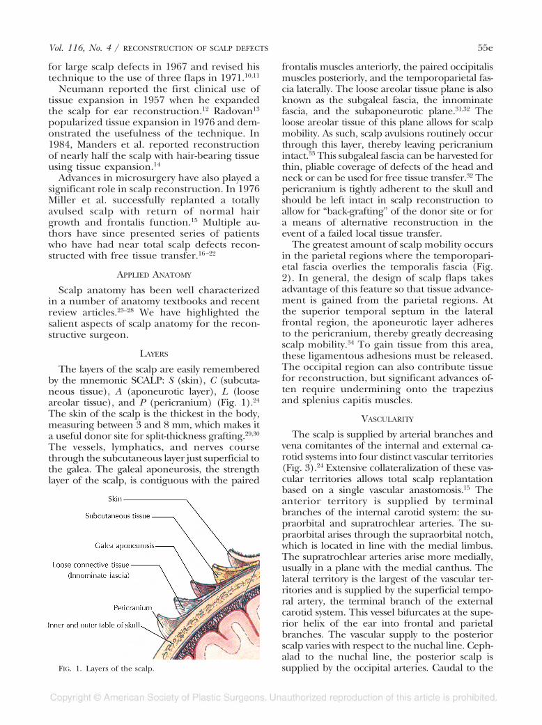
The layers of the scalp are easily rememberedby the mnemonic SCALP: S (skin), C (subcuta-neous tissue), A (aponeurotic layer), L (looseareolar tissue), and P (pericranium) (Fig. 1).24
The skin of the scalp is the thickest in the body,measuring between 3 and 8 mm, which makes ita useful donor site for split-thickness grafting.29,30
The vessels, lymphatics, and nerves coursethrough the subcutaneous layer just superficial tothe galea. The galeal aponeurosis, the strengthlayer of the scalp, is contiguous with the paired
frontalis muscles anteriorly, the paired occipitalismuscles posteriorly, and the temporoparietal fas-cia laterally. The loose areolar tissue plane is alsoknown as the subgaleal fascia, the innominatefascia, and the subaponeurotic plane.31,32 Theloose areolar tissue of this plane allows for scalpmobility. As such, scalp avulsions routinely occurthrough this layer, thereby leaving pericraniumintact.33 This subgaleal fascia can be harvested forthin, pliable coverage of defects of the head andneck or can be used for free tissue transfer.32 Thepericranium is tightly adherent to the skull andshould be left intact in scalp reconstruction toallow for “back-grafting” of the donor site or fora means of alternative reconstruction in theevent of a failed local tissue transfer.
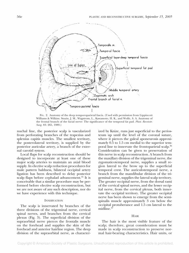
The greatest amount of scalp mobility occursin the parietal regions where the temporopari-etal fascia overlies the temporalis fascia (Fig.2). In general, the design of scalp flaps takesadvantage of this feature so that tissue advance-ment is gained from the parietal regions. Atthe superior temporal septum in the lateralfrontal region, the aponeurotic layer adheresto the pericranium, thereby greatly decreasingscalp mobility.34 To gain tissue from this area,these ligamentous adhesions must be released.The occipital region can also contribute tissuefor reconstruction, but significant advances of-ten require undermining onto the trapeziusand splenius capitis muscles.
VASCULARITY
The scalp is supplied by arterial branches andvena comitantes of the internal and external ca-rotid systems into four distinct vascular territories(Fig. 3).24 Extensive collateralization of these vas-cular territories allows total scalp replantationbased on a single vascular anastomosis.15 Theanterior territory is supplied by terminalbranches of the internal carotid system: the su-praorbital and supratrochlear arteries. The su-praorbital arises through the supraorbital notch,which is located in line with the medial limbus.The supratrochlear arteries arise more medially,usually in a plane with the medial canthus. Thelateral territory is the largest of the vascular ter-ritories and is supplied by the superficial tempo-ral artery, the terminal branch of the externalcarotid system. This vessel bifurcates at the supe-rior helix of the ear into frontal and parietalbranches. The vascular supply to the posteriorscalp varies with respect to the nuchal line. Ceph-alad to the nuchal line, the posterior scalp issupplied by the occipital arteries. Caudal to theFIG. 1. Layers of the scalp.
Vol. 116, No. 4 / RECONSTRUCTION OF SCALP DEFECTS 55e
nuchal line, the posterior scalp is vascularizedfrom perforating branches of the trapezius andsplenius capitis muscles. The smallest territory,the posterolateral territory, is supplied by theposterior auricular artery, a branch of the exter-nal carotid system.
Local flaps for scalp reconstruction should bedesigned to incorporate at least one of thesemajor scalp arteries to maintain an axial bloodsupply. In elective scalp reduction procedures formale pattern baldness, bilateral occipital arteryligation has been described to delay posteriorscalp flaps before cephalad advancement.35 It isconceivable that a similar procedure may be per-formed before elective scalp reconstruction, butwe are not aware of any such description, nor dowe have experience with this technique.
INNERVATION
The scalp is innervated by branches of thethree divisions of the trigeminal nerve, cervicalspinal nerves, and branches from the cervicalplexus (Fig. 3). The superficial division of thesupraorbital nerve pierces the frontalis muscleon the forehead and supplies the skin of theforehead and anterior hairline region. The deepdivision of the supraorbital nerve, as character-
ized by Knize, runs just superficial to the perios-teum up until the level of the coronal suture,where it pierces the galeal aponeurosis approxi-mately 0.5 to 1.5 cm medial to the superior tem-poral line to innervate the frontoparietal scalp.36
Consideration can be given to preservation ofthis nerve in scalp reconstruction. A branch fromthe maxillary division of the trigeminal nerve, thezygomaticotemporal nerve, supplies a small re-gion lateral to the brow up to the superficialtemporal crest. The auriculotemporal nerve, abranch from the mandibular division of the tri-geminal nerve, supplies the lateral scalp territory.The greater occipital nerve, from the dorsal ramiof the cervical spinal nerves, and the lesser occip-ital nerve, from the cervical plexus, both inner-vate the occipital territory. The greater occipitalnerve has been shown to emerge from the semi-spinalis muscle approximately 3 cm below theoccipital protuberance and 1.5 cm lateral to themidline.37
HAIR
The hair is the most visible feature of thescalp; therefore, great consideration must bemade in scalp reconstruction to preserve nor-mal hair-bearing characteristics. Hair units, or
FIG. 2. Anatomy of the deep temporoparietal fascia. (Used with permission from LippincottWilliams & Wilkins. Stuzin, J. M., Wagstrom, L., Kawamoto, H. K., and Wolfe, S. A. Anatomy ofthe frontal branch of the facial nerve: The significance of the temporal fat pad. Plast. Reconstr.Surg. 83: 265, 1989.)
56e PLASTIC AND RECONSTRUCTIVE SURGERY, September 15, 2005

pilosebaceous appendages, are located in thesubcutaneous layer. They are each composedof the follicle, which includes the germinativebulb, the shaft surrounded by an epithelialsheath continuous with the epidermis, the se-baceous gland, and the arrector pili smoothmuscle. The average scalp has more than100,000 hairs.38
Hair physiology is well described in manyreview articles.38,39 Hair follicles continuouslycycle through three stages: anagen (growingphase), catagen (involutional phase), and telo-gen (dormant phase). At any one time, approx-imately 90 to 95 percent of hairs are in theanagen phase, 5 to 10 percent are in the telo-gen phase, and 1 to 2 percent are in the cata-gen phase.40 The anagen phase of scalp hairlasts approximately 1000 days, during whichthe average hair grows 0.3 to 0.4 mm/day (orapproximately 6 inches per year). In contrast,the telogen phase lasts an average of 2 to 3months, whereas the catagen phase lasts only 2to 3 weeks. Up to 100 telogen hairs are losteach day from the head, and approximatelythe same numbers of follicles enter anagen.Therefore, the amount of hair depends on theratio of follicles in anagen versus telogen. In-trinsic and extrinsic factors can influence thisratio.
Extrinsic factors that promote follicular ana-gen arrest include, but are not limited to, phys-iologic or psychological stress, drugs, and childbirth. Intrinsic factors include excessive tension
on wound closure leading to localized ischemiaand follicle involution.
SKIN BIOMECHANICS
Successful scalp reconstruction often re-quires intraoperative use of the intrinsic skinviscoelastic properties, specifically, stress re-laxation and creep. Stress relaxation is definedas the decrease in the amount of force nec-essary to maintain a fixed amount of skinstretch over time.41 Creep is the gain in skinsurface area that results when a constant loadis applied.41
The fundamental basis for these propertieslies in the fact that as force is applied to aleading skin edge, tissue thickness decreasesfrom extrusion of fluid and mucopolysacchar-ides, realignment of dermal collagen bundles,elastic fiber microfragmentation, and mechan-ical stretching of the skin.42 The extent towhich these occur depends on the inherentproperties of the tissue and the amount offorce applied. When used intraoperatively, theviscoelastic properties of the skin can greatlyassist in reconstruction and allow for tension-free closure in circumstances where initial clo-sure is difficult.
PRINCIPLES OF SCALP RECONSTRUCTION
The following basic tenets should be ad-hered to when selecting the appropriatemethod of scalp reconstruction.
FIG. 3. Scalp vascularity and innervation.
Vol. 116, No. 4 / RECONSTRUCTION OF SCALP DEFECTS 57e

Replace Like Tissue with Like Tissue
The best replacement for scalp tissue is scalptissue. There is no other donor site in the bodythat will approximate the same hair-bearingqualities of scalp tissue. The goals of recon-structive surgeons in the past were to obtaincalvarial coverage to prevent calvarial desicca-tion, sequestration, and sepsis. However, today,the reconstructive surgeon should strive for acosmetically appealing result in addition tomerely achieving coverage. This requires atten-tion to hair growth patterns and hairlines sothat reconstruction restores and preserves thenormal anatomy.
Hair transplantation is an alternative tech-nique to excisional surgery that can be used torestore the hair-bearing appearance of smallareas of alopecia whether it be from trauma orfrom skin grafting.43–45 Hair transplantation isperhaps most effectively used as a revisionalsecondary procedure. For example, it can beused to camouflage incisions within hair-bearing scalp or to reestablish a hairline thatmay have been slightly distorted from usingrotational flaps.45 Hair transplantation hasbeen successfully demonstrated in large areasof burn alopecia; however, most reconstructivesurgeons use scalp flaps, tissue expansion, orfree tissue transfer.46
In patients who have developed radiation-induced necrosis of the scalp after receiving ra-diation therapy for cancer, the options for replac-ing “like with like” is limited. Neighboring scalptissue is often fibrotic as a result of radiationtherapy, thereby severely limiting the use of localflaps.47,48 If local flaps are to be used, care shouldbe taken to preserve the vascularity and minimizewound closure tension. Postoperative radiationtherapy is often used for further treatment ofscalp malignancies, and local tissue flaps may notbe as reliable for durable reconstruction.49–51
Therefore, in the scalp wound either previouslysubjected to radiation therapy or in the onco-logic reconstruction that will be irradiated post-operatively, greater consideration should begiven to free tissue transfer instead of localflaps.49–51
Consider Tissue Expansion
Tissue expansion should be considered iflocal tissue rearrangements are inadequate forreconstruction because of the size of the de-fect, traumatized local tissue, unacceptable re-arrangement of hair patterns, or distortion of
the hairline. In these instances, a scalp rotationflap can be used to transfer the defect to a lesscosmetically sensitive area and then “back-graft” the donor site. Simultaneous placementof a tissue expander has been described in theliterature, but this has not been the practice ofthe authors.52
Approximately 50 percent of scalp can bereconstructed with expanded scalp tissue.14 Itdoes require staged operations with a lengthyinterval period and is potentially associatedwith expander complications, which vary from6 to 25 percent.53,54
Stable scalp coverage must be obtained dur-ing the expansion process to prevent calvarialdesiccation and subsequent osteomyelitis. Ifthe periosteum is intact, it can be primarilyskin grafted.55 However, if there is exposedbone denuded of periosteum, the reconstruc-tion becomes more complicated.
Options for the reconstructive surgeon inthese instances include using pericranial flaps,using the loose areolar tissue subgaleal tissue,or burring the outer calvarial table. Pericranialflaps should be designed as large as possible,preferably with a bipedicled base, with incor-poration of at least one major scalp artery.55
They can be rotated to provide for calvarialcoverage and skin grafted immediately. Theloose areolar tissue plane, coined the “sub-galeal fascia” by Tolhurst, has also been de-scribed for coverage of exposed calvaria.32 Hedescribed designing the flap off a major arteryor maintaining a bipedicled base. As a lastresort, the outer table of the calvaria can beburred, thereby exposing the highly vasculardiploic space, which can accept skin graftingimmediately.4 In addition, a cerclage suture,with heavy long-lasting suture material, may beplaced to decrease the size of the defect beforeskin grafting.
When using tissue expansion, the largest ex-pander possible should be placed in a sub-galeal position. The shape of tissue expanderbases affects the amount of tissue gain. VanRappard et al. determined that expanders witha round base, a crescentic base, and a rectan-gular base give tissue gains of 25, 32, and 38percent, respectively.56 The appropriately sizedand shaped expander5 should be selected onthe basis of the individual patient’s defect. Asingle large expander is preferred over multi-ple smaller expanders, as this will give thegreatest gain in tissue per volume of expansionand minimize the infectious risk by limiting the
58e PLASTIC AND RECONSTRUCTIVE SURGERY, September 15, 2005
number of operative sites. However, in scalpreconstruction, multiple expanders are fre-quently used in a single setting to gain thegreatest amount of expansion per operativeprocedure. The expander should be posi-tioned so that the expanded scalp, once ad-vanced, will re-create normal hair patterns. Inthe repair of massive defects, expanded scalpcan be reexpanded with acceptable cosmeticresults.57 Alternatively, an expander after max-imal expansion can be exchanged for a largerexpander before definitive adjacent tissuetransfer to shorten the time required toachieve the aesthetic goal.58 Expansion shouldbe continued until the expanded flap is ap-proximately 20 percent larger than the size ofthe defect to account for tissue recoil duringadvancement.31 The expanded flap size can becalculated by subtracting the base of the ex-pander from the length over the top of theexpander.
In general, tissue expansion is a powerfultechnique for reconstruction of defects notamenable to local tissue transfer but requires alengthy reconstruction. This must be weighedagainst a one-stage, cosmetically inferior recon-struction.
Critical Operative Details
As in all areas of plastic surgery, attention todetail optimizes results. Hemostasis is critical. Lo-cal anesthetic with dilute epinephrine will de-crease intraoperative skin edge bleeding and canbe used to hydrodissect the subgaleal plane. Ifpossible, the use of hemostatic clips on the cutedges of the scalp should be minimized to pre-vent potential follicular damage and subsequentiatrogenic alopecia.59 Electrocautery should beused judiciously at the cut edge of the scalp toprevent thermal injury to hair follicles. Full-thickness hemostatic sutures that are placed cir-cumferentially 1 cm from the proposed incisionhave been described to minimize intraoperativeblood loss.60
Camirand and Doucet in 1995 reported theircomparison between parallel and perpendicularhairline incisions.61 Incisions made perpendicu-lar to the direction of the hair follicle allowedhair to grow through the hairline incision andthus give a softer, more natural appearance.61
Incisions within hair-bearing scalp may be differ-ent. Skin incision made parallel to the directionof the hair follicles may improve cosmesis, asfewer hair follicles are disrupted.62 This may al-low for a less discernible scar in areas of hair-
bearing scalp. In addition, immediate micrograft-ing of hair follicles in a fresh incision has beenproposed to camouflage the incision in hair-bearing tissue.62,63
Excessive tension at wound closure can causealopecia from either hair follicle loss or anagenphase arrest. Therefore, as in all areas of plasticsurgery, the optimal result is obtained with atension-free closure. Again, to minimizewound closure tension, the skin viscoelasticproperties of stress relaxation and creepshould be used. This can be done with eitherhooks and manual force or by rapid intraoper-ative tissue expansion using expanders.64
The galeal aponeurosis is responsible for themajority of resistance to scalp flap advance-ment. By carefully scoring the galea perpendic-ular to the direction of desired tissue gain,additional advancement can be achieved.65
According to Raposio et al., each galeotomycorresponds to a 40 percent reduction in scalpclosing tension and approximately 1.67 mm oftissue gain.65 Ideally, the galea is incised at 1-cmintervals and tested after each score to seewhether adequate tissue length has beengained. Care should be taken to prevent inad-vertent injury to the scalp arteries that lie justsuperficial to the galea. For this reason, weprefer to avoid electrocautery when perform-ing galeal scoring to prevent potential thermalinjury to these vessels.
Proper wound closure requires approxima-tion of the galea, as this is the strength layer ofthe scalp. In scalp reduction surgery for malepattern alopecia, galeal-to-periosteum suturesperipheral to the incision can provide a “pro-gressive-tension suture” effect to further de-crease wound tension and minimize long-termscar widening.66 This technique can be used inscalp reconstruction as well. Disregarding top-ical adhesives, staples are the least ischemic ofskin closure techniques.67,68 They are useful inclosure of hair-bearing scalp because they limitthe amount of follicular ischemia and subse-quent incisional alopecia.
Rotational flaps for reconstruction often re-sult in dog ears. These can be significant andcan create some displeasing scalp contours onthe operative table. When this occurs, resist thetemptation for excision. Clinical experience hasshown that dog-ears uniformly flatten withtime.69–71 In addition, excision of the dog-earonly increases the length-to-width ratio andplaces the distal aspects of the flap at risk.Tension is the greatest culprit for distal flap
Vol. 116, No. 4 / RECONSTRUCTION OF SCALP DEFECTS 59e

ischemia, and excision of dog-ears will not de-crease wound closure tension. If the contourdeformity of the dog-ear is unacceptable to thepatient after a trial period of observation, de-layed excision can usually be performed in anoffice setting without difficulty.
Tailor the Reconstruction to the Patient
As always in plastic surgery, consider patientcomorbidities and preferences in performingreconstructions—the treatment must always betailored to the individual patient. For instance,the schizophrenic patient with a long-standingneglected scalp squamous cell carcinoma andsubsequent large full-thickness wide local exci-sion defect may be better served by a one-stagereconstruction despite distortion of hair pat-terns as opposed to a prolonged tissue expan-sion process.
GUIDELINES TO RECONSTRUCTION
The reconstructive goals and available localtissue options vary depending on defect loca-tion and size. We present our algorithmic ap-proach to these defects.
Anterior Defects
Scalp defects in the anterior region corre-spond to the area posterior to the anteriorhairline and anterior to the plane of the super-ficial temporal vessels that lie just in front of
the root of the helix. The principal goal ofreconstruction in this area is the restoration ofhair-bearing skin to re-create the anterior hair-line. If the patient is bald, flap design can allowscar placement to parallel forehead rhytides.Although vertical scars on the forehead healwell, as is known from experience with para-median forehead flaps for nasal reconstruc-tion, subgaleal dissection onto the foreheadwill necessitate frontalis transection and possi-ble weakness of medial frontalis muscle withsubsequent forehead asymmetry. In addition,scalp flaps should be designed so that the sub-sequent dog-ear will lie on the scalp posteriorvertex or laterally at the temporal crest. Dog-ears in these areas are less cosmetically disfig-uring than dog-ears placed centrally on thehigh forehead. Lastly, any local tissue rear-rangement for reconstruction of this regionshould be designed so as to not distort theremainder of the anterior or temporal hair-line. (Fig. 4).
Small defects (�2 cm2). These defects are ame-nable to direct closure. Depending on the pa-tient’s forehead characteristics, closure can beperformed by using redundant forehead skin,which often secondarily can result in the samecosmetic improvement as a forehead lift. If thepatient has a high hairline, closure may beachieved by preferentially undermining poste-riorly onto the vertex to prevent further ceph-
FIG. 4. Reconstruction Algorithm: Anterior Defects. * Rotation advancement flaps can beutilied to mvoe the defect to a less cosmetically sensitive area, such as the posterior vertex orocciput, with back-grafting and subsequent tissue expansion.
60e PLASTIC AND RECONSTRUCTIVE SURGERY, September 15, 2005

alad migration of the hairline. Additional galea-to-periosteum sutures may be placed to preventscar widening.66 Defects may be enlarged in anelliptical fashion to facilitate closure, but strict3:1 length-to-width ratios are not as crucial be-cause subsequent dog-ears are not problematic.The reconstruction should be designed so thatthe anterior hairline is not disturbed and thefinal scar is camouflaged as a part or a foreheadrhytide or concealed within hair-bearing tissue.Advancement flaps based on subcutaneouspedicles are also possible.71–73
Moderate defects (2 to 25 cm2). Successful re-construction of these defects requires adjacenttissue transfer. For smaller defects of the ante-rior hairline, V-Y flaps, V-Y-S flaps, subcutane-ous pedicled flaps, or rotation advancementflaps can be used ( Fig. 5). 71–74 Bilateral rotationadvancement flaps can be used to place thedog-ear in the location of a natural part. Ahujadescribes the geometric design of bilateral ro-tation-advancement flaps.75 For larger defects ofthe anterior hairline, temporoparietooccipitalflaps or the lateral scalp flap as described forcorrection of male pattern baldness can beused.76–78
For defects just posterior to the anteriorhairline, care should be given to prevent dis-tortion of the hairline. Again V-Y flaps, V-Y-Sflaps, and rotation advancement flaps can beused. In general, scalp rotation flaps for recon-struction of this area will require one largecontralateral rotation flap based on the occip-ital vessels and another smaller rotation flap
based on the ipsilateral superficial temporalvessels. The dog-ear is created on the ipsilateralside at the point of rotation of the superficialtemporal-based flap. The anterior hairline isnot distorted by this technique and the direc-tion of the repositioned hair is usually accept-able.
Large defects (�25 cm2). Temporoparietooc-cipital flaps, as described by Juri, can be used tore-create the anterior hairline.76,77,79 If the an-terior hairline is not involved, large rotationadvancement flaps achieve excellent results. Asfor medium sized defects, one large flap basedon the occipital vessels and a smaller ipsilateralsuperficial temporal flap can be rotated intoresidual defect while directing the dog-ear outlaterally. Alternatively, a large rotation flap withback-grafting of the donor site can be used torestore anterior hair and move the defect pos-teriorly. Subsequent tissue expansion can beused to remove the skin-grafted area.
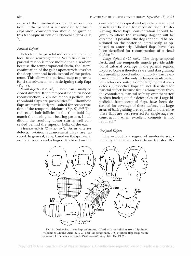
If the defect is extremely large, approxi-mately greater than 50 cm2, the Orticocheaflap is a useful alternative.11,80–82 In this tech-nique, two flaps are used to reconstruct thedefect, each of which is based on the superfi-cial temporal vessels, and one large flap basedon the occipitals is used to fill the donor defect(Fig. 6). Orticochea initially described his tech-nique using four flaps but updated his tech-nique to the use of three flaps to maximize flapvascularity. The cosmetic outcome using Orti-cochea flaps is often inferior to the outcomethat can be obtained with tissue expansion be-
FIG. 5. (Left) Medium anterior scalp defect in a 74-year-old man after Mohs’ excision of a basal cell carcinomawith a 2.5 � 3.0-cm-diameter defect of the anterior scalp. The patient underwent reconstruction with a rotationadvancement flap (center) and is shown (right) 2 weeks postoperatively.
Vol. 116, No. 4 / RECONSTRUCTION OF SCALP DEFECTS 61e
cause of the unnatural resultant hair orienta-tion. If the patient is a candidate for tissueexpansion, consideration should be given tothis technique in lieu of Orticochea flaps (Fig.7).
Parietal Defects
Defects in the parietal scalp are amenable tolocal tissue rearrangement. Scalp tissue in theparietal region is more mobile than elsewherebecause the temporoparietal fascia, the lateralcontinuation of the galea aponeurosis, overliesthe deep temporal fascia instead of the perios-teum. This allows the parietal scalp to providefor tissue advancement in designing scalp flaps(Fig. 8).
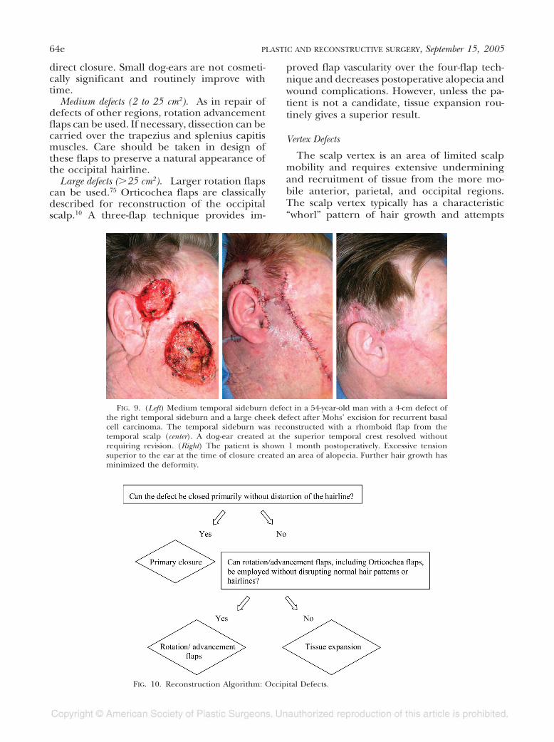
Small defects (�2 cm2). These can usually beclosed directly. If the temporal sideburn needsreconstruction, V-Y, subcutaneous pedicle, andrhomboid flaps are possibilities.83,84 Rhomboidflaps are particularly well suited for reconstruc-tion of the temporal sideburn (Fig. 9).83,84 Theredirected hair follicles in the rhomboid flapmatch the missing hair-bearing pattern. In ad-dition, the resulting donor scar is well con-cealed behind the superior helix of the ear.
Medium defects (2 to 25 cm2). As in anteriordefects, rotation advancement flaps are fa-vored. In general, a flap based on the ipsilateraloccipital vessels and a larger flap based on the
contralateral occipital and superficial temporalvessels can be used for reconstruction. In de-signing these flaps, consideration should begiven to where the resulting dog-ear will bedirected. If possible, the dog-ear should be po-sitioned on the posterior lateral scalp as op-posed to anteriorly. Bilobed flaps have alsobeen described for reconstruction of parietaldefects.85
Large defects (�25 cm2). The deep temporalfascia and the temporalis muscle provide addi-tional calvarial coverage in the parietal region.Exposed bone is therefore rare, and skin graftingcan usually proceed without difficulty. Tissue ex-pansion often is the only technique available forsatisfactory reconstruction of large parietal scalpdefects. Orticochea flaps are not described forparietal defects because tissue advancement fromthe contralateral parietal scalp up over the vertexis often inadequate for defect closure. Large bi-pedicled fronto-occipital flaps have been de-scribed for coverage of these defects, but largeareas of back-grafting are required and thereforethese flaps are best reserved for single-stage re-construction when excellent cosmesis is notrequired.86
Occipital Defects
The occiput is a region of moderate scalpmobility amenable to local tissue transfer. Re-
FIG. 6. Orticochea three-flap technique. (Used with permission from LippincottWilliams & Wilkins. Arnold, P. G., and Rangarathnam, C. S. Multiple-flap scalp recon-struction: Orticochea revisited. Plast. Reconstr. Surg. 69: 607, 1982.)
62e PLASTIC AND RECONSTRUCTIVE SURGERY, September 15, 2005

construction may involve restoration of the oc-cipital hairline or flap design to preserve anintact hairline. The occipital scalp is not ascosmetically sensitive as other more forward-
facing scalp regions and can be camouflagedwith long hair (Fig. 10).
Small defects (�2 cm2). Similar to anterior de-fects, small occipital defects can be closed with
FIG. 7. A 35-year-old woman with a large anterior scalp defect after Mohs’ excision. Reconstruction wasachieved my means of split-thickness skin grafting followed by tissue expansion.
FIG. 8. Reconstruction Algorithm: Parietal Defects.
Vol. 116, No. 4 / RECONSTRUCTION OF SCALP DEFECTS 63e
direct closure. Small dog-ears are not cosmeti-cally significant and routinely improve withtime.
Medium defects (2 to 25 cm2). As in repair ofdefects of other regions, rotation advancementflaps can be used. If necessary, dissection can becarried over the trapezius and splenius capitismuscles. Care should be taken in design ofthese flaps to preserve a natural appearance ofthe occipital hairline.
Large defects (�25 cm2). Larger rotation flapscan be used.75 Orticochea flaps are classicallydescribed for reconstruction of the occipitalscalp.10 A three-flap technique provides im-
proved flap vascularity over the four-flap tech-nique and decreases postoperative alopecia andwound complications. However, unless the pa-tient is not a candidate, tissue expansion rou-tinely gives a superior result.
Vertex Defects
The scalp vertex is an area of limited scalpmobility and requires extensive underminingand recruitment of tissue from the more mo-bile anterior, parietal, and occipital regions.The scalp vertex typically has a characteristic“whorl” pattern of hair growth and attempts
FIG. 9. (Left) Medium temporal sideburn defect in a 54-year-old man with a 4-cm defect ofthe right temporal sideburn and a large cheek defect after Mohs’ excision for recurrent basalcell carcinoma. The temporal sideburn was reconstructed with a rhomboid flap from thetemporal scalp (center). A dog-ear created at the superior temporal crest resolved withoutrequiring revision. (Right) The patient is shown 1 month postoperatively. Excessive tensionsuperior to the ear at the time of closure created an area of alopecia. Further hair growth hasminimized the deformity.
FIG. 10. Reconstruction Algorithm: Occipital Defects.
64e PLASTIC AND RECONSTRUCTIVE SURGERY, September 15, 2005

should be made to try to preserve this arrange-ment (Fig. 11).
Small defects (�2 cm2). For small defects, di-rect closure after subgaleal dissection may bepossible. If not, local flaps such as pinwheelflaps and adjacent rhomboid flaps work partic-ularly well to reconstruct the whorl pattern (Fig.12). Elliptical excision, up to 4 cm wide, withgaleal undermining can be performed similarto scalp reduction for male pattern baldness(Figs. 13 and 14).66
Medium defects (2 to 25 cm2). Pinwheel andrhomboid flaps are less useful but are still pos-
sible alternatives. Usually, larger amounts of un-dermining are required; therefore, double-op-posing rotation advancement flaps are goodalternatives. The incisions can be carried par-allel to the hairline to prevent distortion. Con-sideration should also be given to rotation ad-vancement from the occiput with back-graftingof the donor site. This can be reconstructed ata later time by tissue expansion and transposesthe deformity to a less cosmetically sensitivearea.
Large defects (�25 cm2). The only alternativesfor these defects are very large rotation flaps,
FIG. 11. Reconstruction Algorithm: Vertex Defects.
FIG. 12. Pinwheel flaps. (Used with permission from Lippincott Williams & Wilkins.Dowbak, G. V-Y-S plasty for scalp defects. Plast. Reconstr. Surg. 113: 1889, 2004.)
Vol. 116, No. 4 / RECONSTRUCTION OF SCALP DEFECTS 65e

which require near complete scalp undermin-ing and galeal scoring (Fig. 15). True Ortico-chea flaps are not well suited for repair of thesedefects because the location does not allow for
a large third flap for coverage of the donor-sitedefect. Alternatively, large rotation flaps withsignificant dog-ear deformities can be used withdonor-site back-grafting, if necessary. Despite
FIG. 13. Elliptical closure of vertex defects. Available patterns for scalp reduction.(Used with permission from Lippincott Williams & Wilkins. Bell, M. L. Role of scalpreduction in the treatment of male pattern baldness. Plast. Reconstr. Surg. 69: 272, 1982.)
FIG. 14. (Left) Medium posterior vertex defect in a 77-year-old man who underwent split-thickness skingrafting with a posterior vertex defect after Mohs’ excision of a basal cell carcinoma but suffered graft lossfrom trauma with resulting exposed calvarium. (Center) The defect was reconstructed with an ellipticalexcision of the grafted area, which created a defect 3.5 cm in width. Closure was performed after widesubgaleal undermining with galeal scoring. (Right) The patient is shown 2 months postoperatively. Thesmall dog-ear created on the central vertex improved without requiring revision.
66e PLASTIC AND RECONSTRUCTIVE SURGERY, September 15, 2005
these options, tissue expansion uniformly willyield the best results (Fig. 16).
Nearly Total Defects
Straith and Beers give a colorful account ofa physician using pinch allografts from hisown arm in attempt to reconstruct a scalpavulsion in 1889.9 When this failed, an allo-graft from the patient’s sister and then axenograft from the abdomen of a dog wereattempted unsuccessfully.9 Fortunately, sincethis initial report, many advances have beenmade.
The best technique for reconstruction ofnearly total defects of the scalp is free tissuetransfer. There have been case reports ofsuch defects reconstructed with Integra fol-lowed by skin grafting, but as described, thistechnique requires hyperbaric oxygen ther-apy and has not been supported elsewhere inthe literature.87 Free tissue transfer allows forcomplete scalp reconstruction in a single
stage and can be performed at the time oftumor extirpation. An ample supply of headand neck vessels provide multiple alterna-tives for donor vessels. The most readily avail-able vessels are perhaps the superficial tem-poral vessels. If these are inadequate, otherarterial branches of the external carotid sys-tem and the internal jugular vein may beused. The latissimus dorsi muscle providesarguably the best flap for coverage becauseof its large surface area and long vascularleash (Fig. 17).50 The serratus anterior mus-cle can be included with the latissimus dorsimuscle to increase the flap surface area ifnecessary (Fig. 18). With time, free muscleflaps atrophy and contour nicely to the skulland are less likely to require significant de-bulking than myocutaneous or fasciocutane-ous flaps.50 However, over the long term,muscle flap atrophy may lead to recurrentskull exposure. Composite flaps can preventthis undesirable sequela and therefore may
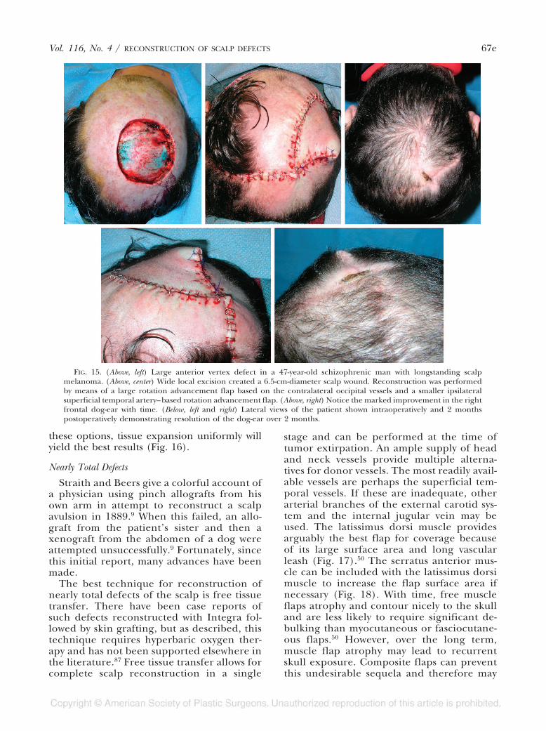
FIG. 15. (Above, left) Large anterior vertex defect in a 47-year-old schizophrenic man with longstanding scalpmelanoma. (Above, center) Wide local excision created a 6.5-cm-diameter scalp wound. Reconstruction was performedby means of a large rotation advancement flap based on the contralateral occipital vessels and a smaller ipsilateralsuperficial temporal artery–based rotation advancement flap. (Above, right) Notice the marked improvement in the rightfrontal dog-ear with time. (Below, left and right) Lateral views of the patient shown intraoperatively and 2 monthspostoperatively demonstrating resolution of the dog-ear over 2 months.
Vol. 116, No. 4 / RECONSTRUCTION OF SCALP DEFECTS 67e
be desirable to muscle-only flaps even if sec-ondary debulking procedures are required.88
Besides free muscle flaps, free tissue transferusing the radial forearm, parascapular, andanterolateral perforator flaps and other freeflaps such as omentum, Scarpa adipofascialflap, and scalp from an identical twin havebeen described.16,19,22,89,90 In addition, whenused for flap coverage, skin grafts should beunmeshed to give the best possible appear-ance. However, most patients will resort towigs (Fig. 19).
DISCUSSION
From ancient management strategies ofscalp avulsion injuries that used trephinationof the outer table of the calvaria2 to the ad-vances in microsurgery that allow for free tissuetransfer, the techniques for scalp reconstruc-tion have paralleled advances in plastic sur-gery. We are now able to obtain coverage over
the calvaria after the most devastating of de-fects; however, the challenge to the reconstruc-tive surgeon today is to do so with excellentcosmetic results. Adhering to the principles ofscalp reconstruction presented in this article,the reconstructive surgeon will be able toachieve this goal. First and foremost, cosmeticscalp reconstruction requires restoration andpreservation of normal hair patterns and hairlines. In addition to obtaining calvarial cover-age, all reconstructive endeavors should strivefor this goal.
Most scalp defects can be reconstructed withlocal tissue rearrangements. Rotation-advance-ment flaps using scalp mobility from the pari-etal regions are the workhorse of reconstruc-tive techniques. Proper design of these flapsrequires preservation of the native hairline,redirection of hair follicles in acceptable pat-terns, incorporation of major vascular pedicles,and closure without excessive tension, which
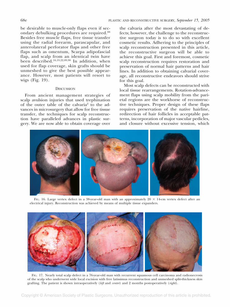
FIG. 16. Large vertex defect in a 38-year-old man with an approximately 20 � 14-cm vertex defect after anelectrical injury. Reconstruction was achieved by means of multiple tissue expanders.
FIG. 17. Nearly total scalp defect in a 76-year-old man with recurrent squamous cell carcinoma and radionecrosisof the scalp who underwent wide local excision with free latissimus reconstruction and unmeshed split-thickness skingrafting. The patient is shown intraoperatively (left and center) and 2 months postoperatively (right).
68e PLASTIC AND RECONSTRUCTIVE SURGERY, September 15, 2005
can be accomplished with galeotomies or useof stress relaxation and creep.
In many instances, local tissues are inade-quate for rearrangement and defect recon-struction. This may occur if the defect isprohibitively large, if local tissues are trauma-tized, or if rotation-advancement flaps wouldrequire disruption of normal hair patternsand hairlines. When this occurs, as long ashair-bearing scalp remains, tissue expansionis the reconstructive technique of choice. Tis-sue expansion can reliably replace damagedscalp tissue with like tissue that maintainsnative hair patterns. If inadequate scalp ispresent to allow for tissue expansion, free
tissue transfer offers the best reconstructiveoption. Muscle flaps are useful for this pur-pose because they contour well to the cal-varia over time as they atrophy from dener-vation.
CONCLUSION
Successful reconstruction of the scalp re-quires careful preoperative planning and pre-cise intraoperative execution. Detailed knowl-edge of scalp anatomy, skin biomechanics, hairphysiology, and the variety of available localtissue rearrangements allows for excellent aes-thetic reconstruction.
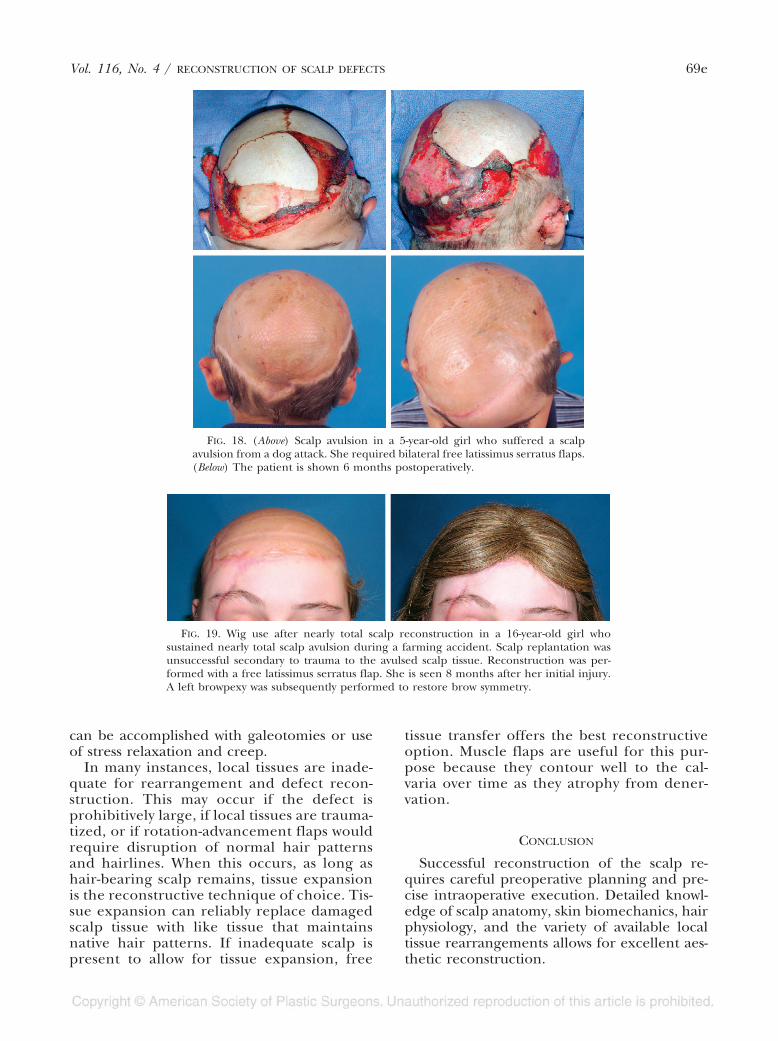
FIG. 18. (Above) Scalp avulsion in a 5-year-old girl who suffered a scalpavulsion from a dog attack. She required bilateral free latissimus serratus flaps.(Below) The patient is shown 6 months postoperatively.
FIG. 19. Wig use after nearly total scalp reconstruction in a 16-year-old girl whosustained nearly total scalp avulsion during a farming accident. Scalp replantation wasunsuccessful secondary to trauma to the avulsed scalp tissue. Reconstruction was per-formed with a free latissimus serratus flap. She is seen 8 months after her initial injury.A left browpexy was subsequently performed to restore brow symmetry.
Vol. 116, No. 4 / RECONSTRUCTION OF SCALP DEFECTS 69e

Rod J. Rohrich, M.D.Department of Plastic SurgeryUniversity of Texas Southwestern Medical Center5323 Harry Hines Blvd., E7.210Dallas, Texas [email protected]
REFERENCES
1. Koss, N., Robson, M., and Krizek, T. J. Scalping injury.Plast. Reconstr. Surg. 55: 439, 1975.
2. Strayer, L. M. Augustin Belloste and the treatment ofavulsion of the scalp. N. Engl. J. Med. 220: 901, 1939.
3. Netolitzky, J. Zur Kasuistik der Hauttransplantation.Wien Med. Wochenshr. 21: 820, 1871.
4. Robinson, E. F. Total avulsion of the scalp. Surg. Gynecol.Obstet. 7: 663, 1908.
5. Marcks, K. M., Trevaskis, A., and Nauss, T. J. Scalp de-fects and their repair. Plast. Reconstr. Surg. 7: 237, 1951.
6. Kazanjian, V. H. Repair of partial losses of the scalp.Plast. Reconstr. Surg. 12: 325, 1953.
7. Gaisford, J. C., Hanna, D. C., and Susen, A. F. Majorresection of scalp and skull for cancer with immediatecomplete reconstruction: 14 cases. Plast. Reconstr. Surg.21: 335, 1958.
8. Coleman, C. C. Scalp flap reconstruction in head andneck cancer patients. Plast. Reconstr. Surg. 24: 45, 1959.
9. Straith, C. L., and Beers, M. D. Scalp avulsions. Plast.Reconstr. Surg. 6: 319, 1950.
10. Orticochea, M. Four-flap scalp reconstruction tech-nique. Br. J. Plast. Surg. 20: 159, 1967.
11. Orticochea, M. New three-flap reconstruction tech-nique. Br. J. Plast. Surg. 24: 184, 1971.
12. Neumann, C. G. The expansion of an area of skin byprogressive distension of a subcutaneous balloon.Plast. Reconstr. Surg. 19: 124, 1957.
13. Radovan, C. Tissue expansion in soft tissue reconstruc-tion. Plast. Reconstr. Surg. 74: 482, 1984.
14. Manders, E. R., Schenden, M. J., Furrey, J. A., et al. Skinexpansion to eliminate large scalp defects. Plast. Re-constr. Surg. 74: 493, 1984.
15. Miller, G. D. H., Anstee, E. J., and Snell, J. A. Successfulreplantation of an avulsed scalp by microvascular anas-tomoses. Plast. Reconstr. Surg. 58: 133, 1976.
16. Ikuta, Y. Microvascular free transfer of omentum. InL. O. Vasconez and B. Strauch (Eds.), Grabb’s Encyclo-pedia of Flaps, 2nd Ed. Philadelphia: Lippincott-Raven,1998. Pp. 42–44.
17. Ohmori, K. Microvascular free transfer of temporo-oc-cipital scalp flaps. In L. O. Vasconez and B. Strauch(Eds.), Grabb’s Encyclopedia of Flaps, 2nd Ed. Philadel-phia: Lippincott-Raven, 1998. Pp. 31–34.
18. Ioannides, C., Fossion, E., and McGrouther, A. D. Re-construction for large defects of the scalp and cra-nium. J. Craniomaxillofac. Surg. 27: 145, 1999.
19. Koshima, I., Inayawa, K., Jitsuiki, Y., Tsuda, K., Moriguchi,T., and Watanabe, A. Scarpa’s adipofascial flap forrepair of wide scalp defects. Ann. Plast. Surg. 36: 88,1996.
20. Lutz, B. S., Wei, F. C., Chen, H. C., et al. Reconstructionof scalp defects with free flaps in 30 cases. Br. J. Plast.Surg. 51: 186, 1998.
21. Pennington, D. G., Stern, H. S., and Lee, K. K. Free flapreconstruction of large defects of the scalp and cal-varium. Plast. Reconstr. Surg. 83: 655, 1989.
22. Chicarelli, Z. N., Ariyan, S., and Cuono, C. B. Single-stage repair of complex scalp and cranial defects withthe free radial forearm flap. Plast. Reconstr. Surg. 77:577, 1986.
23. Abdul-Hassan, H. S., von Drasek Ascher, G., and Acland,R. D. Surgical anatomy and blood supply of the fas-cial layers of the temporal regions. Plast. Reconstr. Surg.77: 17, 1986.
24. Tolhurst, M. D., Carstens, M. H., Greco, R. J., and Hur-witz, D. J. The surgical anatomy of the scalp. Plast.Reconstr. Surg. 87: 603, 1991.
25. Williams, P. H., Warwick, R. H., et al. (Eds.), Gray’s Anat-omy of the Human Body, 36th Ed. Philadelphia: Saun-ders, 1985. P. 530.
26. Anson, B. J., and McVay, C. B. Surgical Anatomy, 6th Ed.Philadelphia: Saunders, 1984. P. 4.
27. Anderson, J. E. (Ed.). Grant’s Atlas of Anatomy, 8th Ed.Baltimore: Williams & Wilkins, 1983.
28. Last, R. J. Surgical Anatomy, Regional and Applied. London:Churchill, 1979. P. 543.
29. Freund, R. M. Scalp, calvarium and forehead recon-struction. In S. J. Aston, R. W. Beasley, and C. H. M.Thorne (Eds.), Grabb and Smith’s Plastic Surgery. Phil-adelphia: Lippincott-Raven, 1997. P. 473.
30. Shestak, K. C., and Ramasastry, S. S. Reconstruction ofdefects of the scalp and skull. In M. Cohen (Ed.),Mastery of Plastic and Reconstructive Surgery. Boston: Lit-tle, Brown, 1994. Pp. 830–841.
31. Elliott, L. F., and Jurkiewicz, M. J. Scalp and calvarium.In M. J. Jurkiewicz, T. J. Krizek, S. J. Mathes, and S.Ariyan (Eds.), Plastic Surgery: Principles and Practice. St.Louis: Mosby, 1990. Pp. 419–440.
32. Carstens, M. H., Greco, R. J., Hurwitz, D. J., and Tolhurst,D. E. Clinical applications of the subgaleal fascia.Plast. Reconstr. Surg. 87: 615, 1991.
33. Marchac, D. Deformities of the forehead, scalp andcranial vault. In J. G. McCarthy (Ed.), Plastic Surgery.Philadelphia: Saunders, 1990. Pp. 1538–1574.
34. Moss, C. J., Mendelson, B. C., and Taylor, G. I. Surgicalanatomy of the ligamentous attachments in the tem-ple and periorbital regions. Plast. Reconstr. Surg. 105:1475, 2000.
35. Brandy, D. A. The effectiveness of occipital artery liga-tions as a priming procedure for extensive scalp-lift-ing. J. Dermatol. Surg. Oncol. 17: 946, 1991.
36. Knize, D. M. Reassessment of the coronal incision andsubgaleal dissection for foreheadplasty. Plast. Reconstr.Surg. 102: 478, 1998.
37. Mosser, S. W., Guyuron, B., Janis, J. E., and Rohrich,R. J. The anatomy of the greater occipital nerve:Implications for the etiology of migraine headaches.Plast. Reconstr. Surg. 113: 693, 2004.
38. Headington, J. T. Telogen effluvium. Arch. Dermatol.129: 356, 1993.
39. Harrison, S., and Sinclair, R. Telogen effluvium. Clin.Exp. Dermatol. 27: 389, 2002.
40. Habif, T. P. Clinical Dermatology, 4th Ed. St. Louis: Mosby,2004. P. 836.
41. Jackson, I. T. General considerations. In I. T. Jackson(Ed.), Local Flaps in Head and Neck Reconstruction. St.Louis: Mosby, 1985. P. 4–5.
42. Sasaki, G. H. Intraoperative sustained limited expan-sion (ISLE) as an immediate reconstructive tech-nique. Clin. Plast. Surg. 14: 563, 1987.
43. Hight, D. W., and Anderton, R. L. Combined use ofpunch graft and an overlay sheet of split-skin graft for
70e PLASTIC AND RECONSTRUCTIVE SURGERY, September 15, 2005

a raw area in the scalp. Plast. Reconstr. Surg. 52: 663,1973.
44. Nordstrom, R. E. A. Punch hair grafting under split-skin grafts on scalps. Plast. Reconstr. Surg. 64: 9, 1979.
45. Barrera, A. The use of micrografts and minigrafts forthe correction of the postrhytidectomy lost sideburn.Plast. Reconstr. Surg. 102: 2237, 1998.
46. Barrera, A. The use of micrografts and minigrafts forthe treatment of burn alopecia. Plast. Reconstr. Surg.103: 581, 1999.
47. Oishi, S. N., and Luce, E. A. The difficult scalp and skullwound. Clin. Plast. Surg. 22: 51, 1995.
48. Nair, S., Giannakopoulos, G., Granick, M., Solomon, M.,McCormack, T., and Black, P. Surgical managementof radiated scalp in patients with recurrent glioma.Neurosurgery 34: 103, 1994.
49. Yuen, J. C., and Hochberg, J. Free flap coverage of scalpdefects following radiation. J. Ark. Med. Soc. 100: 194,2003.
50. Hussussian, C. J., and Reece, G. P. Microsurgical scalpreconstruction in the patient with cancer. Plast. Re-constr. Surg. 109: 1828, 2002.
51. Di Meo, L., and Jones, B. M. Surgical treatment of ra-diation-induced scalp lesions. Br. J. Plast. Surg. 37: 373,1984.
52. Kuwahara, M., Hatoko, M., Tanake, A., Yurugi, S., andMashiba, K. Simultaneous use of a tissue expanderand skin graft in scalp reconstruction. Ann. Plast. Surg.45: 220, 2000.
53. Azzoloni, A., Riberti, C., and Caalca, D. Skin expansionin head and neck reconstructive surgery. Plast. Recon-str. Surg. 90: 799, 1992.
54. Sood, R., and Coleman, J. Scalp and calvarial recon-struction. In E. Eriksson, B. M. Achauer, B. Guyuron,J. J. Coleman, R. C. Russell, and C. A. Vander Kolk(Eds.), Plastic Surgery: Indications, Operations, and Out-comes. St. Louis: Mosby, 2000. Pp. 1519–1539.
55. Terranova, W. The use of periosteal flaps in scalp andforehead reconstruction. Ann. Plast. Surg. 25: 450,1990.
56. van Rappard, J. H., Molenaar, J., Van Doorn, D., etal. Surface-area increase in tissue expansion. Plast.Reconstr. Surg. 82: 833, 1988.
57. Weislander, J. B. Repeated tissue expansion in recon-struction of a huge combined scalp-forehead avulsioninjury. Ann. Plast. Surg. 20: 381, 1988.
58. Bauer, B. Pediatric tissue expansion. In Proceedings of the72nd Annual ASPS/PSEF/ASMS Scientific Meeting, in SanDiego, California, 2003 .
59. Frodel, J. L., and Mabrie, D. Optimal elective scalp in-cision design. Otolaryngol. Head Neck Surg. 121: 374,1999.
60. Sakhai, H. Hemostatic control of scalp incisions. J. Neu-rosurg. 70: 142, 1989.
61. Camirand, A., and Doucet, J. A comparison betweenparallel hairline incisions and perpendicular incisionswhen performing a face lift. Plast. Reconstr. Surg. 99: 10,1995.
62. Seyhan, A., Yoleri, L., and Barutcu, A. Immediate hairtransplantation into a newly closed wound to concealthe final scar on the hair-bearing skin. Plast. Reconstr.Surg. 105: 1866, 2000.
63. Barrera, A. Immediate hair transplantation into anewly closed wound to conceal the final scar onhair-bearing skin (Discussion). Plast. Reconstr. Surg.105: 1871, 2000.
64. Baker, S. R., and Swanson, N. A. Rapid intraoperativetissue expansion in reconstruction of the head andneck. Arch. Otolaryngol. Head Neck Surg. 116: 1431,1990.
65. Raposio, E., Santi, P., and Nordstrom, R. E. A. Effectsof galeostomies on scalp flaps. Ann. Plast. Surg. 41: 17,1998.
66. Raposio, E., Cella, A., Panarese, P., Nordstrom, R. E. A.,and Santi, P. Anchoring galeal flaps for scalp re-duction procedures. Plast. Reconstr. Surg. 102: 2454,1998.
67. Rees, T. D. Is there a place for the staple gun in plasticsurgery? Ann. Plast. Surg. 1: 238, 1978.
68. Bucknall, T. E., Teare, L., and Ellis, H. Skin closure: Acomparison of nylon, polyglycolic acid, and staples.Eur. Surg. Res. 14: 96, 1982.
69. Baker, S. R. Local cutaneous flaps. Otolaryngol. Clin.North Am. 27: 139, 1994.
70. Lesavoy, M. A., Dubrow, T. J., Schwartz, R. J., et al. Ma-nagement of large scalp defects with local pedicleflaps. Plast. Reconstr. Surg. 91: 783, 1993.
71. Byrd, H. S. The use of subcutaneous axial fascial flapsin reconstruction of the head. Ann. Plast. Surg. 4: 191,1980.
72. Hallock, G. G., and Trevaskis, A. E. Refinement of thesubcutaneous pedicle flap for closure of forehead andscalp defects. Plast. Reconstr. Surg. 75: 903, 1985.
73. Sakai, S., Soeda, S., and Terayama, I. Subcutaneouspedicle flaps for scalp defects. Br. J. Plast. Surg. 41: 255,1988.
74. Demir, Z., Velidedeoglu, H., and Celebiogle, S. V-Y-Splasty for scalp defects. Plast. Reconstr. Surg. 112: 1054,2003.
75. Ahuja, R. B. Geometric considerations in the design ofrotation flaps in the scalp and forehead region. Plast.Reconstr. Surg. 81: 900, 1988.
76. Juri, J. Temporoparieto-occipital and temporoparieto-occipitoparietal scalp flaps. In B. Strauch, L. O. Vas-conez, E. J. Hall-Findlay, and W. C. Grabb (Eds.),Grabb’s Encyclopedia of Flaps, 2nd Ed. Philadelphia: Lip-pincott-Raven, 1998. Pp. 19–22.
77. Juri, J. Diagonal frontoparieto-occipital scalp flaps.In B. Strauch, L. O. Vasconez, E. J. Hall-Findlay, andW. C. Grabb (Eds.), Grabb’s Encyclopedia of Flaps, 2ndEd. Philadelphia: Lippincott-Raven, 1998. Pp. 23–25.
78. Elliott, R. A. Lateral scalp flaps for instant results inmale pattern baldness. Plast. Reconstr. Surg. 60: 699,1977.
79. Juri, J., and Juri, C. Aesthetic aspects of reconstructivescalp surgery. Clin. Plast. Surg. 8: 243, 1981.
80. Arnold, P. G., and Rangarathnam, C. S. Multiple-flapscalp reconstruction: Orticochea revisited. Plast. Re-constr. Surg. 69: 605, 1982.
81. Orticochea, M. “Banana peel” scalp, forehead, andnape of neck flaps. In B. Strauch, L. O. Vasconez, E. J.Hall-Findlay, and W. C. Grabb (Eds.), Grabb’s Encyclo-pedia of Flaps, 2nd Ed. Philadelphia: Lippincott-Raven,1998. Pp. 13–18.
82. Yang, C. C., Lin, S., Lai, C. S., and Chou, C. K. Recon-struction of children’s scalp defects with the Ortico-chea flap. Ann. Plast. Surg. 28: 584, 1992.
83. Davis, W. H. Sideburn reconstruction with an arte-rial V-Y hair-bearing scalp flap after the excision of
Vol. 116, No. 4 / RECONSTRUCTION OF SCALP DEFECTS 71e
basal cell carcinoma. Plast. Reconstr. Surg. 106: 94,1999.
84. Ahieja, R. B. Geometric considerations in the design ofrotation flaps in the scalp and forehead region. Plast.Reconstr. Surg. 81: 900, 1988.
85. Iida, N., Ohsumi, N., Tonegawa, M., and Tustsumi,Y. Reconstruction of scalp defects using simpledesigned bilobed flap. Aesthetic Plast. Surg. 24: 137,2000.
86. De Haro, F., and Giraldo, F. Bipedicled fronto-oc-cipital flap for reconstruction of postoncologic de-fects of the lateral scalp. Plast. Reconstr. Surg. 107:506, 2001.
87. Gonyon, D., and Zenn, M. R. Simple approach to theradiated scalp wound using INTEGRA skin substitute.Ann. Plast. Surg. 50: 315, 2003.
88. Hallock, G. G. Long-term superiority of composite ver-sus muscle-only free flaps for skull coverage. Ann.Plast. Surg. 52: 507, 2004.
89. Lutz, B. S. Aesthetic and functional advantages of theanterolateral thigh flap in reconstruction of tumor-related scalp defects. Microsurgery 22: 258, 2002.
90. Buncke, H. J., Hoffman, W. Y., Alpert, B. S., Gordon, L.,and Stefani, A. E. Microvascular transplant of twofree scalp flaps between identical twins. Plast. Reconstr.Surg. 70: 605, 1982.
72e PLASTIC AND RECONSTRUCTIVE SURGERY, September 15, 2005