{Synthesis and efficacy of copper(II) complexes bearing N ...

Clinicopathologic Efficacy of Copper Bromide Plus/YellowLaser (578 nm with 511 nm) for Treatment of Melasma inAsian Patients
HYE IN LEE, MD,� YUN YOUNG LIM, MS,� BEOM JOON KIM, MD, PHD,� MYEUNG NAM KIM, MD,�
HYE JUNG MIN, MS,y JUNG HEE HWANG, MS,y AND KYE YONG SONG, MDy
BACKGROUND Melasma is a common pigmentary disorder in Asians. Although the pathogenesis ofmelasma is not yet fully understood, there are several hypotheses supporting angiogenetic factorsrelated to some types of melasma.
OBJECTIVE To test the efficacy of copper bromide laser in the treatment of Korean women with melasma.
MATERIALS AND METHODS Clinical parameters included physician and patient assessment and Me-lasma Area and Severity Index score. The intensity of pigmentation and erythema was measured using achromometer. To evaluate histopathologic changes, punch biopsies from melasma were obtained fromfour patients. Immunohistochemical staining for Melan-A, endothelin 1, CD34, and vascular endothelialgrowth factor (VEGF) antigen of the melasma lesions was observed.
RESULTS Mean MASI score decreased dramatically after treatment. Patients exhibited telangiectaticerythema within the melasma lesion. The values of L� reflecting intensity of pigmentation increased, andthe values of a� as the measurement of redness decreased after the treatments. Expression of Melan-A,CD34, endothelin-1, and VEGF decreased after treatment.
CONCLUSION The potential application of an antiangiogenetic laser for the treatment of melasmaspecially accompanied by pronounced telangiectasia in Asian skin is a possible treatment option.
The authors have indicated no significant interest with commercial supporters.
Melasma is a common acquired symmetrical
hypermelanosis of sun-exposed areas of the
skin that is common in Asian women. The major
etiological factors are genetic influences, exposure to
ultraviolet (UV) radiation, and sex hormones,1
although the pathogenesis of melasma is not fully
understood.
Recent studies have suggested a possible connection
between vessels and cutaneous pigmentation.
Human melanocytes may respond to angiogenic
factors because normal human melanocytes express
functional vascular endothelial growth factor
(VEGF) receptors.2 Also, it has been reported that
the topical plasmin inhibitor tranexamic acid is
effective in the treatment of UV light–induced
hyperpigmentation.3 Localized microinjection of
tranexamic acid has improved melasma in vivo.4
These in vitro and in vivo findings suggest that
interactions between the altered cutaneous
vasculature and melanocytes may have an influence
on the development of hyperpigmentation in the
overlying epidermis. In some types of melasma,
pronounced telangiectatic erythema confined to
melasma-lesional skin has been observed. Increased
vascularity is one of the major histologic findings
in melasma.5 Interactions between the altered
cutaneous vasculature and melanocytes may
influence the development of melasma.
Traditional therapies, including depigmenting agents
(e.g., hydroquinone, azelaic acid), chemical peels
(e.g., glycolic acid, b-hydroxyl acid, trichloroacetic
acid), topical steroids, and sunscreens have some
& 2010 by the American Society for Dermatologic Surgery, Inc. � Published by Wiley Periodicals, Inc. �ISSN: 1076-0512 � Dermatol Surg 2010;36:1–9 � DOI: 10.1111/j.1524-4725.2010.01564.x
1
Departments of �Dermatology and yPathology, Chung-Ang University College of Medicine, Seoul, Korea

therapeutic effect but are often unsuccessful in the
treatment of refractory melasma. The use of lasers in
the treatment of melasma is controversial. Facial
resurfacing with an erbium laser or a pulsed carbon
dioxide laser, alone or in conjunction with a
Q-switched alexandrite laser, have reportedly been
successful, but they result in significant downtime,
and there is a risk of adverse sequelae.6,7 Fractional
laser therapy with a 1,550-nm erbium fiber laser has
recently been investigated in a pilot study.8
Copper bromide lasers emit a green beam at a
wavelength of 511 nm, which can be used to treat
pigmentary lesions, and a yellow beam at a wave-
length of 578 nm, which can be used to treat vascular
lesions.9,10 This study is a report of the clinical
efficacy and immunohistochemical changes after the
use of copper bromide Plus/Yellow Laser (Norseld
Pty Ltd, Adelaide, Australia) (578 nm with 511 nm)
to treat melasma in Asian patients.
Materials and Methods
Patients
Ten Korean women aged 32 to 51 (mean 40.7) with
melasma were enrolled in this clinical study between
December 2007 and April 2008. Patients aged 30 to
60 with clinically diagnosed melasma were eligible
to participate in this study. The melasma was diag-
nosed through physical examinations and confirmed
using histological examinations.
Pregnant or nursing woman; patients with excessive
photosensitivity to normal sunlight, inflammatory
disease of the skin, open wounds in the area of
treatment, and active herpes simplex; patients
exhibiting symptoms of severe stress; patients
refusing to give informed consent; patients with
facial congenital nevi; patients using oral or topical
medications that can affect the response to visible
light; patients using oral contraceptive pills; patients
who had ever used topical steroids, including triple
combination cream; patients treated with topical
hypopigmenting agents, such as hydroquinone,
tretinoin, kojic acid, and azelaic acid, and other
lasers or intense pulsed light less than 3 months be-
fore were excluded.
The duration of melasma ranged from 6 months to
30 years (mean 9.4 years). Seven patients had Fitz-
patrick skin type III and three skin type IV. Accord-
ing to Wood’s Lamp Assessment, 60% of patients (6/
10) had a mixed-type melasma, and 40% (4/10) had
an epidermal-type melasma. In the distribution of
melasma, seven patients had a malar pattern, and
three had a centrofacial pattern. The centrofacial
pattern was observed in three patients: the melasma
involved the cheeks, forehead, upper lip, and chin.
The malar pattern, located in the malar region, was
observed in seven patients. All subjects were
instructed to avoid the use of bleaching agents during
the course of the treatment and for 3 months of fol-
low-up. They were also instructed on proper sun
protection and the use of broad-spectrum sunscreens.
Informed written consent was obtained from each
patient before skin sampling. The ethical committee
of Chung-Ang University Yong-San Hospital ap-
proved the study. There were no conflicts of interest.
Treatment Protocols
Plus/Yellow Laser was used for all treatments. This
copper bromide laser produces two wavelengths that
can be emitted separately or together: green
(511 nm) and yellow (578 nm). The green and yellow
lasers are simultaneously produced in a 1:9 ratio in
plus mode. The yellow wavelength is adjustable up
to a maximum of 2.1 W. The copper bromide laser
emits quasicontinuous pulse trains with a pulse
width of 24 ns and a pulse repetition rate of 12 kHz.
Treatment fluences ranged from 12 to 14 J/cm2. A
spot size 1.0 mm was used. The emission time was 50
to 60 ms, and the off time was 70 ms, with 7.7 to 8.3
pulses per second and four passes.
Each patient received four treatments at 2-week
intervals administered to the face. A chilled, colorless
ultrasonic gel was applied directly to the skin. No
D E R M AT O L O G I C S U R G E RY2
C L I N I C O PAT H O L O G I C E F F I C A C Y O F C O P P E R B R O M I D E P L U S / Y E L L O W L A S E R

topical or local anesthetic was used in any of the
patients, and the eyes were always protected. The
patients were instructed to avoid the use of any
bleaching or antiwrinkle agents during the course
of the treatment. They were also instructed to avoid
sun exposure and wear broad-spectrum sunscreen
during and after the treatment.
Evaluation Criteria
Evaluation of skin lesions was performed before each
treatment session and 1 month after the final treat-
ment. All patients were followed up at 3 and 6
months after the final treatment. Five standard digital
photographs were taken (EOS 40D, 6.0 megapixels,
Canon, Tokyo, Japan) before each treatment session.
The clinical assessment consisted of the physicians’
overall assessment and patient self-assessment of the
extent of melasma. Clearance was estimated as a
percentage from 0 (no change) to 100 (complete
disappearance of the telangiectasia and pigmented
lesions).
Two investigators independently evaluated Melasma
Area and Severity Index (MASI) scores (Table 1)
before each session and 1 month after the last
session.
For a more objective assessment, the lesional
melasma of 10 patients was evaluated using a skin
color measuring device at the highest point on the
cheekbones before each session and 1 month after
the last session. The intensity of pigmentation and
erythema were measured using skin reflectance
with a tristimulus color analyzer (Chromameter CR-
400, Minolta Co., Tokyo, Japan) and expressed in
the L�a�b� system. This system allows colors to be
quantified according to three axes: white-black or
lightness (L�), red-green or chrome (a�), and yellow-
blue or hue (b�). The L� parameter reflects the in-
tensity of pigmentation, and the a� parameter mea-
sures redness.
To evaluate histopathologic changes, 2 mm punch
biopsies from lesional melasma were obtained from
four patients under local anesthesia before and 3
months after the last treatment. The tissue samples
were prepared for light microscopic study using 10%
formalin fixation. Three-mm-thick paraffin-embed-
ded tissue sections were processed for routine
immunohistochemistry. A hematoxylin and eosin
(H&E) stain was used to study the general histo-
pathological changes in the melasma skin. Melanin
pigment was visualized using Fontana-Masson
staining performed using the usual methods without
an eosin background stain. The immunohistochem-
ical staining was performed on 4- to 5-mm-thick
TABLE 1. Melasma Area and Severity Index (MASI) Scoring System
A: Percentage of the total area involved
0 = none
1 =o10
2 = 10–29
3 = 30–49
4 = 50–69
5 = 70–89
6 = 90–100
D: Darkness of the melasma compared to normal skin
H: Homogeneity of the hyperpigmentation
0 = normal skin color without evidence of involvement
1 = barely visible hyperpigmentation/specks of involvement
2 = mild hyperpigmentation/small patchy areas of involvement o1.5 cm diameter
3 = moderate hyperpigmentation/patches of involvement 42 cm diameter
4 = severe hyperpigmentation/uniform skin involvement without any clear area
MASI score = Forehead 0.3(D 1 H)A 1 Rt. Malar 0.3(D 1 H)A 1 Lt. Malar 0.3(D 1 H)A 1 Chin 0.1(D 1 H)A.
3 6 :* * : 2 0 1 0 3
L E E E T A L

serial paraffin-embedded sections mounted on poly-
L-lysine–coated slides. The sections were depar-
affinized, and the endogenous peroxidase activity
was blocked with 3% hydrogen peroxide for 15
minutes. Then the sections were subjected to antigen
retrieval using pressure cooking in 0.01M citric acid
(pH 6.0, 1251C) for 3 minutes. These sections were
then incubated with monoclonal anti-CD34 anti-
body (1:200 dilution, Cat. No. MS-363, NeoMarker,
Fremont, CA), monoclonal anti-Melan-A antibody
(1:50 dilution, Cat. No. M7196, Dako, Carpinteria,
CA), monoclonal anti-Endothelin 1 antibody (1:500
dilution, Cat. No. MA3-005, Affinity Bioreagents,
Golden, CO), and polyclonal anti-VEGF antibody
(1:100 dilution, Cat. No. Sc-152, Santa Cruz Bio-
technology, Inc., Santa Cruz, CA); incubated for 24
hours at room temperature; and developed using
EnVision Plus reagent (Dako). Amino ethyl carbaz-
ole was used as the substrate. The slides were
counterstained with Mayer’s hematoxylin.
Results
All of the patients completed the full course of the
study. The representative case with a marked re-
sponse is shown in Figure 1. The results of the phy-
sicians’ overall assessments and the patients’
subjective self-assessments have been summarized in
Table 2.
The mean MASI score decreased dramatically, from
12.373.2 before treatment to 9.57 3.5 at the
1-month follow-up visit (po.05). Table 3 summa-
rizes the clinical findings and changes in mean MASI
scores in the 10 patients.
Some degree of telangiectatic erythema was noticed
within the pigmented patches in patients with
melasma. The values of L� were 57.3 1 3.4 in the
melasma lesions and 62.5 1 1.3 in perilesional
normal skin. The values of a� were higher in the
melasma lesions (mean 13.8 1 1.8) than in
perilesional normal skin (mean 11.3 1 1.2).
In the melasma lesion at the highest point of the
cheekbone, L� increased from 56.7 before treatment
to 57.0 after one session; increased to 57.6 after two
sessions, 59.0 after three sessions, and 59.2 after four
sessions; and dropped to 58.4 at the 1-month follow-
up visit (po.01, Figure 2A). The subjects exhibited
telangiectatic erythema within the brownish patches
on physical examination before treatment. The a�
values decreased according to the treatments.
Quantification of a� at the highest point of the
cheekbones revealed a substantial decrease from
14.8 before treatment to 13.5 after one session, 13.3
after two sessions, 12.0 after three sessions, and 10.9
after four sessions and a slight increase to 11.8 at the
1-month follow-up visit (po.01, Figure 2B).
Figure 1. A 42-year-old woman (A) before and (B) 1 month after five copper bromide dual yellow laser treatments.
D E R M AT O L O G I C S U R G E RY4
C L I N I C O PAT H O L O G I C E F F I C A C Y O F C O P P E R B R O M I D E P L U S / Y E L L O W L A S E R

The general histopathological features of pretreat-
ment melasma were compared in the H&E-stained
sections using post-treatment biopsy specimens. All
melasma specimens before treatment showed vary-
ing degrees of epidermal hyperpigmentation, rete
ridge flattening, and epidermal thinning; a slightly
greater number of dermal melanophages; and peri-
vascular lymphohistiocytic infiltration. Basal pig-
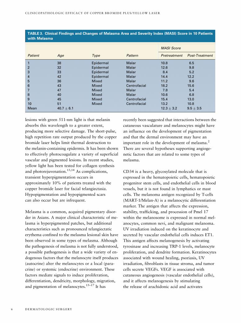
mentation was less after treatment. Fontana-Masson
staining and immunohistochemistry for Melan-A
were performed to investigate changes in melanin in
the melasma lesions after treatment. In the Fontana-
Masson–stained sections, the amount of melanin in
the basal layer of epidermis was lower after the
treatment. Melan-A immunostaining showed a
marked decrease of melanosomes detected by
Melan-A in the epidermis (Figure 3A). To examine
whether vascularity in the melasma lesions was
lower after the treatment, the expression of CD34
and VEGF were examined using immunohisto-
chemistry. CD34 immunostaining showed a marked
decrease in the number and size of blood vessels in
the dermis (Figure 3B). Less positive immunoreac-
tivity against VEGF was noticed in keratinocytes
after treatment (Figure 3C).
Clinically, the treatment was tolerated well without
anesthetics. Transient erythema was observed on the
laser-treated site until 2 days after the laser treat-
ment, but none of the patients noted any long-term
adverse effects, including scarring and postinflam-
matory hyperpigmentation and hypopigmentation.
Long-term follow-up examination using photo-
graphs was done 3 months after the last treatment.
At the 3-month follow-up visit, melasma lesions
show no change from 1 month after the last
treatment. At 6 months after the last treatment,
recurrence of melasma was observed in three pa-
tients (patient numbers 6, 9, and 10).
Discussion
The copper bromide laser is unique because it offers
a dual-wavelength output; 511 nm in the green and
578 nm in the yellow are produced simultaneously. It
emits light with a short pulse duration of 24 ns. The
pulse repetition rate is in the range of 12,000 Hz.
This repetition rate is high enough that the beam
appears to be continuous to the human eye, that is,
quasicontinuous. The individual pulse cannot supply
sufficient thermal energy to coagulate the vessels
being treated. The summation of the thermal energy
from numerous pulses will coagulate the vessels.
Favorable results have been reported when treating
facial telangiectasia with the copper bromide
laser.11,12 The advantage of treating pigmented
Figure 2. (A) At the highest point of the cheekbone, L� in-creased from 56.7 before the treatment to 57.0 after onesession, 57.6 after two sessions, 59.0 after three sessions,and 59.2 after four sessions and dropped to 58.4 at the 1-month follow-up visit (po.01). (B) The a� values decreasedaccording to the treatments. Quantification of a� at thehighest point of the cheekbones revealed a substantial de-crease from 14.8 before the treatment to 13.5 after one ses-sion, 13.3 after two sessions, 12.0 after three sessions, and10.9 after four sessions and a slight increase to 11.8 at the 1-month follow-up visit (po.01).
TABLE 2. Average Improvement (% Change)
Telangiectasia Pigmented Lesions
Patient 82.47 12.6 71.57 11.2
Clinician 76.57 8.5 65.47 8.2
3 6 :* * : 2 0 1 0 5
L E E E T A L
lesions with green 511-nm light is that melanin
absorbs this wavelength to a greater extent,
producing more selective damage. The short-pulse,
high repetition rate output produced by the copper
bromide laser helps limit thermal destruction to
the melanin-containing epidermis. It has been shown
to effectively photocoagulate a variety of superficial
vascular and pigmented lesions. In recent studies,
yellow light has been tested for collagen synthesis
and photorejuvenation.13,14 As complications,
transient hyperpigmentation occurs in
approximately 10% of patients treated with the
copper bromide laser for facial telangiectasia.
Hypopigmentation and hypopigmented scars
can also occur but are infrequent.
Melasma is a common, acquired pigmentary disor-
der in Asians. A major clinical characteristic of me-
lasma is hyperpigmented patches, but additional
characteristics such as pronounced telangiectatic
erythema confined to the melasma lesional skin have
been observed in some types of melasma. Although
the pathogenesis of melasma is not fully understood,
a possible pathogenesis is that a wide variety of en-
dogenous factors that the melanocyte itself produces
(autocrine) alter the melanocytes or a local (para-
crine) or systemic (endocrine) environment. These
factors mediate signals to induce proliferation,
differentiation, dendricity, morphology, migration,
and pigmentation of melanocytes.15–17 It has
recently been suggested that interactions between the
cutaneous vasculature and melanocytes might have
an influence on the development of pigmentation
and that the dermal environment may have an
important role in the development of melasma.2
There are several hypotheses supporting angioge-
netic factors that are related to some types of
melasma.
CD34 is a heavy, glycosylated molecule that is
expressed in the hematopoietic cells, hematopoietic
progenitor stem cells, and endothelial cells in blood
vessels, but it is not found in lymphatics or mast
cells. The melanoma antigen recognized by T-cells
(MART-1/Melan-A) is a melanocytic differentiation
marker. The antigen that affects the expression,
stability, trafficking, and procession of Pmel 17
within the melanosome is expressed in normal mel-
anocytes, common nevi, and malignant melanoma.
UV irradiation induced on the keratinocyte and
secreted by vascular endothelial cells induces ET1.
This antigen affects melanogenesis by activating
tyrosinase and increasing TRP-1 levels, melanocyte
proliferation, and dendrite formation. Keratinocytes
associated with wound healing, psoriasis, UV
irradiation, fibroblasts in tissue stroma, and tumor
cells secrete VEGFs. VEGF is associated with
cutaneous angiogenesis (vascular endothelial cells),
and it affects melanogenesis by stimulating
the release of arachidonic acid and activates
TABLE 3. Clinical Findings and Changes of Melasma Area and Severity Index (MASI) Score in 10 Patients
with Melasma
Patient Age Type Pattern
MASI Score
Pretreatment Post-Treatment
1 38 Epidermal Malar 10.8 6.5
2 32 Epidermal Malar 12.6 9.8
3 33 Epidermal Malar 8.4 5.2
4 42 Epidermal Malar 14.4 12.2
5 36 Mixed Malar 11.2 9.6
6 43 Mixed Centrofacial 18.2 15.6
7 47 Mixed Malar 7.8 5.4
8 40 Mixed Malar 10.6 6.8
9 45 Mixed Centrofacial 15.4 13.0
10 51 Mixed Centrofacial 13.2 10.8
Mean 40.77 6.1 12.37 3.2 9.57 3.5
D E R M AT O L O G I C S U R G E RY6
C L I N I C O PAT H O L O G I C E F F I C A C Y O F C O P P E R B R O M I D E P L U S / Y E L L O W L A S E R
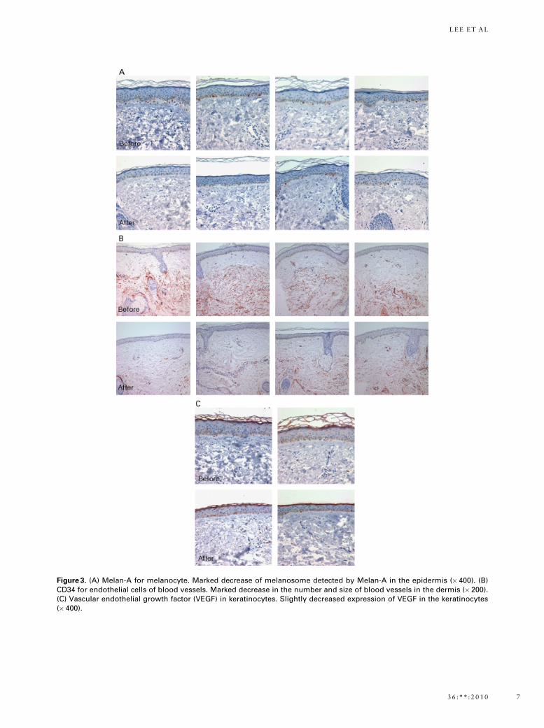
Figure 3. (A) Melan-A for melanocyte. Marked decrease of melanosome detected by Melan-A in the epidermis (�400). (B)CD34 for endothelial cells of blood vessels. Marked decrease in the number and size of blood vessels in the dermis (� 200).(C) Vascular endothelial growth factor (VEGF) in keratinocytes. Slightly decreased expression of VEGF in the keratinocytes(� 400).
3 6 :* * : 2 0 1 0 7
L E E E T A L

phospholipase A2. A possible effect of VEGF on
function of melanocytes is expected in that human
melanocytes express VEGF receptors.
Epidermal keratinocytes have a primary role in the
physiology and pathology of cutaneous angiogene-
sis.18 Moreover, epidermis-derived VEGF is regarded
as a potent angiogenic factor in many cutaneous
diseases.19 Keratinocytes in the skin constitutively
produce VEGF. Its production is up-regulated in
psoriasis, wound healing, and other states of in-
creased skin angiogenesis, as well as by UV irradia-
tion.20–22 In particular, several reports have suggested
that VEGF is induced in human keratinocytes after
UV exposure.23,24 Kim and colleagues25 demon-
strated that acute exposure to UV radiation triggers
angiogenesis via VEGF induction and MEK–ERK1/2
activation and that all-trans retinoic acid (tRA) in-
hibits UV-induced ERK1/2 activation, VEGF upreg-
ulation in keratinocytes, and angiogenesis in human
skin. Also, it was recently reported that the topical
plasmin inhibitor is an effective treatment for UV-
induced hyperpigmentation.3 Lee and colleagues4
suggested that the intralesional localized microinjec-
tion of tranexamic acid can be used as a potentially
new therapeutic modality for the treatment of me-
lasma. The following inflammatory mediators have
been reported to increase melanogenesis: interleukins
(IL-1a, IL-1b, and IL-6), tumor necrosis factor alpha,
eicosanoids (prostaglandins D2, E2, F2, and leuko-
triene B4), and histamine. Tranexamic acid inhibits
UV-induced plasmin activity in keratinocytes by
preventing the binding of plasminogen to the
keratinocytes, which ultimately results in fewer free
arachidonic acids and a diminished ability to produce
prostaglandins, and this decreases melanocyte tyros-
inase activity.
Our results show that expression of VEGF in kera-
tinocytes decreased slightly after treatment with
578-nm copper bromide yellow laser. Therefore, we
expected that the yellow laser would have some
direct or indirect effects on melanogenesis through
the effect on VEGF in keratinocytes, dermal angio-
genesis, and inflammatory mediators, but in vitro
and in vivo studies are needed for demonstrating a
clear mechanism for the antiangiogenic and anti-
melanogenic effects of yellow laser.
Kim and colleagues25 demonstrated that greater
vascularity is one of the major findings in melasma
and that VEGF may be a major angiogenic factor for
altered vessels in melasma. The biological role of
cutaneous blood vessels in the pathogenesis of me-
lasma remains unclear. VEGF is known to stimulate
the release of arachidonic acid and the phosphor-
ylation and activation of cytosolic phospholipase
A2.26 It is possible that the resulting metabolites
from the arachidonic acid pathway affect melano-
genesis.27 Human melanocytes may respond to
angiogenic factors because normal human me-
lanocytes express functional VEGF receptors.2
Therefore, VEGF may have a direct influence on
melanocyte behavior through its receptor.
The copper bromide Plus/Yellow Laser, which pro-
duces green (511 nm) and yellow (578 nm) wave-
lengths, affects the altered dermal vasculature and
epidermal melanin pigmentation in melasma lesions.
It is unclear why melasma is improved after yellow
laser treatment. It is also not clear whether this laser
has a direct effect on the dermal vasculature and
epidermal melanin or an indirect effect on the
epidermal VEGF. In vitro and in vivo studies are
needed to demonstrate a clear mechanism for the
antiangiogenic effects of yellow laser. We suggest
that this antiangiogenetic laser may be a treatment
option for melasma specially accompanied by pro-
nounced telangiectasia. A study using more vascular-
specific lasers such as V-Beam (Candela Corpora-
tion, Wayland, MA) for the treatment of melasma
may provide us with new insights into the patho-
genesis of melasma. Further controlled spilt-face
designed studies are needed to achieve more
improvements for melasma treated with copper
bromide Plus/Yellow Laser.
Acknowledgment This study was supported by the
Chung-Ang University Research Grants in 2010.
D E R M AT O L O G I C S U R G E RY8
C L I N I C O PAT H O L O G I C E F F I C A C Y O F C O P P E R B R O M I D E P L U S / Y E L L O W L A S E R

References
1. Urabe K, Nakayama J, Hori Y. In: Nordlund JJ, Boissy RE,
Hearing VJ, King RA, Ortonne J-P, editors. Melasma pigmentary
system: physiology and pathophysiology. New York: Oxford
University Press; 1997. p. 909–11.
2. Kim EJ, Park HY, Yaar M, et al. Modulation of vascular endo-
thelial growth factor receptors in melanocytes. Exp Dermatol
2005;14:625–33.
3. Maeda K, Naganuma M. Topical trans-4-aminomethylcyclo-
hexanecarboxylic acid prevents ultraviolet radiation-induced pig-
mentation. J Photochem Photobiol B: Biol 1998;47:136–41.
4. Lee JH, Park JG, Lim SH, et al. Localized intradermal microin-
jection of tranexamic acid for treatment of melasma in Asian
patients: a preliminary clinical trial. Dermatol surg 2006;32:
626–33.
5. Kim EH, Kim YC, Lee ES, Kang HY. The vascular characteristics
of melasma. J Dermatol Sci 2007;46:111–6.
6. Manaloto RM, Alster T. Erbium: YAG laser resurfacing for re-
fractory melasma. Dermatol Surg 1999;25:121–3.
7. Angsuwarangsee S, Polnikorn N. Combined ultrapulse CO2 laser
and Q-switched alexandrite laser compared with Q-switched
alexandrite laser alone for refractory melasma: split face design.
Dermatol Surg 2003;29:59–64.
8. Rokhsar CK, Fitzpatrick RE. The treatment of melasma with
fractional photothermolysis: a pilot study. Dermatol Surg
2005;31:1645–50.
9. Spicer MS, Goldberg DJ. Lasers in dermatology. J Am Acad
Dermatol 1996;34:1–25.
10. Dinehart SM, Waner M, Flock S. The copper vapor laser for
treatment of cutaneous vascular and pigmented lesions.
J Dermatol Surg Oncol 1993;19:370–5.
11. Key JM, Maver M. Selective destruction of facial telangiectasia
with a copper vapor laser. Arch Otolaryngol 1992;118:509–13.
12. McCoy S, Hanna M, Anderson P, et al. An evaluation of the
copper-bromide laser for treating telangiectasia. Dermatol Surg
1996;22:551–7.
13. McCoy S. Photorejuvenation of facial skin using a copper bromide
laser at 578 and 511 nm-comparative results of a 2 hemifacial
treatment, Lasers in Medical Science 2003;18:Abstract No.07
14. Cassuto DA. Nonablative photorejuvenation with a scanned
copper bromide laser. Lasers in Surgery and Medicine 2003;
ASLMS: Abstract No.54
15. Slominski A, Wortsman J, Mazurkiewicz JE, et al. Detection of
proopiomelanocortin-derived antigens in normal and pathologic
human skin. J Lab Cutan Med 1993;122:658–66.
16. Slominski A, Heasley D, Mazurkiewicz JE, et al. Expression of
proopiomelanocortin (POMC)-derived melanocyte-stimulating
hormone (MSH) peptides in skin of basal cell carcinoma patients.
Hum Pathol 1996;30:208–15.
17. Nagahama M, Funasaka Y, Fernandez-Frez ML, et al. Immuno-
reactivity of a-melanocyte-stimulating hormone,
adrenocorticotrophic hormone and b-endorphin in cutaneous
malignant melanoma and benign melanocytic naevi. Br J
Dermatol 1998;138:981–5.
18. Malhotra R, Stenn KS, Fernandez LA, Braverman IM. Angiogenic
properties of normal and psoriatic skin associate with epidermis,
not dermis. Lab Invest 1989;61:162–5.
19. Bowden J, Brennan PA, Umar T, Cronin A. Expression of vascular
endothelial growth factor in basal cell carcinoma and cutaneous
squamous cell carcinoma of the head and neck. J Cutan Pathol
2002;29:585–9.
20. Yano K, Kajiya K, Ishiwata M, et al. Ultraviolet B-induced skin
angiogenesis is associated with a switch in the balance of vascular
endothelial growth factor and thrombospondin-1 expression.
J Invest Dermatol 2004;122:201–2.
21. Lauer G, Sollberg S, Cole M, et al. Expression and proteolysis of
vascular endothelial growth factor is increased in chronic wounds.
J Invest Dermatol 2000;115:12–8.
22. Gille J, Reisinger K, Asbe-Vollkopf A, et al. Ultraviolet-A-induced
transactivation of the vascular endothelial growth factor gene in
HaCaT keratinocytes is conveyed by activator protein-2 tran-
scription factor. J Invest Dermatol 2000;115:30–6.
23. Mildner M, Weninger W, Trautinger F, et al. UVA and UVB ra-
diation differentially regulate vascular endothelial growth factor
expression in keratinocyte-derived cell lines and in human kera-
tinocytes. Photochem Photobiol 1999;70:674–9.
24. Blaudschun R, Brenneisen P, Wlaschek M, et al. The first peak
of the UVB irradiation-dependent biphasic induction of
vascular endothelial growth factor (VEGF) is due to phosphor-
ylation of the epidermal growth factor receptor and independent
of autocrine transforming growth factor alpha. FEBS Lett
2000;474:195–200.
25. Kim MS, Kim YK, Eun HC, et al. All-trans retinoic acid antag-
onizes UV-induced VEGF production and angiogenesis via the
inhibition of ERK activation in human skin keratinocytes. J Invest
Dermatol 2006;126:2697–706.
26. Wheeler-Jones C, Abu-Ghazaleh R, Cospedal R, et al. Vascular
endothelial growth factor stimulates prostacyclin production and
activation of cytosolic phopholipase A2 in endothelial cells via
p42/p44 mitogen-activated protein kinase. FEBS Lett
1997;420:28–32.
27. Abdel-Malek Z, Kadekaro AL. Human pigmentation: its
regulation by ultraviolet light and by endocrine, paracrine, and
autocrine factors. In: Nordlund JJ, Boissy RE, Hearing VJ, King
RA, Oetting WS, Ortonne JP, editors. The pigmentary system.
Oxford: Blackwell Publishing Ltd; 2006. p. 410–20.
Address correspondence and reprint requests to: Prof.Beom Joon Kim, MD, PhD, Department of Dermatology,Chung-Ang University College of medicine, 65-207Hangangro 3-ka, Yongsan-gu, Seoul 140-757, SouthKorea, or e-mail: [email protected]
3 6 :* * : 2 0 1 0 9
L E E E T A L