Clinico-radiological Correlation of Bone Changes in ...ijl.org.in/2016/1 W Mohammad et al...
13
Leprosy is a medical- social disease, it is associated with stigma in the society due to the resulting deformities in some persons. Although stigma has decreased after the widespread use of MDT, some disabilities do occur which are mostly due to late initiation of treatment and inappropriate care. Besides the nerve and skin involvement bone changes have been reported to be common in leprosy. These bony changes need to be understood in the present MDT era specially in the context of clinical spectrum and duration of disease/ deformities. Fifty clinically diagnosed and histologically classified leprosy patients with deformities/ disabilities of either hands/feet/ face who attended the OPD of Department of Dermatology, Venereology and Leprosy, Government Medical College, Amritsar were examined and evaluated in the study. Radiological examination of hands, feet and skull was done in each case and the bone changes in hands and feet; and skull and paranasal sinus changes were correlated with clinical parameters. Bone changes were observed in 90% of cases radiologically. Specific bone changes in hands and feet, non-specific bone changes in hands, feet, skull and paranasal sinuses were seen in 66%, 82% and 32% of cases respectively. Common specific bone changes in hands and feet observed were primary periostitis (14%), honey combing (46%), bone cyst (36%), thinning and irregularity of cortex (28%) and area of bone destruction (20%); Among the non-specific bone changes observed were contracted fingers/claw hands/claw toes (64%) and absorption of terminal phalanges (40%). The maxillary sinus, and paranasal sinus changes were the most common radiological findings observed in skull. The study of the radiological changes may help the clinicians to understand the gravity of the situation and undertake steps for timely prevention of permanent loss of function and the occurrence of deformities and disabilities. Key words : Leprosy, Deformities, Radiological changes http://www.ijl.org.in Original Article Received : Accepted : 26.06.2015 28.02.2016 Indian J Lepr 2016, 88 : 83-95 © Hind Kusht Nivaran Sangh, New Delhi 1 W Mohammad, MBBS, MD, At present working as Medical Officer, District Hospital, Fatehgarh Sahib, Punjab 2 SK Malhotra, MBBS, MD, Professor & Head of Department of Dermatology, Venereology and Leprosy, Government Medical College, Amritsar, Punjab 3 PK Garg, MBBS, Junior Resident, Department of Dermatology, Venereology and Leprosy, Government Medical College, Amritsar, Punjab Corresponding author: Dr SK Malhotra, Address: HIG 943, Sector-3, Ranjit Avenue, Amritsar, Punjab, India - 143001 Email: [email protected] Clinico-radiological Correlation of Bone Changes in Leprosy Patients Presenting with Disabilities/Deformities 1 2 3 W Mohammad , SK Malhotra , PK Garg Introduction Leprosy is a chronic granulomatous disease caused by Mycobacterium leprae, an acid fast rod shaped bacillus which was discovered by Armauer Hansen in 1873 in Norway. The disease mainly affects the peripheral nerves and the skin and
Transcript of Clinico-radiological Correlation of Bone Changes in ...ijl.org.in/2016/1 W Mohammad et al...

Leprosy is a medical- social disease, it is associated with stigma in the society due to the resulting deformities
in some persons. Although stigma has decreased after the widespread use of MDT, some disabilities do occur
which are mostly due to late initiation of treatment and inappropriate care. Besides the nerve and skin
involvement bone changes have been reported to be common in leprosy. These bony changes need to be
understood in the present MDT era specially in the context of clinical spectrum and duration of disease/
deformities. Fifty clinically diagnosed and histologically classified leprosy patients with deformities/
disabilities of either hands/feet/ face who attended the OPD of Department of Dermatology, Venereology and
Leprosy, Government Medical College, Amritsar were examined and evaluated in the study. Radiological
examination of hands, feet and skull was done in each case and the bone changes in hands and feet; and skull
and paranasal sinus changes were correlated with clinical parameters. Bone changes were observed in 90% of
cases radiologically. Specific bone changes in hands and feet, non-specific bone changes in hands, feet, skull
and paranasal sinuses were seen in 66%, 82% and 32% of cases respectively. Common specific bone changes
in hands and feet observed were primary periostitis (14%), honey combing (46%), bone cyst (36%), thinning
and irregularity of cortex (28%) and area of bone destruction (20%); Among the non-specific bone changes
observed were contracted fingers/claw hands/claw toes (64%) and absorption of terminal phalanges (40%).
The maxillary sinus, and paranasal sinus changes were the most common radiological findings observed in
skull. The study of the radiological changes may help the clinicians to understand the gravity of the situation
and undertake steps for timely prevention of permanent loss of function and the occurrence of deformities
and disabilities.
Key words : Leprosy, Deformities, Radiological changes
http://www.ijl.org.in
Original Article
Received : Accepted : 26.06.2015 28.02.2016
Indian J Lepr 2016, 88 : 83-95© Hind Kusht Nivaran Sangh, New Delhi
1 W Mohammad, MBBS, MD, At present working as Medical Officer, District Hospital, Fatehgarh Sahib, Punjab2 SK Malhotra, MBBS, MD, Professor & Head of Department of Dermatology, Venereology and Leprosy,
Government Medical College, Amritsar, Punjab3 PK Garg, MBBS, Junior Resident, Department of Dermatology, Venereology and Leprosy, Government Medical College,
Amritsar, Punjab
Corresponding author: Dr SK Malhotra, Address: HIG 943, Sector-3, Ranjit Avenue, Amritsar, Punjab, India - 143001
Email: [email protected]
Clinico-radiological Correlation of Bone Changes in LeprosyPatients Presenting with Disabilities/Deformities
1 2 3W Mohammad , SK Malhotra , PK Garg
Introduction
Leprosy is a chronic granulomatous disease
caused by Mycobacterium leprae, an acid fast rod
shaped bacillus which was discovered by Armauer
Hansen in 1873 in Norway. The disease mainly
affects the peripheral nerves and the skin and

Mohammad et al84
sometimes other tissues, notably the eyes, the
mucosa of the upper respiratory tract, muscles,
bones and testes. Clinical spectrum of leprosy can
vary from the presence of an insignificant area of
hypopigmented skin that heals spontaneously, to
widespread involvement of skin, damage to
peripheral nerves, eyes, bones, muscles and
other tissues with the development of defor-
mities and disability. The whole spectrum from
the Indeterminate, Tuberculoid to the Lepro-
matous disease has been divided into five types
based on clinical, bacteriological, histological
and immunological features (Ridley and Jopling
1966, IAL classification 1982). Pure Neuritic type
is seen especially in India (IAL classification 1982).
The disease itself is not directly responsible for
most of the deformities. Leprosy character-
istically results in extensive nerve damage leading
to varying loss of sensation of touch, temperature
and pain and so the patient unknowingly
damages and deform himself/herself. Deformity
is defined as alteration in the form, shape or
appearance of a part of the body, whereas
disability is deterioration in the ability or capacity
of an individual (Dharmendra 1978). Deformities
of limbs in leprosy are of two types, primary and
secondary. Primary deformities are directly the
results of the invasion of the tissues by M. leprae
or the result of reaction of the body to the
infection. The most important primary disability is
the loss of nerves and nerve endings resulting in
loss of sensation, damage to the part, most often
repeatedly, paralysis and deformities. The loss of
sensation makes the patient liable to injuries
because of continuous use of insensitive hands
and feet which are repeatedly injured/wounded
and may also be secondarily infected. This
results in ulceration and absorption of digits,
amputations which lead to severe disability and is
a cause of stigma to the patient and the society.
Brand (1966) emphasized on the impact of
paralysis of nerves and reported that nonspecific
infection and trauma are the reasons for bone
resorption in 98% of cases (MacMoran and
Brand 1987).
Disabilities and deformities in leprosy patients
are not an inevitable result of the disease except
in few, such as collapse of the nose or loss of
eyebrows in untreated advanced lepromatous
leprosy. The majority of disabilities and defor-
mities are attributable to peripheral nerve
damage which occurs in two particular phases
of the disease, namely in the long untreated
patients and during the course of both Type I and
TypeII lepra reactions (Pearson and Ross 1975).
Early diagnosis, regular and adequate treatment,
and early recognition of decreasing nerve
function are of utmost importance for prevention
and possible reversal of nerve damage.
M. leprae has a predilection for infecting tissues in
the cooler areas of the body, consequently the
characteristic bone lesions are seen in the acral
parts of the body. Bone changes attributed to
leprosy may be divided into two groups, 'specific'
and 'non-specific' in origin. Specific bone lesions
are the direct effect of M. leprae invasion (leprous
osteitis) whereas non specific lesions are the
result of trauma and infection of the tissues
denervated by leprous infection (Paterson 1961,
Lechat 1962).
With the widespread use of multi-drug therapy
(MDT), clinical profile of disease is changing. For
providing proper care we need to study the profile
of disease in different settings specially the extent
of involvement of different organs. The main aim
of this study was to evaluate the pattern of bone
changes in leprosy patients with disabilities/
deformities of hands, feet and face and to
correlate the clinical findings and duration of
disease/deformities with radiological changes in
them.

Clinico-radiological Correlation of Bone Changes in Leprosy Patients Presenting with Disabilities/Deformities 85
Materials and Methods
Fifty clinically diagnosed leprosy patients with
deformities/disabilities of either hands/feet
and/or face irrespective of their age, sex,
occupation and treatment status were selected
for this study. These patients were attending the
leprosy clinic of Skin and STD Department, Guru
Nanak Dev Hospital/Govt. Medical College,
Amritsar. Ethical clearance for the study protocol
was obtained from the Institutional Ethical
Committee of the Medical College, Amritsar. An
informed consent was obtained from all the
patients included in the study.
Clinical evaluation was done with particular
reference to the age, sex, occupation, duration
and type of leprosy, duration of deformity, lepra
reaction, trophic ulcers and other secondary
changes. Routine investigations including slit
skin smear and skin biopsy were done in each case
for were done in each case which included
anteroposterior and lateral views of the hands,
feet and skull and Water's view for ascertaining
the involvement of paranasal sinuses.
Disability index (DI), which indicates severity of
disabilities/deformities was calculated using the
formula proposed by Bechelli and Domingquez
(1971). DI is related to the maximum grades of
disabilities. The grades of deformities/disabilities
of right and left sides of hands, feet and face were
added and it was divided by the number of sites
studied i.e. 6 (Table 1). Disability index was
calculated in these leprosy patients having
disabilities/deformities according to WHO
disability grading system (WHO 1970). Various
radiological changes in hands, feet and skull were
then correlated with the clinical parameters such
as age, sex, occupation, type of leprosy, duration
of disease, deformity, disability index, reaction
and treatment status.
Results
Important findings of the present study are
summarized in Tables 1 to 8 and illustrated in
Figures 1 to 6. The mean age of the patients
included, was 41.7 years (range from 10-90
years). Genderwise distribution of cases was
34 males and 16 females Majority of patients
Table 1 : Method of Calculation of Disability Index [DI] (Bechelli and Dominoguez 1971)
Sign Hand Foot Eye EyeRight Left Right Left Right Left
Trophic ulcer Max 2; Max 2; Conjunctivitis/ Max 2;Minimum zero Minimum zero keratitis Minimum zero
Mobile claw/foot drop Lagopthalmos
Fixed deformity, Min 2: Max 3 Min 2: Max 3 Iritis/Iridocyclitis/ Min 2: Max 3absorption, severe loss ofstiff joints vision
Total
Fig 1 : Types of injuries in the anaesthetichands and feet

Mohammad et al86
31 (62%) were in the age group of 20-59 years.
Profession wise predominant among them were
labourers (24 i.e. 48%;) followed by housewives
(14; i.e. 28%) 5 office workers (10%), 3 patients
were students, 3 farmers (6% each) and one
businessman (2%). A good number of patients
(23; 46%) were having disease from the last 1-5
years while the rest (27; 54%) were suffering from
the diseases for more than 5 years. Forty three
patients had MB disease (LL-21, BL-7, BB-3,
Polyneuritc 12) while rest had BT disease (BT-7).
No case of tuberculoid type (TT) was seen in the
study group.
The duration of presence of deformities/ disabilities in these leprosy patients was for< 1 year in 14 patients (28%); it ranged between
1-5 years in 20 (40%) patients followed by more
than 5 years in 16 (32%) of cases. Majority of
patients 36 (72%) were receiving regular
treatment for a variable period of time. Fourteen
(28%) cases were untreated and presented with
deformities. Majority of patients (84%) presented
with weakness, followed by anaesthesia of hands
and feet in 78% of cases and claw hands and toes
in 74% of cases (Table 5). One case was also having
Type I lepra reaction and 4 cases had Type II lepra
reaction. A substantial proportion of patients (i.e.
21; 42%) had a disability index between 0.16 to
0.5, one (2%) case had a disability index ranging
between 1.66-2.0, and the rest had between >0.5
to 1.66 (28; 56%).
Fig 2 : Skiagram of left hand with absorption
of middle & distal phalanges of index, middle
and ring finger; eccentric absorption of corres-
ponding proximal phalanges; clawing of little
finger and thumb; Right hand skiagramshows
absorption of middle and distal phalanges of
middle and little finger; eccentric absorption of
proximal phalanx of middle finger; arthrodesis
of proximal and distal interphalangeal joint
of ring finger; concentric and eccentric
absorption of distal and middle phalanges of
ring finger.
Fig 3 : Showing left ulnar claw hand
Fig 4 : Ray showing bony changes in hand with
clawing of left little finger

Clinico-radiological Correlation of Bone Changes in Leprosy Patients Presenting with Disabilities/Deformities 87
The overall prevalence of bone changes in these
patients was 90% (45/50 patients). Several
patients had more than one type of bone
changes, as well they involved multiple limbs
Table 2 : Specific bone changes observed radiologically in hands and feet
Specific bone changes No. of cases showing radiological changesin hands and feet Hands Feet Both hands Total In percentage
alone alone and feet number ofcases
Primary periostitis 2 5 - 7 14.0
Honey combing 4 8 11 23 46.0
Bone cyst 4 11 3 18 36.0
Enlargement of nutrient foramen 2 1 - 3 6.0
Thinning and irregularity of cortex 4 4 6 14 28.0
Concentric cortical erosion 1 4 - 5 10.0
Area of bone destruction 2 7 1 10 20.0
Sub-articular erosion 0 1 2 3 6.0
Sclerosis 5 3 - 8 16.0
Any other - - - - -
Fig 5 : Showing depressed bridge of nose in
lepromatous leprosy
Fig 6 : Showing diffuse infiltration of face, loss
of eye brows, destruction of nasal septum

Mohammad et al88
Table 3 : The non specific bone changes seen radiologically in hands and feet
Non-specific bone changes No. of cases showing radiological changesin hands and feet Hands Feet Both hands Total In percentage
alone alone and feet number ofcases
Absorption of the following
o Terminal phalanges 5 5 10 20 40.0
o Middle phalanges 3 10 3 16 32.0
o Proximal phalanges - 8 1 9 18.0
o Metacarpals - - - - -
o Metatarsals - 1 - 1 2.0
Soft tissue changes - 7 1 8 16.0
Concentric absorption 4 9 3 16 32.0
Contracted fingers / 24 3 5 32 64.0
claw hand/claw toes
Tuft erosion 3 7 6 16 32.0
Arthritis 5 3 1 9 18.0
Subluxation and/or dislocation 5 7 4 16 32.0
Cupping of joints - 8 3 11 22.0
Fractures - 2 - 2 4.0
Secondary periostitis 1 3 - 4 8.0
Osteomyelitis - 3 - 3 6.0
Disintegration of tarsal bones - 4 - 4 8.0
Eccentric absorption 1 6 2 9 18.0
Others - 16 - 16 32.0
Table 4 : Bony changes detected radiologically in skull and paranasal sinuses
Right Left
Maxillary antrum
· Diffuse opacity 4 5
· Local mucosal thickening 5 4
· Generalized mucosal thickening 9 6
Ethmoidal sinus sinus 1 -
Frontal sinus - -
Atrophy/loss of anterior nasal spine - -
Atrophy/loss of alveolar process of maxilla - -
(Table 2). These radiological changes were
further sub-grouped into specific and non-
specific bone changes in hands, feet; skull and
paranasal sinus.
Specific bone changes in hands and feet : Specific
bone changes in hands and feet were seen
in 66% of patients. Various changes with their
frequencies are shown in Table 2.
Non specific bone changes in hands and feet : Non
specific bone changes in anaesthetic hands and
feet (Fig 1), claw hand (3) are shown in Fig 2
and 4 respectively, these are also summarized in
Table 3. Such changes were seen in 82% of
patients. Several of patients with specific bone
changes also had bone changes attributable to
non-specific reasons and multiple limbs were
involved in these non specific changes. The
individual changes with their frequencies are
shown in Table 3.
Skull and paranasal sinus changes : Among the
bone changes in the skull, majority of bone
changes were seen in maxillary antrum (Fig. 5)
followed by ethmoidal sinus in one case only.
Clinically extensive facial and nasal involvement
was visible in some of such cases (Fig. 6). Various
changes with their frequencies are shown in
(Table 4).
Clinico-radiological correlation
All the patients i.e. 100% showing specific bone
changes in hands and feet and 46% who had
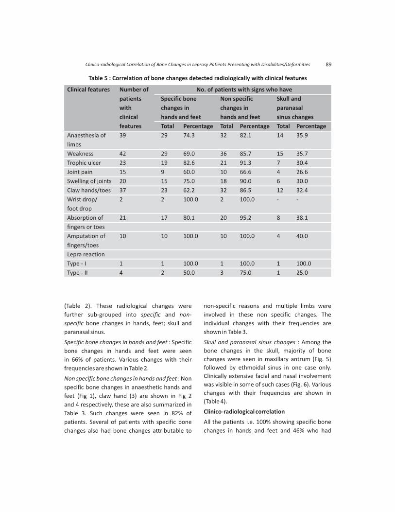
Table 5 : Correlation of bone changes detected radiologically with clinical features
Clinical features Number of No. of patients with signs who have
patients Specific bone Non specific Skull and
with changes in changes in paranasal
clinical hands and feet hands and feet sinus changes
features Total Percentage Total Percentage Total Percentage
Anaesthesia of 39 29 74.3 32 82.1 14 35.9
limbs
Weakness 42 29 69.0 36 85.7 15 35.7
Trophic ulcer 23 19 82.6 21 91.3 7 30.4
Joint pain 15 9 60.0 10 66.6 4 26.6
Swelling of joints 20 15 75.0 18 90.0 6 30.0
Claw hands/toes 37 23 62.2 32 86.5 12 32.4
Wrist drop/ 2 2 100.0 2 100.0 - -
foot drop
Absorption of 21 17 80.1 20 95.2 8 38.1
fingers or toes
Amputation of 10 10 100.0 10 100.0 4 40.0
fingers/toes
Lepra reaction
Type - I 1 1 100.0 1 100.0 1 100.0
Type - II 4 2 50.0 3 75.0 1 25.0
Clinico-radiological Correlation of Bone Changes in Leprosy Patients Presenting with Disabilities/Deformities 89

changes in skull and paranasal sinus changes were
in the age group of more than 60 years. While
100% of patients in the age group of 0-19 years
showed only non specific bone changes in hands
and feet. There was no significant difference in
specific bone changes and skull and paranasal
sinus changes (p >0.05) in the males as compared
to females. The differences were significant in
case of non-specific bone changes (p<0.05), being
more in women than in men. Occupation wise
specific bone changes in hands and feet were
seen in 100% cases of farmers while nonspecific
bone changes in hands, feet, skull and paranasal
sinuses was noted in them. These were seen in
100% of cases of students and businessmen. In
the present study, the highest percentage i.e.
90% of patients showing specific and non specific
bone changes in hands and feet belonged to LL
type of leprosy while highest percentage i.e. 42%
of patients having skull and paranasal sinus
changes were of the LL and BL types.
As summarised in Table 6, specific bone changes
in hands and feet were seen in 100 % of patients
with amputation of fingers/toes, wrist drop/foot
drop and Type I lepra reaction; 82.6% with trophic
ulcer; 80.1% with absorption of fingers/toes; 75%
with swelling of joints; and 50% with Type II lepra
reaction. Non-specific bone changes in hands and
feet were seen in all cases with wrist drop/foot
drop, amputation of fingers/toes and Type I
reaction. Skull and paranasal sinus changes were
seen in one case of Type I reaction 4/10 cases with
amputation of fingers/toes, 8/21 cases with
absorption of fingers or toes. Three out of four
cases of Type II lepra reaction also showed this
type of bone changes.
Table 6 : Comparison of frequency of various specific bone changes in hands and feetby various investigators with present study
18S.No. Specific bone changes Paterson Chhabriya Thappa Choudhuri Present8 23 9in hands and feet et al et al et al study
1. Bone cyst 2.8% 22.0% 10.5% 22.7% 36.0%
2. Subarticular erosion - 10.0% 10.5% 10.0% 6.0%
3. Enlargement of nutrient 1.5% 2.0% 5.3% 4.5% 6.0%
foramens
4. Primary periostitis - 4.0% 1.3% 28.2% 14.0%
5. Concentric cortical erosion 0.2% 8.0% 1.3% 10.0% 10.0%
6. Honeycombing 3.0% 6.0% - - 46.0%
7. Thinning and irregularity - 4.0% - - 28.0%
of cortex
8. Ill defined areas of bone 1.0% 8.0% - - 20.0%
destruction
9. Sclerosis with
·No deformity 2.6% 14.0% - - 16.0%
·With deformity 1.6% - - - -
10. Pseudocysts 5.0% - - - -
11. Subarticular collapse 1.2% - - - -
12. Cortical areas destroyed 0.7% - - - -
Mohammad et al90

Table 7 : Comparison of frequency of various non specific bone changes in hands and feetby various investigators with present study
18S.No. Non-specific bone changes Paterson Chhabriya Thappa Choudhuri Present8 23 9in hands and feet et al et al et al study
1. Absorption of
o Terminal phalanges 8.0% 84.0% 59.2% 48.2% 40.0%
o Middle phalanges 8.0% 72.0% 34.2% 27.2% 32.0%
o Proximal phalanges 10.0% 60.0% 19.7% 13.6% 18.0%
o Metacarpals - 22.0% 10.5% 10.9% 2.0%
Heads of bones 5.5%
Shafts of bones 7.4%
2. Soft tissue changes - 74.0% 39.5% 44.5% 16.0%
3. Concentric absorption 14.0% 68.0% 39.5% 32.7% 32.0%
4. Contracted fingers/claw hand/ - 38.0% 36.8% 22.7% 64.0%
claw toes
5. Tuft erosion of terminal phalanx 27.0% 56.0% 15.8% 13.6% 32.0%
6. Arthritis
o Acute 4.7%
o Chronic 10.0% 10.0% 14.5% 26.4% 18.0%
7. Subluxation and/or dislocation 4.5% 28.0% 10.5% 18.2% 32.0%
8. Cupping of joints 0.2% 6.0% 7.9% 6.4% 22.0%
9. Fractures - 4.0% 6.6% 3.6% 4.0%
10. Secondary periostitis 9.0% 18.0% 6.6% 3.6% 8.0%
11. Osteomyelitis 5.6% 14.0% 5.3% 4.5% 6.0%
12. Disintegration of tarsal bones 2.1% - 1.3% 1.8% 8.0%
13. Eccentric absorption - 2% - 2.7% 18.0%
14. Acute osteitis 1.0% - - - -
15. Others - - - 22.7% 32.0%
Table 8 : Comparison of the frequency of skull and paranasal sinus changes observedin various studies with present study
S. Skull and paranasal Barnetson Chhabriya Hauhmar Choudhuri Present2 8 12 9No. sinus changes J et al et al et al study
1. Maxillary antrum
Diffuse opacity - 21.4% 1.8% 10%
Local mucosal thickening 87.5% - 28.6% - 10%
Generalised mucosal thickening 100% - 28.4% 4.5% 18%
2. Ethmoidal sinus change 44% - - 0.9% 2%
3. Frontal sinus change 31% - - 1.8% -
4. Atrophy/loss of anterior nasal spine - 10% - - -
5. Atrophy/loss of alveolar process - - - - -
of maxilla
Clinico-radiological Correlation of Bone Changes in Leprosy Patients Presenting with Disabilities/Deformities 91

Discussion
Bone lesions occurring in leprosy patients have
been recognized as an important feature of the
disease for many years (Gass and Rishi 1934,
Barnetson 1950 and 1951, Kozuma 1953,
Job1963, Thapa et al 1992, Choudhuri et al 1999).
Many of these lesions are secondary and not
specifically caused by the leprosy bacillus.
Radiological studies undertaken in selected cases,
will help the clinician to determine the extent of
bone involvement and to suggest the method of
treatment likely to be effective in preventing a
permanent loss of function.
It has long been evident that direct leprous
involvement of bones is one of the factors in bone
absorption. Gass and Rishi (1934) found acid fast
bacilli in the bone marrow. Direct leprous invasion
could result in bone atrophy and destruction
(Kozuma 1959, Job 1963). However these
observations were mostly in the pre MDT era.
One of the most important factors in bone
absorption is leprous osteitis and osteomyelitis
following ulceration and secondary infection.
Even if there is no ulceration of the skin, the
metatarso-phalangeal joints and calcaneum are
the most affected pressure points in such patients
(Skinsnes et al 1972). Specific leprous changes in
the veins lead to disturbance in the normal
tonicity and blood flow and may contribute to
thrombosis at these sites, resulting in ischemia.
These vascular changes may be contributing to
mutilation and deformities of hands and feet
which occur in leprosy (Bansal et al 1987). There is
close relationship between anaesthesia and distal
absorption (Lechat 1962). The disturbance of
reflex vasomotor response following leprous
neuritis is considered to be one of the important
factors in the pathogenesis of neurotrophic
atrophy (Barnetson 1950, 1951). Thus various
probable factors initiating and sustaining defor-
mities of leprosy and bone absorption are
peripheral nerve damage, specific leprous
inflammation, immunologic reactions and
secondary influences (Skinsnes et al 1972).
Bone changes are reported in variable frequ-
encies in different studies. In the present study,
90% patients had bone changes whereas in
the study of Chamberlain et al (1931), Paterson
(1955), Basu (1962), Thappa et al (1992) and
Choudhuri et al (1999), it was 15%, 95%, 91%,
82.9% and 87.3%, respectively. In fact, the
frequency of bone changes is more closely related
to severity of disabilities. In different studies,
specific bone changes in hands and feet were
between 3-44.5%, non-specific bone changes in
hands, feet were between 45-78.9% and skull &
paranasal sinus changes were observed in 9.1%
respectively (Paterson 1961, Chhabriya et al 1985,
Thappa et al 1992, Choudhuri et al 1999).
Frequency of such changes observed in the
present study are compared with various
reported studies in Tables 6,7 and 8. Variations
in the frequencies among various studies
may be due to different time periods reflective
of diagnostic and therapeutic interventions,
geographical factors as well as method of
inclusion of cases in different studies. This
justified the need to continue to address the issue
in different settings as done in the present study.
In the present study, an increase in specific bone
changes in hands, feet, skull and paranasal
sinus was observed with increasing age. This
observation was almost similar to that reported
by Choudhuri et al (1999) and at variance to those
by Paterson (1961) and Thappa et al (1992). There
was no significant difference in specific bone
changes in hands feet, skull and paranasal sinus
changes (p>0.05) in males in comparison to
females while it was significant in case of non-
specific bone changes in hands and feet (p<0.05).
This difference in gender is similar to that
reported by earlier authors. There was some
Mohammad et al92

correlation between farmer occupation and
types of bone changes, however, this and the
other professions were not sufficiently repre-
sented to draw any concrete conclusions.
In the present study, higher percentage of all
types of bone changes were seen in LL and BL
types of leprosy which reflects the extensive
nature of involvement in these types of cases.
An increase in duration of disease (> 5 years)
resulted in higher incidence of specific and non-
specific bone changes in hands and feet.
However, skull and paranasal sinus changes were
seen in patients with disease duration of < 1 year.
Non-specific bone changes in hands, feet, skull
and paranasal sinus changes increased with
increasing duration of deformities which were
similar to those reported by various authors. All
types of bone changes increased with increasing
disability index.
In the present study, it was observed, that higher
percentage of specific bone changes in hands and
feet were observed in untreated patients, while
non-specific bone changes in hands, feet, skull
and paranasal sinus changes were observed in
those under treatment for a variable periods of
time. These changes were not statistically
significant (p>0.05). Similar findings have been
reported by earlier quoted workers as well.
Paterson (1961) found non-specific bone changes
in hands and feet in 56% patients with
anaesthesia, in 80% patients with contractures
and in 87-94% patients with ulceration and
scarring (Table 5). Thappa et al (1992) observed
non-specific bone changes in hands and feet
in 81.7% of patients with anaesthesia, 83.3%
with paralysis, 100% with contractures, 90.5%
with infection and ulceration and 100% with
absorption of fingers and/or toes. In the present
study, 82% patients with anaesthesia, 86.5%
patients with contractures and 91.3% patients
with trophic ulcers showed non-specific bone
changes in hands and feet. Furthermore,
regarding specific bone changes, our obser-
vations are also similar to those reported by
Paterson (1961) and Thappa et al (1992). Thappa
et al (1992) reported that no significant cor-
relation between specific bone changes in hands
and feet and various clinical features. However, in
the present study specific bone changes were
seen in 100% of patients with amputation of
fingers/toes, wrist drop/foot drop and type I lepra
reaction, 82.6% with trophic ulcer and 80.1% with
absorption of fingers and toes (Table 5).
Hauhner et al (1992) observed a correlation
between the presence of nasal deformity and
antral damage. He observed antral changes in all
the patients with nasal deformity while only 40%
patients without nasal deformity showed antral
changes. Choudhuri et al (1999) found skull and
paranasal sinus changes in 9.1% of cases with
various types of disabilities and deformities but
he did not correlate them with clinical features. In
the present study, skull and paranasal sinus
changes were seen in 100% cases of Type I lepra
reaction (one case only), 20 (40%) cases with
amputation of fingers/toes and 19 (38.0%) cases
with absorption of fingers/toes. One out of four
cases of Type II lepra reaction also showed this
type of bone changes (Table 5).
In the present study, 72% of patients showing
specific bone changes had duration of deformity
of more than 1 year and also maximum per-
centage of non specific bone changes were seen
in patients with duration of deformity of 1-5
years. Further, in our cases these are clearly linked
to extent of disease, 86% being MB cases. As bony
changes may be result of persisting deformity and
also may contribute to its evolution, prevention
and early management of disease and its
complications including deformities are likely to
have impact on reducing bony changes. The
highest percentage of specific bone changes as
Clinico-radiological Correlation of Bone Changes in Leprosy Patients Presenting with Disabilities/Deformities 93

well as non specific bone changes in hands and
feet were seen in patients with disability index
(DI) of 1.16-2.0. All skull and paranasal sinus
changes were also seen in patients with DI of
1.16-1.5. There was no significant difference
in the specific, non-specific bone changes in
skull and paranasal sinus in patients receiving
treatment (for a variable period) and those who
were untreated (p>0.05). These changes in the
skull and paranasal sinuses may have evolved
over a long period thus may take longer period
for healing/regeneration if at all this occurs. As
nonspecific infection and trauma are considered
to be main reasons for bone resorption in
most of cases (MacMoran and Brand 1987), it is
conceivable that such proper care of limbs in such
cases can make a difference.
It is recommended that all patients of leprosy
should be subjected to early radiological
examination of hands, feet, skull and paranasal
sinuses for early detection of various types of
bone changes, proper monitoring and early
institution of appropriate medical and treatment
as well as other preventive measures that will be
helpful in preventing deformities/disabilities. As
prevention or reduction of deformity is important
for the personal, economic and emotional
welfare of leprosy patients, usefulness of such
monitoring and interventions should be studied
in well designed prospective studies.
References
1. Bansal R, Kaur S, Kumar B et al (1987). Venous
involvement in leprosy; A venographic and
histopathological correlation. Int J Lepr. 55:
499-506.
2. Barnetson J (1950). Skin temperature studies in
neural leprosy. Trans Roy Soc Trop Med Hyg. 43:
539-44.
3. Barnetson J (1951). Osseus changes in neural
leprosy : radiological findings. Acta Radiol. 34:
47-56.
4. Basu SP (1962). Radiological observation in
leprosy. Indian Practitioner. 15: 53-9.
5. Bechelli LM, Martinez-Dominquez V (1971).
Disability index for Leprosy patients. Bull WHO.
44: 709-13.
6. Brand PW (1966). Paralysis of nerves in leprosy. Int
J Lepr. 34: 184-6.
7. Chamberlain WE, Wayson NE, Garland LH (1931).
The bone and joint changes; a roentgenological
study. Radiology. 17: 930-9.
8. Chhabriya BD, Sharma NK, Aggarwal GR (1985).
Bone changes in leprosy. Int J Lepr. 57: 632-9.
9. Choudhuri H, Thappa DM, Kumar RH et al (1999).
Bone changes in leprosy patients with disabilities/
Deformities (A clinico-radiological correlation).
Indian J Lepr. 7: 203-15.
10. Dharmendra (1978). Leprosy. 1. Kothari Medical
Publishing House, Bombay, pp 197-204.
11. Gass HH and Rishi OP (1934). Examination of bone
marrow for M leprae. Lepr India. 6: 8.
12. Hauhnar CZ, Kaur S, Sharma VK et al (1992). A
clinical and radiological study of the maxillary
antrum in lepromatous leprosy. Indian J Lepr. 64:
487-94.
13. Indian Association of Leprologists (1982). Clinical,
histological and immunological features of the
five type classification by Indian Association of
Leprologists. Lepr India. 54: 22-32.
14. Job CK (1963). Pathology of leprous osteomyelitis.
Int J Lepr. 31: 26-33.
15. Kozuma A (1959). A study of bone marrow in
leprosy. J Kyushu Hemat Soc. 9: 32-48.
16. Lechat MF (1962). Bone lesions in leprosy. Int J
Lepr. 30: 125-37.
17. MacMoran JW, Brand PW (1987). Bone loss in
limbs with decreased or absent sensation: ten
year follow-up of the hands in leprosy. Skeletal
Radiol. 16: 452-9.
18. Paterson DE (1955). Radiological bone changes
and angiographic findings in leprosy with special
reference to pathogenesis of atrophic conditions
of digits. J Fac Radiol. 7: 35-6.
Mohammad et al94

19. Paterson DE (1961). Bone changes in leprosy, their
incidence, progress prevention and arrest. Indian J
Lepr. 29: 393-422.
20. Pearson JMH and Ross WF (1975). Nerve involve-
ment in leprosy - pathology, differential diagnosis
and principles of management. Lepr Rev. 46:
199-212.
21. Ridley DS and Jopling WH (1966). Classification of
leprosy according to immunity - a five group
system. Int J Lepr. 34: 255-73.
22. Skinsnes OK, Sakurai I, Aquine TI (1972). Patho-
genesis of extremity deformity in leprosy. Int J
Lepr. 40: 375-88.
23. Thappa DM, Sharma VK, Kaur S et al (1992).
Radiological changes in hands and feet in disabled
leprosy patients: A clinico-radiological co-relation.
Indian JLepr. 64: 58-66.
24. WHO (1970). WHO Technical Report Series. 459:
206-30.
How to cite this article : (2016). Correlation of Bone Changes in Leprosy Patients Presenting with Disabilities/Deformities. J Lepr. 88 : 83-95.
Mohammad W, Malhotra SK and Garg PK Clinico-radiological Indian
Clinico-radiological Correlation of Bone Changes in Leprosy Patients Presenting with Disabilities/Deformities 95