Dignity and Symptom Control Rachel Sheils GSFCH Conference 10.7.2009.
Upload
francine-singletonCategory
view
215download
0
Clinico-pathological conference:Gynae Oncology
Friday Dec 7th 2007
Alex Laios,
Orla Sheils,
John O’Leary

HISTORY
• 43 yr old, Irish lady, married, P0+0
• Consulted GP with a 3/12 Hx of:– Abdominal distention (increasing abdominal girth)– Intermittent abdominal pain, progressively worsening
(like tightness across the abdomen)– Loss of appetite– Weight loss associated with lower abdominal
discomfort of ~3/52 duration– 1 recent episode of SOB and dry cough– No change in urinary or bowel habits

Questions
• What are the possible causes of increasing abdominal girth?
• What is the possible cause of weight loss in this woman?
• Why does this woman have shortness of breath and dry cough?

Questions
• What is the next step in managing this patient?
• What investigations would be ordered in this case?

Ultrasound examination of the abdomen-pelvis [ordered by GP]
• Massive ascites • 9 cm large complex cystic mass probably arising
from the pelvis, with multiple septations• Left ovary could not be visualized• Left hydronephrosis
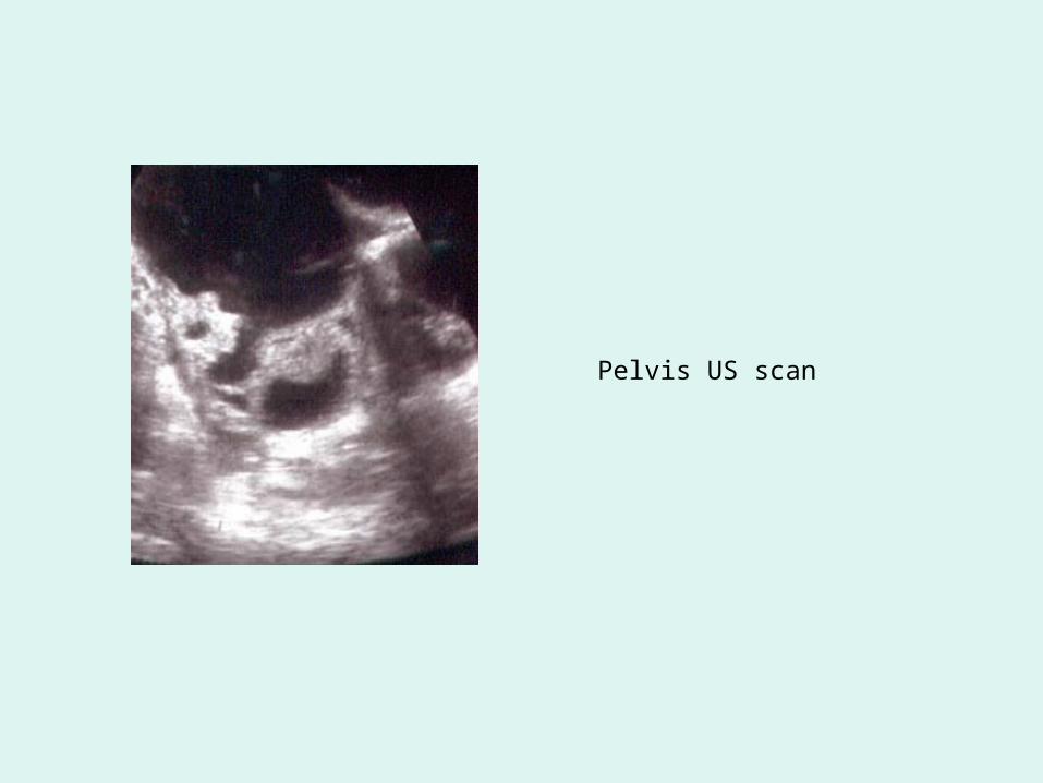
Pelvis US scan

Referral to gynae oncology service
Physical examination • Thin lady, previously healthy• No lymphadenopathy• Breast examination was normal• Lung fields clear on auscultation• Abdominal distention to 28 weeks size by a mass of poor
mobility arising from pelvis and upper abdominal fullness, suggesting omental disease
• Clinical ascites• Distended pouch of Douglas with thickening on recto-
vaginal examination

Medical and Gynaecologic History
Medical Hx:– HTN, Ulcerative colitis (previously on long term steroids but no
evidence of DEXA osteopenia)– Medications: Centyl, Lipitor– Allergies: Penicillin
Surgical Hx: Arthroscopy, cholecystectomy Family Hx: Bowel Ca (father), breast Ca (mother)Gynae Hx:
– Menarche at age 12y– Regular cycles, no dysmennorhea, LMP 2/52 ago– Last Cx smear 3 years ago– Never on OCP
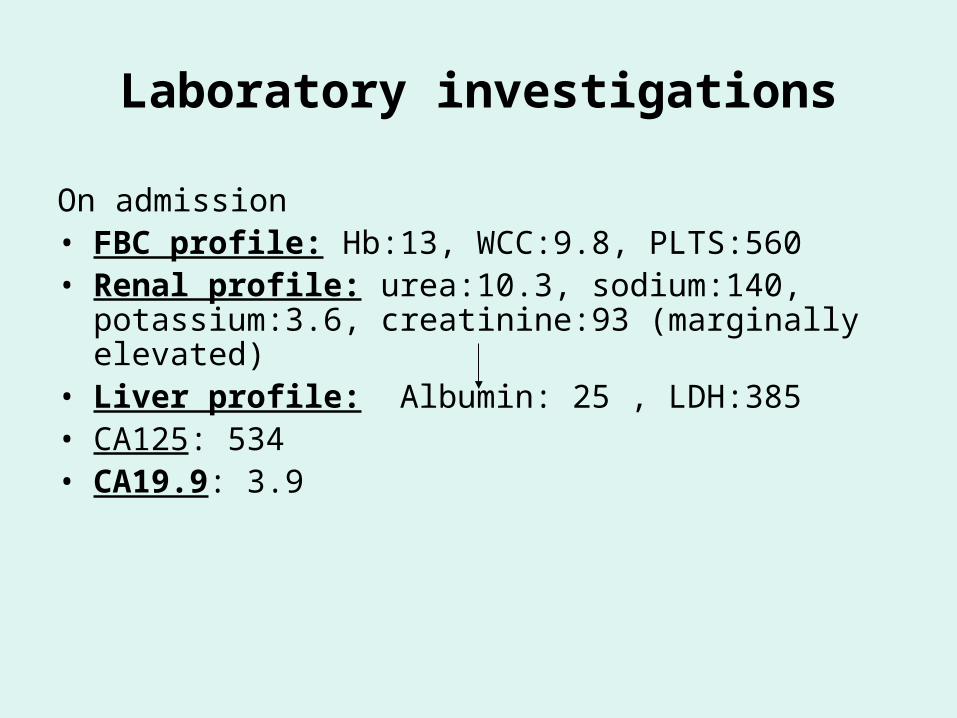
Laboratory investigations
On admission• FBC profile: Hb:13, WCC:9.8, PLTS:560• Renal profile: urea:10.3, sodium:140, potassium:3.6,
creatinine:93 (marginally elevated)• Liver profile: Albumin: 25 , LDH:385• CA125: 534• CA19.9: 3.9
Questions
• What is your provisional diagnosis?
• Can you identify any risk factors from her medical history?
• What is your interpretation of her blood results?– Albumin– urea, creatinine– Hb, plts
Radiology investigations
• CXR: – Lung fields appear clear– No cardiomegaly – No pleural effusion
• CT TAP (chest abdomen pelvis) – 11 X 12.5cm complex pelvic mass arising from the left ovary– Massive ascites– Omental cake– No evidence of retroperitoneal lymphadenopathy– Left hydronephrosis– Splenic hilar and peritoneal nodes
• 3-D colour Doppler• FDG-PET
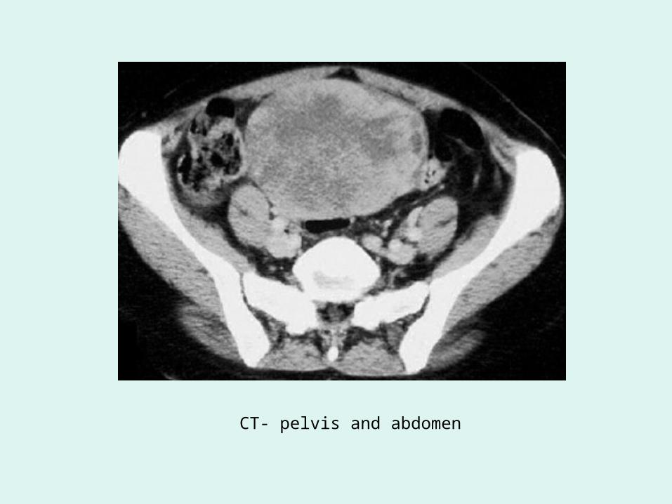
CT- pelvis and abdomen
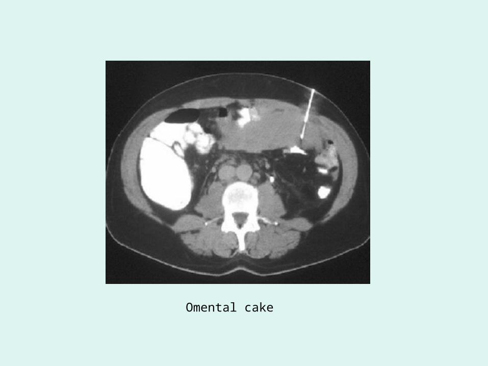
Omental cake
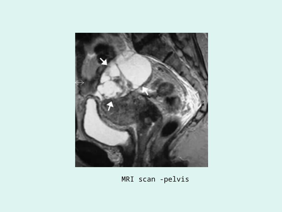
MRI scan -pelvis
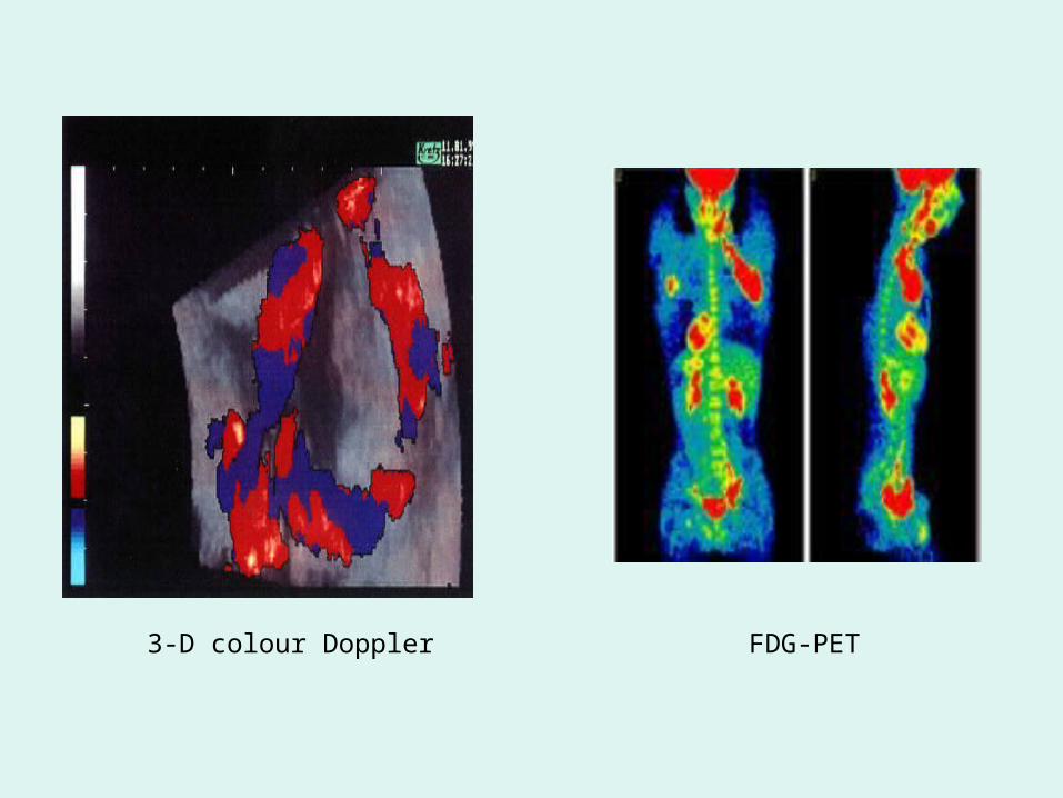
3-D colour Doppler FDG-PET
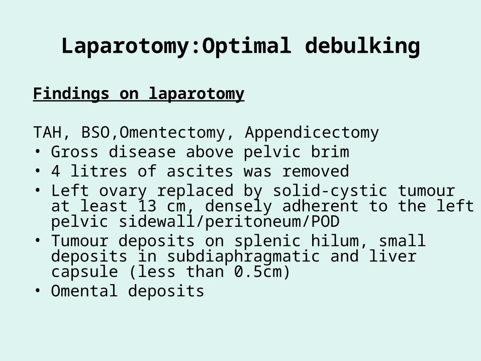
Laparotomy:Optimal debulking
Findings on laparotomy
TAH, BSO,Omentectomy, Appendicectomy• Gross disease above pelvic brim• 4 litres of ascites was removed• Left ovary replaced by solid-cystic tumour at least 13 cm,
densely adherent to the left pelvic sidewall/peritoneum/POD• Tumour deposits on splenic hilum, small deposits in
subdiaphragmatic and liver capsule (less than 0.5cm)• Omental deposits
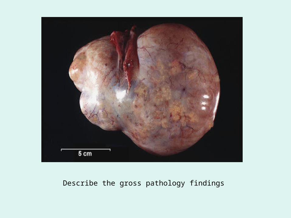
Describe the gross pathology findings

Peritoneal fluid
What does this show?
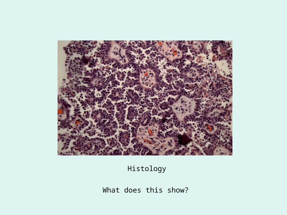
Histology
What does this show?
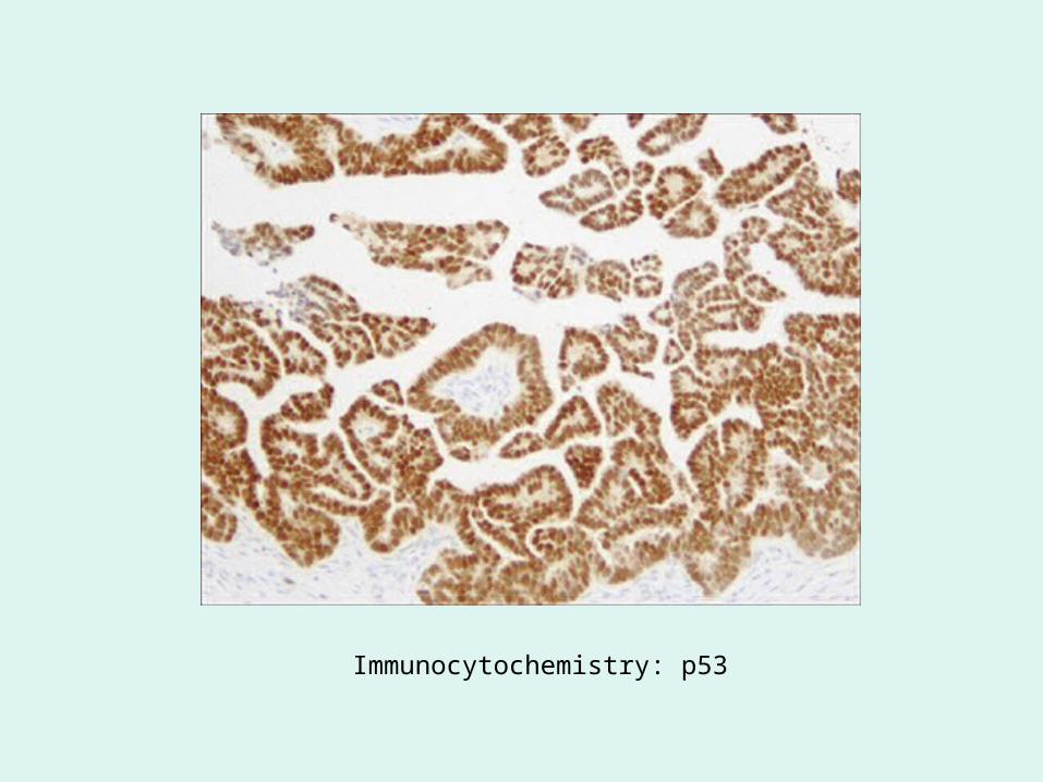
Immunocytochemistry: p53
Pathological diagnosis
• Papillary serous cystadenocarcinoma of the left ovary– TNM stage pT3, N1, Mx– FIGO stage IIIC
HISTORY
• Uneventful recovery• Histology available at day 9• Referred to medical oncologists for adjuvant
chemotherapy• Discharged on day 13• Returned 6 weeks after surgery for initiation of
chemotherapy
HISTORY
• Received 6 cycles of Carboplatin and Taxol– Question: what do these agents exactly do?
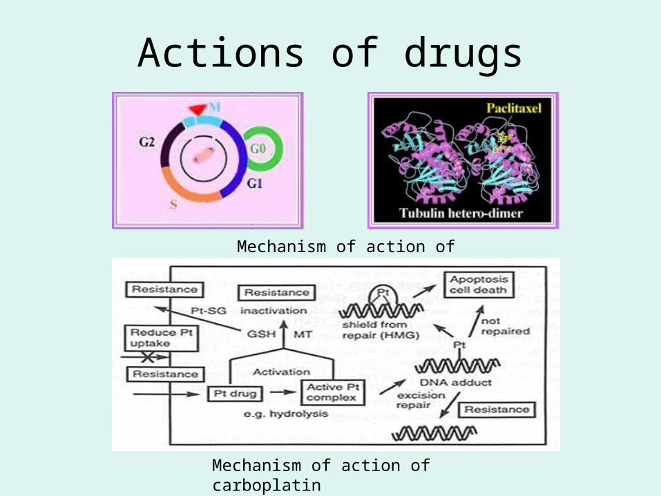
Actions of drugs
Mechanism of action of taxol
Mechanism of action of carboplatin
HISTORY
• Chemotherapy completed 3 months later• Remained well and returned for combined
follow-up with Gynae-Oncologists and Medical Oncologists– Question: what is entailed in the medical follow-up?
Follow-up
• History
• Clinical examination
• CA-125
HISTORY
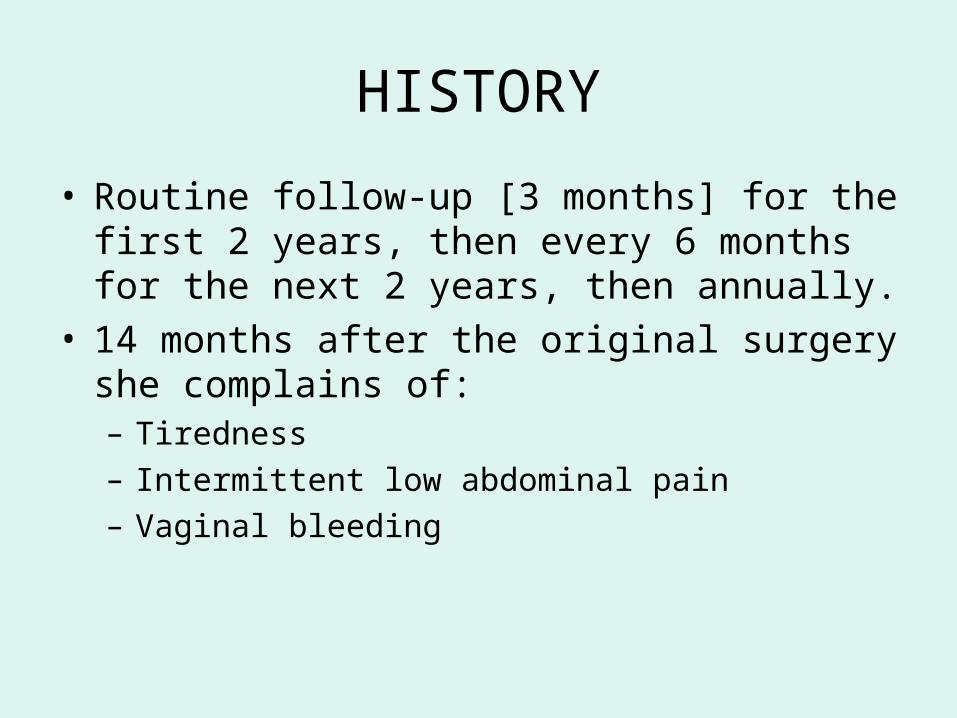
• Routine follow-up [3 months] for the first 2 years, then every 6 months for the next 2 years, then annually.
• 14 months after the original surgery she complains of:– Tiredness– Intermittent low abdominal pain– Vaginal bleeding
Questions
• Why does this patient have a vaginal bleeding?
• What is the cause of the intermittent abdominal pain?

HISTORY
• On clinical examination, two nodules are identified close to the vaginal vault
• Raising CA125• CT of thorax, abdomen and pelvis performed
– Two small soft tissue masses suspicious for disease recurrence seen at the vaginal vault
• Biopsy performed of vaginal lesions
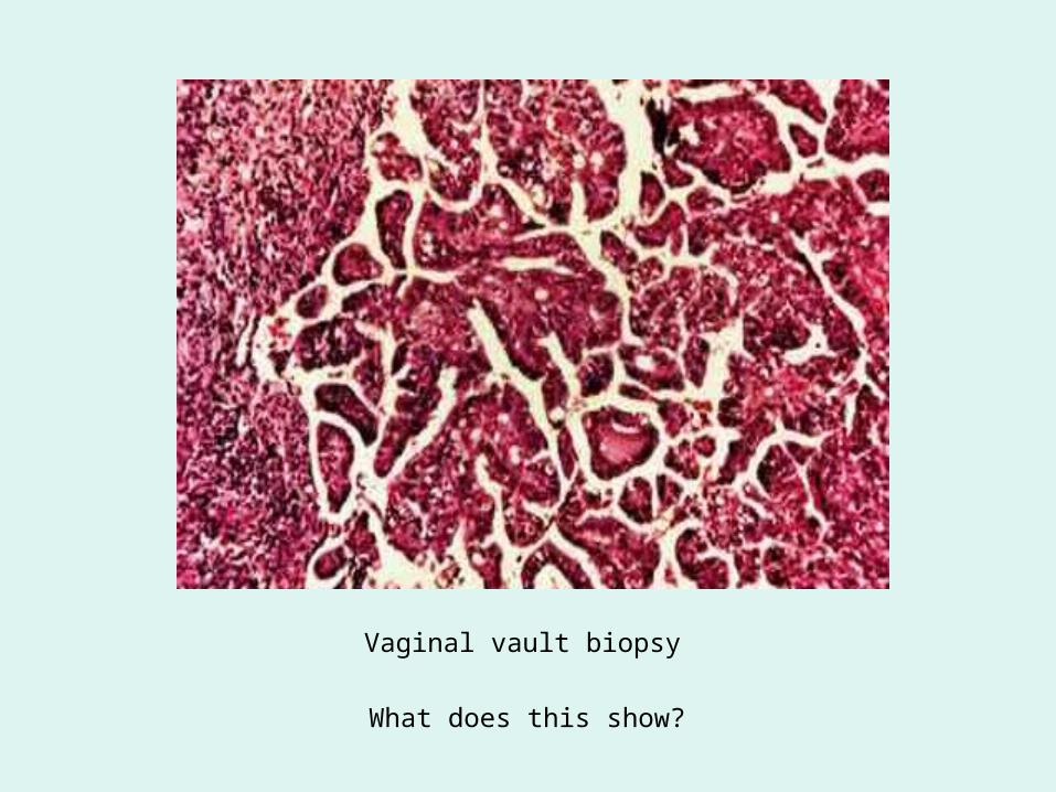
Vaginal vault biopsy
What does this show?

Relapse
• Will the patient benefit from the same chemotherapy?
• Will she benefit from excision of the nodules?
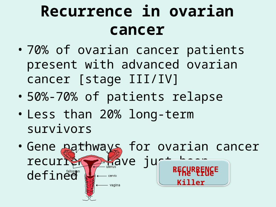
Recurrence in ovarian cancer
• 70% of ovarian cancer patients present with advanced ovarian cancer [stage III/IV]
• 50%-70% of patients relapse
• Less than 20% long-term survivors
• Gene pathways for ovarian cancer recurrence have just been defined
“The true Killer”RECURRENCE
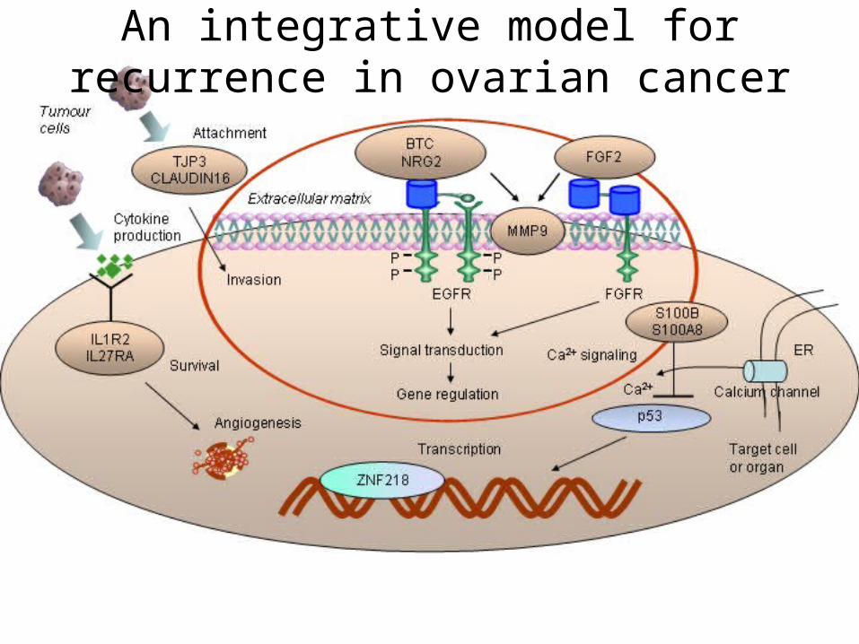
An integrative model for recurrence in ovarian cancer
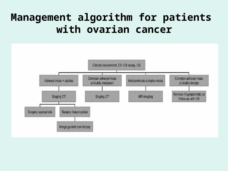
Management algorithm for patients with ovarian cancer
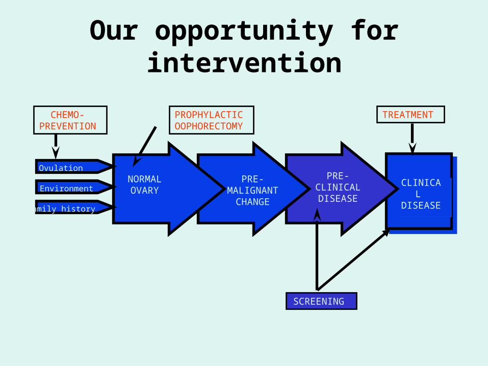
Our opportunity for intervention
CLINICAL DISEASE
CLINICAL DISEASE
NORMAL OVARY
PRE- MALIGNANT
CHANGE
PRE- CLINICAL DISEASE
Family history
CHEMO- PREVENTION
PROPHYLACTIC OOPHORECTOMY
SCREENING
TREATMENT
Environment
Ovulation

Module network procedure
Pre- processing
Image trait selection
Disease traits
Gene expression data
Image traits
Expression data
Clustering
Gene partition
Functional modules
Annotation analysis
Graphic presentation
Independent Validation
Classification program learning
Post- processing
Genes
Life sciences
Information sciences
Life and Information sciences
Pathological data
Proteomic data
MRI3-D colour doppler CT FDG-PET