CLINICAL&EVALUATION&OF& … Evaluation Of... · Blinkreflex& • Blinkreflex( glabellarreflex,&...
34
CLINICAL EVALUATION OF TRIGEMINAL NERVE FUNCTION
Transcript of CLINICAL&EVALUATION&OF& … Evaluation Of... · Blinkreflex& • Blinkreflex( glabellarreflex,&...
CLINICAL EVALUATION OF TRIGEMINAL NERVE FUNCTION
Sensory Evalua;on
• Exterocep;ve sensa;on (pain, light touch, heat, and cold)
• three Vth divisions : tested individually and compared with opposite side.
• Lesions of individual divisions (distal to the gasserian ganglion) : result in sensory loss confined to the cutaneous supply of that division
• Lesions at or proximal to the gasserian ganglion :affects whole I/L face.
• Lesions within the brainstem or upper cervical cord :onion-‐skin distribu;on of sensory loss,
• Dissocia;on of sensa;on on the face (pain and temperature vs. touch sensa;on) differen;ates lesions affec;ng the spinal tract and nucleus of the Vth nerve from lesions affec;ng the main sensory nucleus.
• The cutaneous area over the angle of the mandible is supplied by the second and third cervical roots (by way of the great auricular nerve) and not by the Vth nerve

Motor Evalua;on
supplies the muscles of mas;ca;on. clench the jaw (Ms and T), move the jaw from side to side against
resistance (lateral pterygoids), and protrude the jaw.
• With nuclear or infranuclear lesions of the motor division of the Vth nerve (LMN ) – I/L T and M muscles do not contract when the jaw is clenched,
– the jaw deviates to the paralyzed side when the mouth is opened (due to contrac;on of the C/L intact LP muscle), and
– the jaw cannot be deviated toward the nonparalyzed side (due to I/L LP paresis).
– Atrophy and fascicula;on of the mas;catory muscles may also be evident.
• Other muscles supplied by the Vth nerve (mylohyoid, anterior belly of the digastric, tensor tympani, tensor veli pala;ni) are difficult to evaluate clinically.
• Flaccidity of floor of mouth due to mylohyoid and digastric paralysis may be evident on palpa;on, and
• Paralysis of tensor tympani: result in difficulty in hearing high notes.
• Reflex Evalua;on : – corneal reflex – Sternutatory reflex and – jaw jerk (M reflex).
• Corneal reflex: – Afferent arc : travels through the ophthalmic (upper cornea) and maxillary (lower cornea)
– Efferent arc : I/L(direct reflex) and C/L (consensual reflex) facial nerve to the orbicularis oculi muscles.
– Lesions of Vth nerve (peripheral or pontomedullary trigeminal pathways) result in loss of I/L and C/L responses.
– Suprasegmental modula0on of this reflex : a parietal lobe lesion (involving the perisylvian por0on of the postcentral gyrus) may result in a C/L loss of the corneal reflex
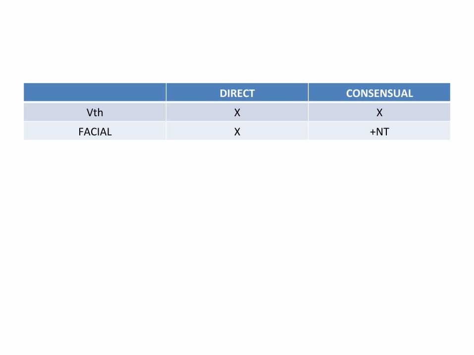
DIRECT CONSENSUAL
Vth X X
FACIAL X +NT
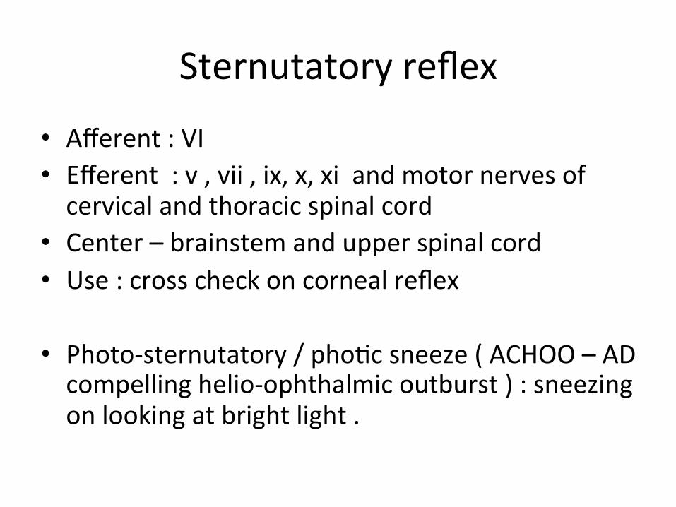
Sternutatory reflex
• Afferent : VI • Efferent : v , vii , ix, x, xi and motor nerves of cervical and thoracic spinal cord
• Center – brainstem and upper spinal cord • Use : cross check on corneal reflex
• Photo-‐sternutatory / pho;c sneeze ( ACHOO – AD compelling helio-‐ophthalmic outburst ) : sneezing on looking at bright light .
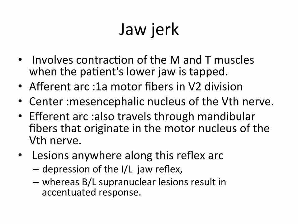
Jaw jerk • Involves contrac;on of the M and T muscles when the pa;ent's lower jaw is tapped.
• Afferent arc :1a motor fibers in V2 division • Center :mesencephalic nucleus of the Vth nerve. • Efferent arc :also travels through mandibular fibers that originate in the motor nucleus of the Vth nerve.
• Lesions anywhere along this reflex arc – depression of the I/L jaw reflex, – whereas B/L supranuclear lesions result in accentuated response.
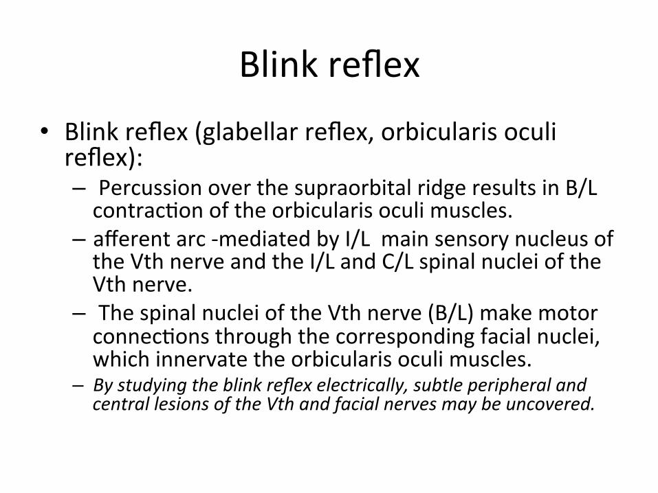
Blink reflex • Blink reflex (glabellar reflex, orbicularis oculi reflex): – Percussion over the supraorbital ridge results in B/L contrac;on of the orbicularis oculi muscles.
– afferent arc -‐mediated by I/L main sensory nucleus of the Vth nerve and the I/L and C/L spinal nuclei of the Vth nerve.
– The spinal nuclei of the Vth nerve (B/L) make motor connec;ons through the corresponding facial nuclei, which innervate the orbicularis oculi muscles.
– By studying the blink reflex electrically, subtle peripheral and central lesions of the Vth and facial nerves may be uncovered.
Corneo-‐mandibular reflex • Consists of B/L eye blink and a brisk C/L anterolateral jaw movement induced by corneal s;mula;on
• ? Associated movt rather than true reflex • s/o supranuclear interrup;on of I/L cor;co-‐trigeminal tract ( only eye sign in ALS )
• In the corneomandibular reflex, the jaw movement is primarily related to the blink rather than the corneal s;mulus
Localiza;on of Lesions Affec;ng Cranial Nerve V
Supranuclear Lesions • Supranuclear control of Vth motor func;on is B/L; however, the C/L hemisphere exerts predominant control on the voluntary ac;vity of the M
• Cor;cobulbar fibers originate in the lower frontal motor cortex, descend through the CR, IC, and cerebral peduncle, and then decussate in the pons to supply the motor nucleus of the Vth nerve.
• Lesions interrup;ng this pathway may result in C/L Vth motor paresis (e.g., devia)on of the jaw away from the lesion), but because of the B/L innerva;on, paresis may be mild.
Supranuclear Lesion : • B/L upper motor neuron lesions (pseudobulbar palsy) result in profound Vth motor paresis, oden with an exaggerated jaw reflex. Mas;ca;on is then markedly impaired.
• Thalamic lesions may result in anesthesia of the C/L face.
• Parietal lesions may be a/w depression of the C/L corneal reflex, even when facial sensa;on is otherwise intact
Nuclear Lesions
• Involve other brainstem structures, and therefore – long tract signs, and – other cranial nerve involvement
• I/L paresis, atrophy, and fascicula;ons of the muscles of mas;ca;on occur.
• A pon)ne localiza)on of this mas)catory paresis is suggested by associated findings:
– contralateral hemiplegia (due to affec;on of the basis pon;s),
– ipsilateral hemianesthesia of the face (due to affec;on of the main sensory nucleus of the trigeminal nerve),
– contralateral hemisensory loss of the limbs and trunk (due to spinothalamic tract affec;on), and
– Internuclear ophthalmoplegia (secondary to medial longitudinal fasciculus damage) and
– ipsilateral Horner syndrome (due to involvement of descending sympathe;c fibers)
• The nucleus of the spinal tract of the trigeminal nerve extends from the caudal end of the pons to the third or fourth cervical spinal cord level.
• Therefore, lesions affec;ng the caudal pons, lateral medulla, or upper cervical cord result in : – ipsilateral facial analgesia, hypesthesia, and thermoanesthesia.
– Because the lateral spinothalamic tract lies in close proximity to the trigeminal spinal nucleus,it is oden associated with contralateral trunk and extremity hypalgesia and thermoanesthesia.
onion-‐skin paeern of sensory loss
• Lower medullary or upper cervical spinal nuclear lesions result in sensory disturbance that affects the peripheral (lateral) forehead, cheek, and jaw (onion-‐skin paeern of sensory loss).
• This onion-‐skin segmental distribu;on reflects the rostral-‐caudal somatotopic arrangement of the cutaneous distribu;on of the spinal nucleus (e.g., perioral area -‐rostral; lateral face -‐caudal).
lateral medullary (Wallenberg) syndrome/PICA Syndrome )
• spinal nucleus of the trigeminal nerve is characteris;cally affected
• most oden secondary to brainstem infarc;on due to intracranial vertebral artery occlusion
• Classically aeributed to PICA occlusion, but in 80-‐85% of cases the vertebral artery is also involved“.
• Acute onset
• Lesions affec)ng the mesencephalic nucleus of the trigeminal nerve cause no apparent neurologic signs and symptoms, except perhaps depression of the ipsilateral jaw jerk (masseter reflex).
Lesions Affec;ng the Preganglionic Trigeminal Nerve Roots
• In its cisternal course, the preganglionic trigeminal nerve root may be damaged by – tumor (meningioma, schwannoma, metastasis, nasopharyngeal carcinoma),
– infec;on (granulomatous, infec;ous, or carcinomatous meningi;s),
– trauma, or – aneurysm.
• Preganglionic trigeminal nerve involvement is suggested by the involvement of the neighboring cranial nerves (especially cranial nerves VI, VII, and VIII).
• The trigeminal roots may be involved by extension of pathologic processes (usually acous;c neuroma or meningioma) located in the cerebellopon;ne angle.
• associated with ipsilateral ;nnitus, deafness, and ver;go (due to involvement of cranial nerve VIII).
• Facial nerve paralysis, • ipsilateral ataxia, and nystagmus (due to involvement of the cerebellar peduncles and cerebellum),
• ipsilateral lateral rectus paralysis (Vith CN involvement), • and, rarely, affec;on of cranial nerves IX through XII may also occur.
Trigeminal neuralgia (;c douloureux, Fothergill's disease)
• syndrome of sudden, excrucia;ng, lancina;ng, paroxysmal, and usually unilateral pains in the distribu;on of one or more of the divisions (oden the maxillary or mandibular) of the trigeminal nerve
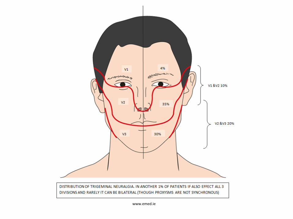
• Incidence:4-‐5/100,000 • Female predominance (m: f = 1:2 ~2:3) • Mean age: 50 yrs. • Right side more than the led. • B/L : exceedingly rare, except in Mul;ple sclerosis. • Pain from (V2):referred to upper lip, nose, and cheek. • (V3): lower lip. • Tic pain confined to the ophthalmic division (V1) is dis;nctly
uncommon.
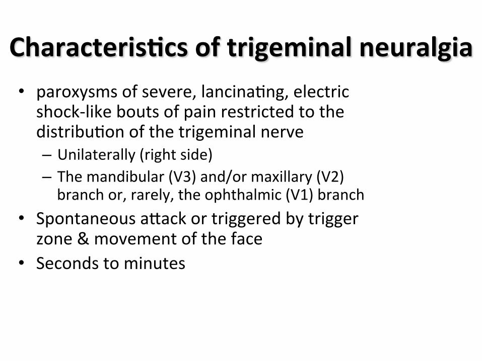
Characteris<cs of trigeminal neuralgia • paroxysms of severe, lancina;ng, electric shock-‐like bouts of pain restricted to the distribu;on of the trigeminal nerve – Unilaterally (right side) – The mandibular (V3) and/or maxillary (V2) branch or, rarely, the ophthalmic (V1) branch
• Spontaneous aeack or triggered by trigger zone & movement of the face
• Seconds to minutes
www.emed.ie
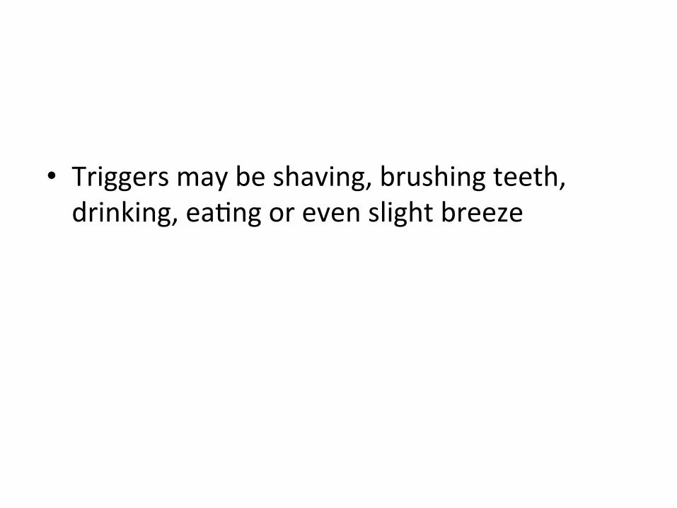
• Triggers may be shaving, brushing teeth, drinking, ea;ng or even slight breeze

Diagnos<c criteria for classic trigeminal neuralgia
• Paroxysmal attacks of pain lasting from a fraction of a second to two minutes that affect one or more divisions of the trigeminal nerve
• Pain has at least one of the following characteristics – intense, sharp, superficial, or stabbing
– precipitated from trigger areas or by trigger factors
• Attacks are similar in individual patients
• No neurological deficit is clinically evident
• Not attributed to another disorder
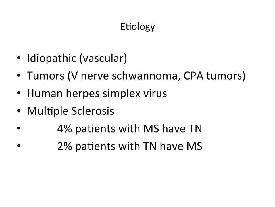
E;ology
• Idiopathic (vascular) • Tumors (V nerve schwannoma, CPA tumors) • Human herpes simplex virus • Mul;ple Sclerosis • 4% pa;ents with MS have TN • 2% pa;ents with TN have MS
Vascular • An aberrant loop of artery, or less commonly vein, is found to
be compressing the root entry zone of the trigeminal nerve in 80-‐90% of pa;ents at surgery
• The nerve is demyelinated next to the compressing vessel
• Elimina;ng the compression provides long term relief
• Sensory func;on recovers ader decompression
• Compression by tumours or the demyelina;ng plaques of mul;ple sclerosis produce similar lesions of the root entry zone
Raeder's Paratrigeminal Syndrome
• Two essen;al components:
• unilateral oculosympathe;c paresis (AKA par;al Horner's syndrome (HS), this usually lacks anhidrosis, and ptosis also)
• homolateral trigeminal nerve involvement (usually ;c-‐like pain, but may be analgesia or masseter weakness; pain, if present, must be ;c-‐like and does not include e.g. unilateral head, face or vascular pain)
• Localizing value: region adjacent to trigeminal nerve in middle fossa. The cause is oden not determined, but may rarely be due to aneurysm" compressing V1 with sympathe;cs.
GRADENIGO'S SYNDROME
AKA apical petrosi;s: Mastoidi;s with involvement of petrous apex (if pneuma;zed). Classic triad:
• Abducens-‐palsy:from inflamma;on of 6th nerve at Dorello's canal,which is where it enters the cavernous sinus just medial to the petrous apex
• retro-‐orbital pain: due to inflamma;on of V1 draining ear
• I/L ear discharge
The Cavernous Sinus Syndrome
• Lesions within the cavernous sinus may damage the ophthalmic and maxillary divisions of the trigeminal nerve and the abducens, trochlear, and oculomotor nerves.
• Total unilateral ophthalmoplegia • associated with pain, paresthesias, and sensory loss in the distribu;on of the ophthalmic and, less oden, the maxillary divisions of the trigeminal nerve.
• Occasionally, oculosympathe;c paresis (without anhidrosis) may also occur.
• Because the mandibular nerve is spared, no mas;catory paresis is evident.
The Superior Orbital Fissure Syndrome
• The abducens, trochlear, and oculomotor nerves as well as the ophthalmic division of the trigeminal nerve pass through the superior orbital fissure.
• complete (external and internal) ophthalmoplegia • pain, paresthesias, and sensory loss in the ophthalmic cutaneous
distribu;on. • Occasionally, oculosympathe;c paresis (without anhidrosis) may occur
because of the involvement of the sympathe;c fibers. • Exophthalmos, due to blockade of the ophthalmic veins, and • blindness, due to extension of the pathologic process to involve the op;c
canal, may also occur. • Except for the occasional instance of involvement of the maxillary
division of the trigeminal nerve in the cavernous sinus syndrome, the superior orbital fissure syndrome and the cavernous sinus syndrome usually cannot be differen)ated clinically without the use of neuroradiologic procedures.