Clinical Study Two Ports Laparoscopic Inguinal...
6
Clinical Study Two Ports Laparoscopic Inguinal Hernia Repair in Children Medhat M. Ibrahim Pediatric Surgery Unit, Faculty of Medicine, Al-Azhar University, Nasr City, Cairo 11884, Egypt Correspondence should be addressed to Medhat M. Ibrahim; dr medhat [email protected] Received 30 August 2014; Revised 10 January 2015; Accepted 10 January 2015 Academic Editor: Peng Hui Wang Copyright © 2015 Medhat M. Ibrahim. is is an open access article distributed under the Creative Commons Attribution License, which permits unrestricted use, distribution, and reproduction in any medium, provided the original work is properly cited. Introduction. Several laparoscopic treatment techniques were designed for improving the outcome over the last decade. e various techniques differ in their approach to the inguinal internal ring, suturing and knotting techniques, number of ports used in the procedures, and mode of dissection of the hernia sac. Patients and Surgical Technique. 90 children were subjected to surgery and they undergone two-port laparoscopic repair of inguinal hernia in children. Technique feasibility in relation to other modalities of repair was the aim of this work. 90 children including 75 males and 15 females underwent surgery. Hernia in 55 cases was right- sided and in 15 leſt-sided. Two patients had recurrent hernia following open hernia repair. 70 (77.7%) cases were suffering unilateral hernia and 20 (22.2%) patients had bilateral hernia. Out of the 20 cases 5 cases were diagnosed by laparoscope (25%). e patients’ median age was 18 months. e mean operative time for unilateral repairs was 15 to 20 minutes and bilateral was 21 to 30 minutes. ere was no conversion. e complications were as follows: one case was recurrent right inguinal hernia and the second was stitch sinus. Discussion. e results confirm the safety and efficacy of two ports laparoscopic hernia repair in congenital inguinal hernia in relation to other modalities of treatment. 1. Introduction During recent years, the trend toward laparoscopic approach for hernia repair in children has been increasingly justified. e ability to detect and repair the contralateral opening of internal rings simultaneously, along with safe high ligation of the hernia sac without injury of the vas deference or the spermatic vessels, make laparoscopic approach a reliable alternative to the conventional open techniques [1]. e universally known limitations of the laparoscopic surgery are as follows. (1) Most of these methods employ a laparoscope inserted via an umbilical incision and two lateral ports for instruments to ligate the hernia defect [2]. e necessity for intra-abdominal skills, such as intracorporeal suturing, knot tying, and manipulation of the suture on a needle may be time-consuming and cumbersome. (2) Recurrence rate aſter laparoscopic surgery is generally known to be higher than aſter open surgery [2]. With the increase in laparoscopic inguinal hernia repair, several treatment techniques have developed over the past two decades, aimed at improving the outcome [3]; the various techniques differ in their approach to the inguinal internal ring, suturing and knotting techniques, number of ports used in the procedures, endoscopic instruments used, mode of dissection of the hernia sac, and extracorporeal and intracorporeal suturing and knotting techniques [4]. is study is an early experience of two ports laparoscopic repair of inguinal hernia. I used two 5 mm ports which reduce the port numbers and size, the purse string suturing, and extracorporeal knotting of the hernia sac. 2. Materials and Surgical Technique is study was conducted from April 2009 to April 2013. 90 children with inguinal hernia were subjected to full clinical evaluation and routine investigations. e main outcome measurements were correlation between clinical diagnosis, abdominal ultrasound results and the laparoscopic evaluation of contralateral hernia, feasibility of two ports laparoscopic repair of the inguinal hernia, conversion rate, need for additional port, operative time, postoperative pain and requirement of analgesia, hospital stay, recurrence rate, and fat of nonexcised hernia sac and development of post- operative hydrocele. e ethical committee approved the technique. Written consent was obtained from the family Hindawi Publishing Corporation Minimally Invasive Surgery Volume 2015, Article ID 821680, 5 pages http://dx.doi.org/10.1155/2015/821680
Transcript of Clinical Study Two Ports Laparoscopic Inguinal...

Clinical StudyTwo Ports Laparoscopic Inguinal Hernia Repair in Children
Medhat M. Ibrahim
Pediatric Surgery Unit, Faculty of Medicine, Al-Azhar University, Nasr City, Cairo 11884, Egypt
Correspondence should be addressed to Medhat M. Ibrahim; dr medhat [email protected]
Received 30 August 2014; Revised 10 January 2015; Accepted 10 January 2015
Academic Editor: Peng Hui Wang
Copyright © 2015 Medhat M. Ibrahim.This is an open access article distributed under the Creative Commons Attribution License,which permits unrestricted use, distribution, and reproduction in any medium, provided the original work is properly cited.
Introduction. Several laparoscopic treatment techniques were designed for improving the outcome over the last decade.The varioustechniques differ in their approach to the inguinal internal ring, suturing and knotting techniques, number of ports used in theprocedures, and mode of dissection of the hernia sac. Patients and Surgical Technique. 90 children were subjected to surgery andthey undergone two-port laparoscopic repair of inguinal hernia in children. Technique feasibility in relation to other modalities ofrepair was the aim of this work. 90 children including 75 males and 15 females underwent surgery. Hernia in 55 cases was right-sided and in 15 left-sided. Two patients had recurrent hernia following open hernia repair. 70 (77.7%) cases were suffering unilateralhernia and 20 (22.2%) patients had bilateral hernia. Out of the 20 cases 5 cases were diagnosed by laparoscope (25%). The patients’median age was 18 months. The mean operative time for unilateral repairs was 15 to 20 minutes and bilateral was 21 to 30 minutes.There was no conversion.The complications were as follows: one case was recurrent right inguinal hernia and the second was stitchsinus. Discussion. The results confirm the safety and efficacy of two ports laparoscopic hernia repair in congenital inguinal herniain relation to other modalities of treatment.
1. Introduction
During recent years, the trend toward laparoscopic approachfor hernia repair in children has been increasingly justified.The ability to detect and repair the contralateral opening ofinternal rings simultaneously, along with safe high ligationof the hernia sac without injury of the vas deference orthe spermatic vessels, make laparoscopic approach a reliablealternative to the conventional open techniques [1].
The universally known limitations of the laparoscopicsurgery are as follows. (1) Most of these methods employ alaparoscope inserted via an umbilical incision and two lateralports for instruments to ligate the hernia defect [2]. Thenecessity for intra-abdominal skills, such as intracorporealsuturing, knot tying, and manipulation of the suture ona needle may be time-consuming and cumbersome. (2)Recurrence rate after laparoscopic surgery is generally knownto be higher than after open surgery [2].
With the increase in laparoscopic inguinal hernia repair,several treatment techniques have developed over the pasttwo decades, aimed at improving the outcome [3]; the varioustechniques differ in their approach to the inguinal internalring, suturing and knotting techniques, number of ports
used in the procedures, endoscopic instruments used, modeof dissection of the hernia sac, and extracorporeal andintracorporeal suturing and knotting techniques [4].
This study is an early experience of two ports laparoscopicrepair of inguinal hernia. I used two 5mmports which reducethe port numbers and size, the purse string suturing, andextracorporeal knotting of the hernia sac.
2. Materials and Surgical Technique
This study was conducted from April 2009 to April 2013.90 children with inguinal hernia were subjected to fullclinical evaluation and routine investigations. The mainoutcome measurements were correlation between clinicaldiagnosis, abdominal ultrasound results and the laparoscopicevaluation of contralateral hernia, feasibility of two portslaparoscopic repair of the inguinal hernia, conversion rate,need for additional port, operative time, postoperative painand requirement of analgesia, hospital stay, recurrence rate,and fat of nonexcised hernia sac and development of post-operative hydrocele. The ethical committee approved thetechnique. Written consent was obtained from the family
Hindawi Publishing CorporationMinimally Invasive SurgeryVolume 2015, Article ID 821680, 5 pageshttp://dx.doi.org/10.1155/2015/821680
2 Minimally Invasive Surgery
Figure 1: Port position used in right congenital inguinal hernia.
Figure 2: Needle passing to the peritoneal cavity.
after getting full information about the surgery and thepostoperative squeal. All patients received one dose of antibi-otic prophylaxis in the form of ceftriaxone 50mg/kg at thetime of induction of anesthesia. Under general endotrachealanesthesia, patients were scraped and draped. The surgeonposition was at the child head. ENDOPATH XCEL port (5mm) with 00 scope inserted supra umbilical by close tech-nique. Pneumo-peritoneum pressure adjusted at 8–12mmHg according to child condition. Initial visualization of bothinternal rings was done for the bilateral hernia diagnosis.Seconded working port 5mm was inserted according tothe unilaterally and bilaterally hernia under visualization(Figure 1). Patient position was changed to trendelenburgposition 45∘ which helped in reduction of the hernia contentsand gave better visualization of the rings. Proline 3/0 suturemounted needle was passed from the outside through skinand abdominal wall muscles to the peritoneal cavity undervisualization (Figure 2). Laparoscopic needle holder graspsthe needle inside the abdominal cavity.
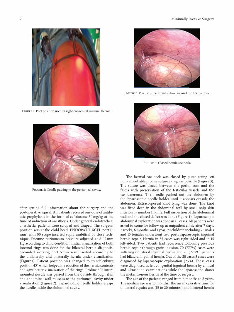
Figure 3: Proline purse string suture around the hernia neck.
Figure 4: Closed hernia sac neck.
The hernial sac neck was closed by purse string 3/0non- absorbable proline suture as high as possible (Figure 3).The suture was placed between the peritoneum and thefascia with preservation of the testicular vessels and thevas deference. The needle pushed out the abdomen bythe laparoscopic needle holder until it appears outside theabdomen. Extracorporeal knot tying was done. The knotwas fixed deep in the abdominal wall by small snip skinincision by number 11 knife. Full inspection of the abdominalwall and the closed defect was done (Figure 4). Laparoscopicabdominal explorationwas done in all cases. All patients wereasked to come for follow-up at outpatient clinic after 7 days,2 weeks, 6 months, and 1 year. 90 children including 75 malesand 15 females underwent two ports laparoscopic inguinalhernia repair. Hernia in 55 cases was right-sided and in 15left-sided. Two patients had recurrence following previoushernia repair through groin incision. 70 (77.7%) cases weresuffering unilateral inguinal hernia and 20 (22.2%) patientshad bilateral inguinal hernia. Out of the 20 cases 5 cases werediagnosed by laparoscopic exploration (25%). These caseswere diagnosed as left congenital inguinal hernia by clinicaland ultrasound examinations while the laparoscope showsthe metachronous hernia at the time of surgery.
The age of the patients ranged from 6 months to 8 years.The median age was 18 months. The mean operative time forunilateral repairs was (15 to 20 minutes) and bilateral hernia

Minimally Invasive Surgery 3
repair was (21 to 30 minutes). The scars on the abdominalwall were small andminute (5mm incision for umbilical port,5mm working port and stitch site). There was no conversionor third port insertion. During follow-up there was onepatient who got noncomplicated right recurrent inguinalhernia after two weeks from surgery. This patient underwentopen right herniotomy where the hernia sac was not closedand proline stitch did not surround all the circumferences ofthe sac. Another case gets stitch sinuses at the proline knotsite, which required open surgical removal of the stitch andopen herniotomy at the same time. In this case the hernia sacwas closed completely by the proline stitch. Once the prolinestitch removed the sac opened, so, herniotomy and Transfixation was done.There were no other complications such astesticular atrophy or secondary hydrocele. All patients wererecovered smoothly from anesthesia and there was no needfor analgesia. All patients were discharged home two hoursafter full recovery from anesthesia.
3. Discussion
Inguinal hernia in pediatric age group is a common problemand all the pediatric surgeons are fully familiar with thevarious aspects of its traditional surgical repair through thegroin incision which has a high success rate and acceptablecosmetic results with few complications [4].
By far one of the drawbacks of this conventional tech-nique is inability to rule out the contralateral patent pro-cesses vaginalis and synchronous hernia. With the advent ofminimal access surgery, many pediatric surgeons accepted it,as a suitable and reliable alternative to the open techniques,considering its superiority for handling tissues during repairof recurrent inguinal hernias and also for its capabilities inregard to justifying and managing the synchronous subtlecontralateral hernia [5]. However, there are still some issuesregarding the introduction of laparoscopic inguinal herniarepair as the gold standard method, especially taking intoconsideration the possible longer operative time and theinevitable need for three separate ports which is the casein routine laparoscopic herniotomy techniques. In theseworks only two ports were used to do laparoscopic her-nia repair in children, which increases the benefit of thelaparoscopic herniotomy. The modified, new laparoscopictechnique improved the diagnosis of ipsilateral hernia, usingextracorporeal tying, that yields excellent cosmetic result.
Out of the 20 cases 5 (25%) case were diagnosed by lapar-oscopic exploration. These cases were diagnosed as left con-genital inguinal hernia clinically and ultrasound examina-tions cannot detect the metachronous hernia in the rightside prior to surgery. Laparoscope was superior to ultrasoundin diagnosis of the metachronous hernia, while in otherstudies significant number of children (20%) presentingwith unilateral hernias have contralateral patent processesvaginalis [5, 6]. The options for detection of contralateralpatent processes vaginalis are many, namely, routine bilateralopen surgery explorations [7], use of ultrasonography [8],laparoscopy [9], and the wait and watch policy [10].
Although laparoscopy proves advantageous over opensurgery by precise detection and simultaneous repair of con-tralateral patent processes vaginalis, its management remainsa contentious issue.The current consensus amongst surgeonspracticing open surgery favors operating on the symptomaticside alone [9] as the rate ofmetachronous hernia is significantthat it necessitates subsequent surgery in a twentieth ofpatients [11].Therefore, this advantage of laparoscopic herniarepair may be significant in clinical practice as it gives gooddiagnosis and also repair for the metachronous hernia.
In open surgery, time is consumed in gaining access,obtaining adequate exposure, identification of the herniasac, and dissection of the cord structures from the sacwithout harming the important cord structures [10]. Inlaparoscopic surgery, approaching from within makes thearea of interest bloodless, and the magnification rendersanatomy splendidly clear, making surgery precise [12]. Butthe time limiting step remains intracorporeal suturing thatplaces considerable demands on the requirement of handeye coordination, especially while negotiating the posteriorand medial hemicircumference of the internal ring, overthe iliac and inferior epigastric vessels [11]. With growingexperience [11] and in this study the subfascia purse stringsuture and extracorporeal subcutaneous knotting with twoports markedly reduce the operative time over the traditionalthree ports laparoscopic hernia repair with intracorporealsuture ligation. In this study the mean operative time forunilateral repairs was (15 to 20 minutes) and bilateral herniarepair was (21 to 30 minutes); the mean duration of surgerywas markedly less than the operative time noted in Lukongstudy [3]. The extracorporeal suturing was effective easy notneed for special tool or surgical skills which also proved byother studys [11, 12].
The difference in postoperative pain following opensurgery and laparoscopic surgery is subject to controversy.Some report less pain while others report greater pain inthe immediate postoperative period following laparoscopicsurgery compared with open surgery [13]. Bharathi R. foundpain perception following either procedure to be similar.Parietal pain predominates in open surgery can well becontrolled by caudal analgesia. On the other hand, painperception is multimodal and multifactorial in laparoscopicsurgery [14]. In addition to parietal pain caused by port place-ment, capnoperitoneumcauses visceral pain due to stretching(peritoneal and diaphragmatic) and acidosis [14]. Neitherthe use of smaller ports nor the use of caudal analgesiawould completely obliterate pain following laparoscopy [14].Therefore, the decrease in the size of the incision does notnecessarily translate into a proportionate decrease in pain.Hence, the difference in postoperative pain between laparo-scopic surgery and open surgery is not significant enough torate either surgery superior. In this study, the intraperitonealgas pressure was located less than in other studies [14] dueto the less number of ports and no need for intra-abdominalsurgical maneuvers. Which reduced the post-operative pain.All patient was not need for post-operative analgesia.
Many new techniques have recently taken place in pedi-atric laparoscopic inguinal hernia surgery. Lee and Liang

4 Minimally Invasive Surgery
[15] introduced the Endo-needle designed specifically forlaparoscopic extraperitoneal closure of the patent processusvaginalis. Endo and Ukiyama performed microlaparoscopichigh ligation in 450 patients with good results.They reportedno complications related to the procedure and a remarkablylow recurrence rate (0.88%) [16]. Prasad et al. used a stainlesssteel curved awl and a 1.7mm telescope to safely performneedlescopic inguinal herniorrhaphy [17]. Shalaby et al. usedRN for closure of IIR in 150 patients successfully withexcellent cosmetic results without any recurrence [18]. Chanand Tam [19] introduced the saline injection and needlesign to reduce the recurrence rate in his series. Bharathiet al. [20] used the TNH technique in 67 repairs, and 146repairs were performed using the SEAL technique. Theystated that SEAL resulted in marked reduction of operativetime when compared to the TNH technique (unilateral: 15versus 25 minutes; bilateral: 25 versus 40 minutes). Theyadded that avoiding the vas deferens and gonadal vesselsduring the SEAL repair in boys may leave a small gap atthe internal ring as well as leaving the hernia sac in situ,which has the potential to contribute to a higher incidence ofrecurrence in male patients. They believe that ligation of theinternal ring leads to scarring and obliteration of the spacedistally. This would explain the relatively low incidence ofpostoperative hydrocele. Fluid accumulating in the distal sacpostoperatively often reabsorbs spontaneously and does notnecessitate additional intervention [20]. In this study therewas no hydrocele detected as a postoperative complicationin spite of no excision of the sac and the recurrence ratewas 1% recurrence. Several piercings of the peritoneum byRN around the neck of the sac may add fixation of thesuture at this level that prevents migration of the suturedistally initiating recurrence. It may result in creation ofadhesions of the sac preventing hydrocele formation. Thismay explain the high incidence of recurrence in the seriesof Manoharan et al. [9] and Bharathi et al. [20] that applythe subcutaneous suture around the internal ring withoutpiercing the peritoneum. However, Chan and Tam [19]reported a very low recurrence rate (1%) after refinement ofthe technique by using TNH and injecting saline around thevas and vessels and using the needle sign to avoid damageto the testicular vessel and vas. They claimed that presenceof a complete ring around IIR prevents recurrence. Prasadet al. reported no recurrence in their early small series of 8cases [17]. Shalaby et al. reported no recurrence in their series[18]. The results of this study confirm the safety and efficacyof laparoscopic hernia repair with two ports in congenitalinguinal hernia in children. It resulted in good diagnosis andmanagement of metachronous hernia and marked reductionof operative time, less recurrence, no hydrocele formation,excellent cosmetic, and no postoperative pain results.
Conflict of Interests
The author has no conflict of interests to report and receivedno financial support for this study.
References
[1] R. Saranga Bharathi, Comparative study of laparoscopic versusconventional surgery for congenital inguinal hernia in children[M.S. thesis], University of Pune, 2008.
[2] Y.-T. Chang, “Technical refinements in single-port laparoscopicsurgery of inguinal hernia in infants and children,” DiagnosticandTherapeutic Endoscopy, vol. 2010, Article ID 392847, 6 pages,2010.
[3] C. S. Lukong, “Surgical techniques of laparoscopic inguinalhernia repair in childhood: a critical appraisal,” Journal ofSurgical Technique and Case Report, vol. 4, no. 1, pp. 1–5, 2012.
[4] P. Kapur, M. G. Caty, and P. L. Glick, “Pediatric hernias andhydroceles,” Pediatric Clinics of North America, vol. 45, no. 4,pp. 773–789, 1998.
[5] F. Schier, P. Montupet, and C. Esposito, “Laparoscopic inguinalherniorrhaphy in children: a three-center experience with 933repairs,” Journal of Pediatric Surgery, vol. 37, no. 3, pp. 395–397,2002.
[6] D. Ozgediz, K. Roayaie, H. Lee et al., “Subcutaneous endoscop-ically assisted ligation (SEAL) of the internal ring for repair ofinguinal hernias in children: report of a new technique and earlyresults,” Surgical Endoscopy, vol. 21, no. 8, pp. 1327–1331, 2007.
[7] F. Rathauser, “Historical overview of the bilateral approach topediatric inguinal hernias,” The American Journal of Surgery,vol. 150, no. 5, pp. 527–532, 1985.
[8] D. M. Miltenburg, J. G. Nuchtern, T. Jaksic, C. Kozinetiz, andM. L. Brandt, “Laparoscopic evaluation of the pediatric inguinalhernia—a meta-analysis,” Journal of Pediatric Surgery, vol. 33,no. 6, pp. 874–879, 1998.
[9] S. Manoharan, U. Samarakkody, M. Kulkarni, R. Blakelock, andS. Brown, “Evidence-based change of practice in the manage-ment of unilateral inguinal hernia,” Journal of Pediatric Surgery,vol. 40, no. 7, pp. 1163–1166, 2005.
[10] C. Esposito, L.Montinaro, F. Alicchio, A. Savanelli, T. Armenise,and A. Settimi, “Laparoscopic treatment of inguinal hernia inthe first year of life,” Journal of Laparoendoscopic and AdvancedSurgical Techniques, vol. 20, no. 5, pp. 473–476, 2010.
[11] F. Schier, “Laparoscopic inguinal hernia repair-a prospectivepersonal series of 542 children,” Journal of Pediatric Surgery, vol.41, no. 6, pp. 1081–1084, 2006.
[12] P. Chinnaswamy, V. Malladi, K. V. Jani et al., “Laparoscopicinguinal hernia repair in children,” Journal of the Society ofLaparoendoscopic Surgeons, vol. 9, no. 4, pp. 393–398, 2005.
[13] P. Ekstein, A. Szold, B. Sagie, N. Werbin, J. M. Klausner, and A.A. Weinbroum, “Laparoscopic surgery may be associated withsevere pain and high analgesia requirements in the immediatepostoperative period,” Annals of Surgery, vol. 243, no. 1, pp. 41–46, 2006.
[14] V. L. Wills and D. R. Hunt, “Pain after laparoscopic cholecys-tectomy,” British Journal of Surgery, vol. 87, no. 3, pp. 273–284,2000.
[15] Y. Lee and J. Liang, “Experience with 450 cases of micro-laparoscopic herniotomy in infants and children,” PediatricEndosurgery and Innovative Techniques, vol. 6, no. 1, pp. 25–28,2002.
[16] M. Endo and E. Ukiyama, “Laparoscopic closure of patentprocessus vaginalis in girlswith inguinal hernia using a speciallydevised suture needle,” Pediatric Endosurgery & InnovativeTechniques, vol. 5, no. 2, pp. 187–191, 2001.
[17] R. Prasad, H. N. Lovvorn III, G. M. Wadie, and T. E. Lobe,“Early experience with needleoscopic inguinal herniorrhaphy

Minimally Invasive Surgery 5
in children,” Journal of Pediatric Surgery, vol. 38, no. 7, pp. 1055–1058, 2003.
[18] R. Y. Shalaby, M. Fawy, S. M. Soliman, and A. Dorgham, “A newsimplified technique for needlescopic inguinal herniorrhaphyin children,” Journal of Pediatric Surgery, vol. 41, no. 4, pp. 863–867, 2006.
[19] K. L. Chan and P. K. H. Tam, “Technical refinements in lapar-oscopic repair of childhood inguinal hernias,” Surgical Endos-copy and Other Interventional Techniques, vol. 18, no. 6, pp. 957–960, 2004.
[20] R. S. Bharathi, A. K. Dabas, M. Arora, and V. Baskaran, “Lapar-oscopic ligation of internal ring—three ports versus single-porttechnique: are working ports necessary?” Journal of Laparoen-doscopic and Advanced Surgical Techniques, vol. 18, no. 6, pp.891–894, 2008.

Submit your manuscripts athttp://www.hindawi.com
Stem CellsInternational
Hindawi Publishing Corporationhttp://www.hindawi.com Volume 2014
Hindawi Publishing Corporationhttp://www.hindawi.com Volume 2014
MEDIATORSINFLAMMATION
of
Hindawi Publishing Corporationhttp://www.hindawi.com Volume 2014
Behavioural Neurology
EndocrinologyInternational Journal of
Hindawi Publishing Corporationhttp://www.hindawi.com Volume 2014
Hindawi Publishing Corporationhttp://www.hindawi.com Volume 2014
Disease Markers
Hindawi Publishing Corporationhttp://www.hindawi.com Volume 2014
BioMed Research International
OncologyJournal of
Hindawi Publishing Corporationhttp://www.hindawi.com Volume 2014
Hindawi Publishing Corporationhttp://www.hindawi.com Volume 2014
Oxidative Medicine and Cellular Longevity
Hindawi Publishing Corporationhttp://www.hindawi.com Volume 2014
PPAR Research
The Scientific World JournalHindawi Publishing Corporation http://www.hindawi.com Volume 2014
Immunology ResearchHindawi Publishing Corporationhttp://www.hindawi.com Volume 2014
Journal of
ObesityJournal of
Hindawi Publishing Corporationhttp://www.hindawi.com Volume 2014
Hindawi Publishing Corporationhttp://www.hindawi.com Volume 2014
Computational and Mathematical Methods in Medicine
OphthalmologyJournal of
Hindawi Publishing Corporationhttp://www.hindawi.com Volume 2014
Diabetes ResearchJournal of
Hindawi Publishing Corporationhttp://www.hindawi.com Volume 2014
Hindawi Publishing Corporationhttp://www.hindawi.com Volume 2014
Research and TreatmentAIDS
Hindawi Publishing Corporationhttp://www.hindawi.com Volume 2014
Gastroenterology Research and Practice
Hindawi Publishing Corporationhttp://www.hindawi.com Volume 2014
Parkinson’s Disease
Evidence-Based Complementary and Alternative Medicine
Volume 2014Hindawi Publishing Corporationhttp://www.hindawi.com